Academic Profile
Statistics
Similar Authors
Papers on arXiv

Deformable image registration establishes non-linear spatial correspondences between fixed and moving images. Deep learning-based deformable registration methods have been widely studied in recent yea...
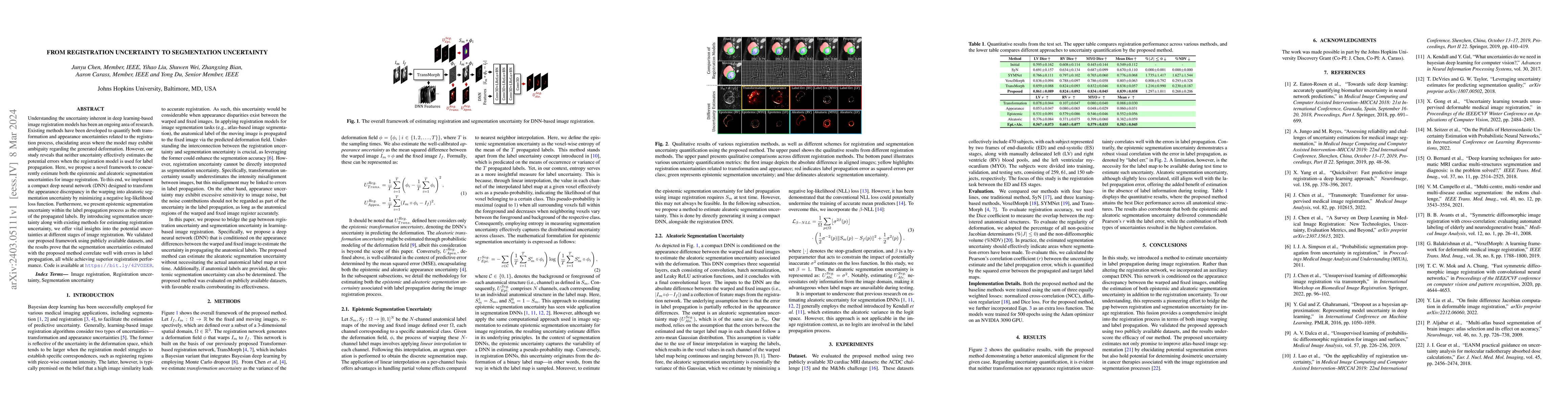
Understanding the uncertainty inherent in deep learning-based image registration models has been an ongoing area of research. Existing methods have been developed to quantify both transformation and...
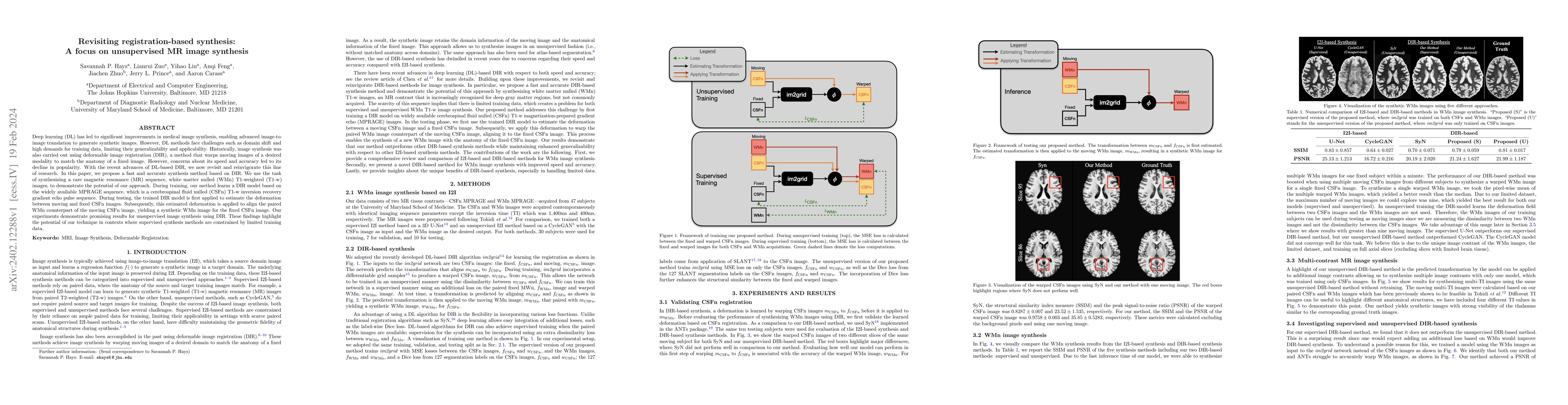
Deep learning (DL) has led to significant improvements in medical image synthesis, enabling advanced image-to-image translation to generate synthetic images. However, DL methods face challenges such...
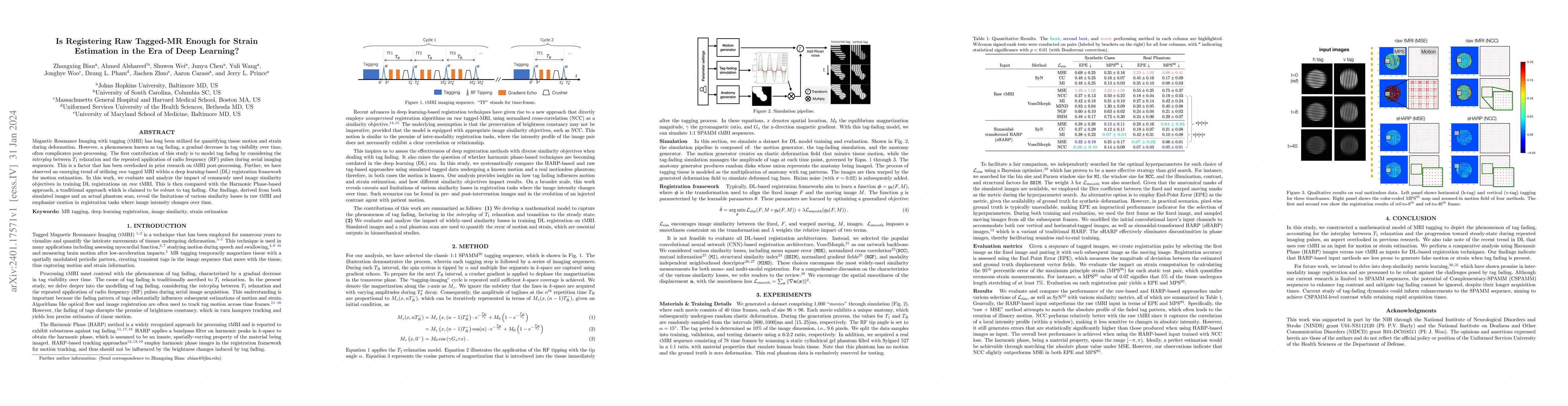
Magnetic Resonance Imaging with tagging (tMRI) has long been utilized for quantifying tissue motion and strain during deformation. However, a phenomenon known as tag fading, a gradual decrease in ta...
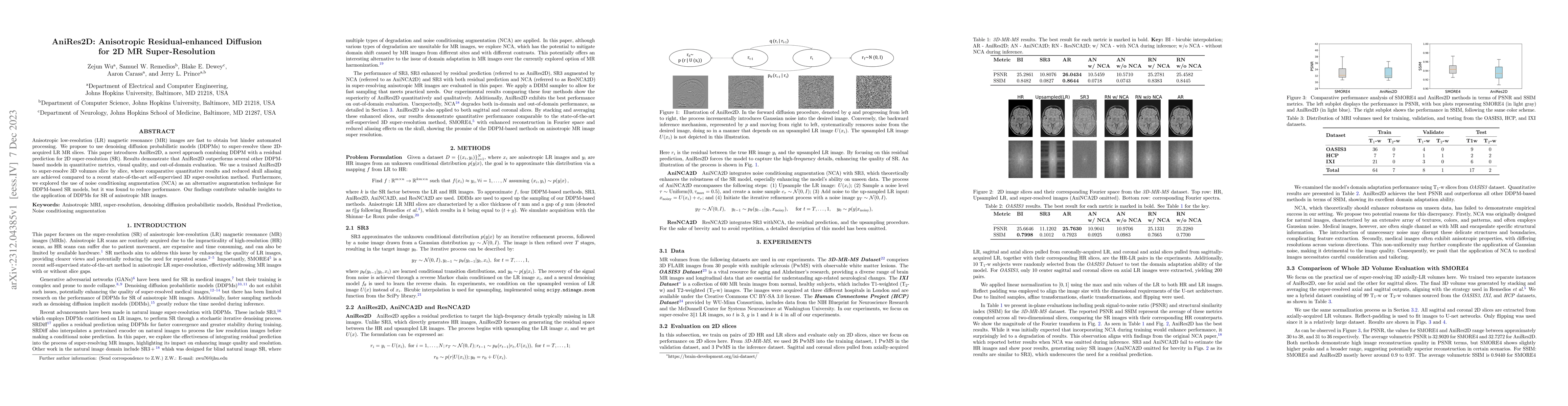
Anisotropic low-resolution (LR) magnetic resonance (MR) images are fast to obtain but hinder automated processing. We propose to use denoising diffusion probabilistic models (DDPMs) to super-resolve...
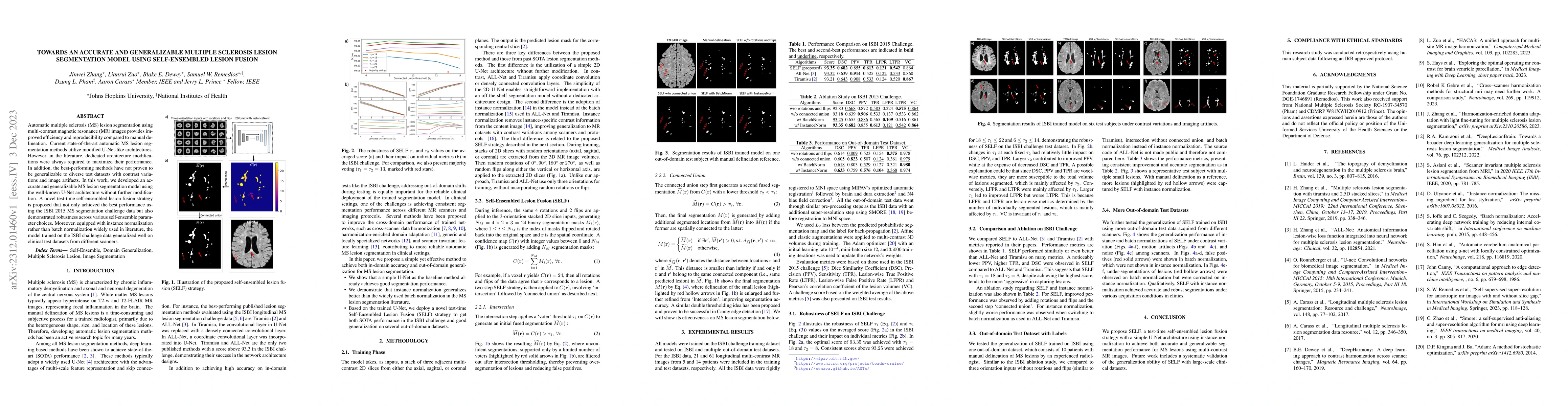
Automatic multiple sclerosis (MS) lesion segmentation using multi-contrast magnetic resonance (MR) images provides improved efficiency and reproducibility compared to manual delineation. Current sta...
Deep learning algorithms utilizing magnetic resonance (MR) images have demonstrated cutting-edge proficiency in autonomously segmenting multiple sclerosis (MS) lesions. Despite their achievements, t...
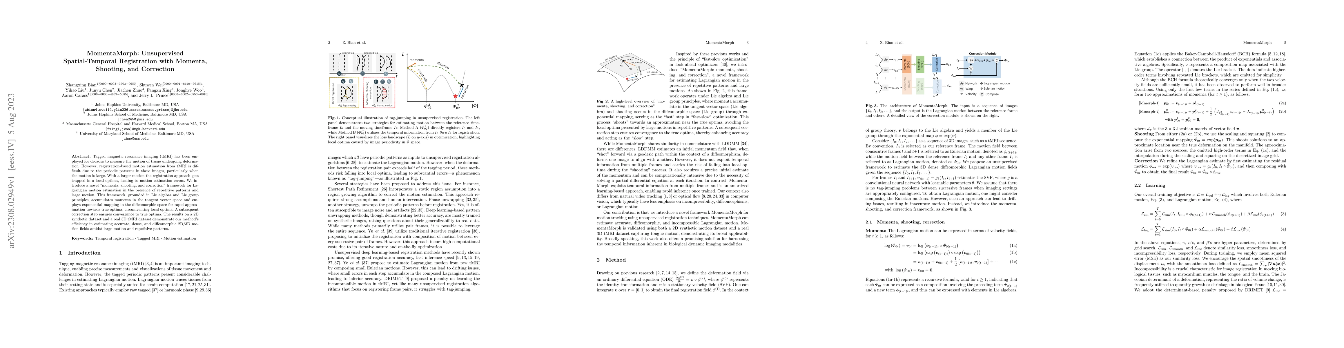
Tagged magnetic resonance imaging (tMRI) has been employed for decades to measure the motion of tissue undergoing deformation. However, registration-based motion estimation from tMRI is difficult du...
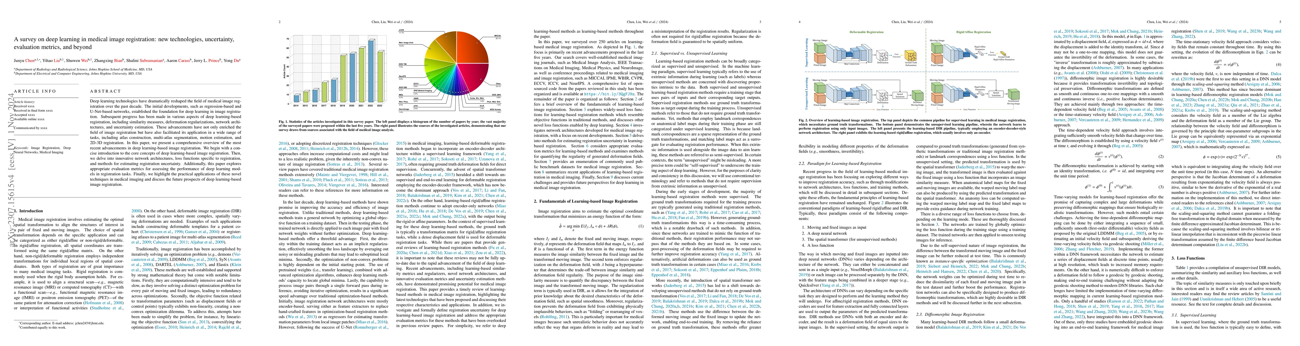
Deep learning technologies have dramatically reshaped the field of medical image registration over the past decade. The initial developments, such as regression-based and U-Net-based networks, estab...
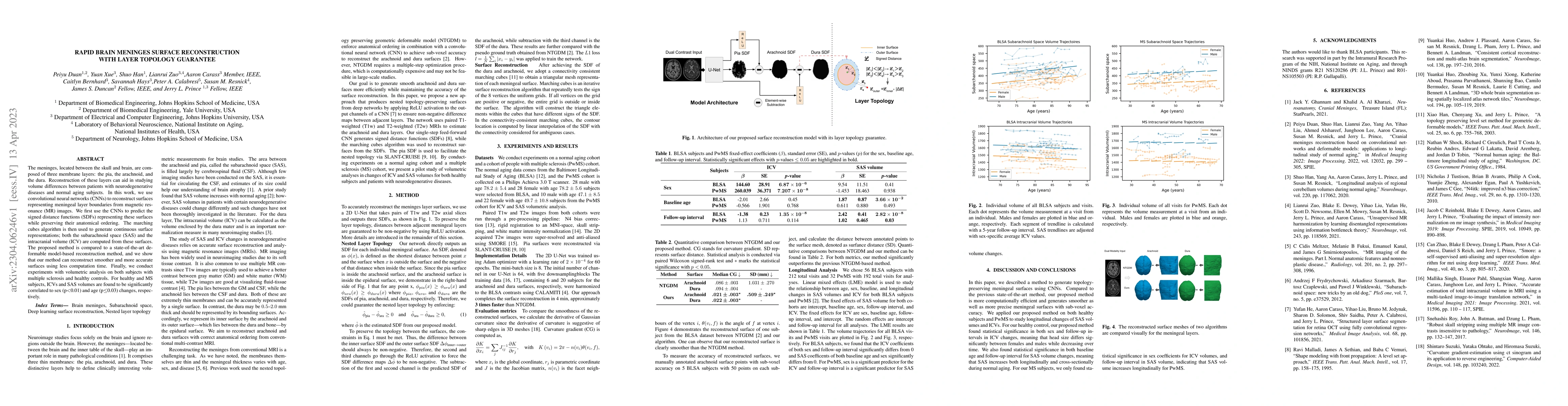
The meninges, located between the skull and brain, are composed of three membrane layers: the pia, the arachnoid, and the dura. Reconstruction of these layers can aid in studying volume differences ...
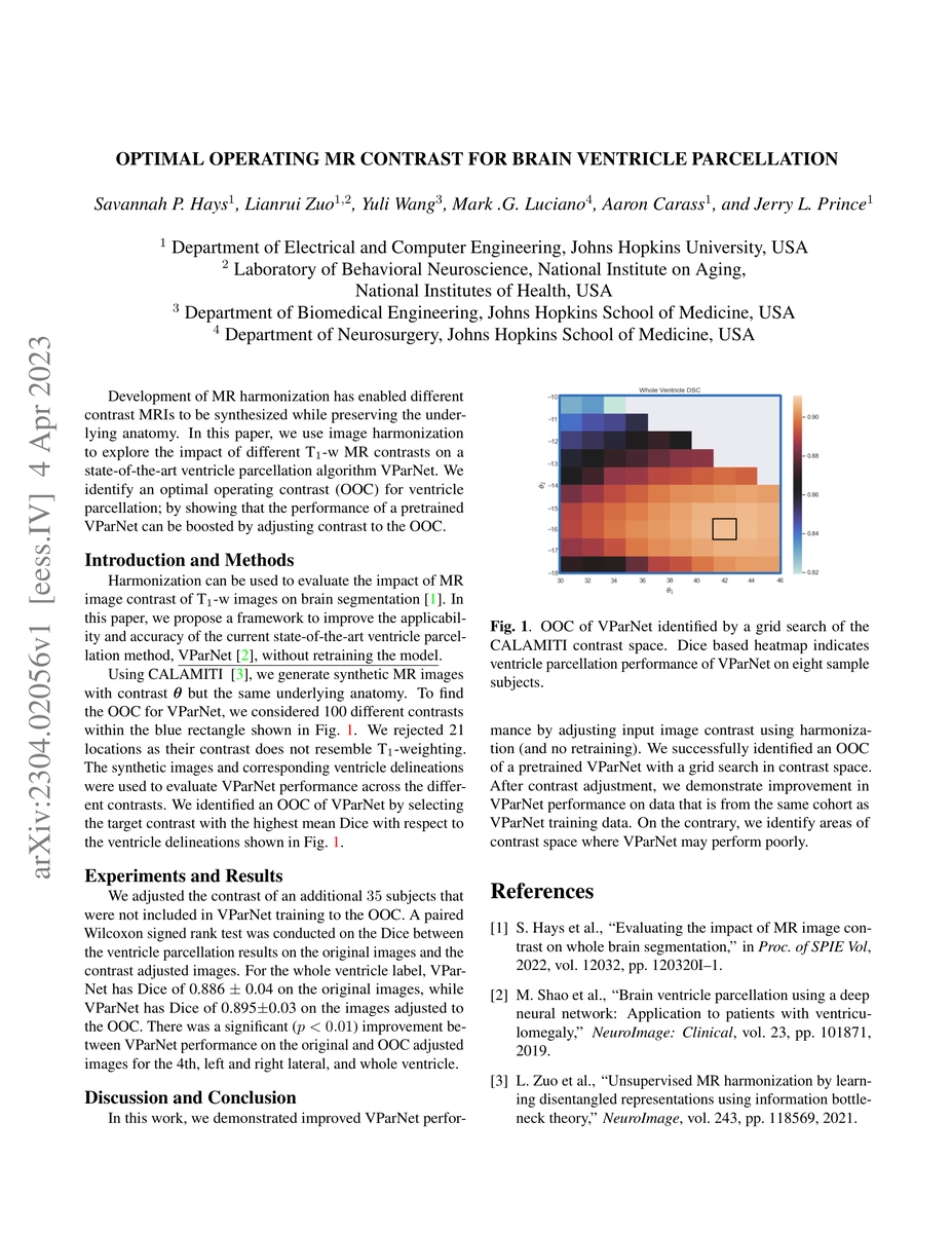
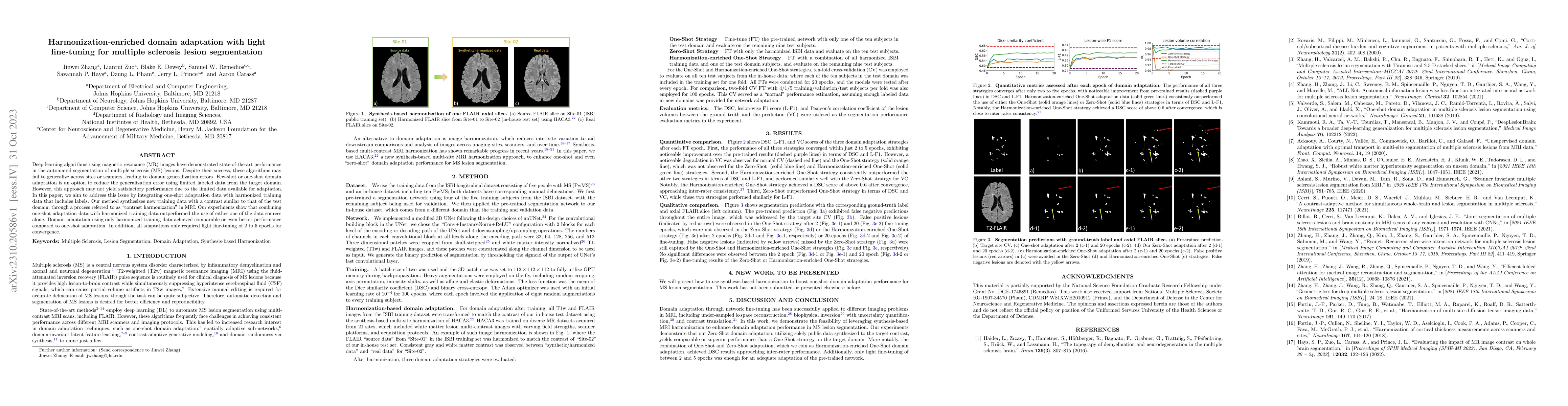
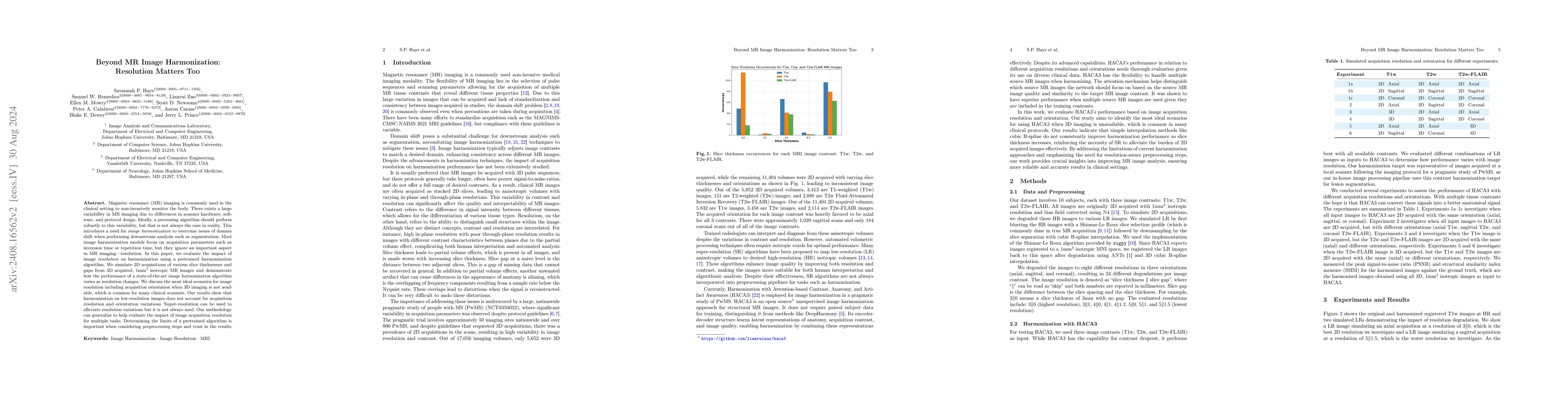
Development of MR harmonization has enabled different contrast MRIs to be synthesized while preserving the underlying anatomy. In this paper, we use image harmonization to explore the impact of diff...
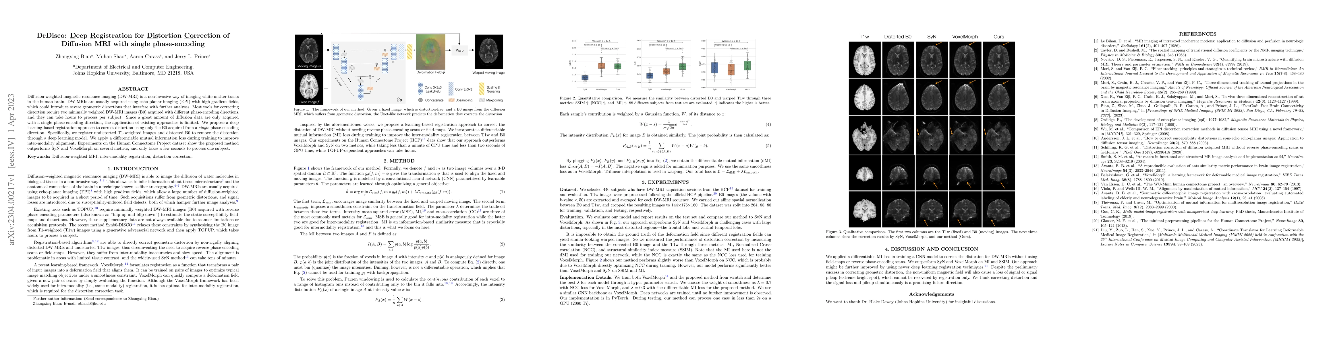
Diffusion-weighted magnetic resonance imaging (DW-MRI) is a non-invasive way of imaging white matter tracts in the human brain. DW-MRIs are usually acquired using echo-planar imaging (EPI) with high...
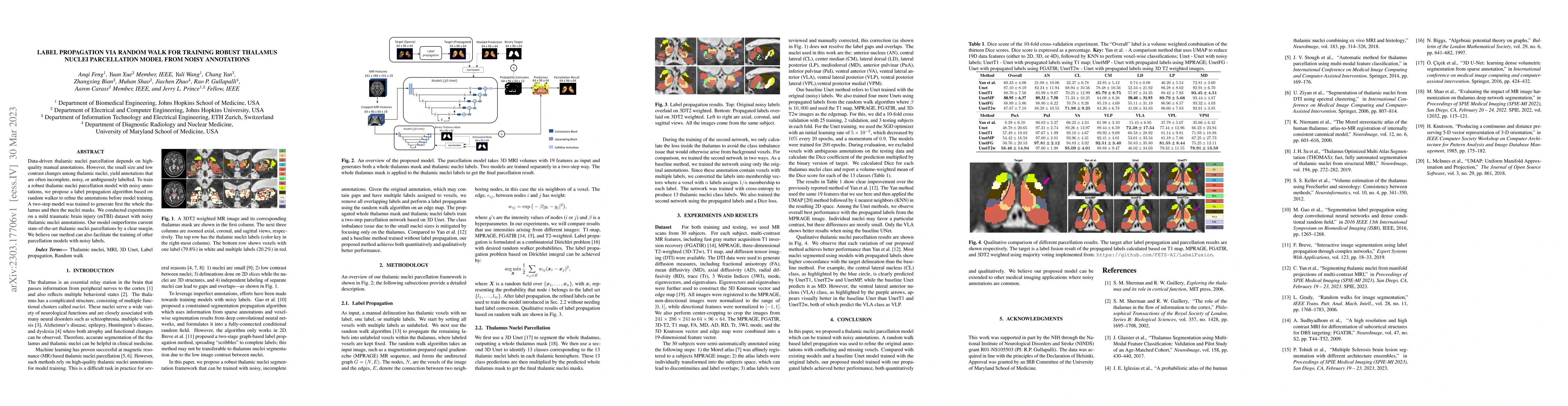
Data-driven thalamic nuclei parcellation depends on high-quality manual annotations. However, the small size and low contrast changes among thalamic nuclei, yield annotations that are often incomple...
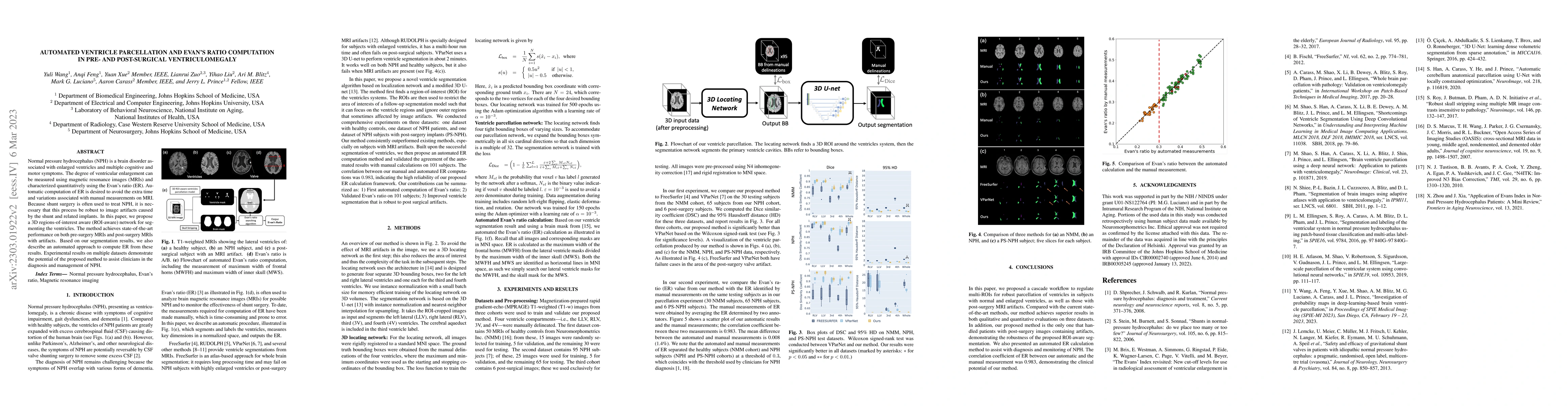
Normal pressure hydrocephalus~(NPH) is a brain disorder associated with enlarged ventricles and multiple cognitive and motor symptoms. The degree of ventricular enlargement can be measured using mag...
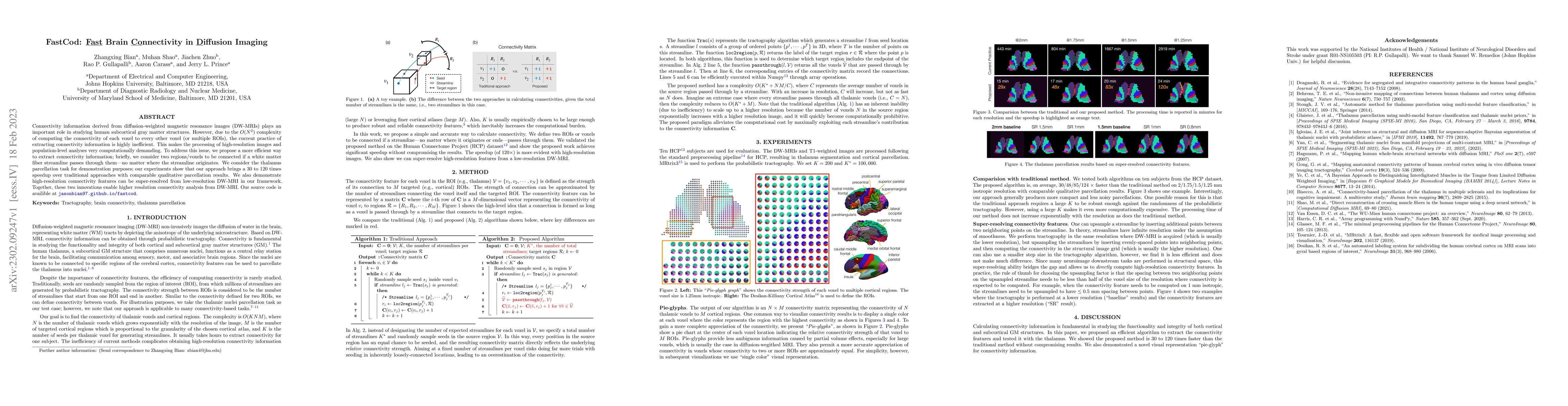
Connectivity information derived from diffusion-weighted magnetic resonance images~(DW-MRIs) plays an important role in studying human subcortical gray matter structures. However, due to the $O(N^2)...
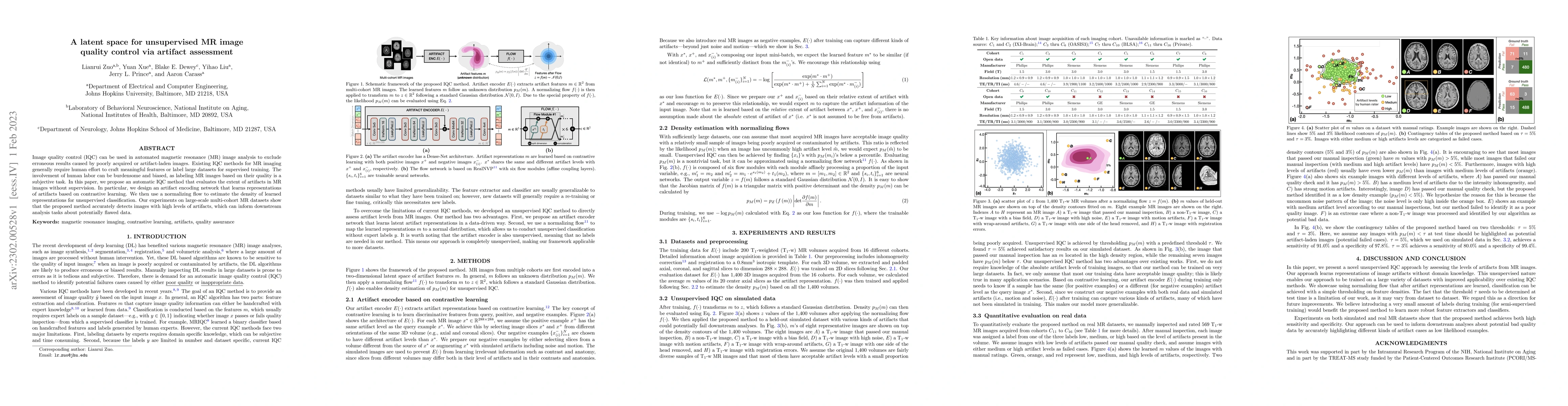
Image quality control (IQC) can be used in automated magnetic resonance (MR) image analysis to exclude erroneous results caused by poorly acquired or artifact-laden images. Existing IQC methods for ...
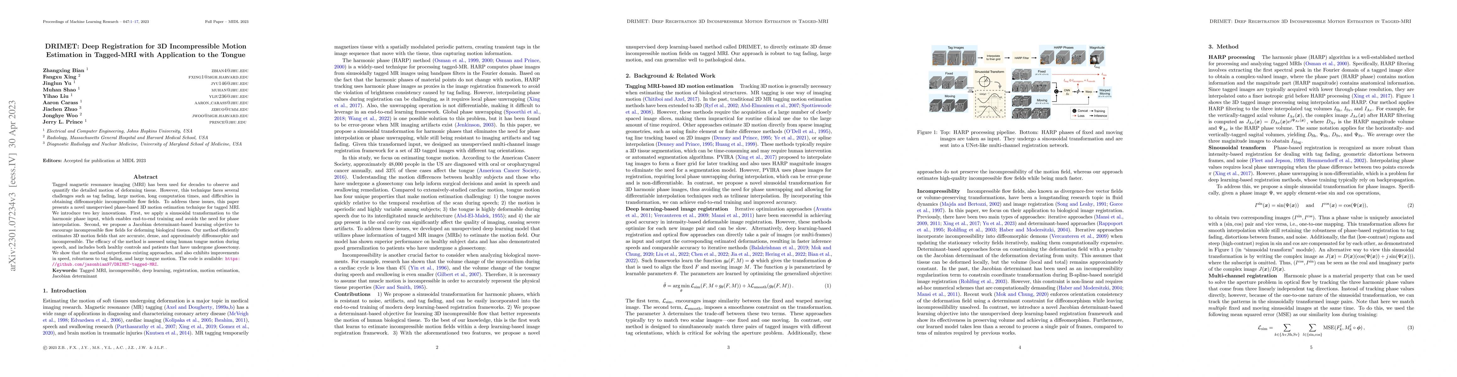
Tagged magnetic resonance imaging~(MRI) has been used for decades to observe and quantify the detailed motion of deforming tissue. However, this technique faces several challenges such as tag fading...
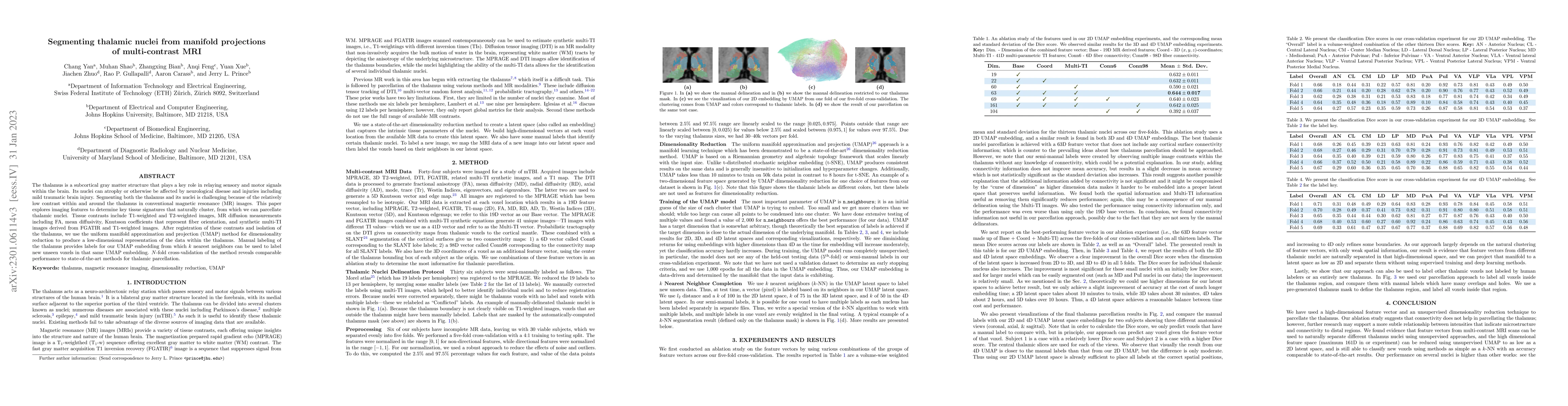
The thalamus is a subcortical gray matter structure that plays a key role in relaying sensory and motor signals within the brain. Its nuclei can atrophy or otherwise be affected by neurological dise...
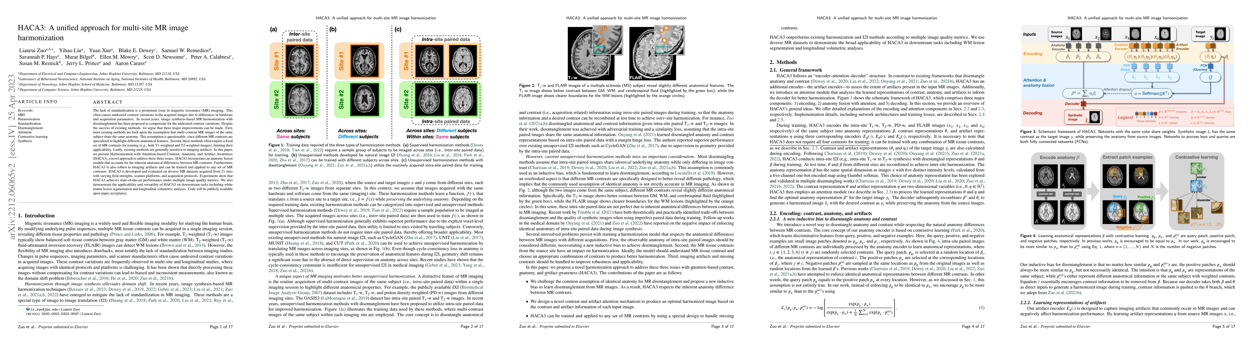
The lack of standardization is a prominent issue in magnetic resonance (MR) imaging. This often causes undesired contrast variations in the acquired images due to differences in hardware and acquisi...
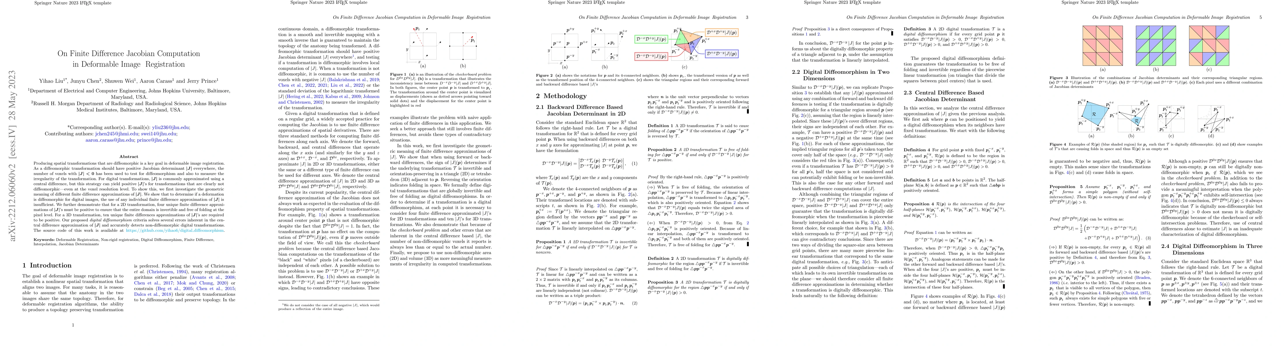
Producing spatial transformations that are diffeomorphic is a key goal in deformable image registration. As a diffeomorphic transformation should have positive Jacobian determinant |J| everywhere, t...
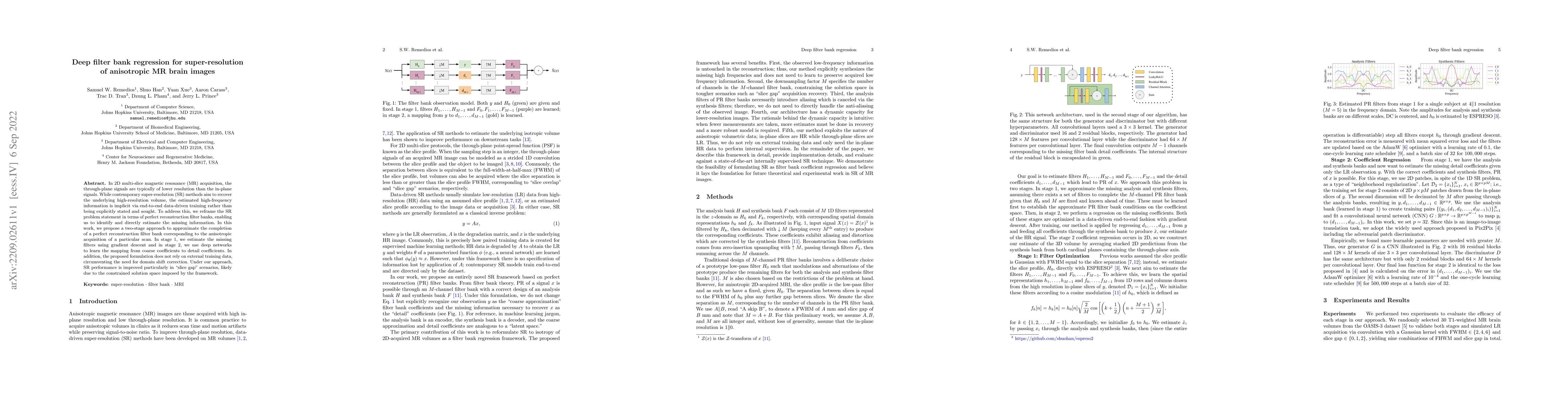
In 2D multi-slice magnetic resonance (MR) acquisition, the through-plane signals are typically of lower resolution than the in-plane signals. While contemporary super-resolution (SR) methods aim to ...
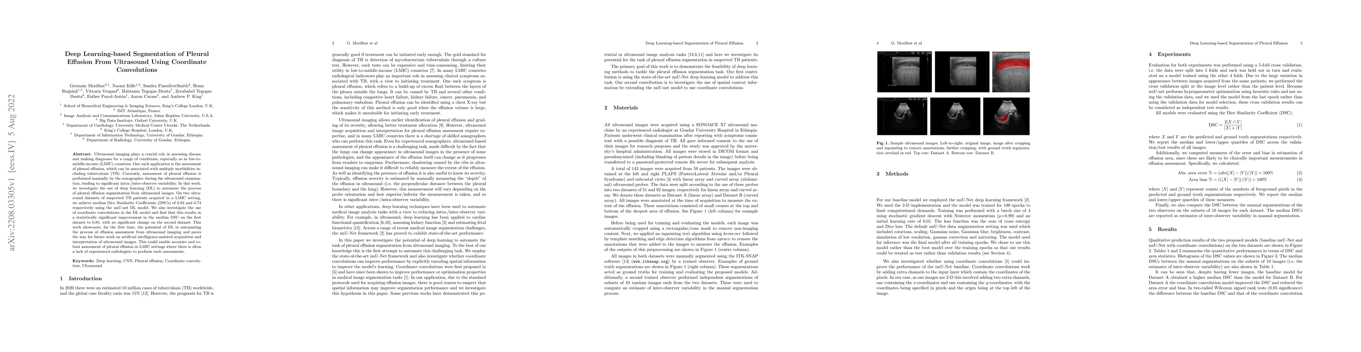
In many low-to-middle income (LMIC) countries, ultrasound is used for assessment of pleural effusion. Typically, the extent of the effusion is manually measured by a sonographer, leading to signific...
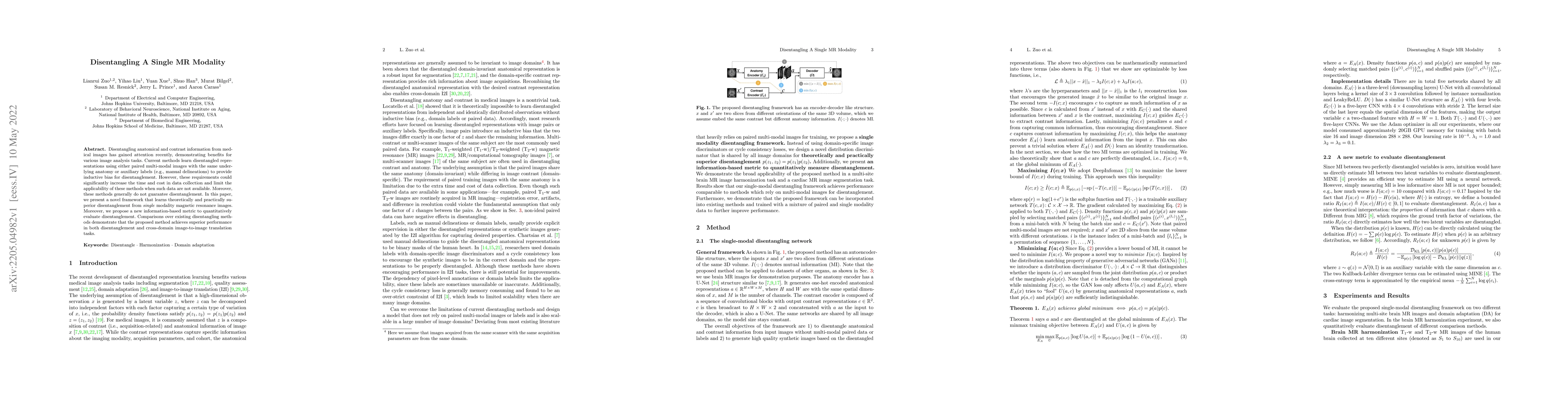
Disentangling anatomical and contrast information from medical images has gained attention recently, demonstrating benefits for various image analysis tasks. Current methods learn disentangled repre...
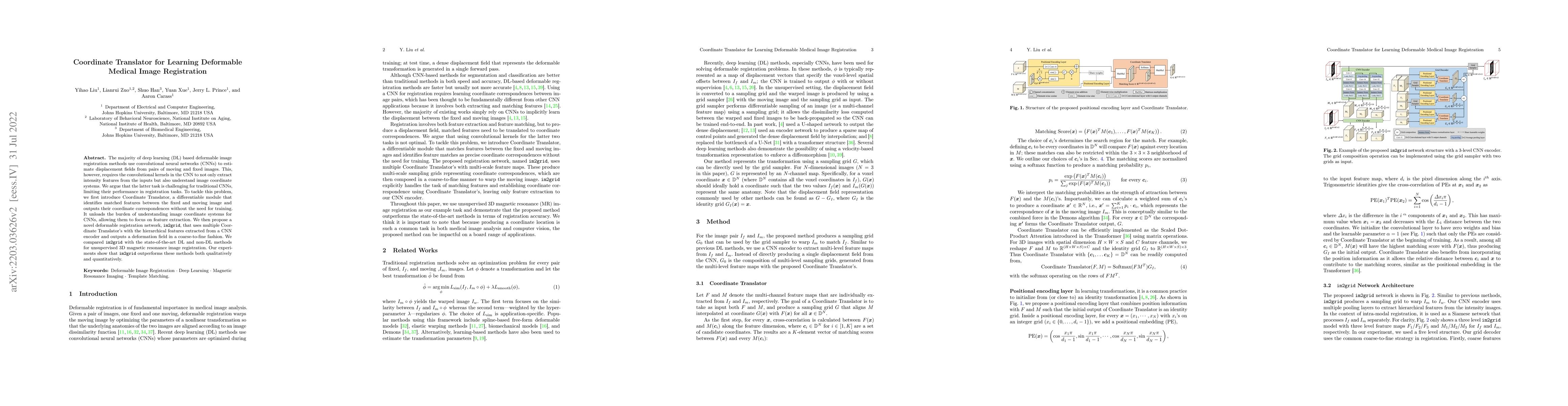
The majority of deep learning (DL) based deformable image registration methods use convolutional neural networks (CNNs) to estimate displacement fields from pairs of moving and fixed images. This, h...
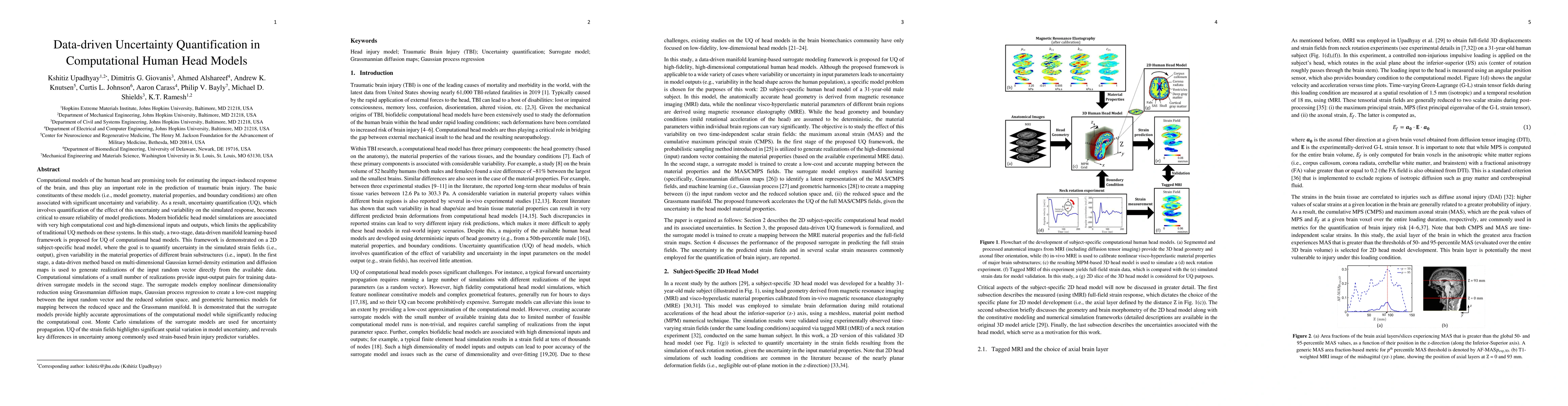
Computational models of the human head are promising tools for estimating the impact-induced response of brain, and thus play an important role in the prediction of traumatic brain injury. Modern bi...
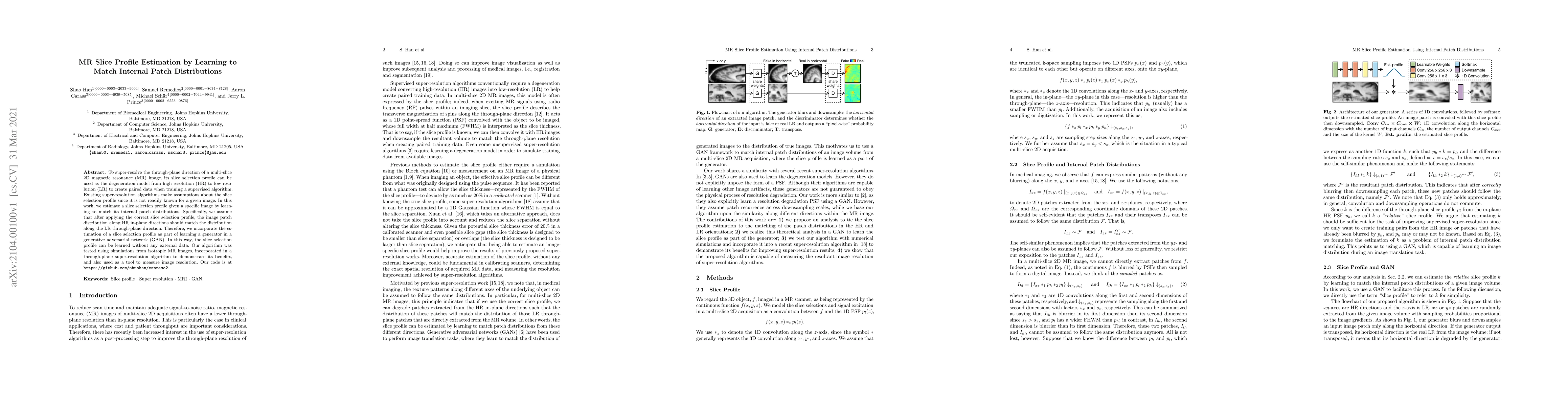
To super-resolve the through-plane direction of a multi-slice 2D magnetic resonance (MR) image, its slice selection profile can be used as the degeneration model from high resolution (HR) to low res...
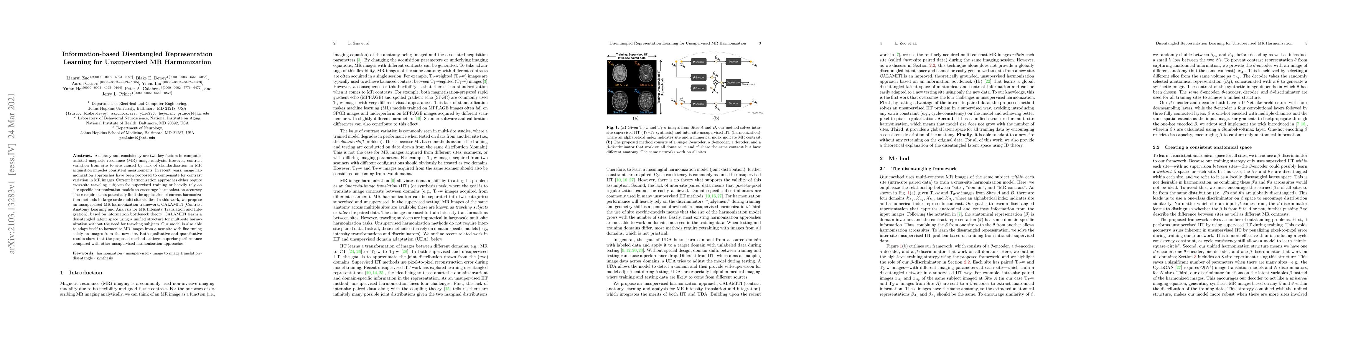
Accuracy and consistency are two key factors in computer-assisted magnetic resonance (MR) image analysis. However, contrast variation from site to site caused by lack of standardization in MR acquis...
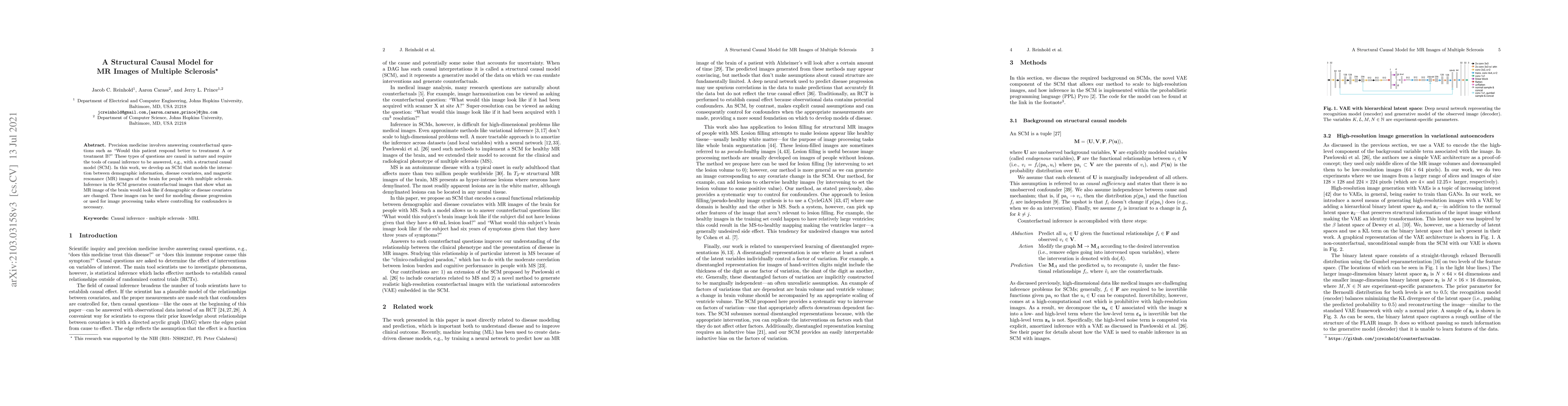
Precision medicine involves answering counterfactual questions such as "Would this patient respond better to treatment A or treatment B?" These types of questions are causal in nature and require th...
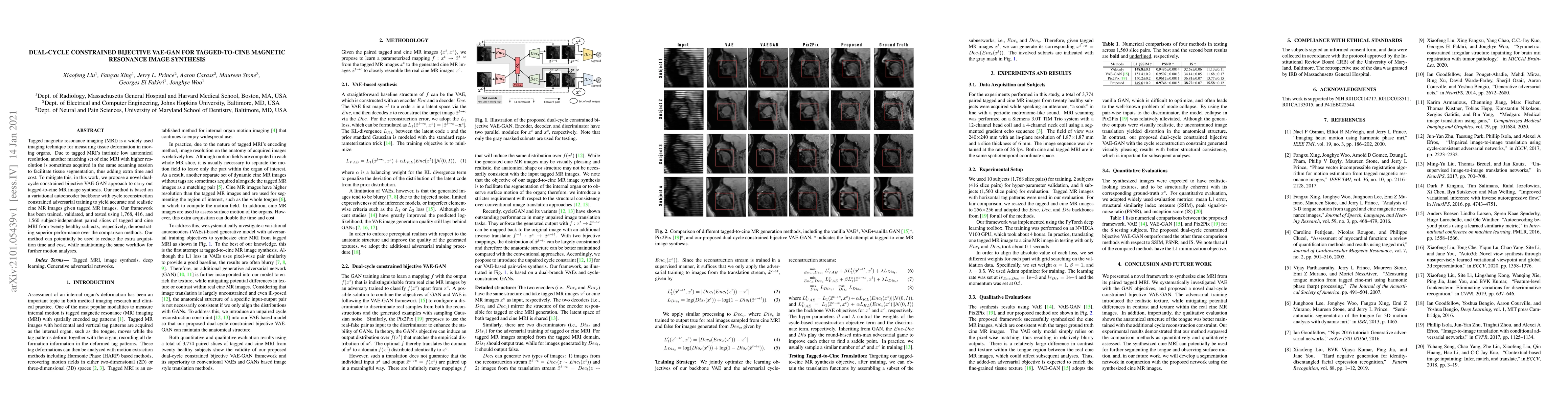
Tagged magnetic resonance imaging (MRI) is a widely used imaging technique for measuring tissue deformation in moving organs. Due to tagged MRI's intrinsic low anatomical resolution, another matchin...
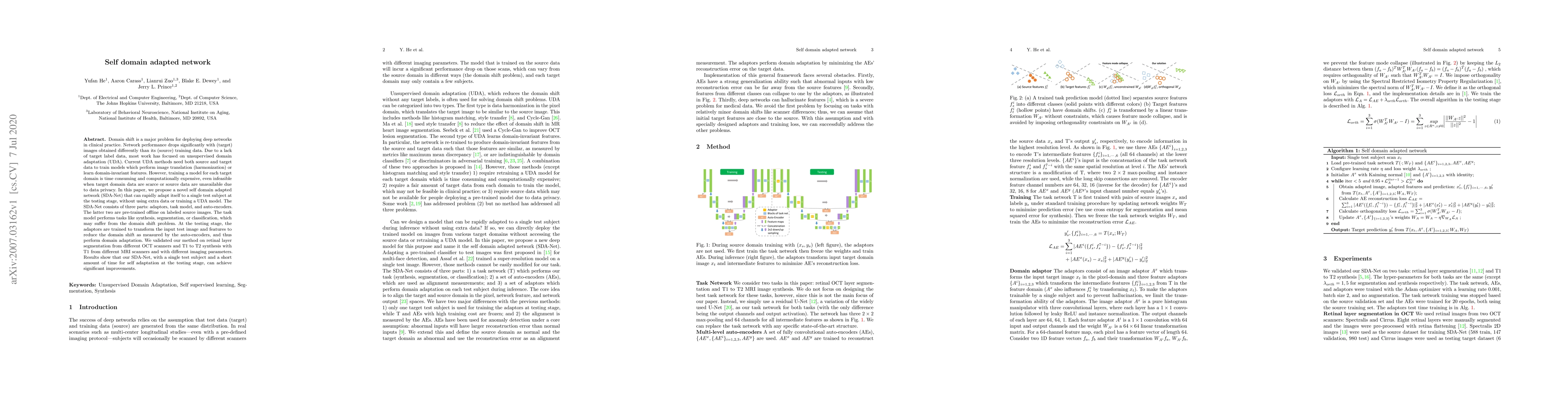
Domain shift is a major problem for deploying deep networks in clinical practice. Network performance drops significantly with (target) images obtained differently than its (source) training data. D...
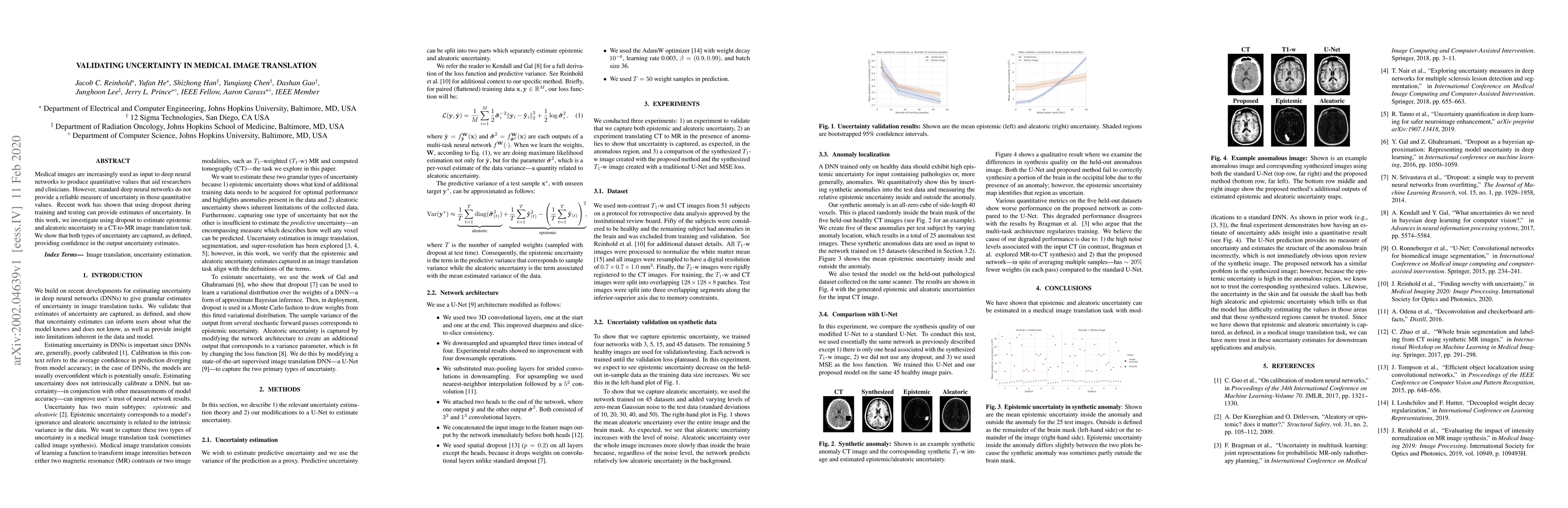
Medical images are increasingly used as input to deep neural networks to produce quantitative values that aid researchers and clinicians. However, standard deep neural networks do not provide a reli...
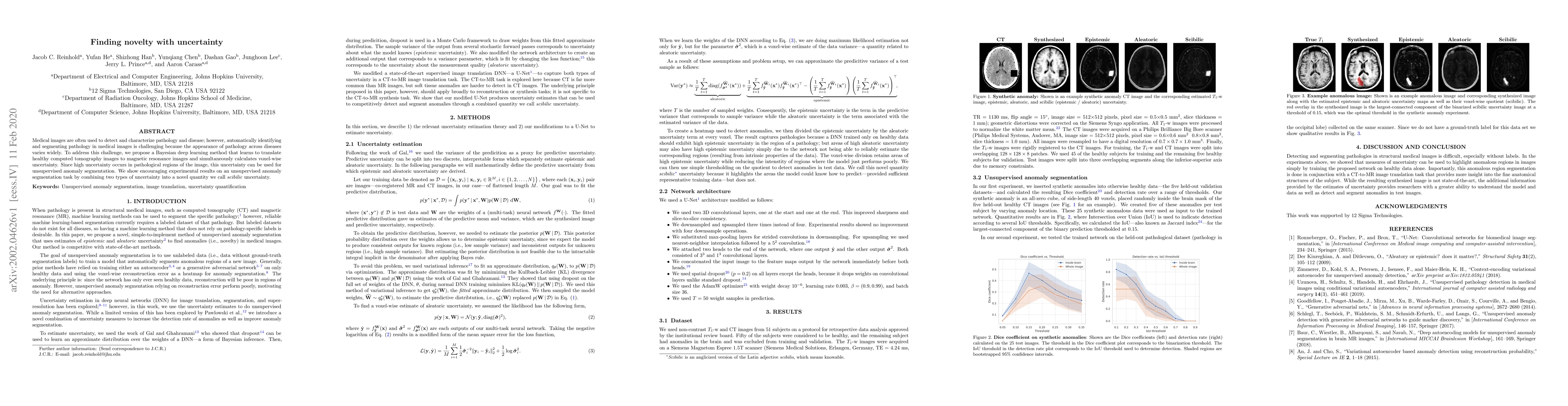
Medical images are often used to detect and characterize pathology and disease; however, automatically identifying and segmenting pathology in medical images is challenging because the appearance of...
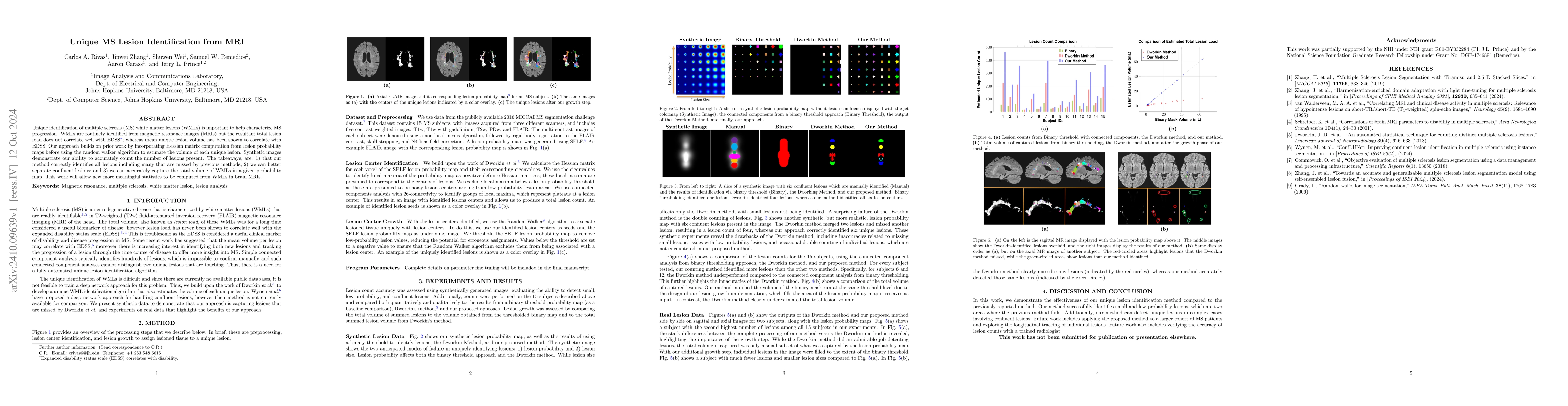
Unique identification of multiple sclerosis (MS) white matter lesions (WMLs) is important to help characterize MS progression. WMLs are routinely identified from magnetic resonance images (MRIs) but t...
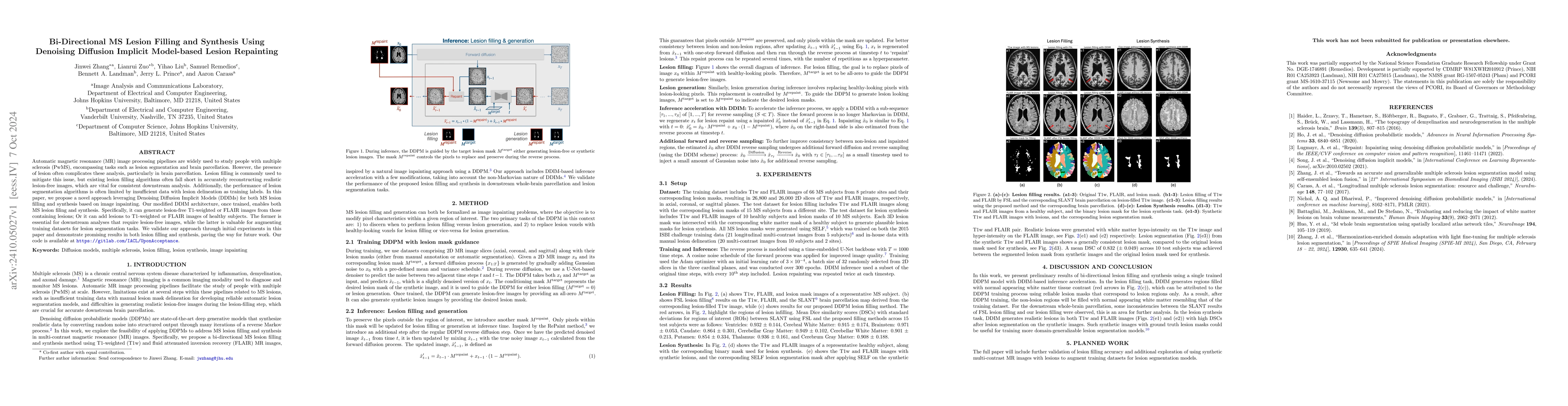
Automatic magnetic resonance (MR) image processing pipelines are widely used to study people with multiple sclerosis (PwMS), encompassing tasks such as lesion segmentation and brain parcellation. Howe...
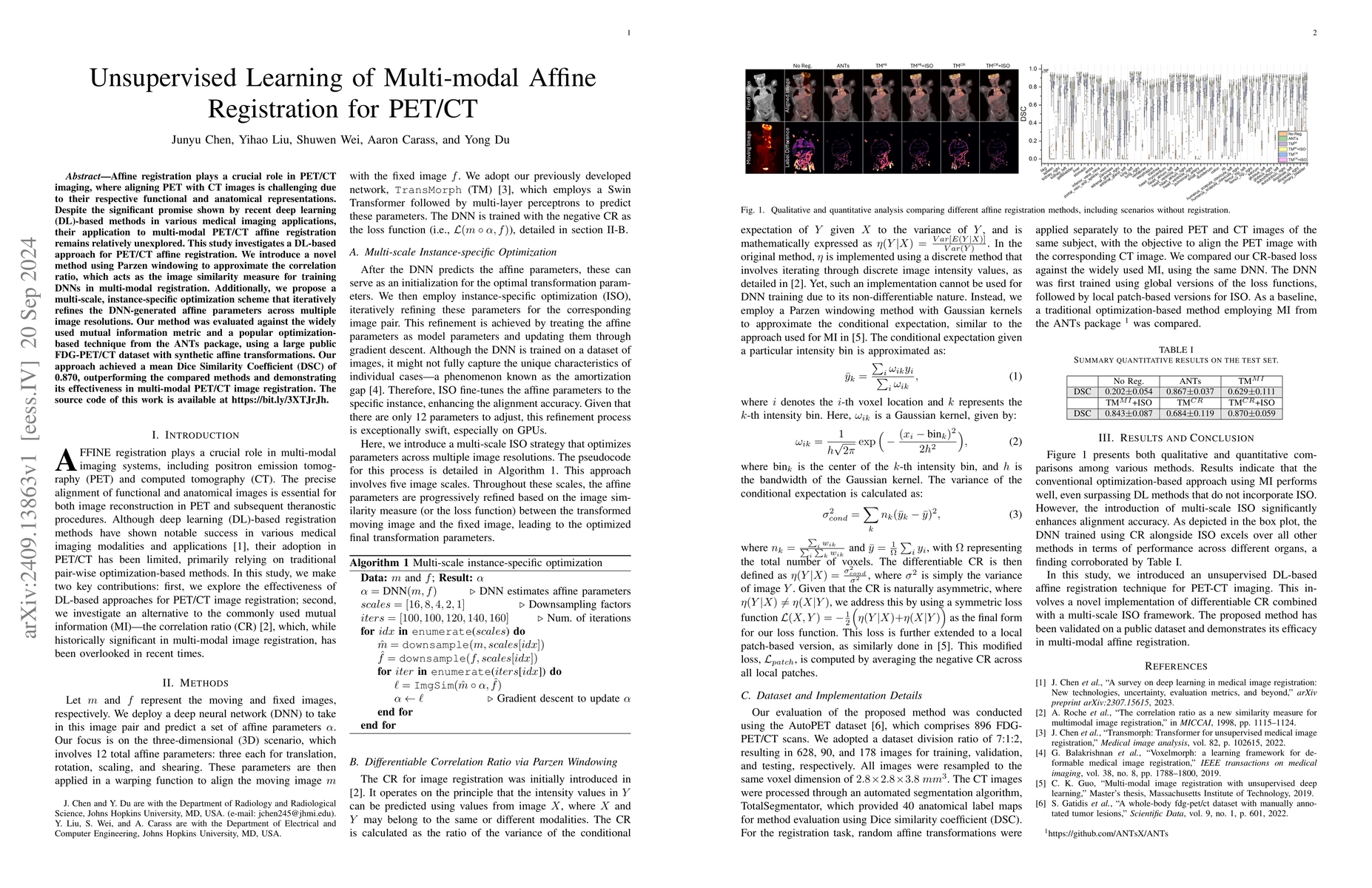
Affine registration plays a crucial role in PET/CT imaging, where aligning PET with CT images is challenging due to their respective functional and anatomical representations. Despite the significant ...
Magnetic resonance (MR) imaging is commonly used in the clinical setting to non-invasively monitor the body. There exists a large variability in MR imaging due to differences in scanner hardware, soft...
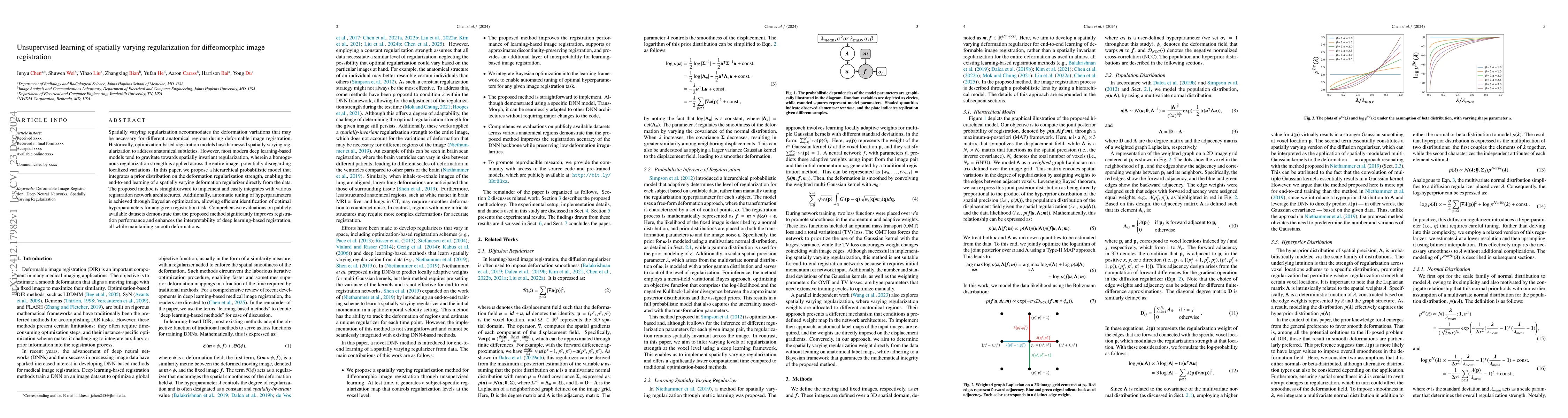
Spatially varying regularization accommodates the deformation variations that may be necessary for different anatomical regions during deformable image registration. Historically, optimization-based r...
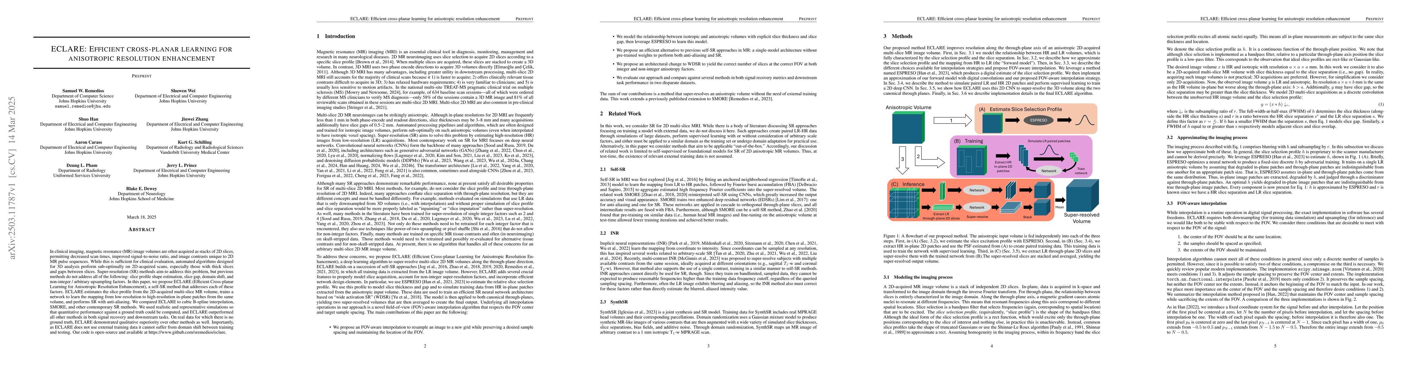
In clinical imaging, magnetic resonance (MR) image volumes are often acquired as stacks of 2D slices, permitting decreased scan times, improved signal-to-noise ratio, and image contrasts unique to 2D ...
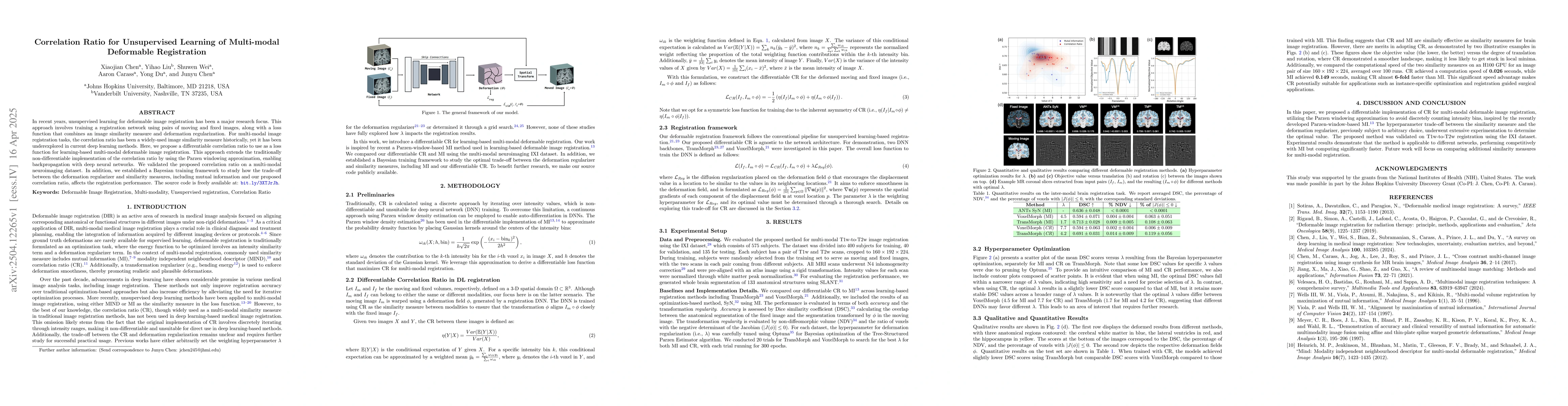
In recent years, unsupervised learning for deformable image registration has been a major research focus. This approach involves training a registration network using pairs of moving and fixed images,...
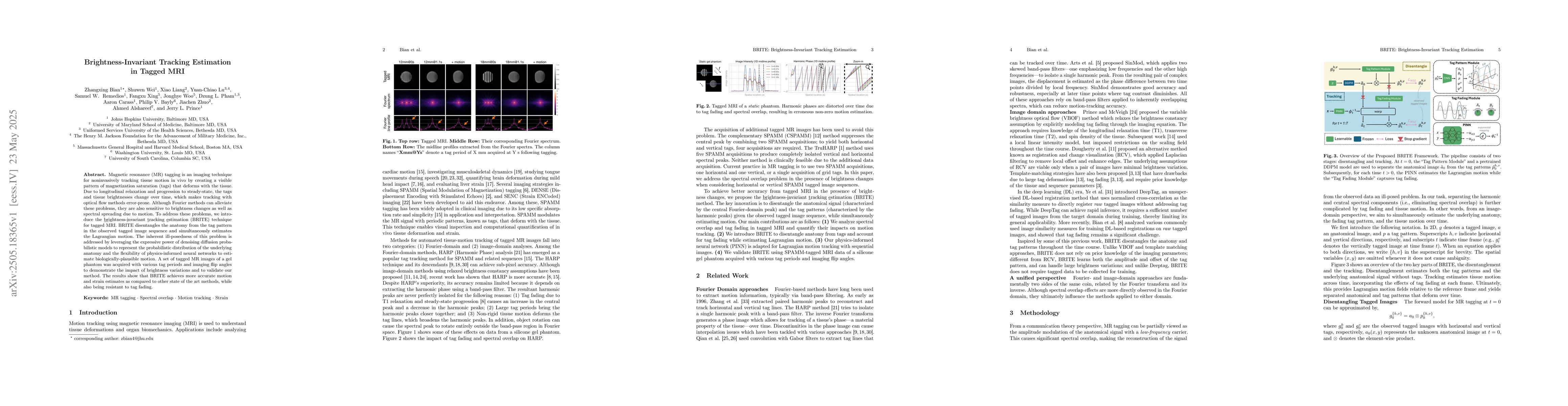
Magnetic resonance (MR) tagging is an imaging technique for noninvasively tracking tissue motion in vivo by creating a visible pattern of magnetization saturation (tags) that deforms with the tissue. ...
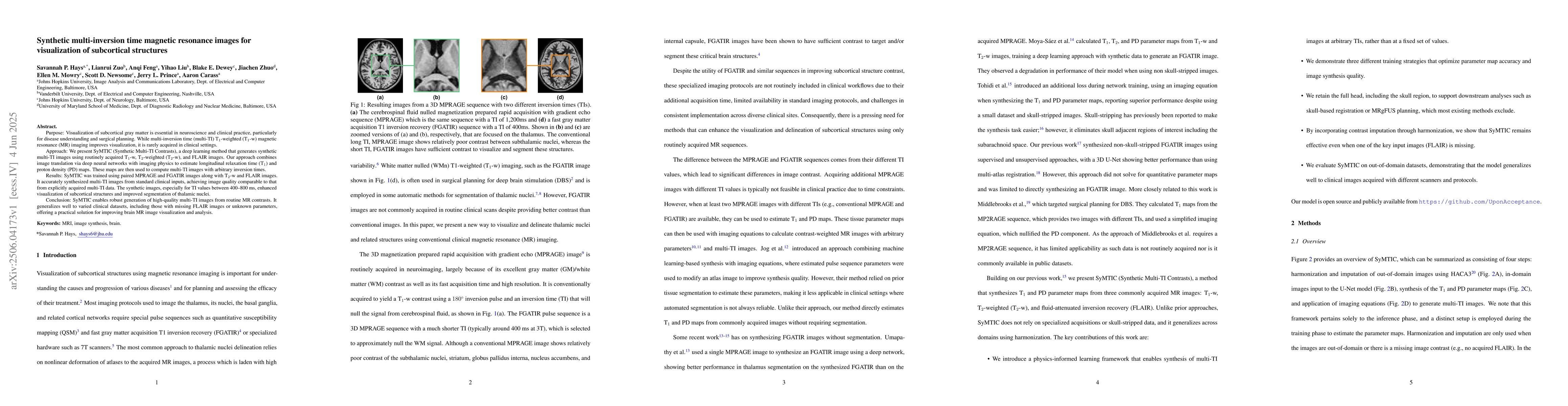
Purpose: Visualization of subcortical gray matter is essential in neuroscience and clinical practice, particularly for disease understanding and surgical planning.While multi-inversion time (multi-TI)...
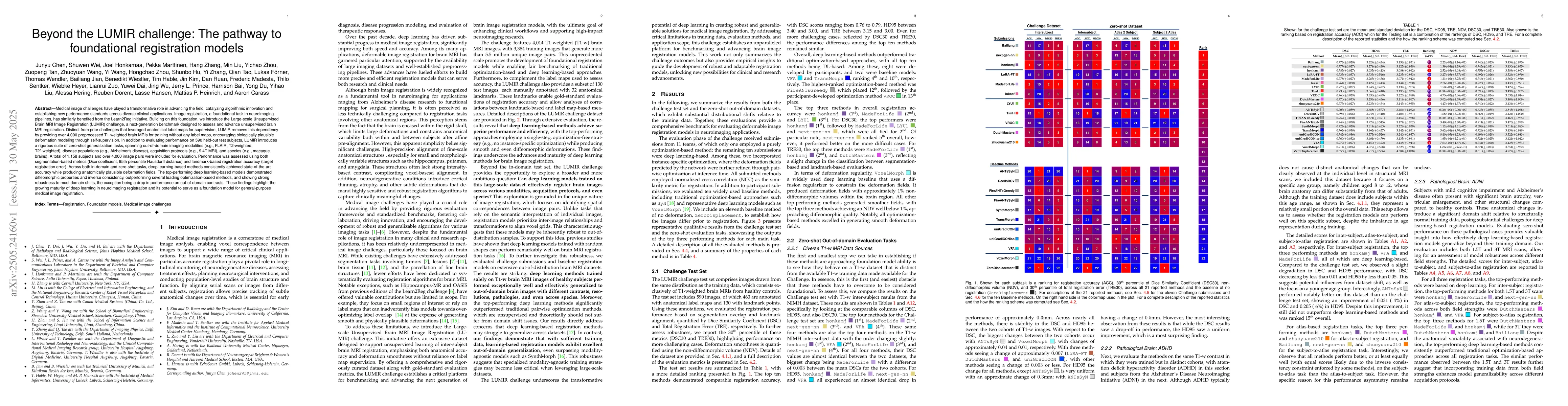
Medical image challenges have played a transformative role in advancing the field, catalyzing algorithmic innovation and establishing new performance standards across diverse clinical applications. Im...
Recent advances in deep learning-based medical image registration have shown that training deep neural networks~(DNNs) does not necessarily require medical images. Previous work showed that DNNs train...
Automated segmentation of multiple sclerosis (MS) lesions using multicontrast magnetic resonance (MR) images improves efficiency and reproducibility compared to manual delineation, with deep learning ...
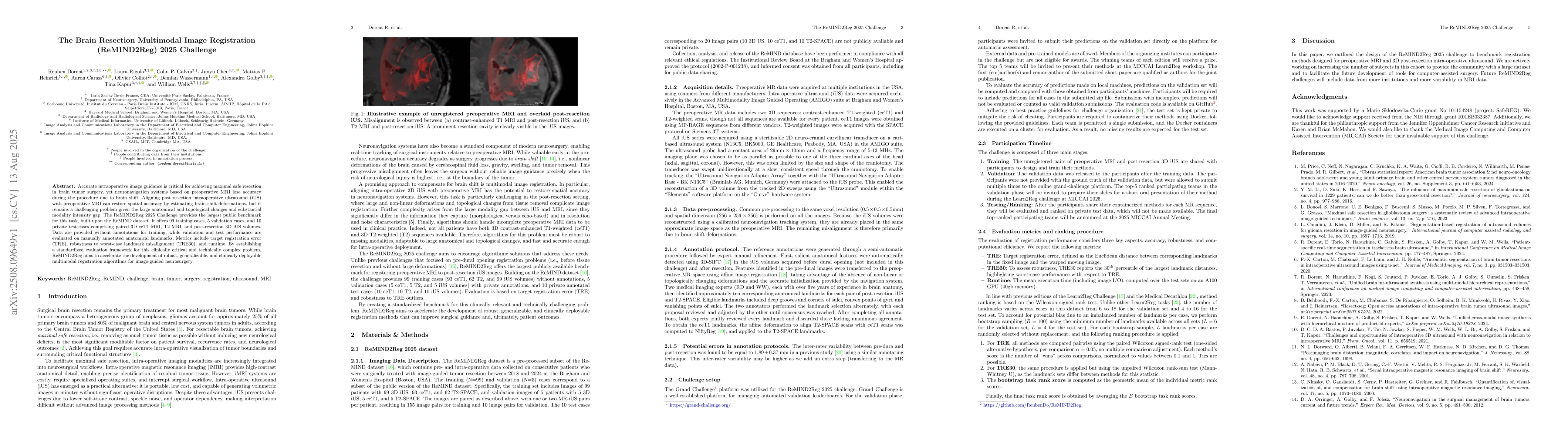
Accurate intraoperative image guidance is critical for achieving maximal safe resection in brain tumor surgery, yet neuronavigation systems based on preoperative MRI lose accuracy during the procedure...
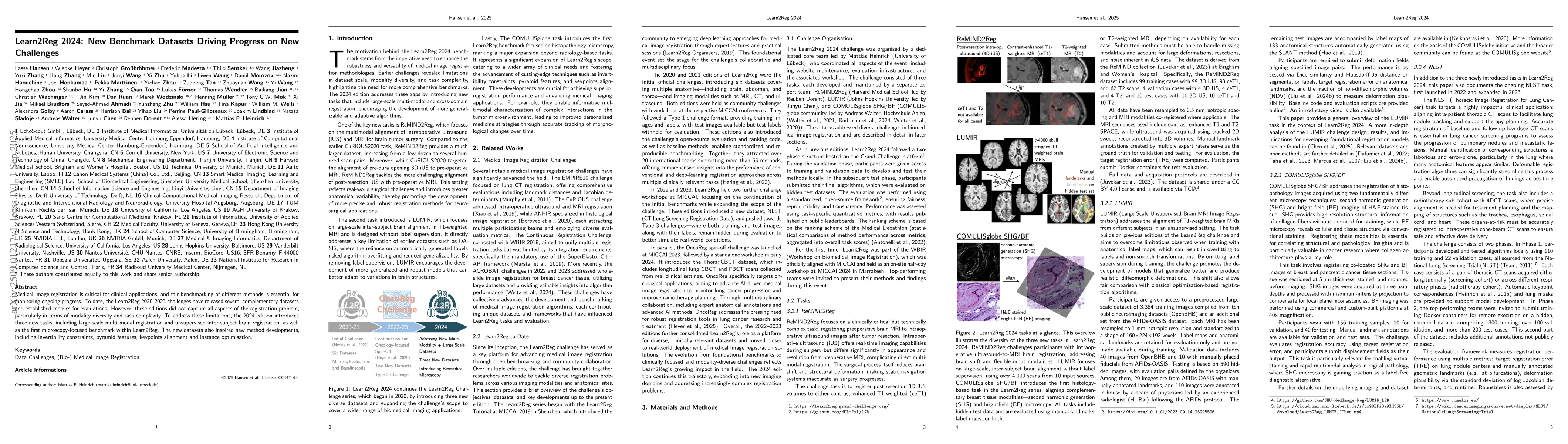
Medical image registration is critical for clinical applications, and fair benchmarking of different methods is essential for monitoring ongoing progress. To date, the Learn2Reg 2020-2023 challenges h...
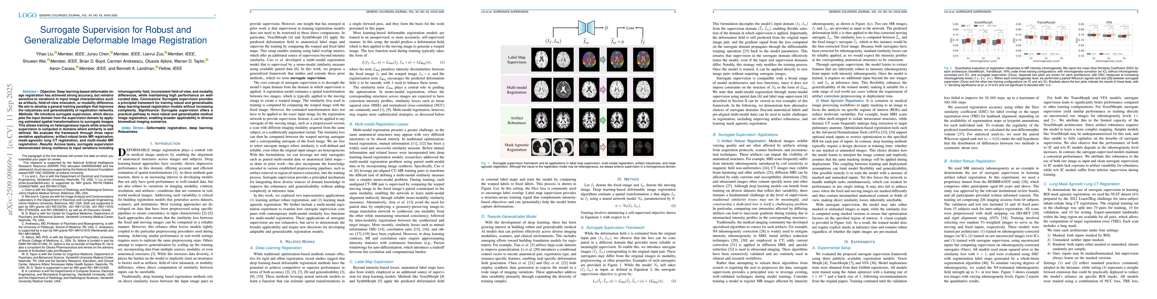
Objective: Deep learning-based deformable image registration has achieved strong accuracy, but remains sensitive to variations in input image characteristics such as artifacts, field-of-view mismatch,...
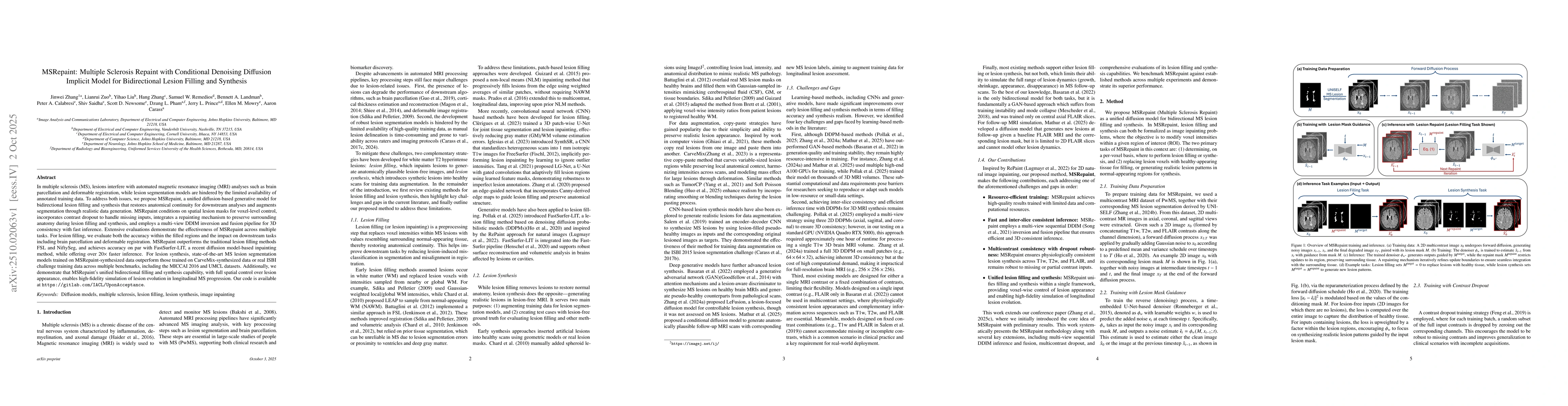
In multiple sclerosis, lesions interfere with automated magnetic resonance imaging analyses such as brain parcellation and deformable registration, while lesion segmentation models are hindered by the...
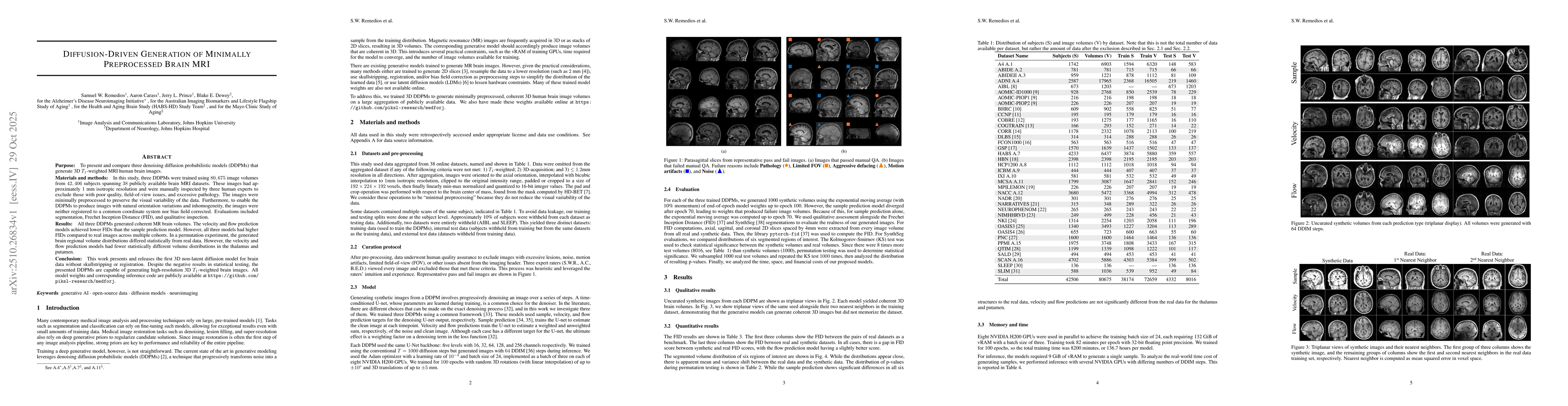
The purpose of this study is to present and compare three denoising diffusion probabilistic models (DDPMs) that generate 3D $T_1$-weighted MRI human brain images. Three DDPMs were trained using 80,675...
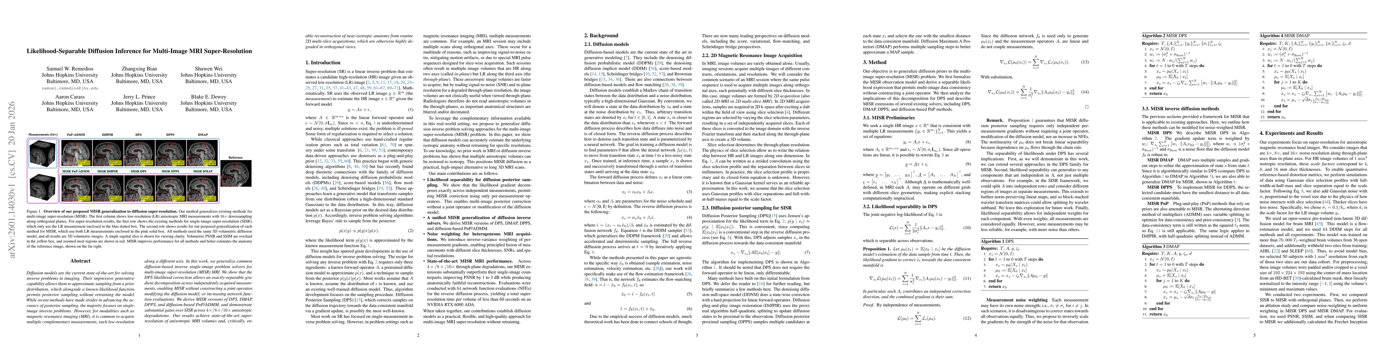
Diffusion models are the current state-of-the-art for solving inverse problems in imaging. Their impressive generative capability allows them to approximate sampling from a prior distribution, which a...
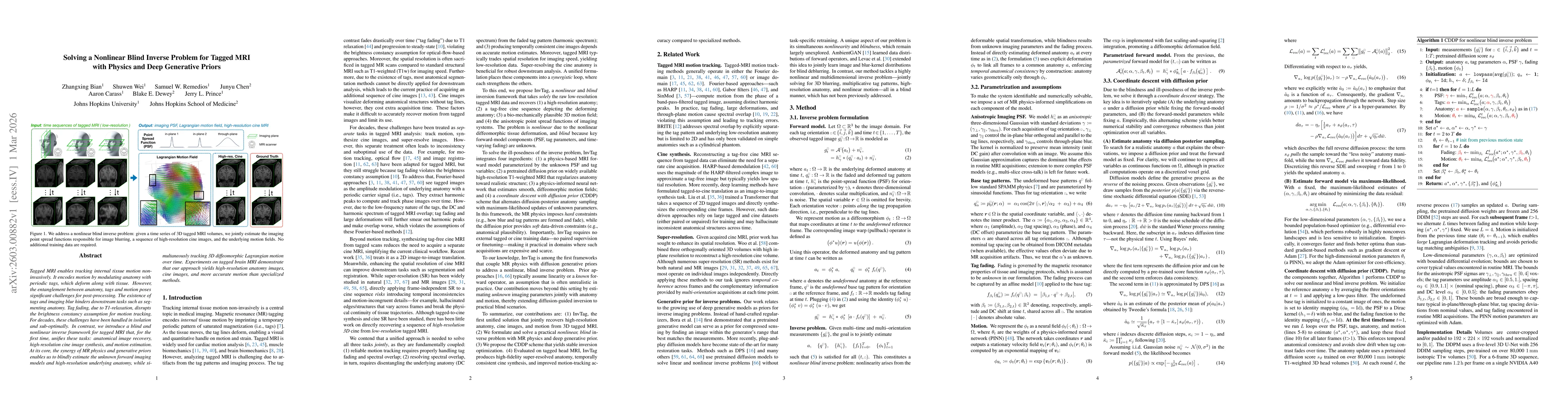
Tagged MRI enables tracking internal tissue motion non-invasively. It encodes motion by modulating anatomy with periodic tags, which deform along with tissue. However, the entanglement between anatomy...
Reliable harmonization of heterogeneous magnetic resonance~(MR) image datasets, especially those acquired in pragmatic clinical trials, is critical to advance multi-center neuroimaging studies and tra...