Academic Profile
Statistics
Similar Authors
Papers on arXiv
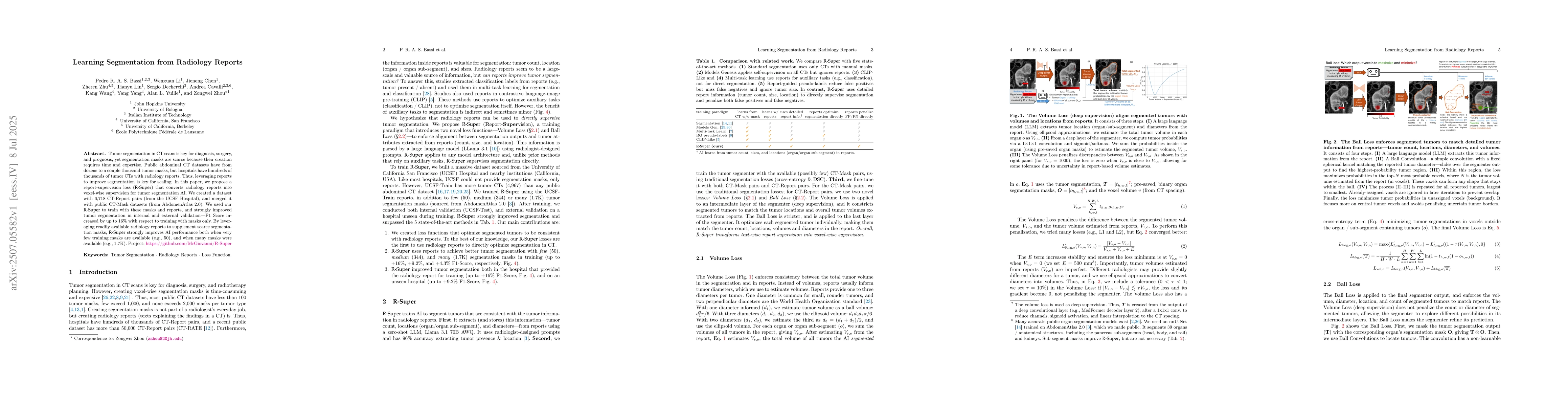
Prior to the deep learning era, shape was commonly used to describe the objects. Nowadays, state-of-the-art (SOTA) algorithms in medical imaging are predominantly diverging from computer vision, whe...
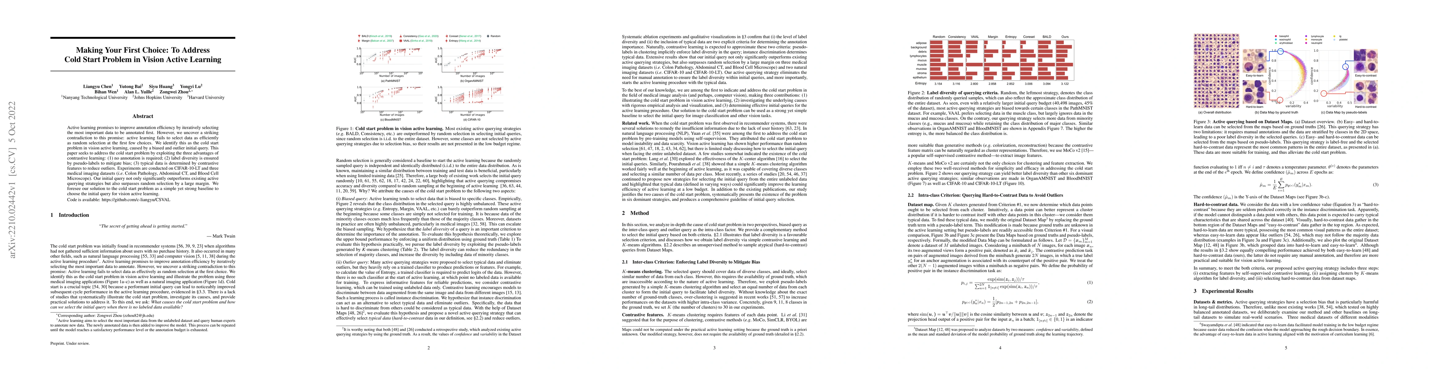
Active learning promises to improve annotation efficiency by iteratively selecting the most important data to be annotated first. However, we uncover a striking contradiction to this promise: active...
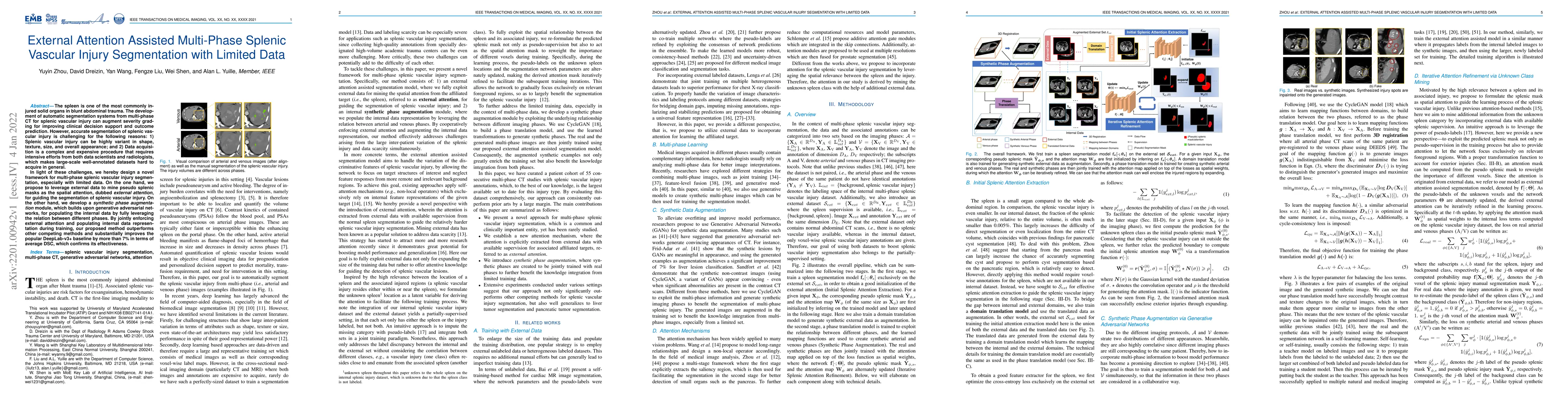
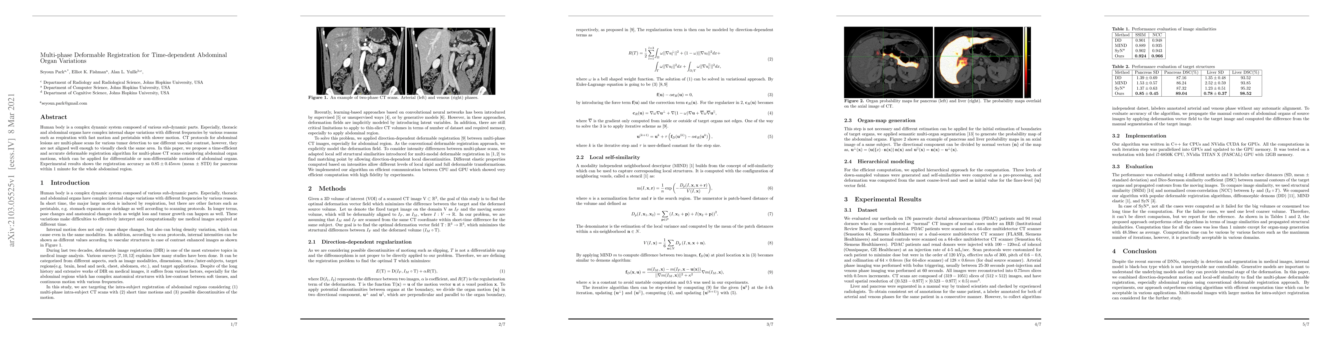
The spleen is one of the most commonly injured solid organs in blunt abdominal trauma. The development of automatic segmentation systems from multi-phase CT for splenic vascular injury can augment s...
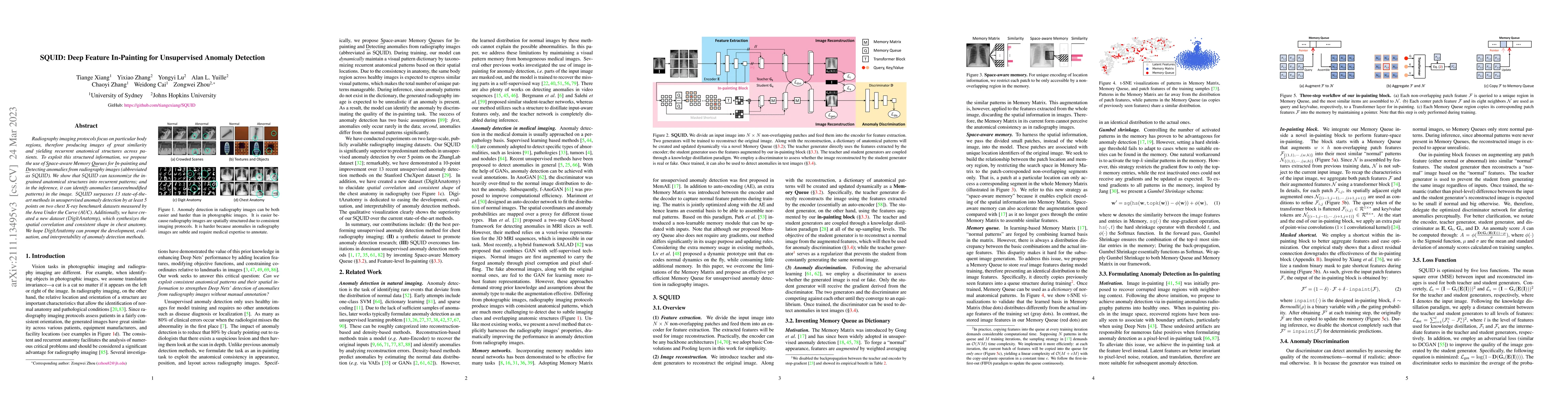
Radiography imaging protocols focus on particular body regions, therefore producing images of great similarity and yielding recurrent anatomical structures across patients. To exploit this structure...
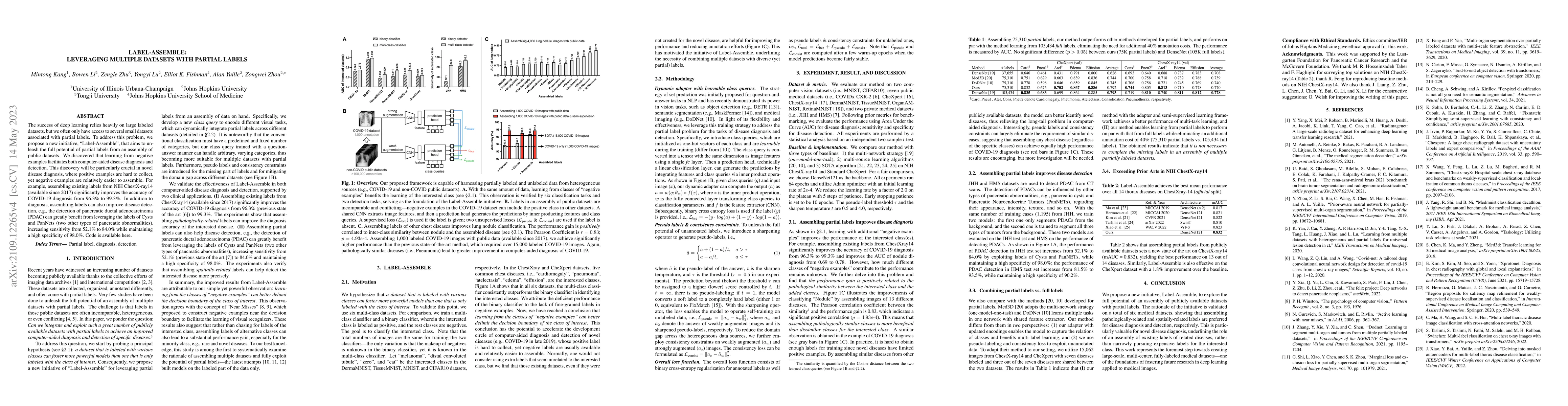
The success of deep learning relies heavily on large labeled datasets, but we often only have access to several small datasets associated with partial labels. To address this problem, we propose a n...
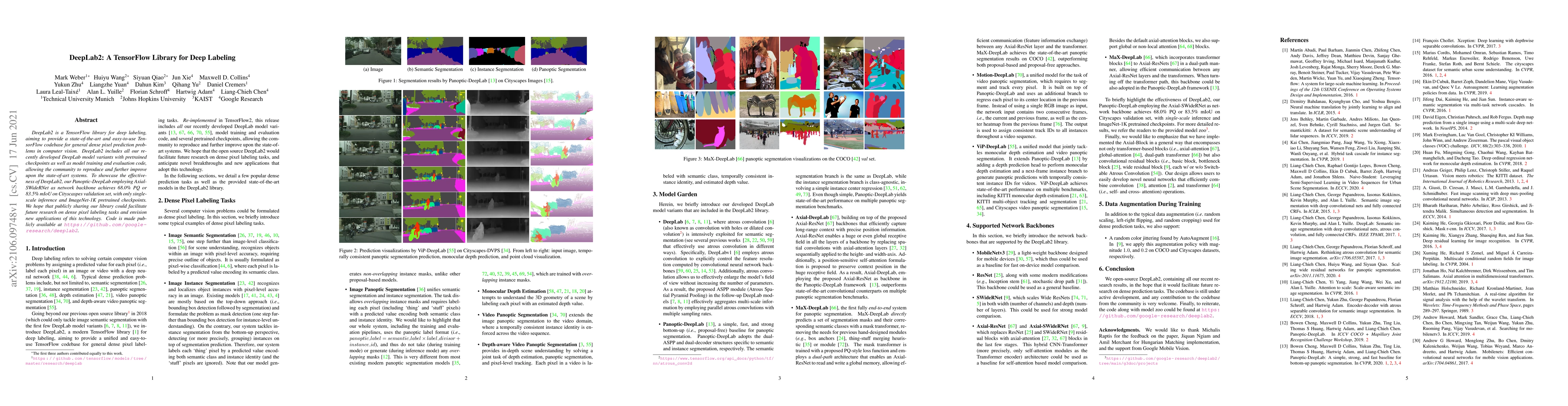
DeepLab2 is a TensorFlow library for deep labeling, aiming to provide a state-of-the-art and easy-to-use TensorFlow codebase for general dense pixel prediction problems in computer vision. DeepLab2 ...
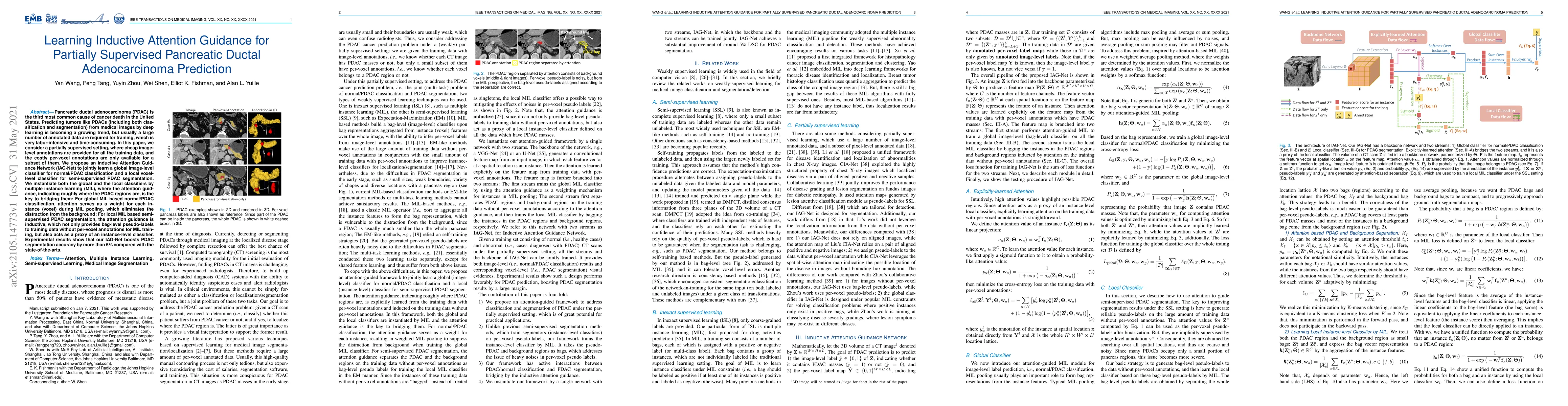
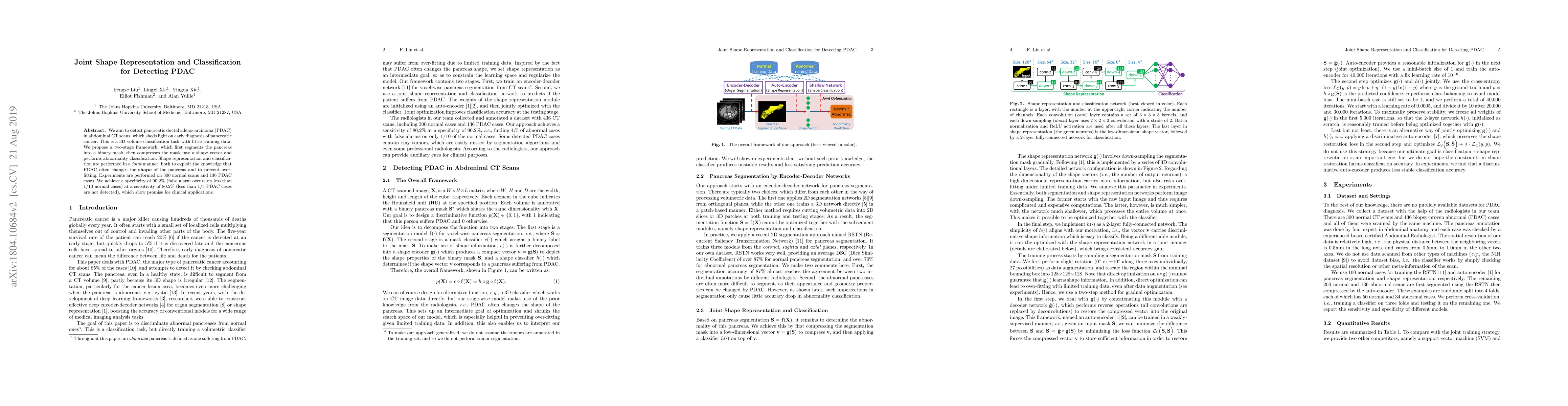
Pancreatic ductal adenocarcinoma (PDAC) is the third most common cause of cancer death in the United States. Predicting tumors like PDACs (including both classification and segmentation) from medica...
Human body is a complex dynamic system composed of various sub-dynamic parts. Especially, thoracic and abdominal organs have complex internal shape variations with different frequencies by various r...
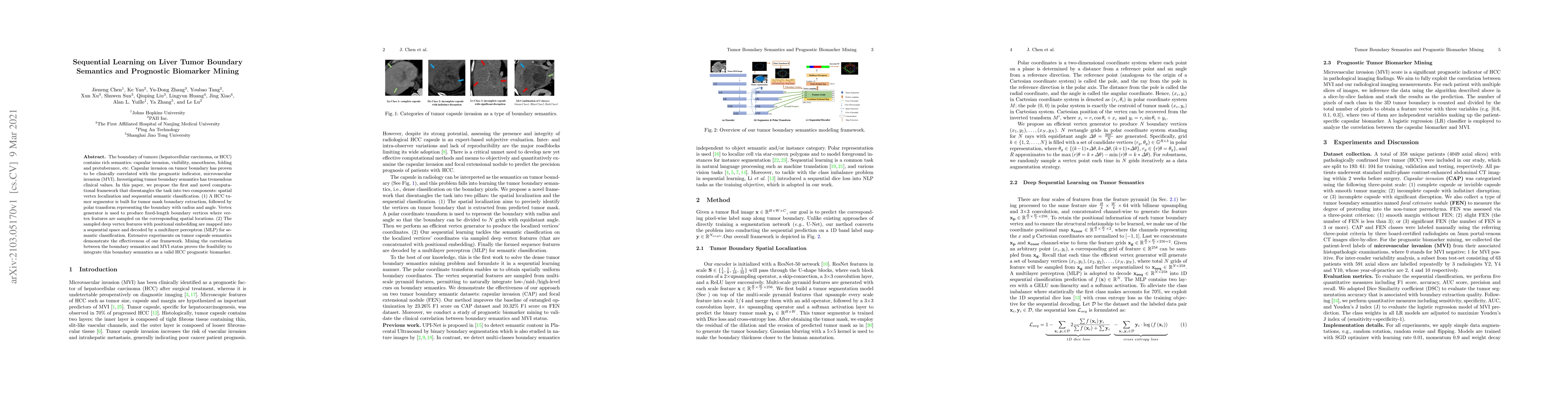
The boundary of tumors (hepatocellular carcinoma, or HCC) contains rich semantics: capsular invasion, visibility, smoothness, folding and protuberance, etc. Capsular invasion on tumor boundary has p...
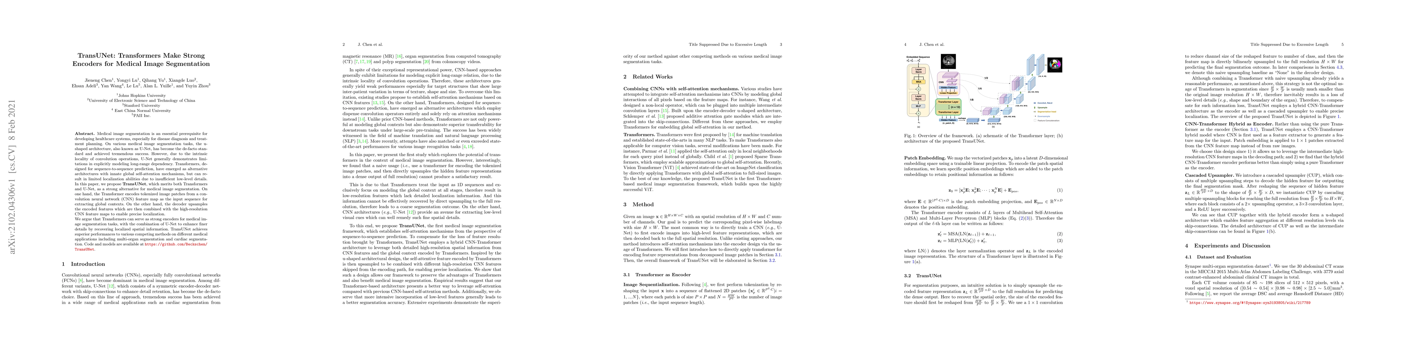
Medical image segmentation is an essential prerequisite for developing healthcare systems, especially for disease diagnosis and treatment planning. On various medical image segmentation tasks, the u...
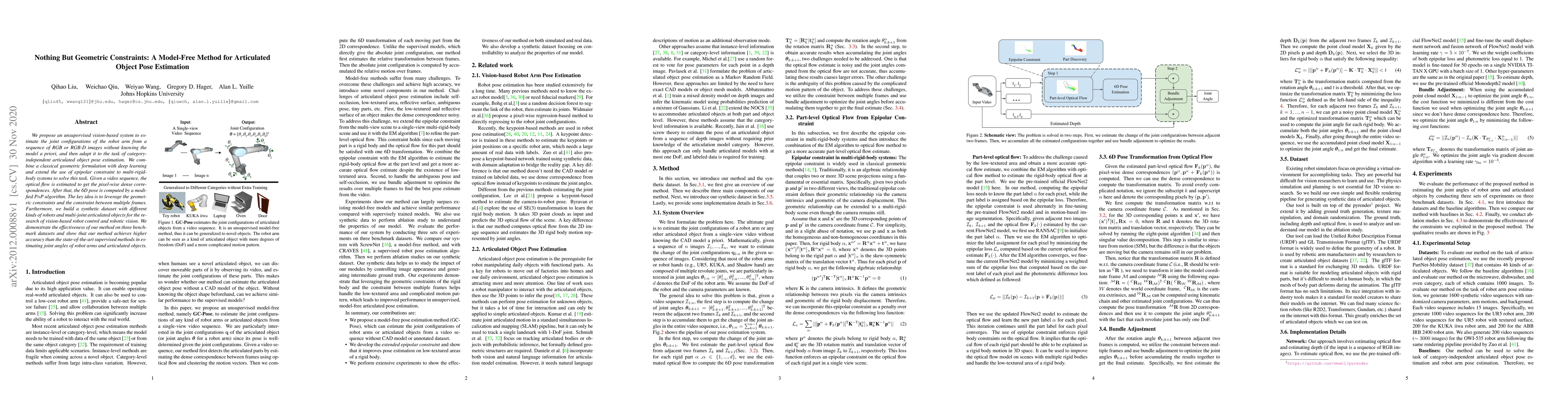
We propose an unsupervised vision-based system to estimate the joint configurations of the robot arm from a sequence of RGB or RGB-D images without knowing the model a priori, and then adapt it to t...
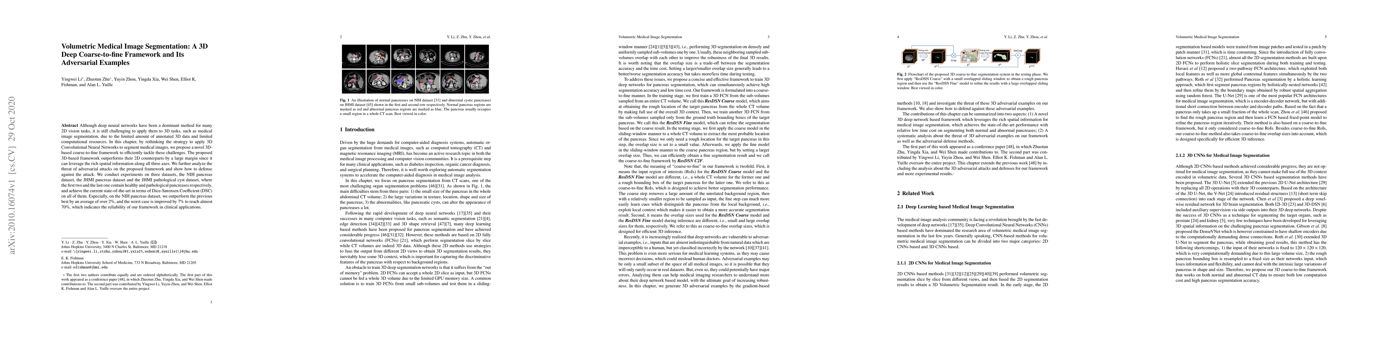
Although deep neural networks have been a dominant method for many 2D vision tasks, it is still challenging to apply them to 3D tasks, such as medical image segmentation, due to the limited amount o...
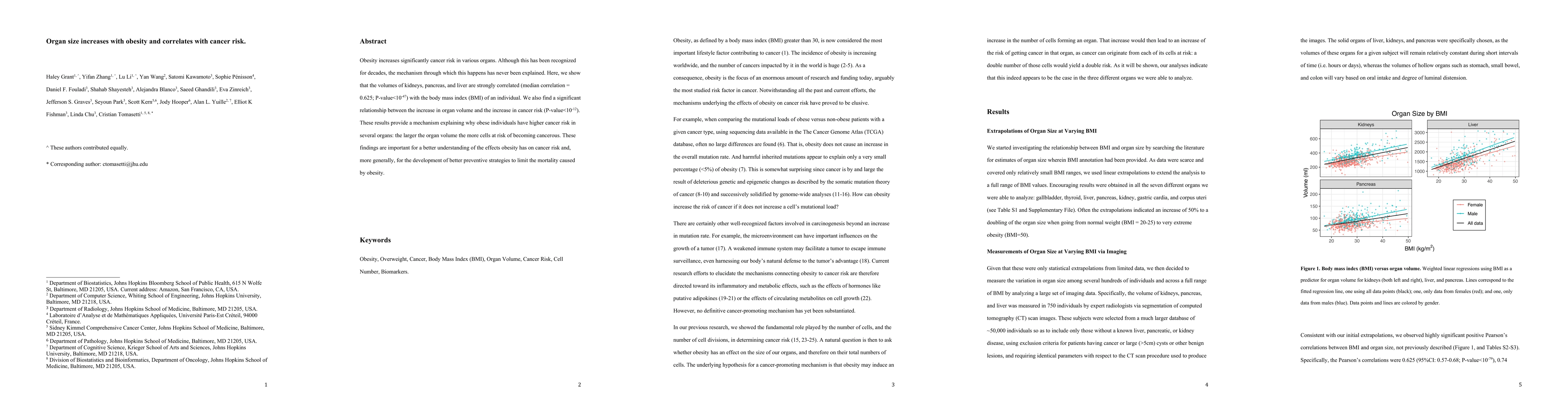
Obesity increases significantly cancer risk in various organs. Although this has been recognized for decades, the mechanism through which this happens has never been explained. Here, we show that th...
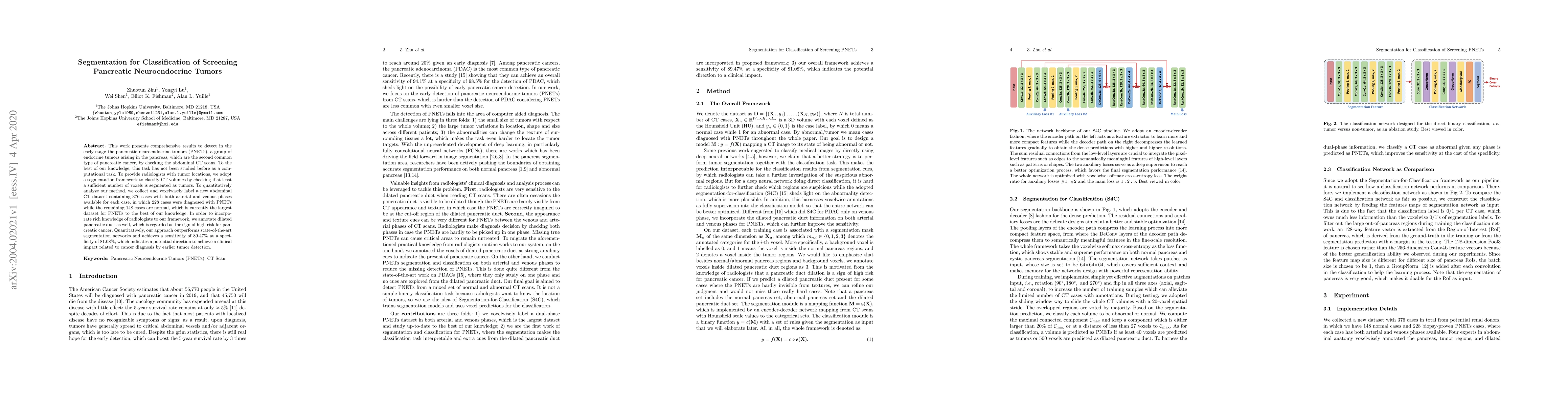
This work presents comprehensive results to detect in the early stage the pancreatic neuroendocrine tumors (PNETs), a group of endocrine tumors arising in the pancreas, which are the second common t...
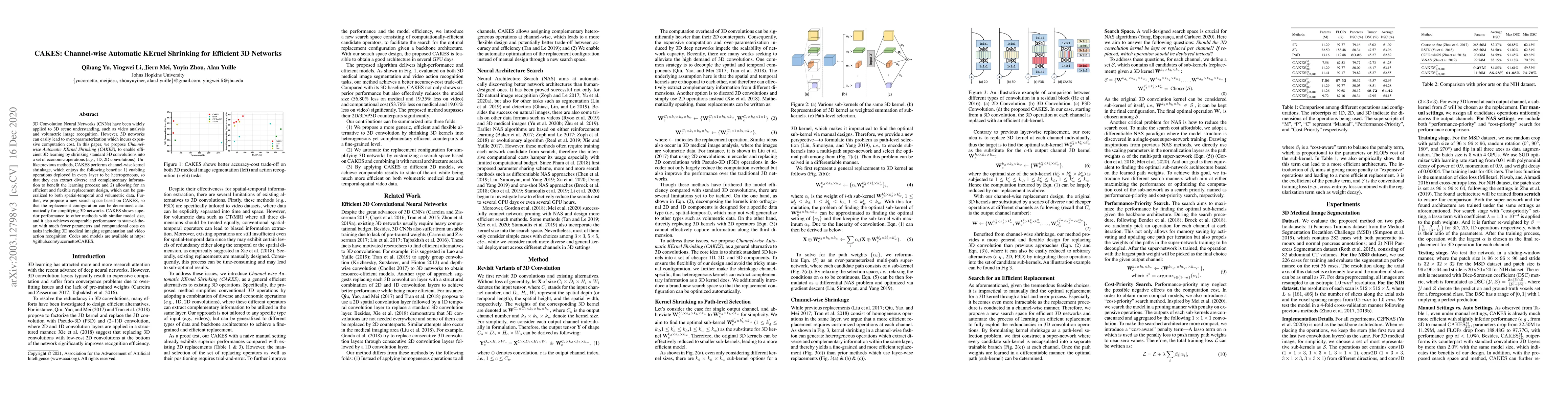
3D Convolution Neural Networks (CNNs) have been widely applied to 3D scene understanding, such as video analysis and volumetric image recognition. However, 3D networks can easily lead to over-parame...
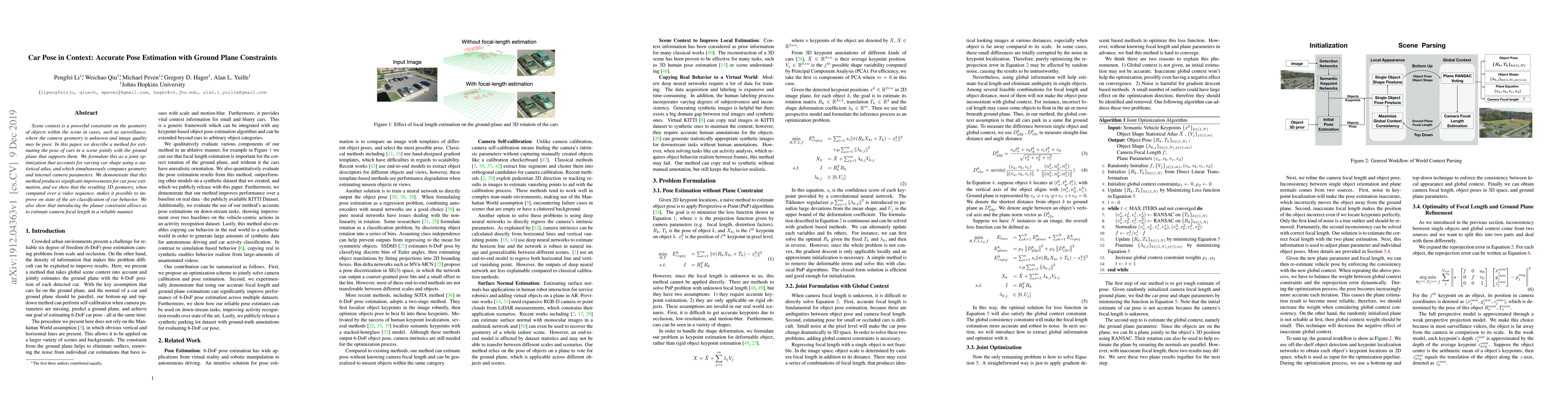
Scene context is a powerful constraint on the geometry of objects within the scene in cases, such as surveillance, where the camera geometry is unknown and image quality may be poor. In this paper, ...
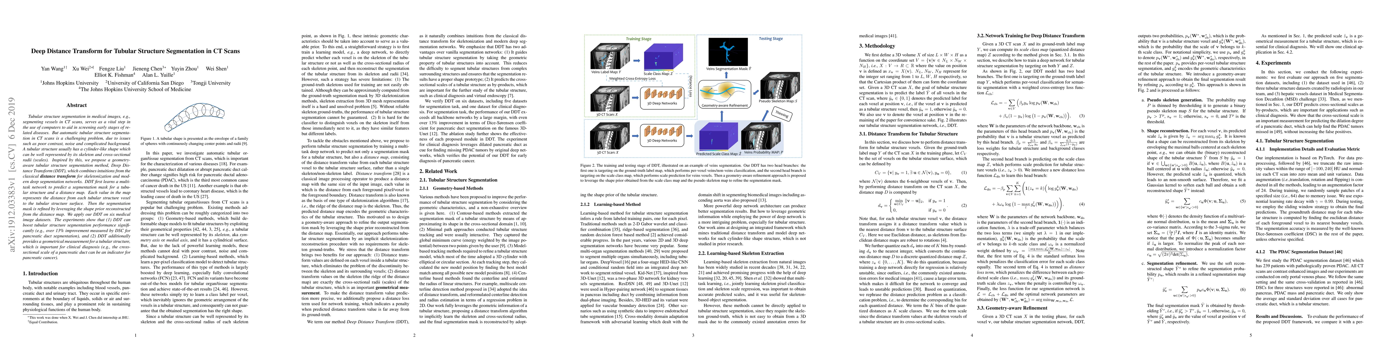
Tubular structure segmentation in medical images, e.g., segmenting vessels in CT scans, serves as a vital step in the use of computers to aid in screening early stages of related diseases. But autom...
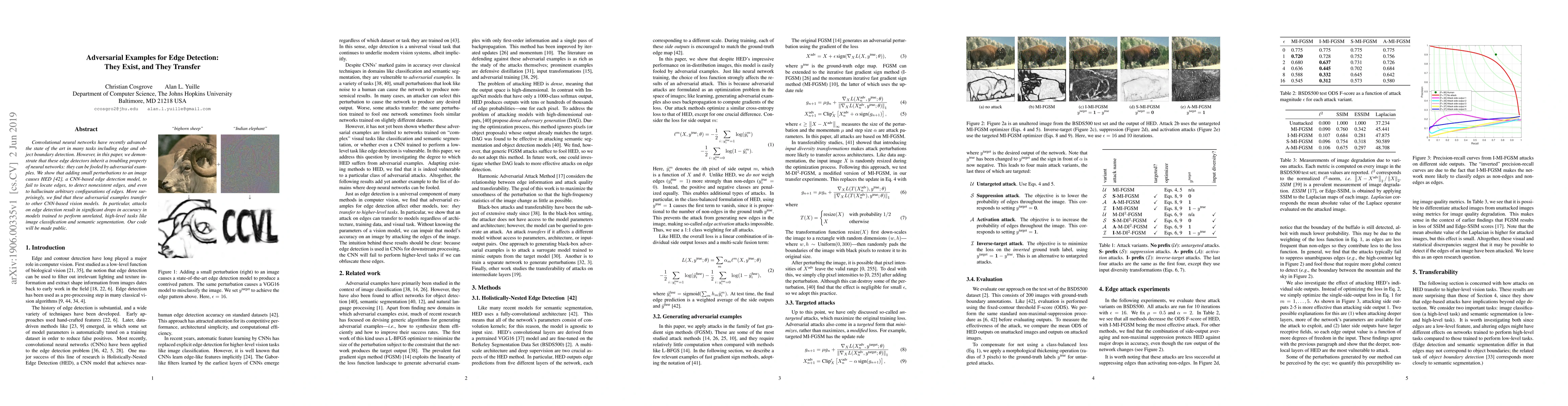
Convolutional neural networks have recently advanced the state of the art in many tasks including edge and object boundary detection. However, in this paper, we demonstrate that these edge detectors...
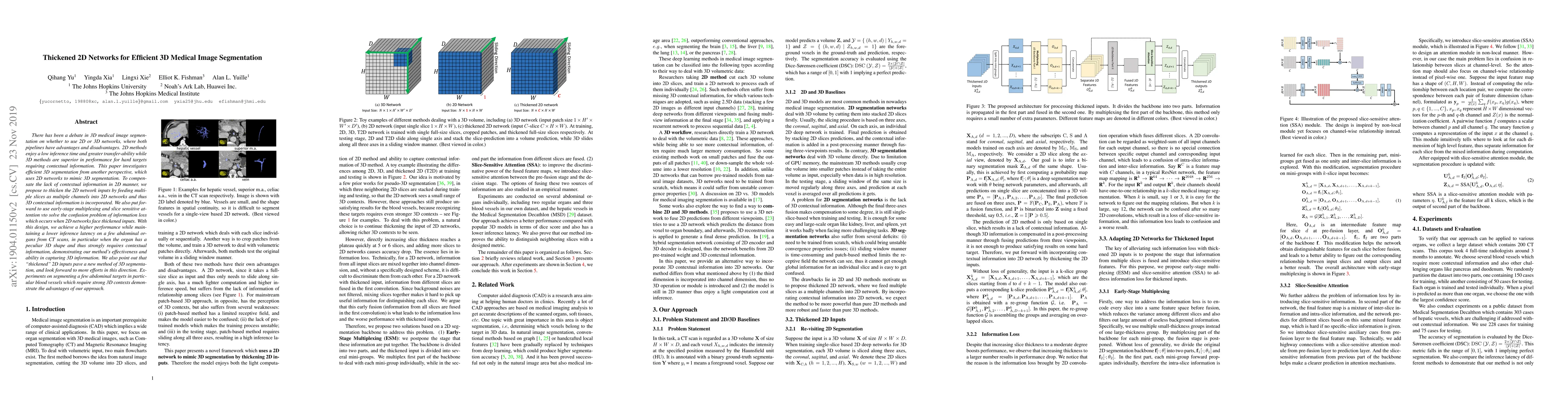
There has been a debate in 3D medical image segmentation on whether to use 2D or 3D networks, where both pipelines have advantages and disadvantages. 2D methods enjoy a low inference time and greate...
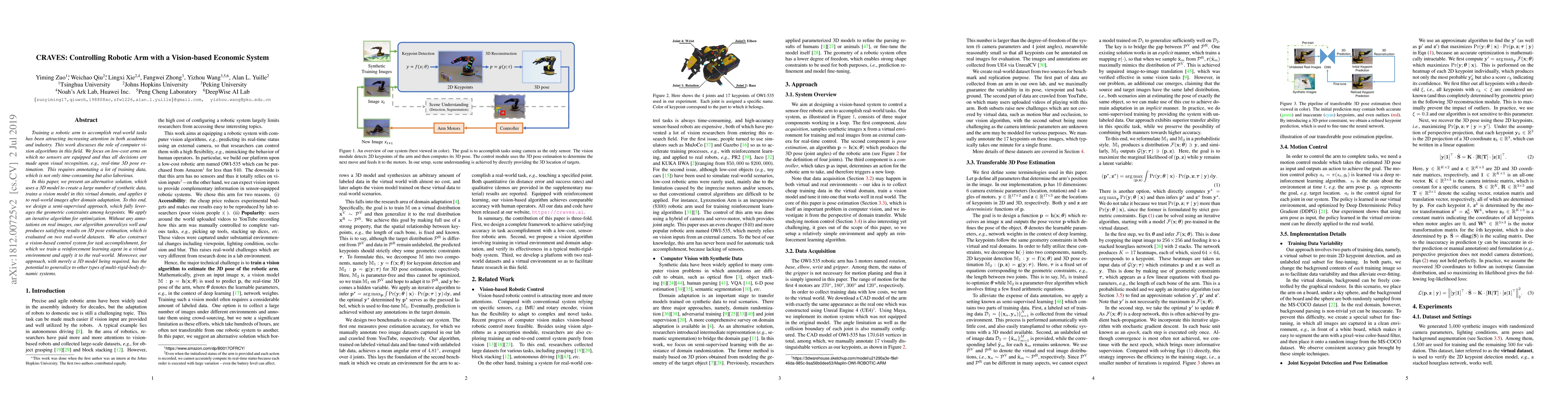
Training a robotic arm to accomplish real-world tasks has been attracting increasing attention in both academia and industry. This work discusses the role of computer vision algorithms in this field...
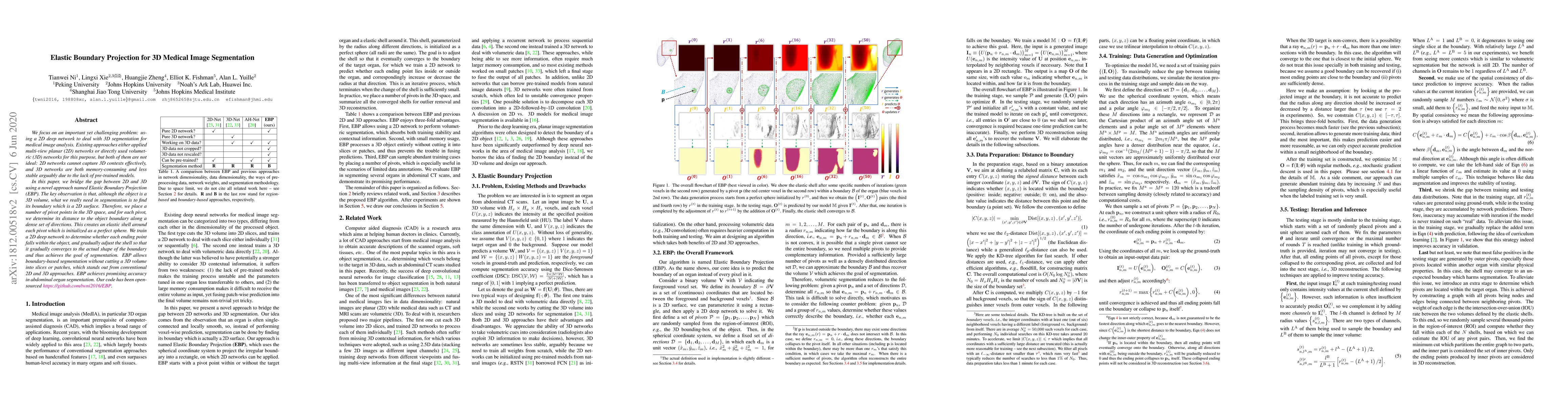
We focus on an important yet challenging problem: using a 2D deep network to deal with 3D segmentation for medical image analysis. Existing approaches either applied multi-view planar (2D) networks ...
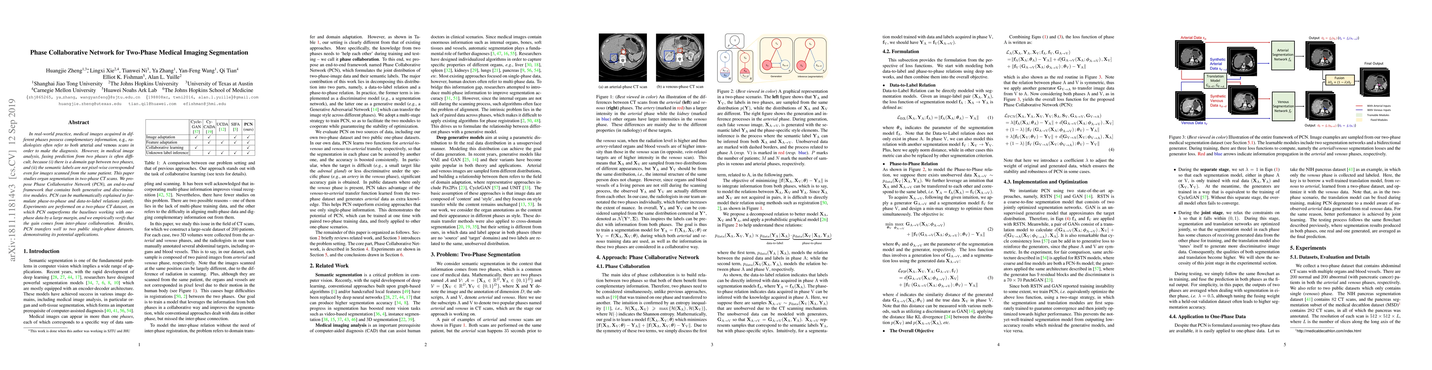
In real-world practice, medical images acquired in different phases possess complementary information, {\em e.g.}, radiologists often refer to both arterial and venous scans in order to make the dia...
This is an opinion paper about the strengths and weaknesses of Deep Nets for vision. They are at the heart of the enormous recent progress in artificial intelligence and are of growing importance in...
We aim to detect pancreatic ductal adenocarcinoma (PDAC) in abdominal CT scans, which sheds light on early diagnosis of pancreatic cancer. This is a 3D volume classification task with little trainin...
How can we test AI performance? This question seems trivial, but it isn't. Standard benchmarks often have problems such as in-distribution and small-size test sets, oversimplified metrics, unfair comp...
Building trusted datasets is critical for transparent and responsible Medical AI (MAI) research, but creating even small, high-quality datasets can take years of effort from multidisciplinary teams. T...
Precision medicine in the quantitative management of chronic diseases and oncology would be greatly improved if the Computed Tomography (CT) scan of any patient could be segmented, parsed and analyzed...
Widely adopted evaluation metrics for sparse-view CT reconstruction--such as Structural Similarity Index Measure and Peak Signal-to-Noise Ratio--prioritize pixel-wise fidelity but often fail to captur...
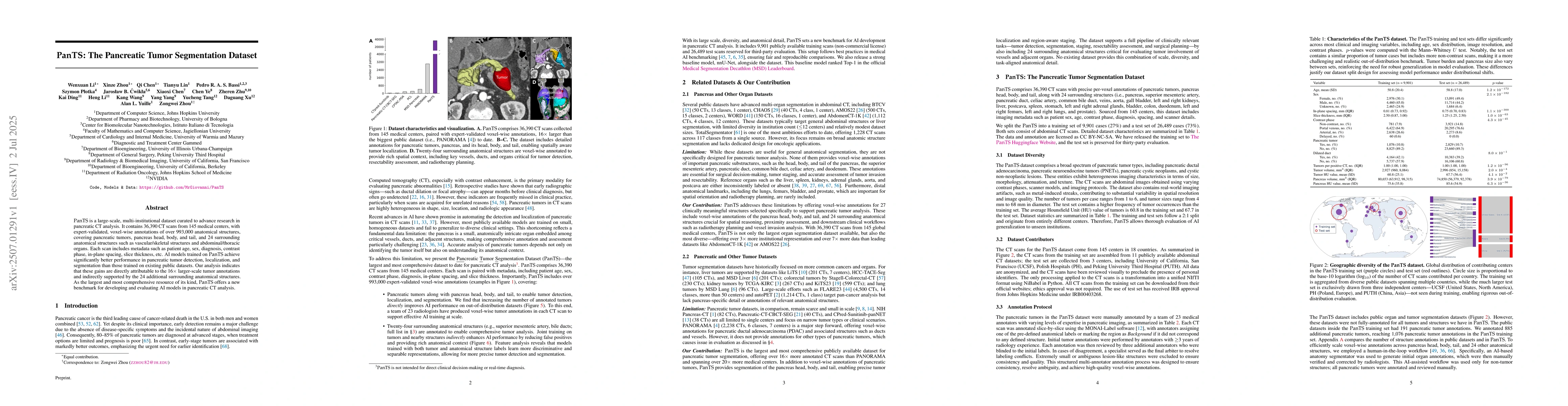
PanTS is a large-scale, multi-institutional dataset curated to advance research in pancreatic CT analysis. It contains 36,390 CT scans from 145 medical centers, with expert-validated, voxel-wise annot...
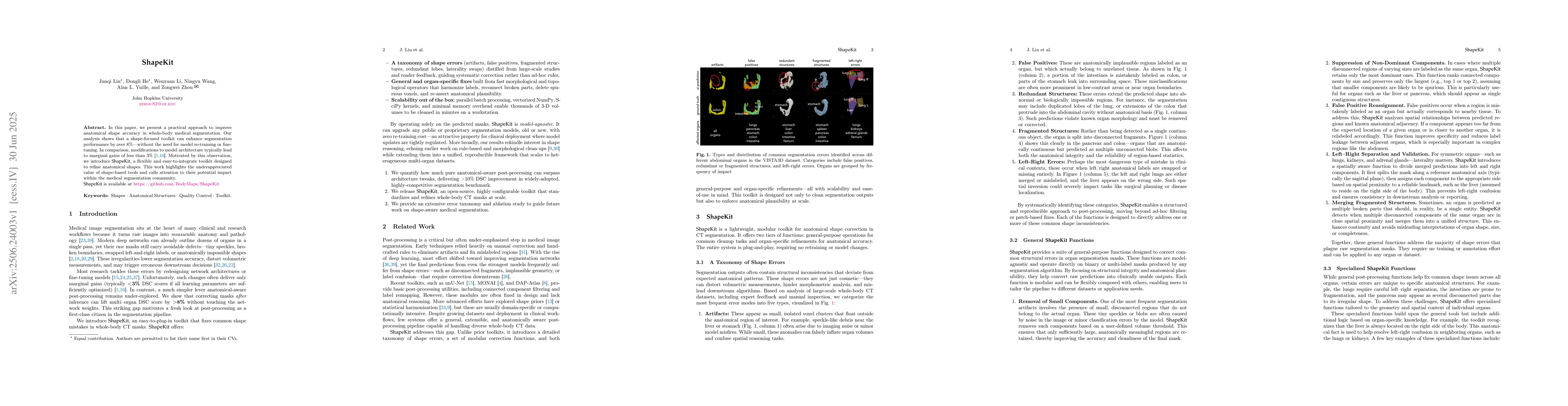
In this paper, we present a practical approach to improve anatomical shape accuracy in whole-body medical segmentation. Our analysis shows that a shape-focused toolkit can enhance segmentation perform...
Tumor segmentation in CT scans is key for diagnosis, surgery, and prognosis, yet segmentation masks are scarce because their creation requires time and expertise. Public abdominal CT datasets have fro...
Early tumor detection save lives. Each year, more than 300 million computed tomography (CT) scans are performed worldwide, offering a vast opportunity for effective cancer screening. However, detectin...
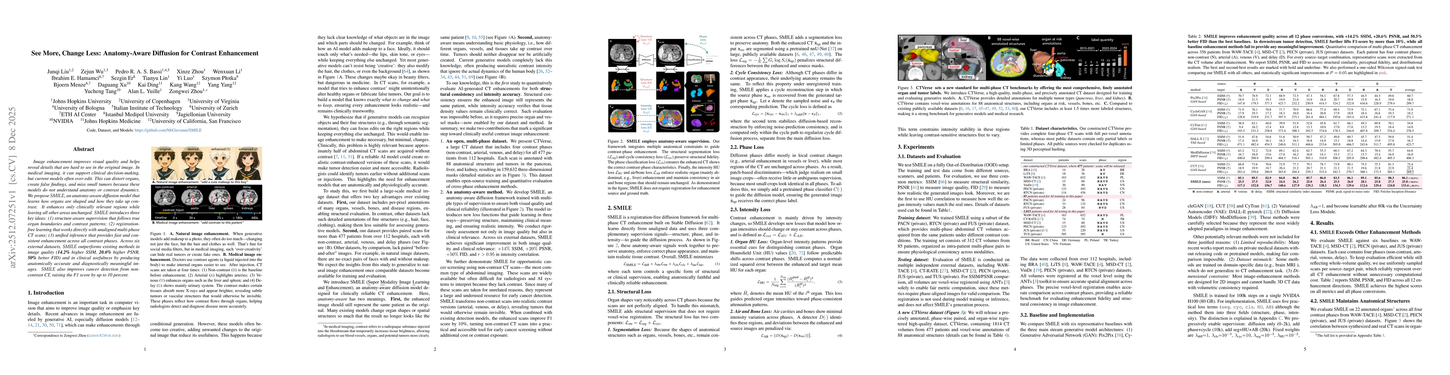
Image enhancement improves visual quality and helps reveal details that are hard to see in the original image. In medical imaging, it can support clinical decision-making, but current models often ove...
Open challenges have become the de facto standard for comparative ranking of medical AI methods. Despite their importance, medical AI leaderboards exhibit three persistent limitations: (1) score gaps ...
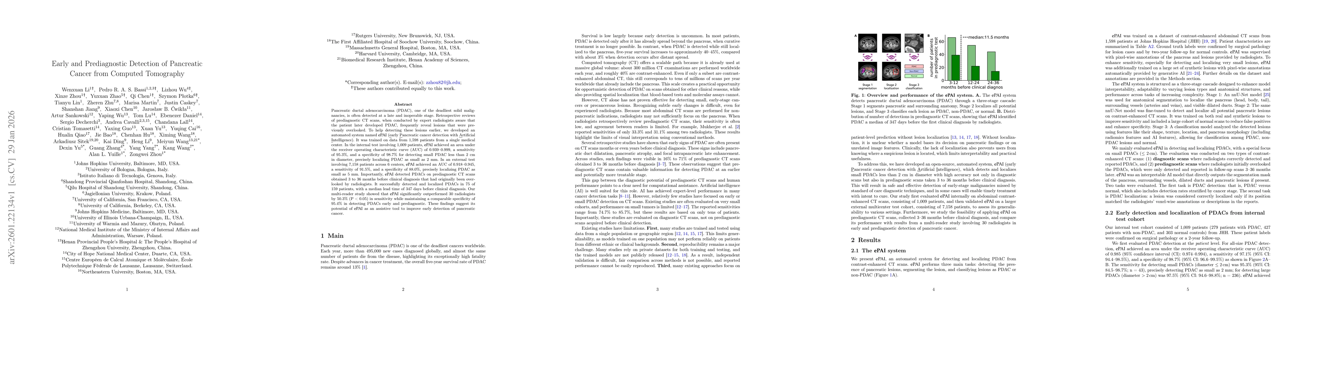
Pancreatic ductal adenocarcinoma (PDAC), one of the deadliest solid malignancies, is often detected at a late and inoperable stage. Retrospective reviews of prediagnostic CT scans, when conducted by e...
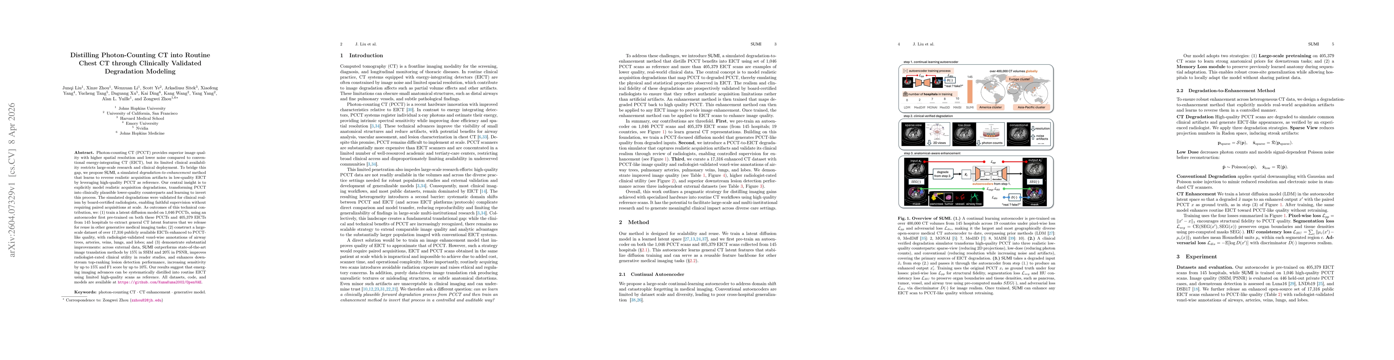
Photon-counting CT (PCCT) provides superior image quality with higher spatial resolution and lower noise compared to conventional energy-integrating CT (EICT), but its limited clinical availability re...
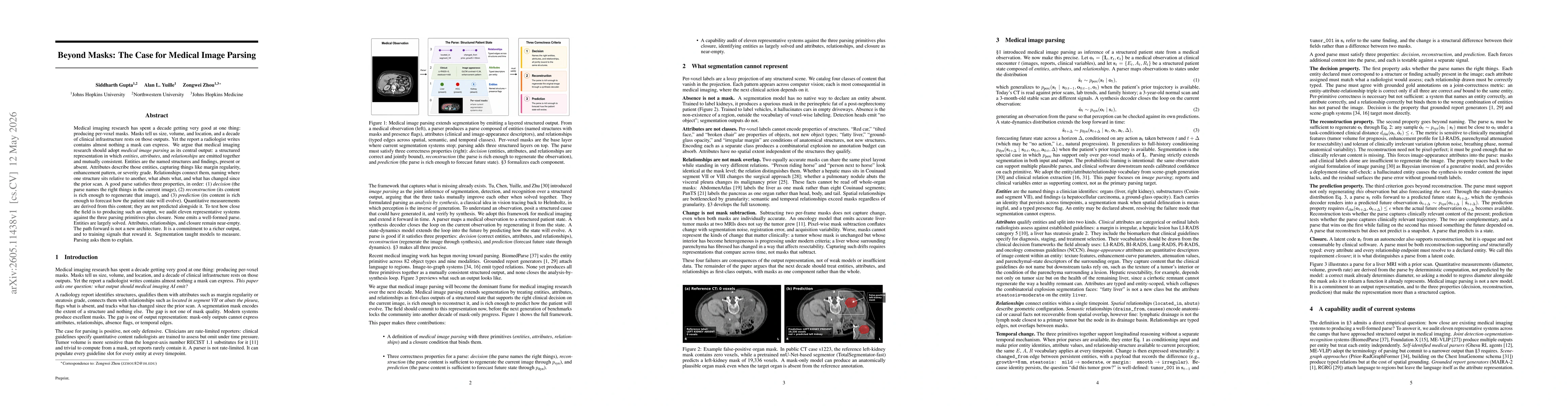
Medical imaging research has spent a decade getting very good at one thing: producing per-voxel masks. Masks tell us size, volume, and location, and a decade of clinical infrastructure rests on those ...
Cancer screening is a reasoning task. A radiologist observes findings, compares them to prior scans, integrates clinical context, and reaches a diagnostic conclusion confirmed by pathology. We present...
Artificial intelligence (AI) has achieved remarkable success in medical imaging, but it is widely recognized that these models often perform inconsistently across real-world clinical settings. Such in...