Academic Profile
Statistics
Similar Authors
Papers on arXiv
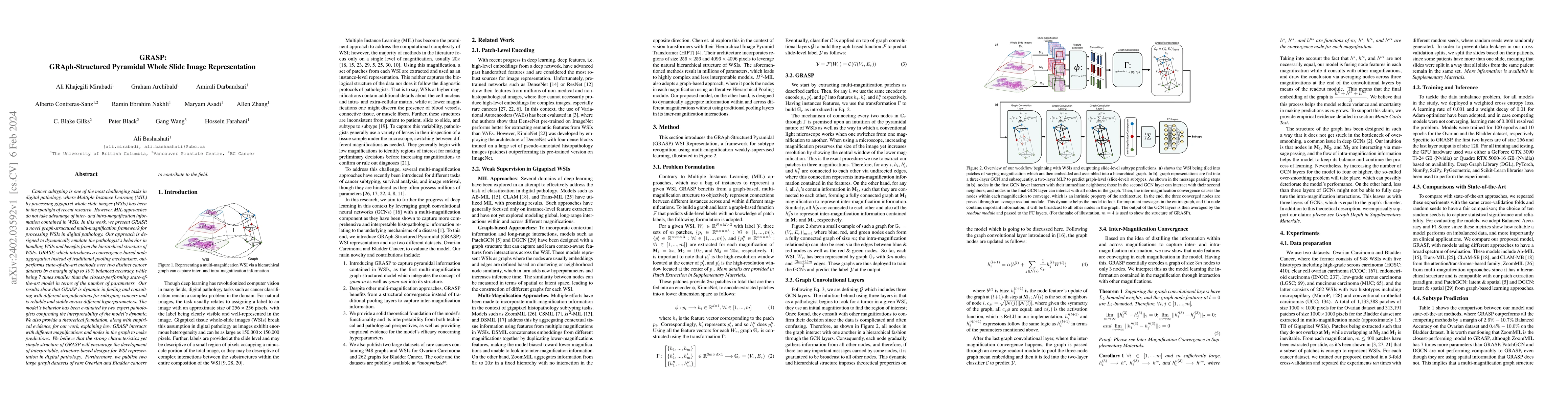
Cancer subtyping is one of the most challenging tasks in digital pathology, where Multiple Instance Learning (MIL) by processing gigapixel whole slide images (WSIs) has been in the spotlight of rece...
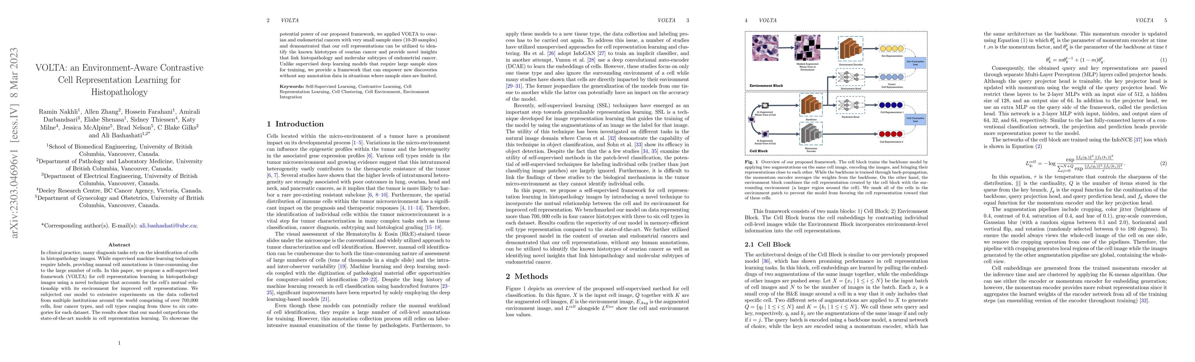
In clinical practice, many diagnosis tasks rely on the identification of cells in histopathology images. While supervised machine learning techniques require labels, providing manual cell annotation...
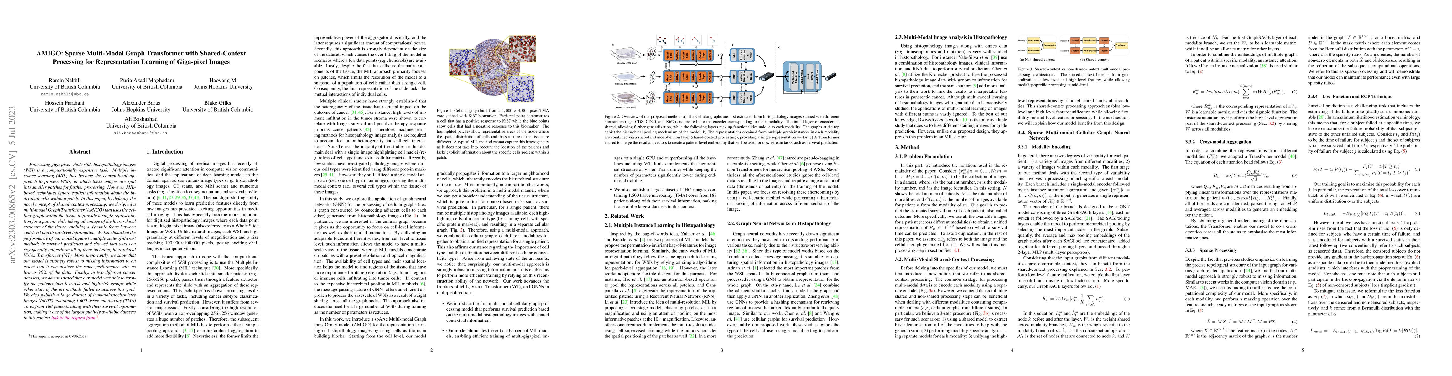
Processing giga-pixel whole slide histopathology images (WSI) is a computationally expensive task. Multiple instance learning (MIL) has become the conventional approach to process WSIs, in which the...
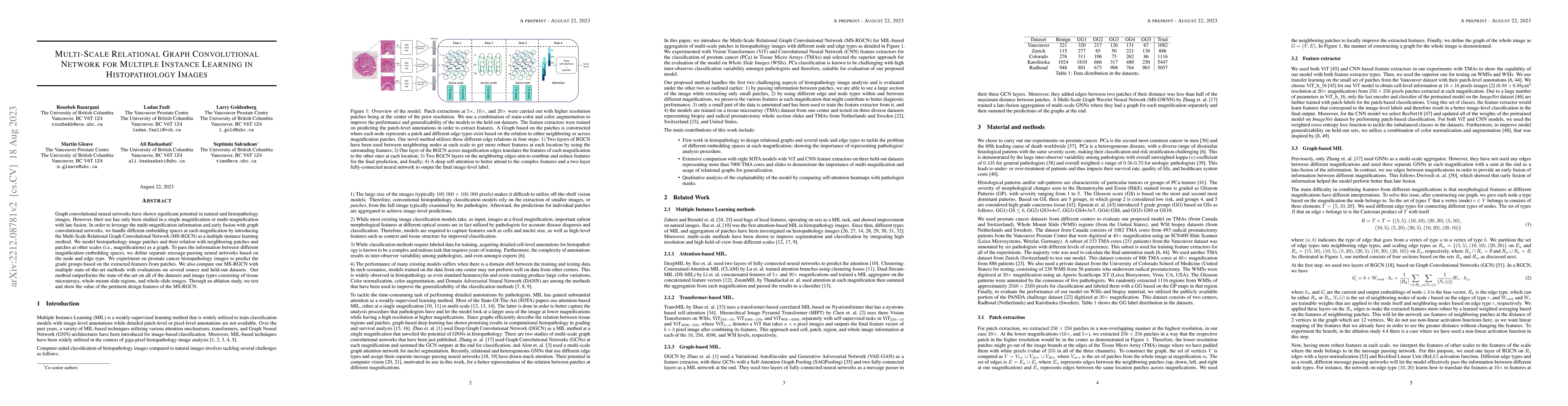
Graph convolutional neural networks have shown significant potential in natural and histopathology images. However, their use has only been studied in a single magnification or multi-magnification w...
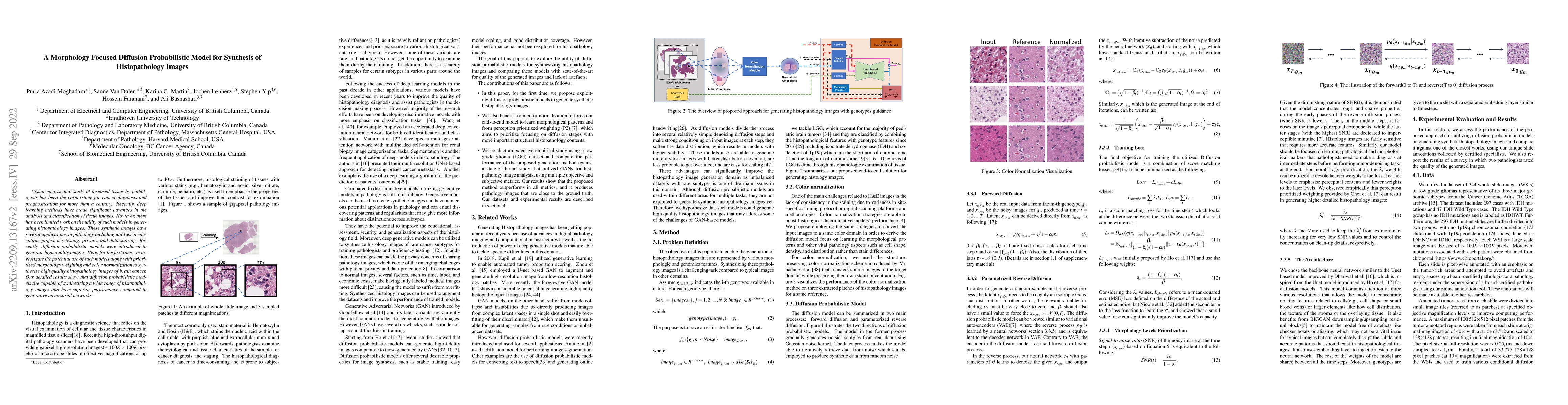
Visual microscopic study of diseased tissue by pathologists has been the cornerstone for cancer diagnosis and prognostication for more than a century. Recently, deep learning methods have made signi...
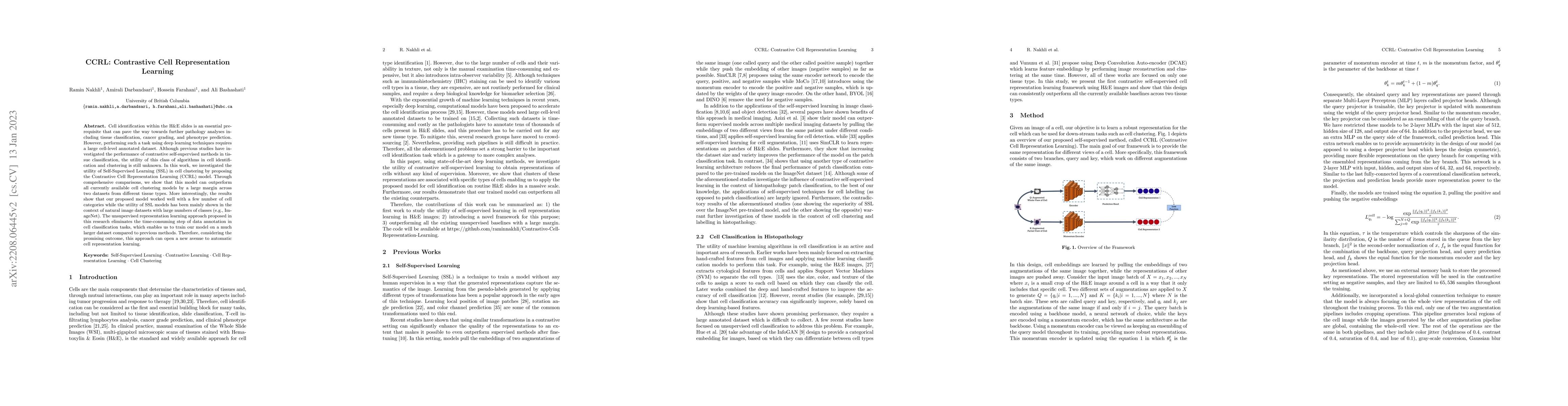
Cell identification within the H&E slides is an essential prerequisite that can pave the way towards further pathology analyses including tissue classification, cancer grading, and phenotype predict...
Bevacizumab is a widely studied targeted therapeutic drug used in conjunction with standard chemotherapy for the treatment of recurrent ovarian cancer. While its administration has shown to increase t...
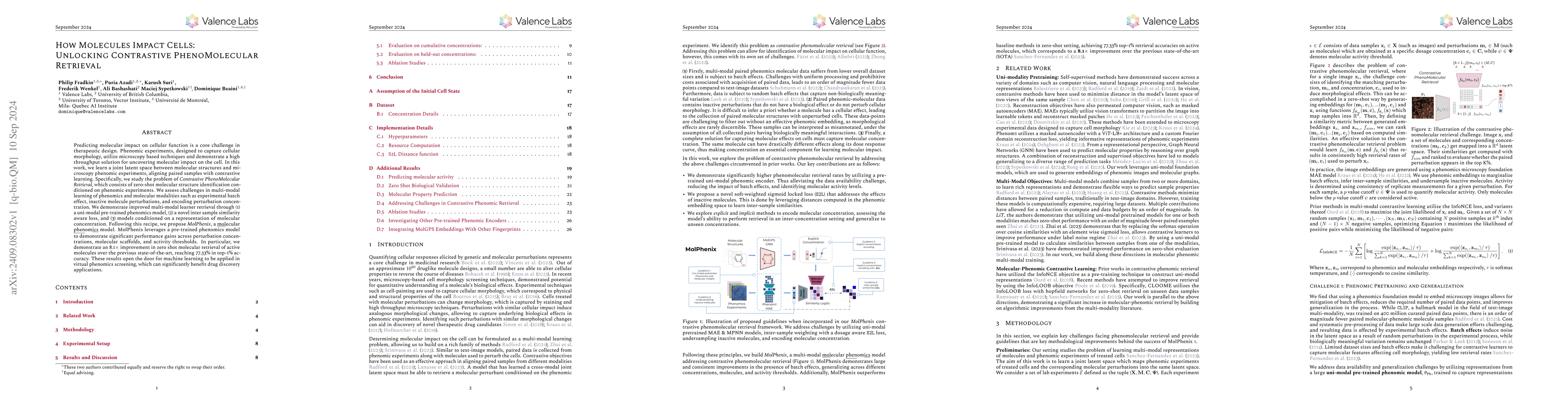
Predicting molecular impact on cellular function is a core challenge in therapeutic design. Phenomic experiments, designed to capture cellular morphology, utilize microscopy based techniques and demon...
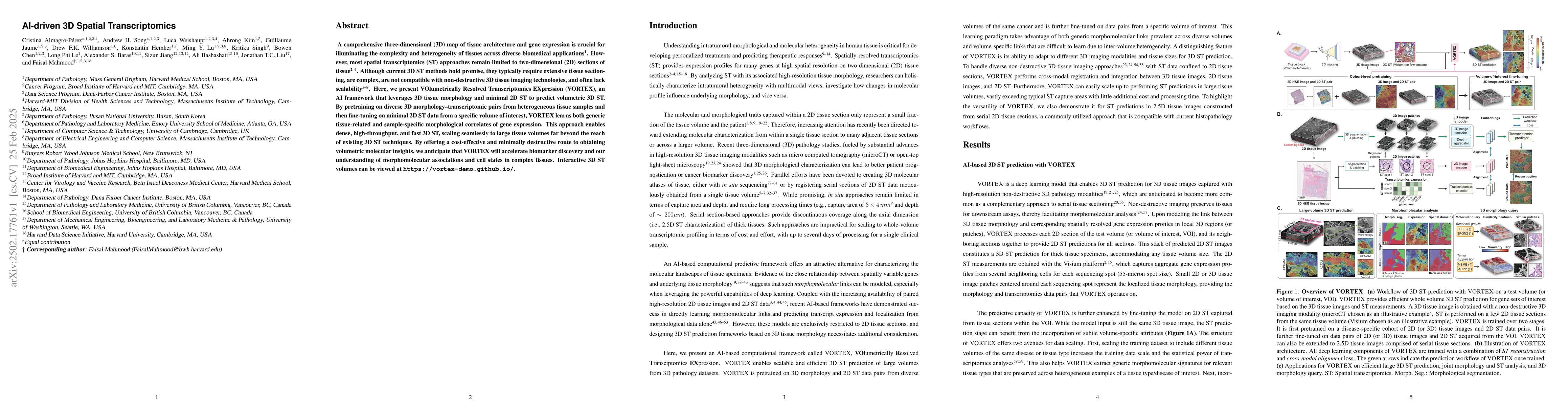
A comprehensive three-dimensional (3D) map of tissue architecture and gene expression is crucial for illuminating the complexity and heterogeneity of tissues across diverse biomedical applications. Ho...