Academic Profile
Statistics
Similar Authors
Papers on arXiv
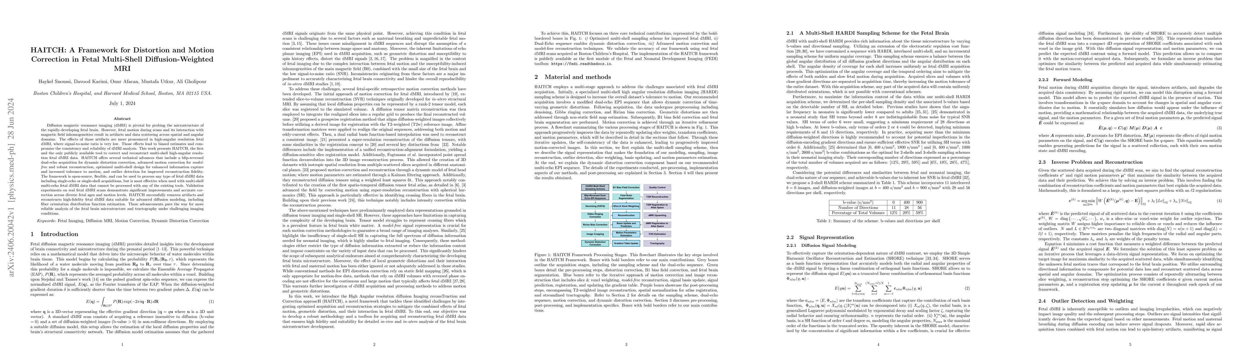
Diffusion magnetic resonance imaging (dMRI) is pivotal for probing the microstructure of the rapidly-developing fetal brain. However, fetal motion during scans and its interaction with magnetic field ...
Diffusion-weighted Magnetic Resonance Imaging (dMRI) is increasingly used to study the fetal brain in utero. An important computation enabled by dMRI is streamline tractography, which has unique app...
Deep learning models have shown great promise in estimating tissue microstructure from limited diffusion magnetic resonance imaging data. However, these models face domain shift challenges when test...
Fetal brain extraction is a necessary first step in most computational fetal brain MRI pipelines. However, it has been a very challenging task due to non-standard fetal head pose, fetal movements du...
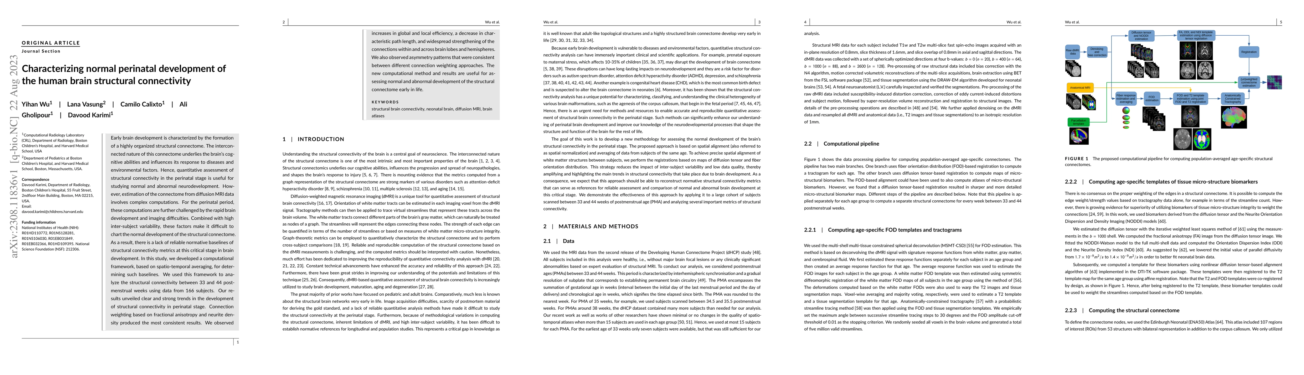
Early brain development is characterized by the formation of a highly organized structural connectome. The interconnected nature of this connectome underlies the brain's cognitive abilities and infl...
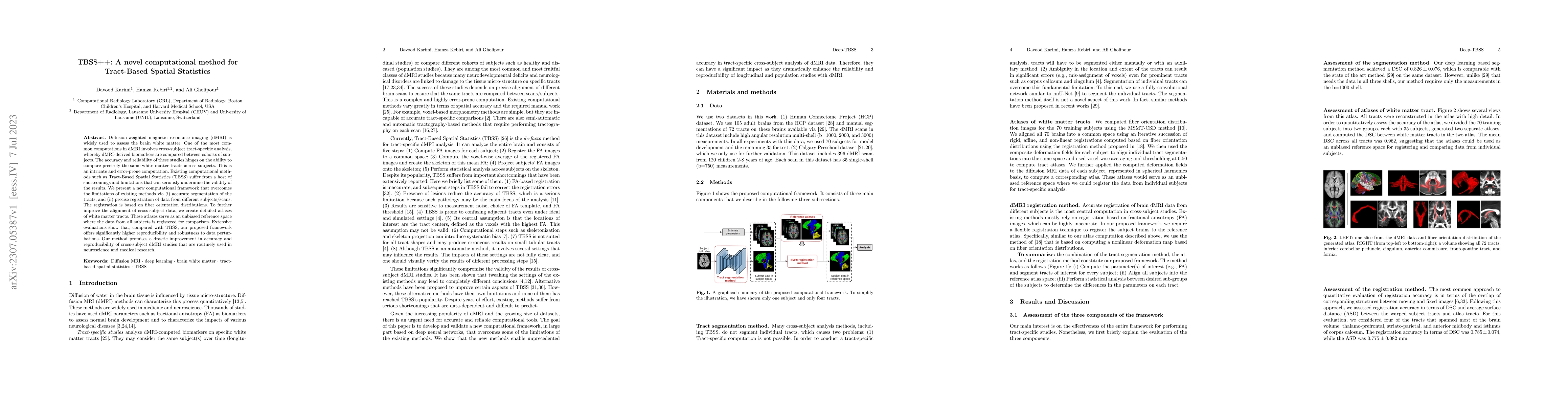
Diffusion-weighted magnetic resonance imaging (dMRI) is widely used to assess the brain white matter. One of the most common computations in dMRI involves cross-subject tract-specific analysis, wher...
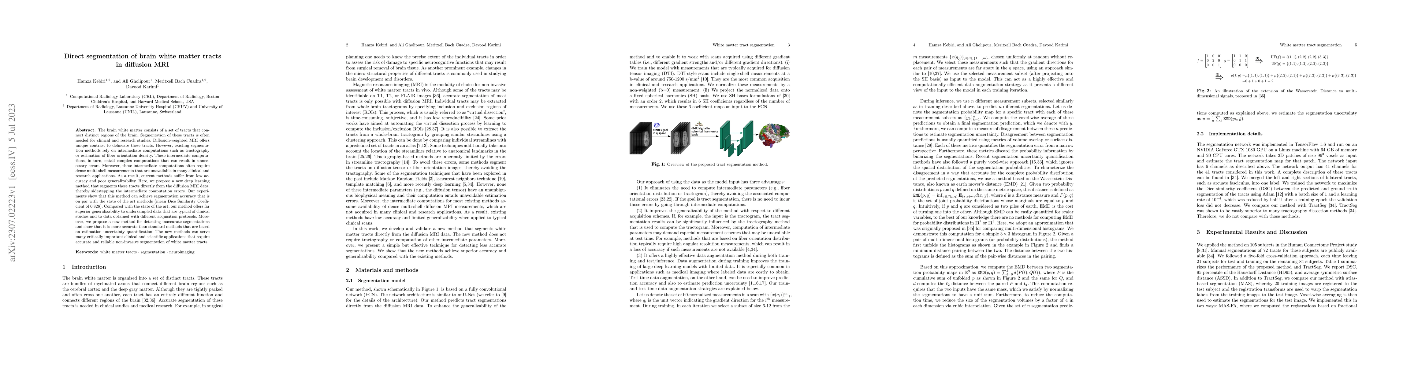
The brain white matter consists of a set of tracts that connect distinct regions of the brain. Segmentation of these tracts is often needed for clinical and research studies. Diffusion-weighted MRI ...
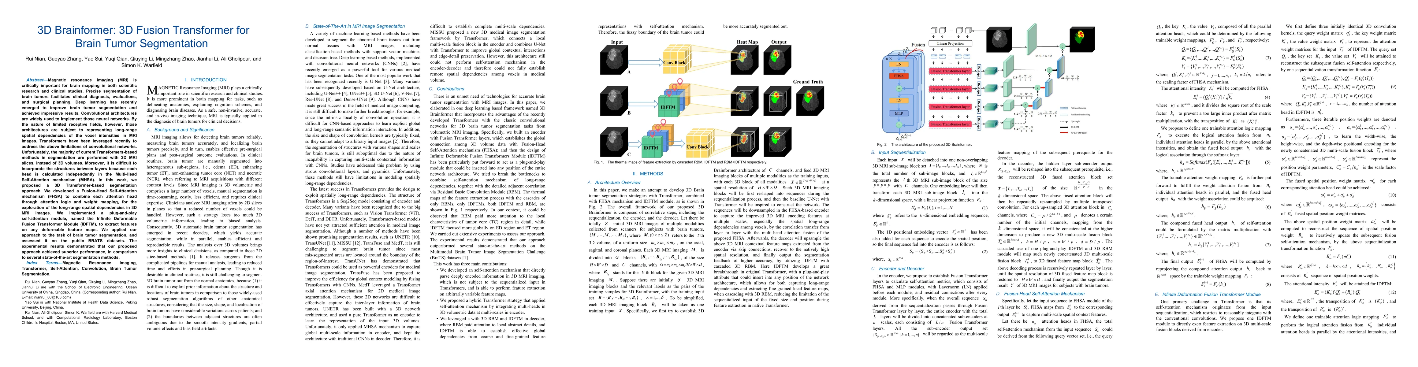
Magnetic resonance imaging (MRI) is critically important for brain mapping in both scientific research and clinical studies. Precise segmentation of brain tumors facilitates clinical diagnosis, eval...
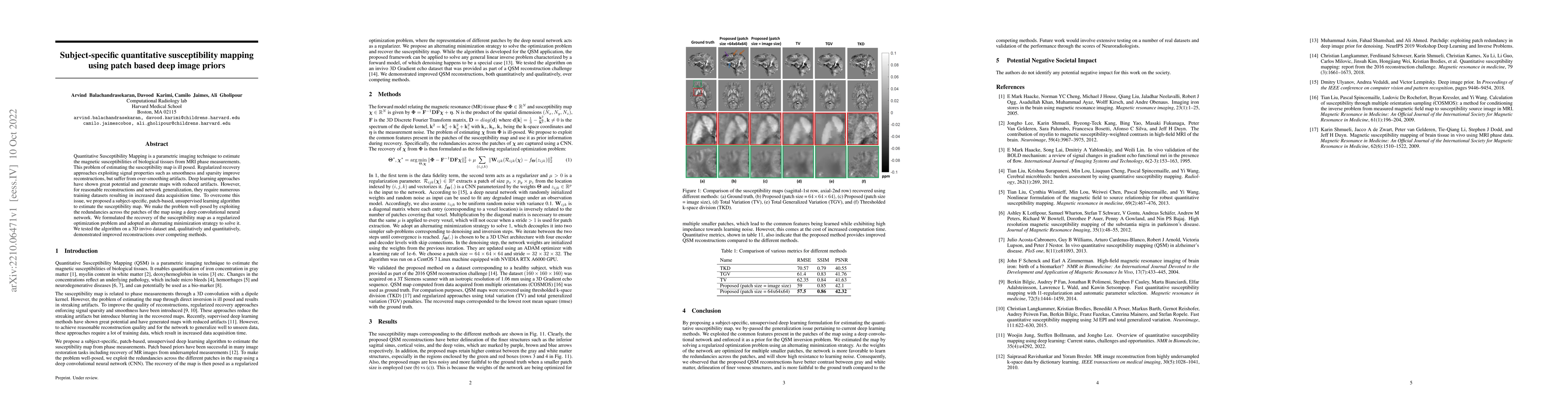
Quantitative Susceptibility Mapping is a parametric imaging technique to estimate the magnetic susceptibilities of biological tissues from MRI phase measurements. This problem of estimating the susc...
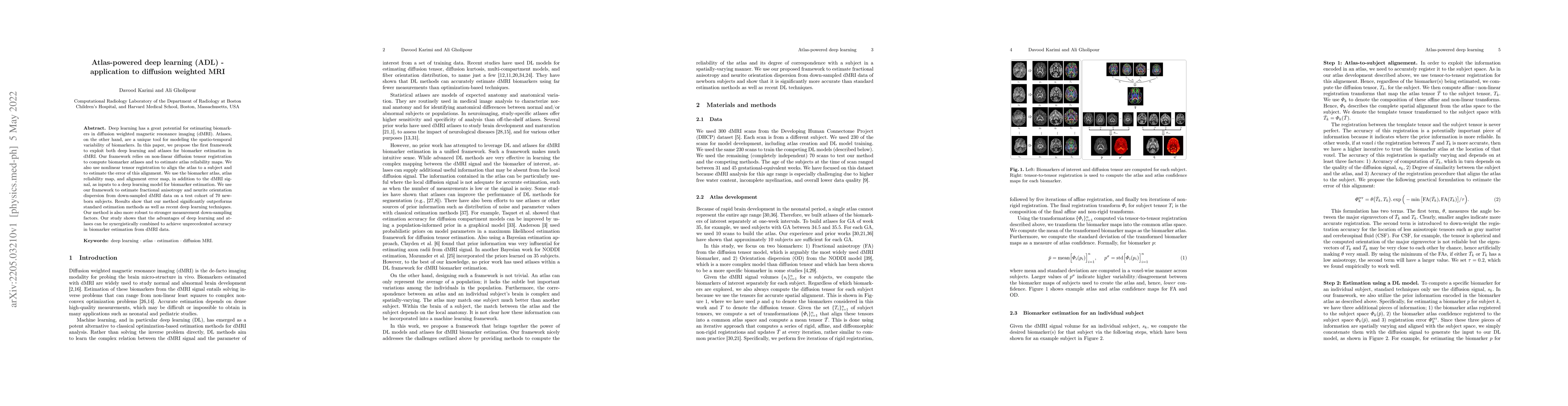
Deep learning has a great potential for estimating biomarkers in diffusion weighted magnetic resonance imaging (dMRI). Atlases, on the other hand, are a unique tool for modeling the spatio-temporal ...
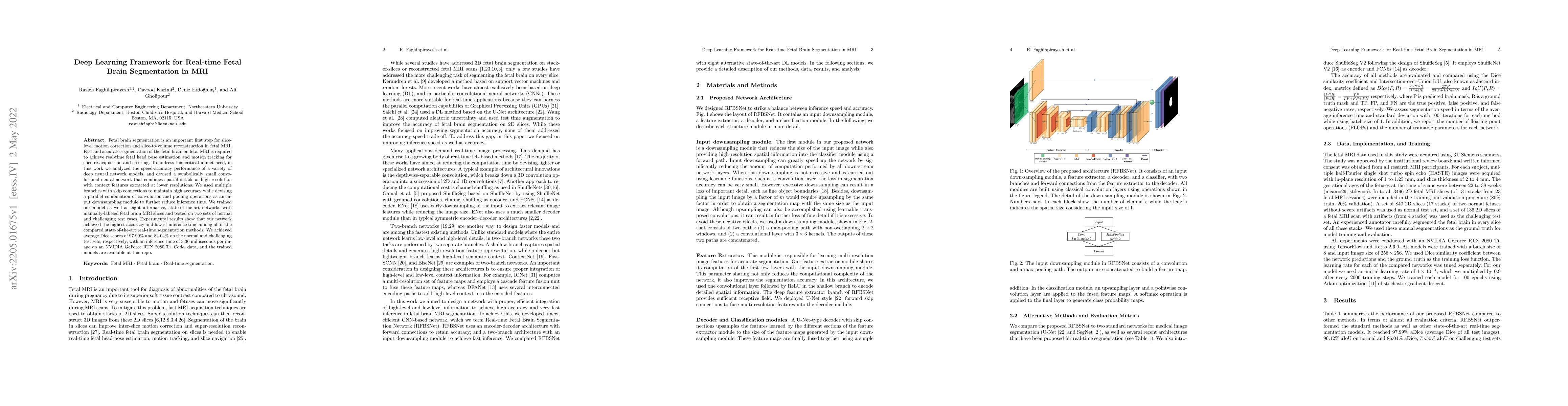
Fetal brain segmentation is an important first step for slice-level motion correction and slice-to-volume reconstruction in fetal MRI. Fast and accurate segmentation of the fetal brain on fetal MRI ...
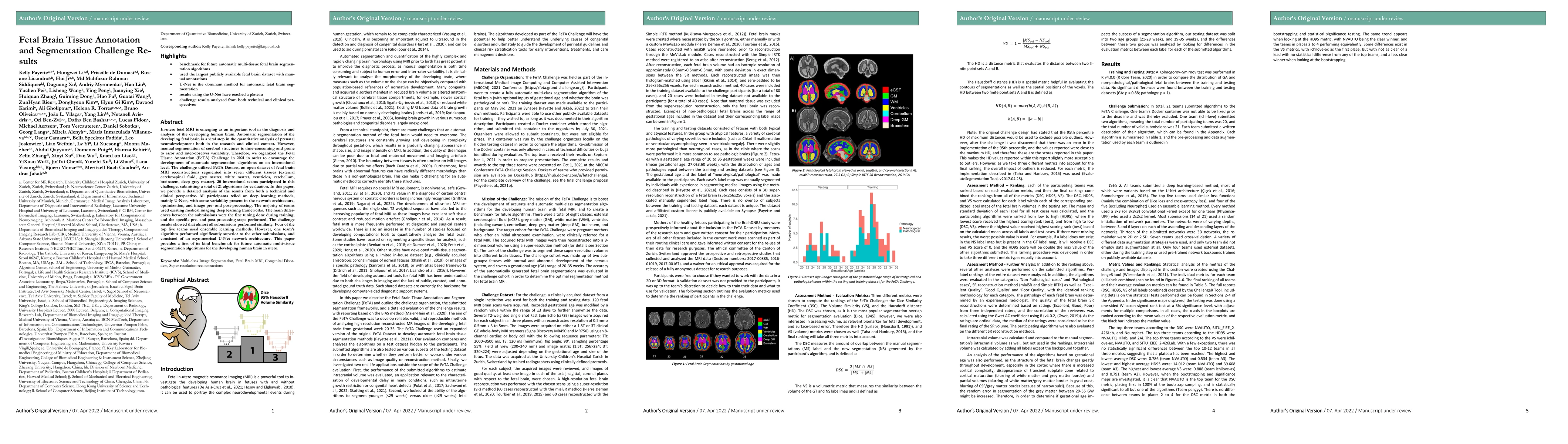
In-utero fetal MRI is emerging as an important tool in the diagnosis and analysis of the developing human brain. Automatic segmentation of the developing fetal brain is a vital step in the quantitat...
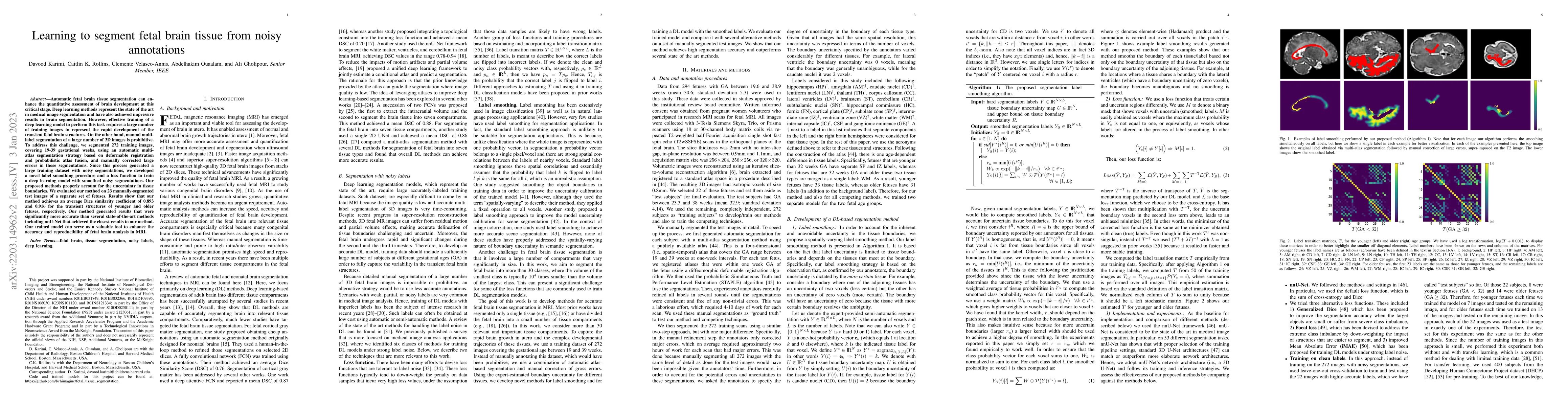
Automatic fetal brain tissue segmentation can enhance the quantitative assessment of brain development at this critical stage. Deep learning methods represent the state of the art in medical image s...
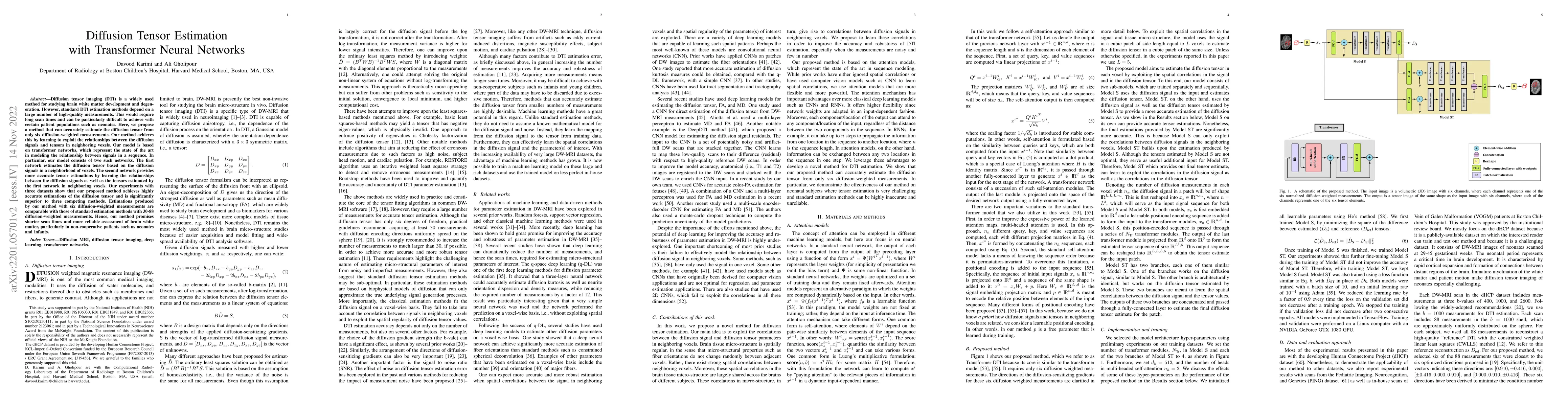
Diffusion tensor imaging (DTI) is a widely used method for studying brain white matter development and degeneration. However, standard DTI estimation methods depend on a large number of high-quality...
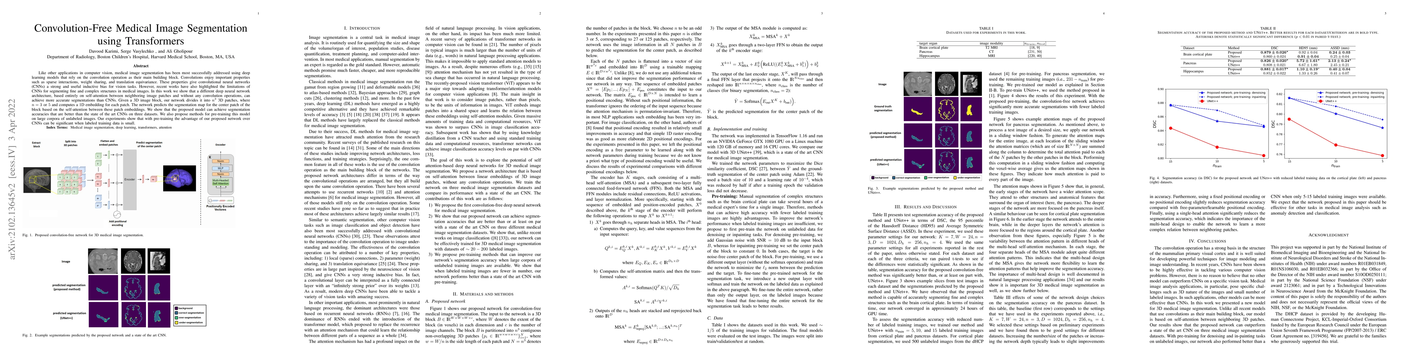
Like other applications in computer vision, medical image segmentation has been most successfully addressed using deep learning models that rely on the convolution operation as their main building b...
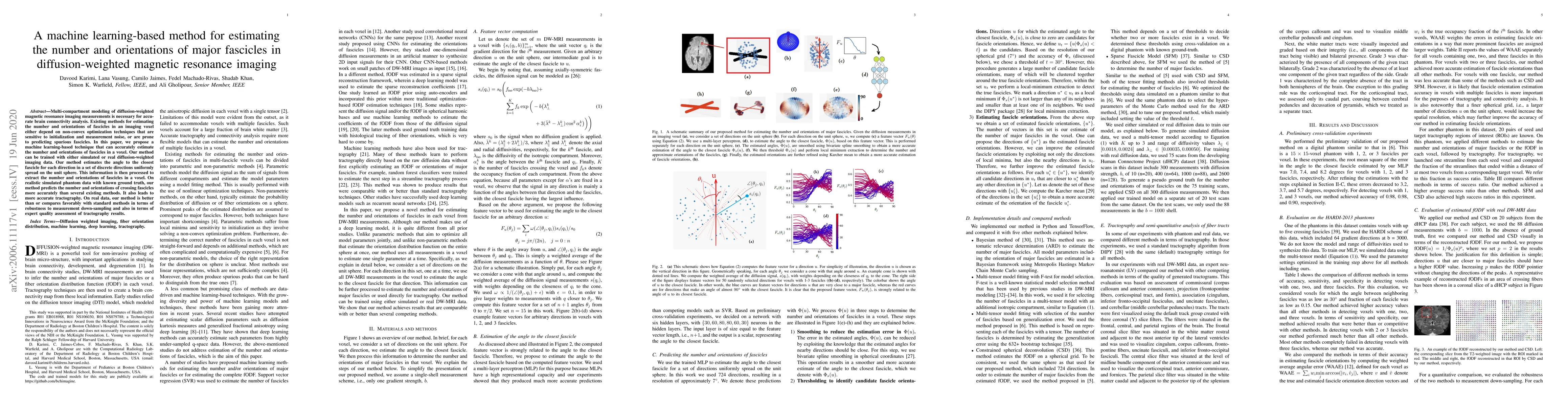
Multi-compartment modeling of diffusion-weighted magnetic resonance imaging measurements is necessary for accurate brain connectivity analysis. Existing methods for estimating the number and orienta...
Transfer learning is widely used for training machine learning models. Here, we study the role of transfer learning for training fully convolutional networks (FCNs) for medical image segmentation. O...
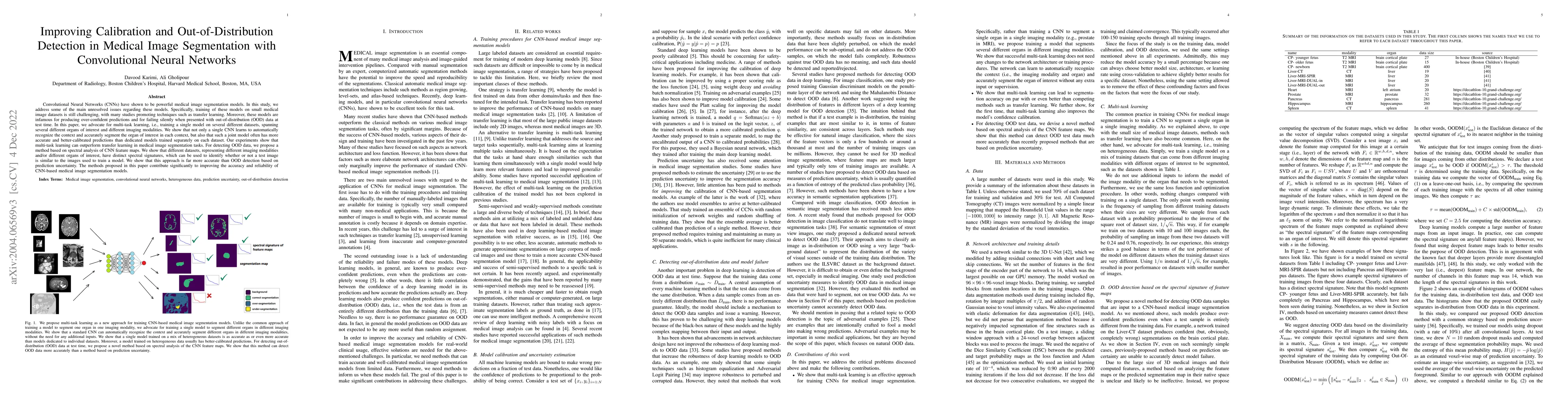
Convolutional Neural Networks (CNNs) have shown to be powerful medical image segmentation models. In this study, we address some of the main unresolved issues regarding these models. Specifically, t...
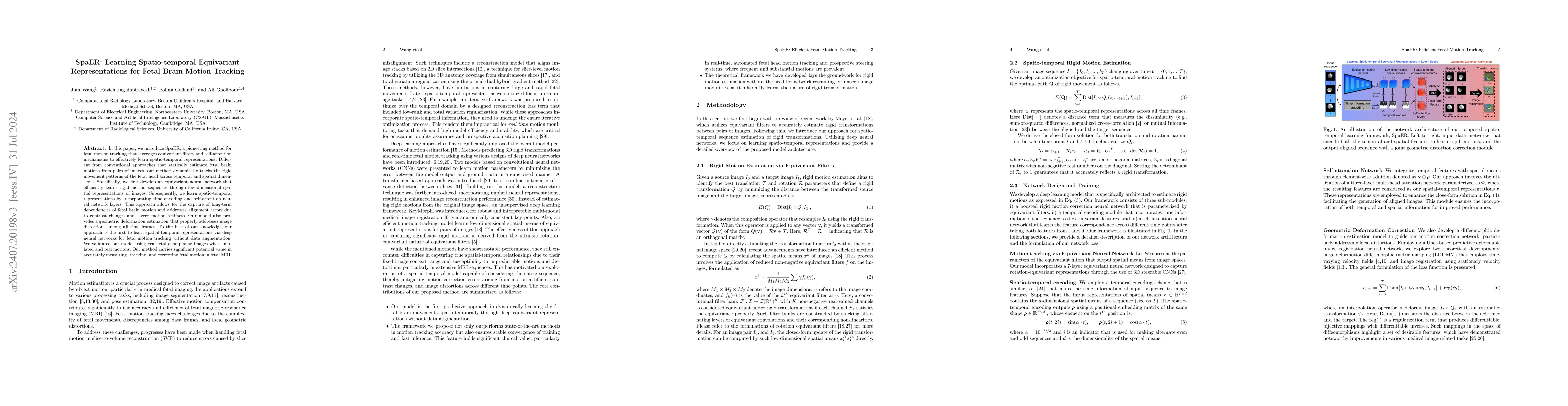
In this paper, we introduce SpaER, a pioneering method for fetal motion tracking that leverages equivariant filters and self-attention mechanisms to effectively learn spatio-temporal representations. ...
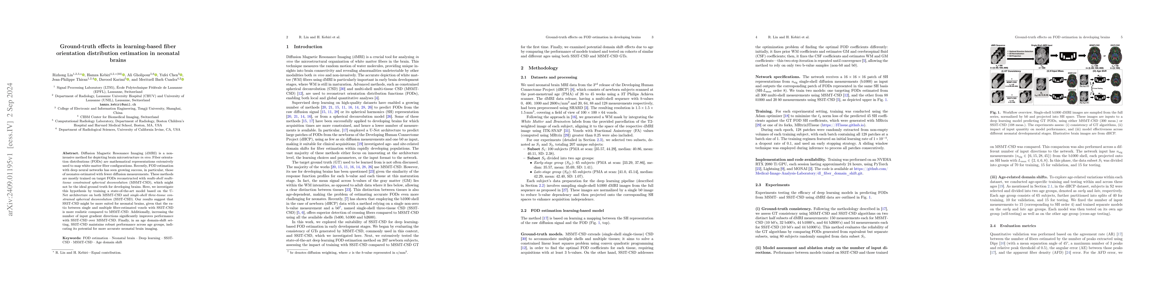
Diffusion Magnetic Resonance Imaging (dMRI) is a non-invasive method for depicting brain microstructure in vivo. Fiber orientation distributions (FODs) are mathematical representations extensively use...
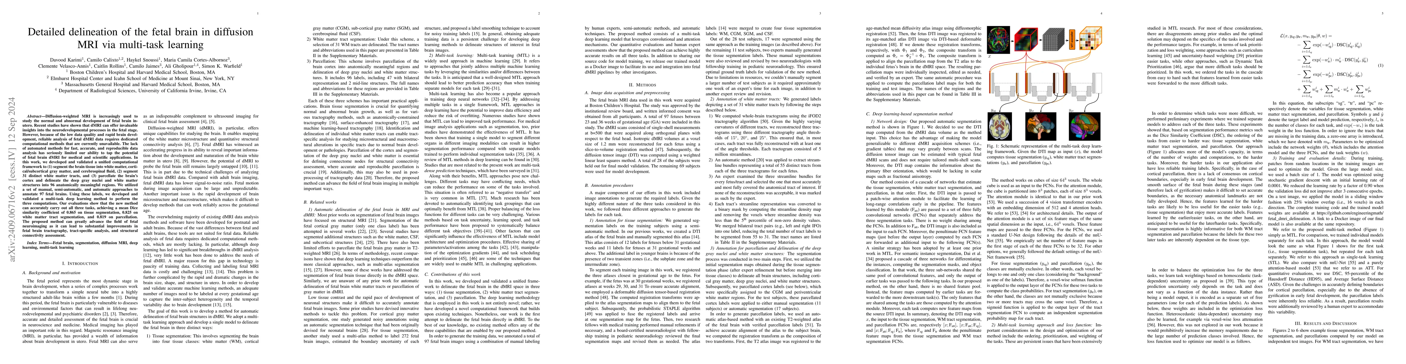
Diffusion-weighted MRI is increasingly used to study the normal and abnormal development of fetal brain in-utero. Recent studies have shown that dMRI can offer invaluable insights into the neurodevelo...
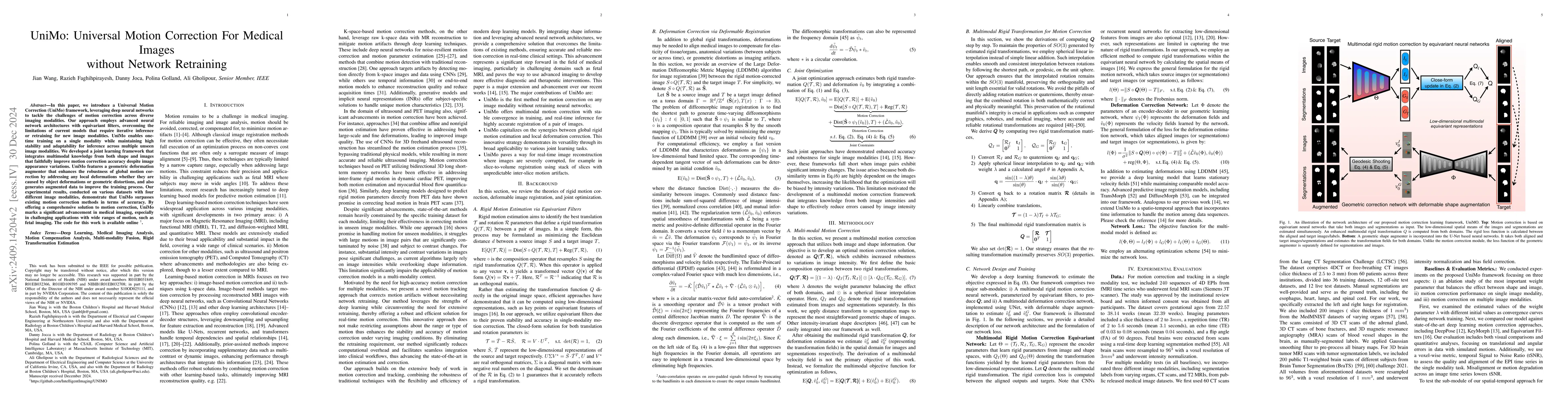
In this paper, we introduce a Universal Motion Correction (UniMo) framework, leveraging deep neural networks to tackle the challenges of motion correction across diverse imaging modalities. Our approa...
There is a growing interest in using diffusion MRI to study the white matter tracts and structural connectivity of the fetal brain. Recent progress in data acquisition and processing suggests that thi...
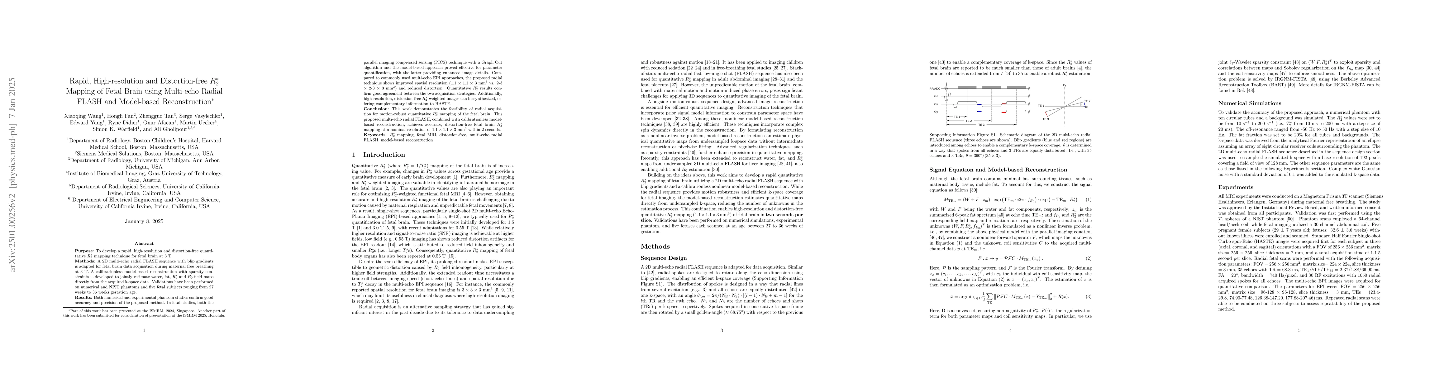
Purpose: To develop a rapid, high-resolution and distortion-free quantitative $R_{2}^{*}$ mapping technique for fetal brain at 3 T. Methods: A 2D multi-echo radial FLASH sequence with blip gradients...
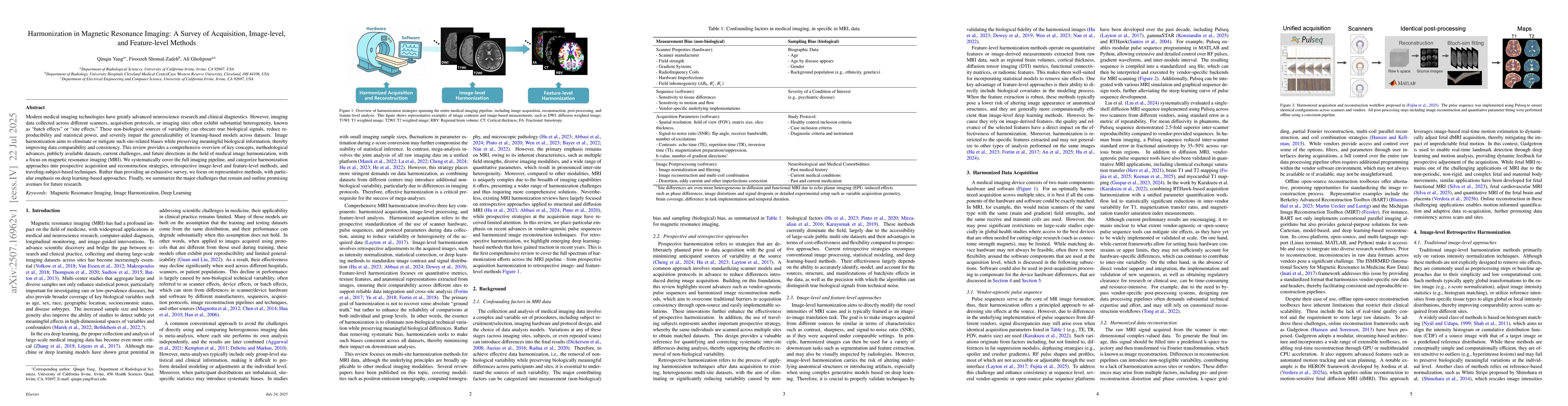
Modern medical imaging technologies have greatly advanced neuroscience research and clinical diagnostics. However, imaging data collected across different scanners, acquisition protocols, or imaging s...
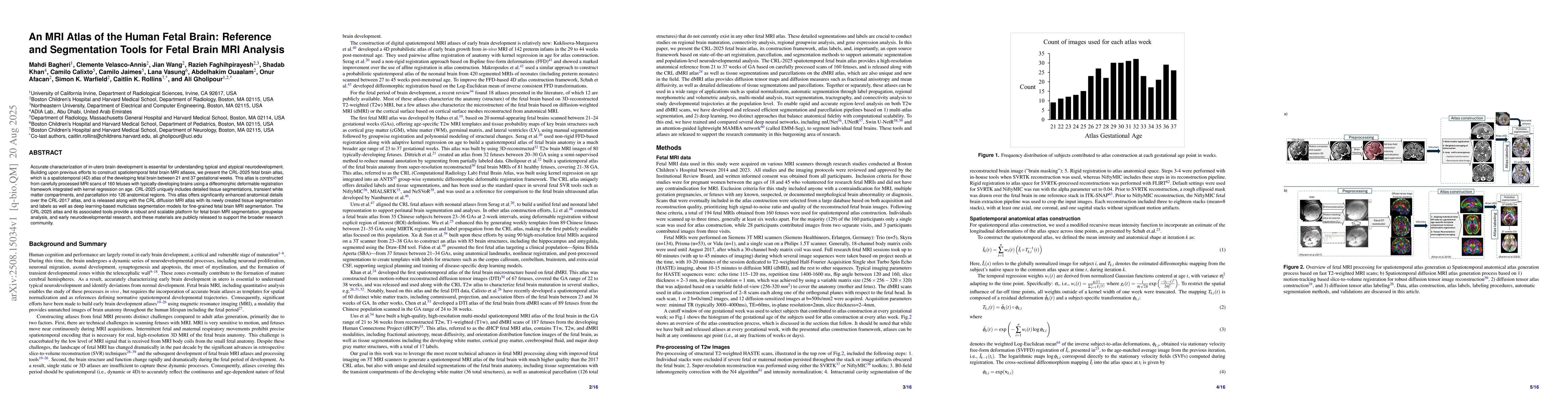
Accurate characterization of in-utero brain development is essential for understanding typical and atypical neurodevelopment. Building upon previous efforts to construct spatiotemporal fetal brain MRI...