Academic Profile
Statistics
Similar Authors
Papers on arXiv
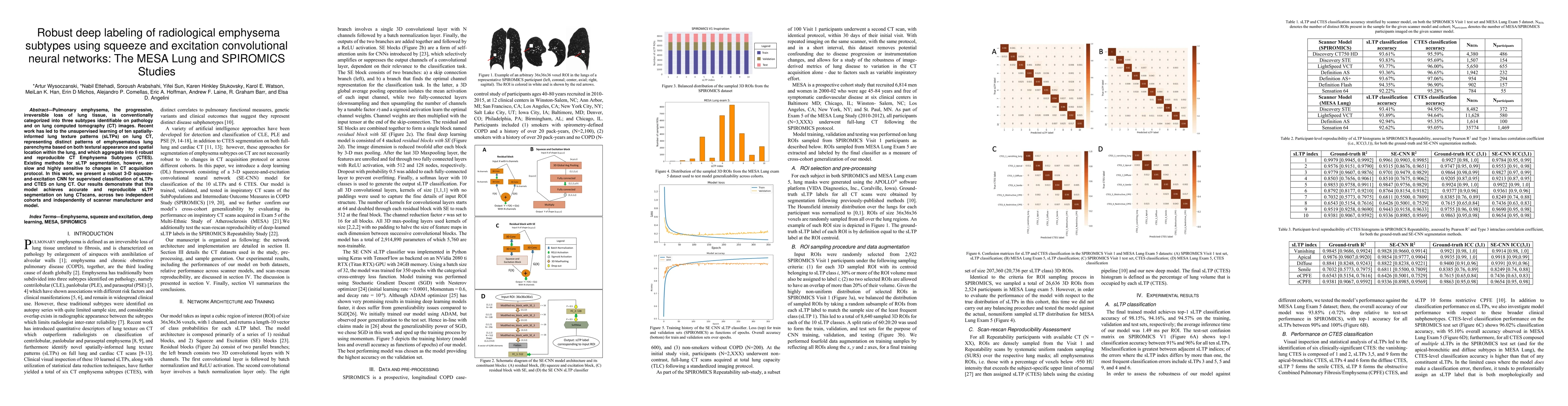
Pulmonary emphysema, the progressive, irreversible loss of lung tissue, is conventionally categorized into three subtypes identifiable on pathology and on lung computed tomography (CT) images. Recen...
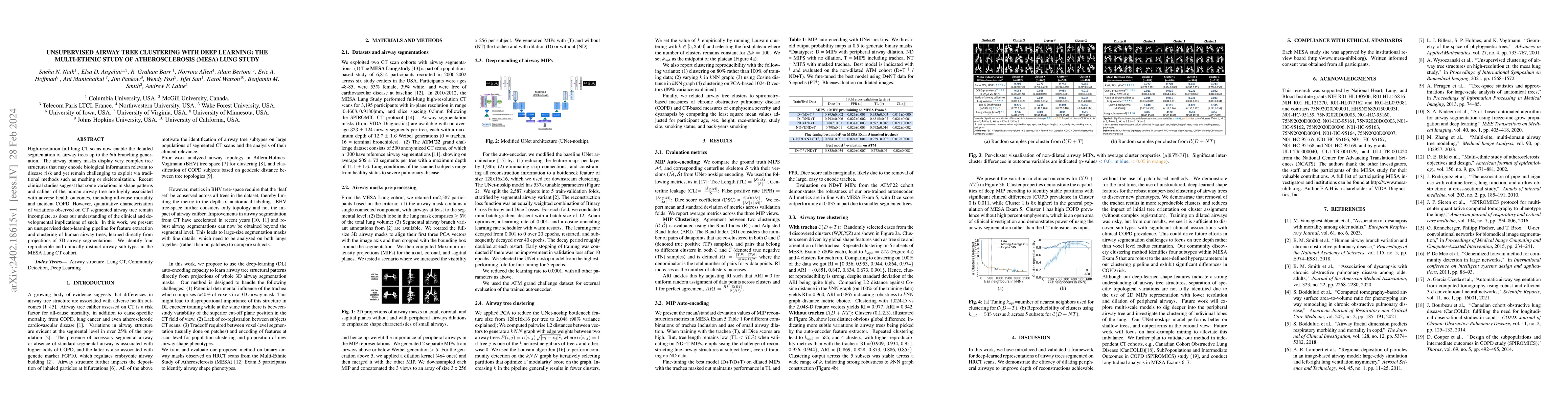
High-resolution full lung CT scans now enable the detailed segmentation of airway trees up to the 6th branching generation. The airway binary masks display very complex tree structures that may enco...
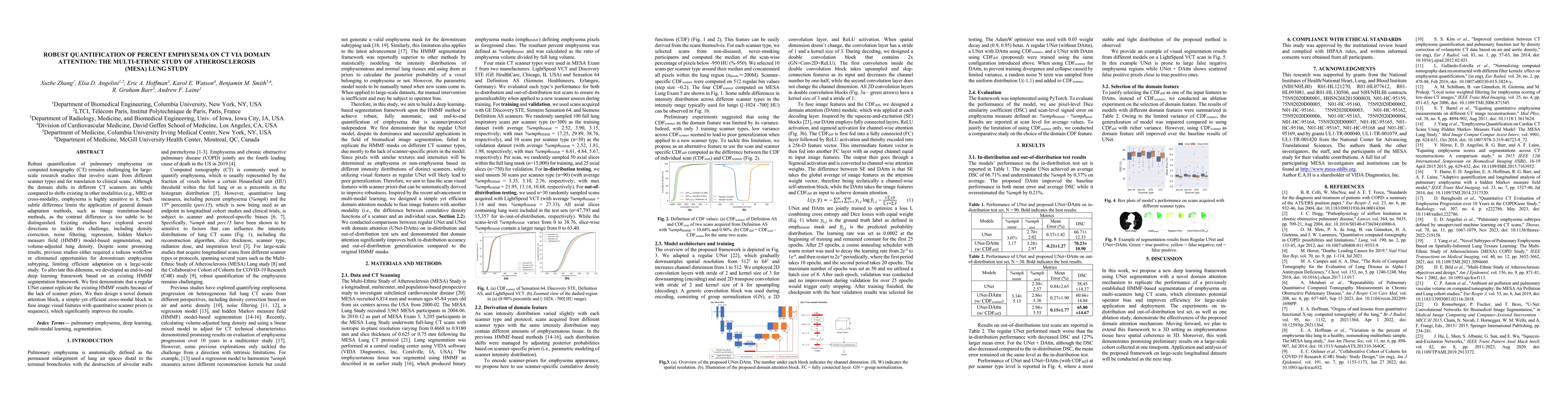
Robust quantification of pulmonary emphysema on computed tomography (CT) remains challenging for large-scale research studies that involve scans from different scanner types and for translation to c...
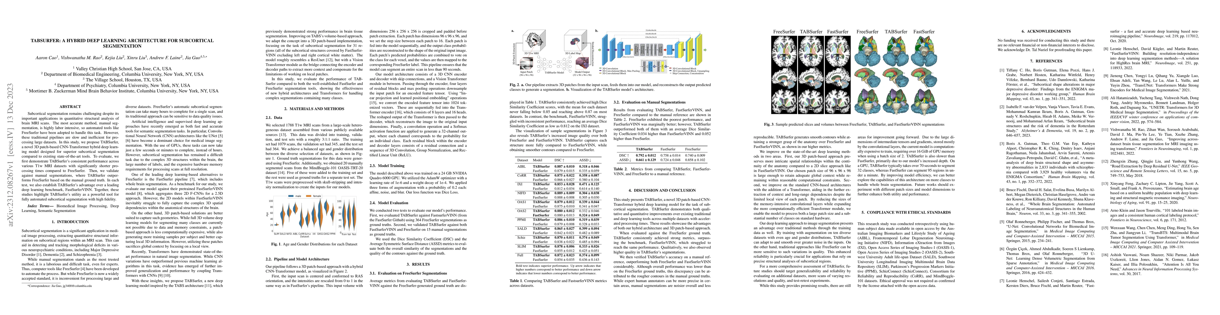
Subcortical segmentation remains challenging despite its important applications in quantitative structural analysis of brain MRI scans. The most accurate method, manual segmentation, is highly labor...
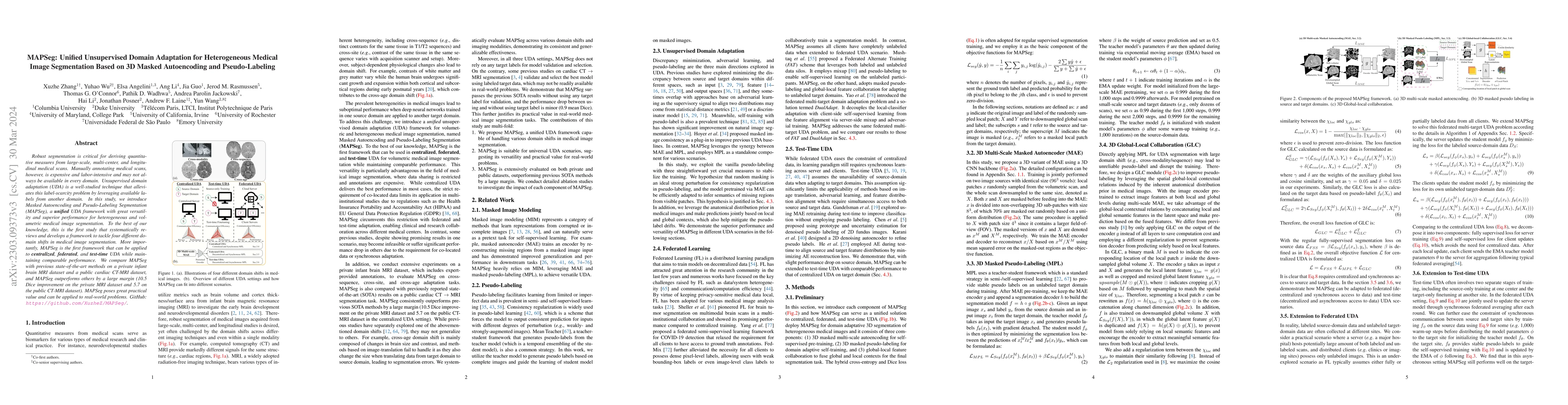
Robust segmentation is critical for deriving quantitative measures from large-scale, multi-center, and longitudinal medical scans. Manually annotating medical scans, however, is expensive and labor-...
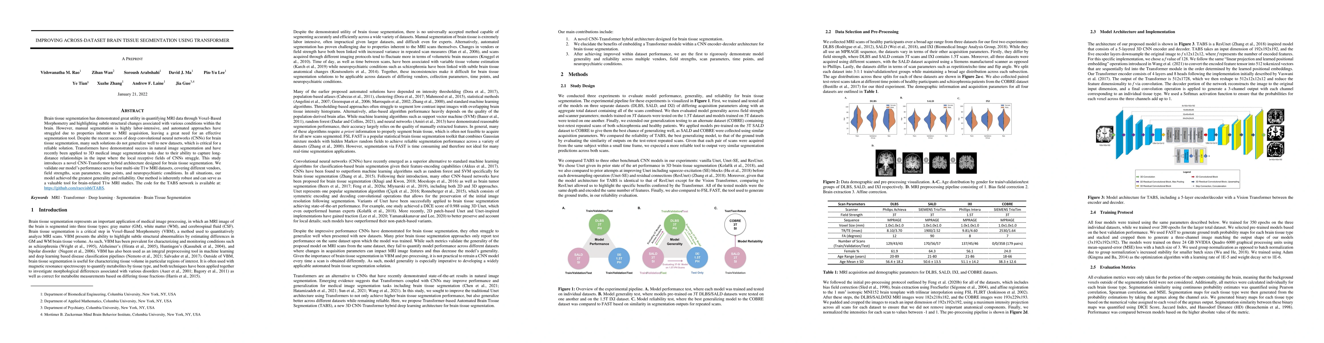
Brain tissue segmentation has demonstrated great utility in quantifying MRI data through Voxel-Based Morphometry and highlighting subtle structural changes associated with various conditions within ...
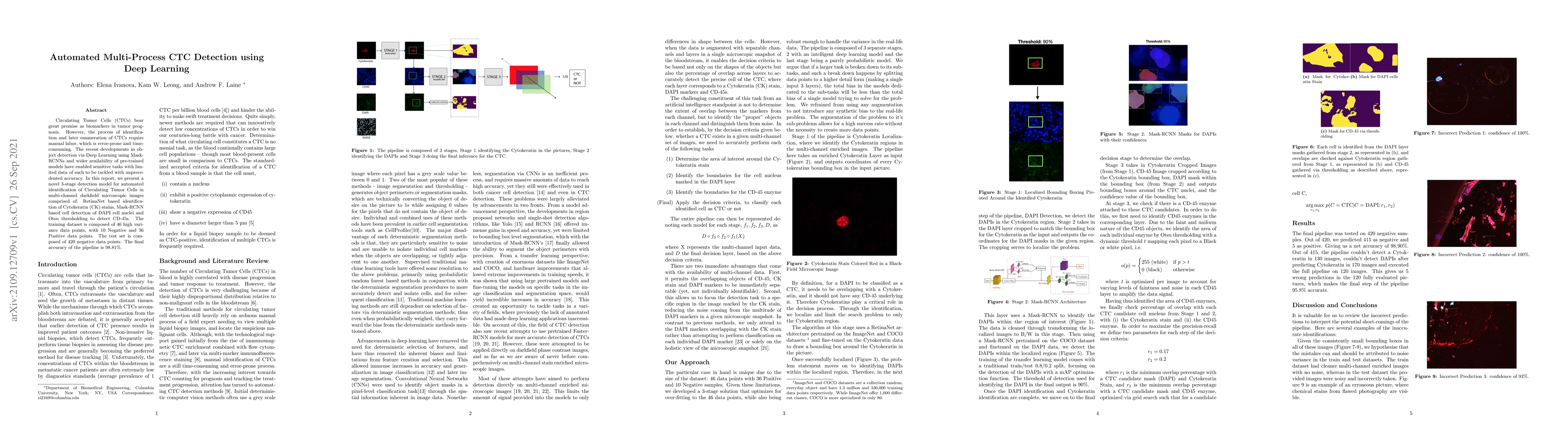
Circulating Tumor Cells (CTCs) bear great promise as biomarkers in tumor prognosis. However, the process of identification and later enumeration of CTCs require manual labor, which is error-prone an...
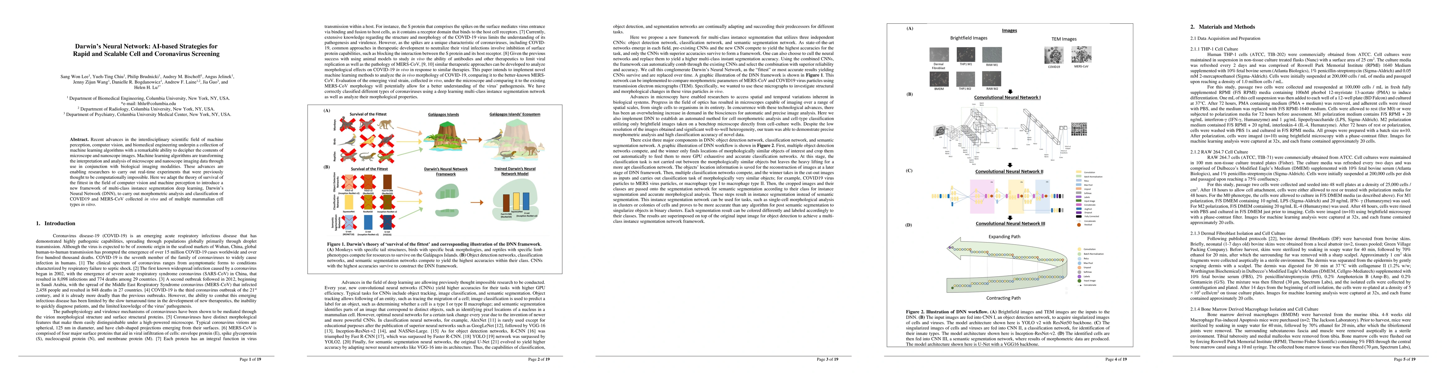
Recent advances in the interdisciplinary scientific field of machine perception, computer vision, and biomedical engineering underpin a collection of machine learning algorithms with a remarkable ab...
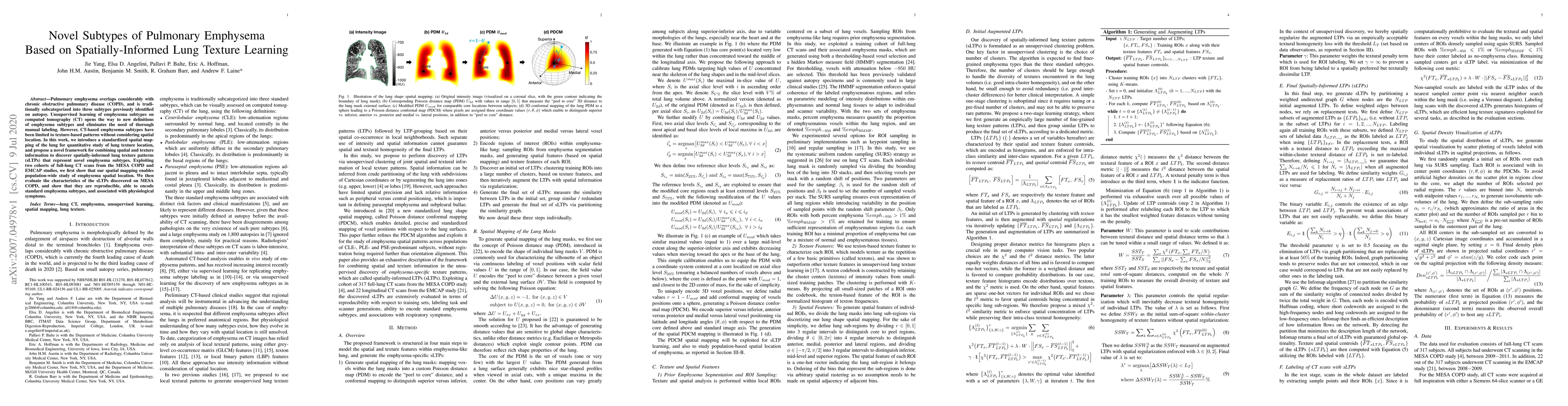
Pulmonary emphysema overlaps considerably with chronic obstructive pulmonary disease (COPD), and is traditionally subcategorized into three subtypes previously identified on autopsy. Unsupervised le...
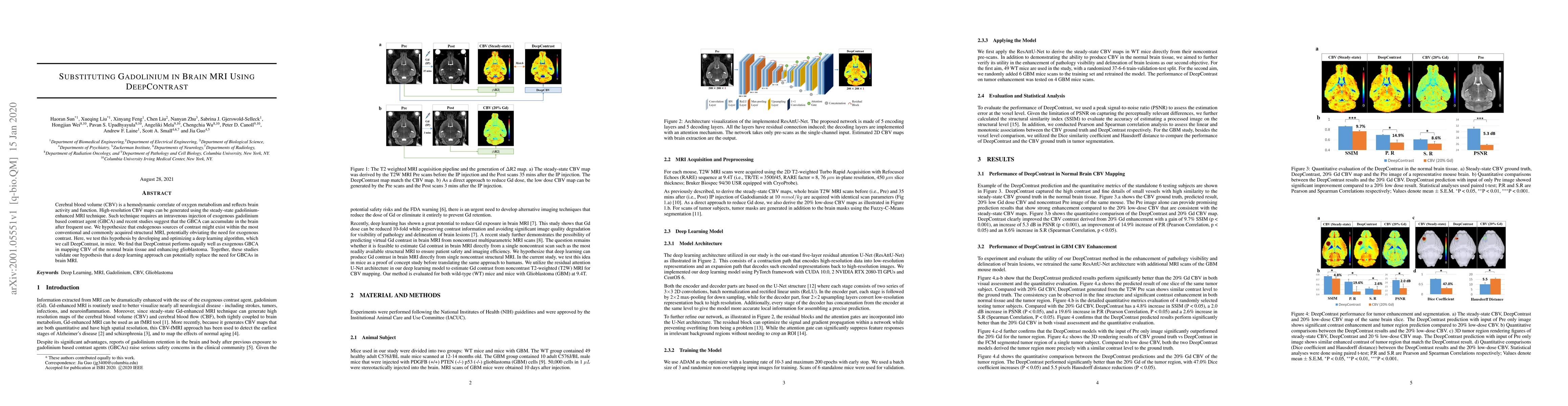
Cerebral blood volume (CBV) is a hemodynamic correlate of oxygen metabolism and reflects brain activity and function. High-resolution CBV maps can be generated using the steady-state gadolinium-enha...
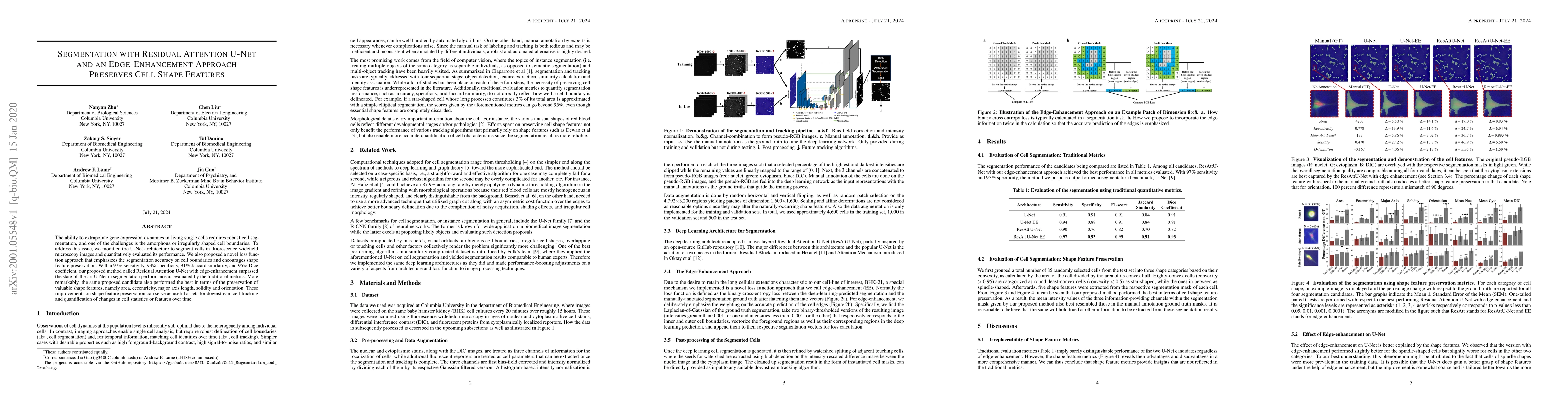
The ability to extrapolate gene expression dynamics in living single cells requires robust cell segmentation, and one of the challenges is the amorphous or irregularly shaped cell boundaries. To add...
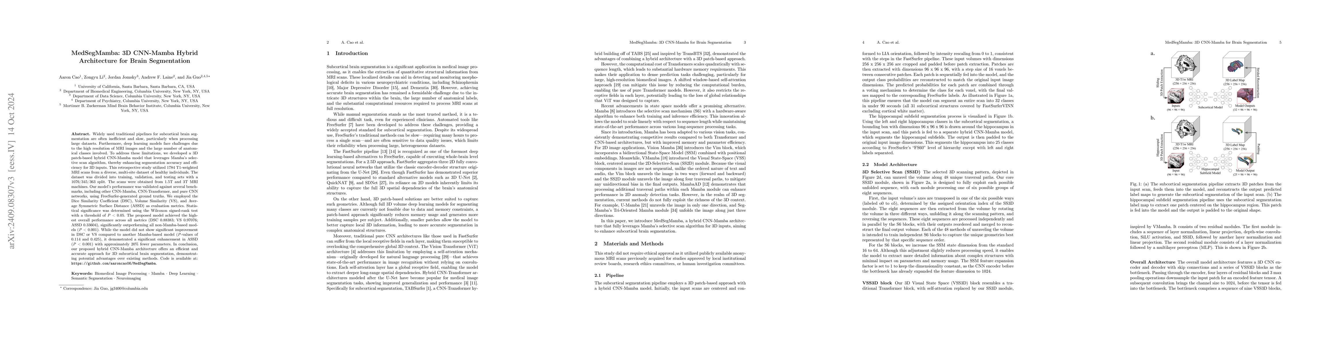
Widely used traditional pipelines for subcortical brain segmentation are often inefficient and slow, particularly when processing large datasets. Furthermore, deep learning models face challenges due ...
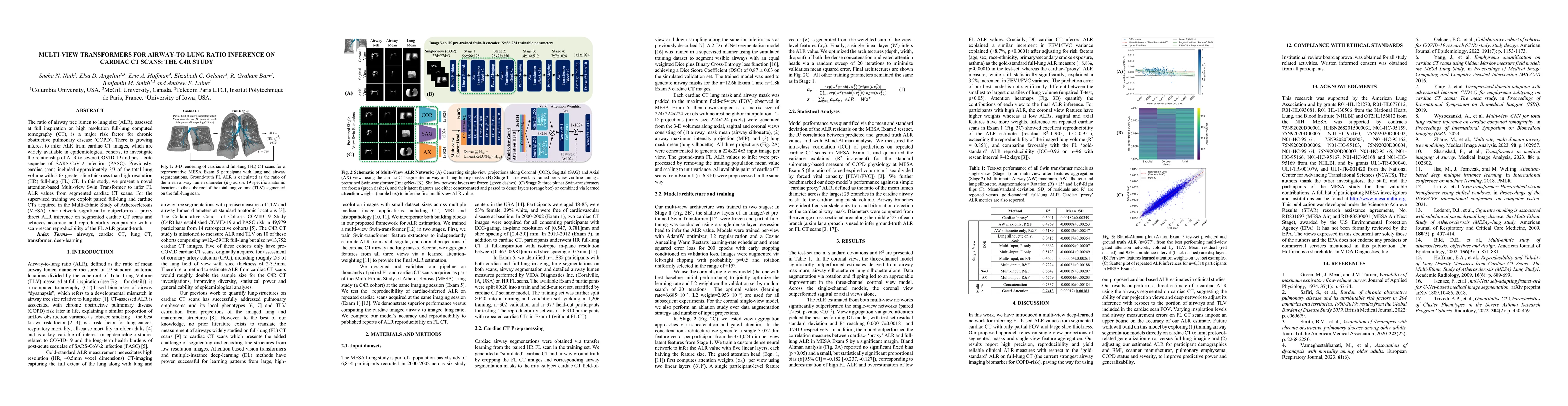
The ratio of airway tree lumen to lung size (ALR), assessed at full inspiration on high resolution full-lung computed tomography (CT), is a major risk factor for chronic obstructive pulmonary disease ...
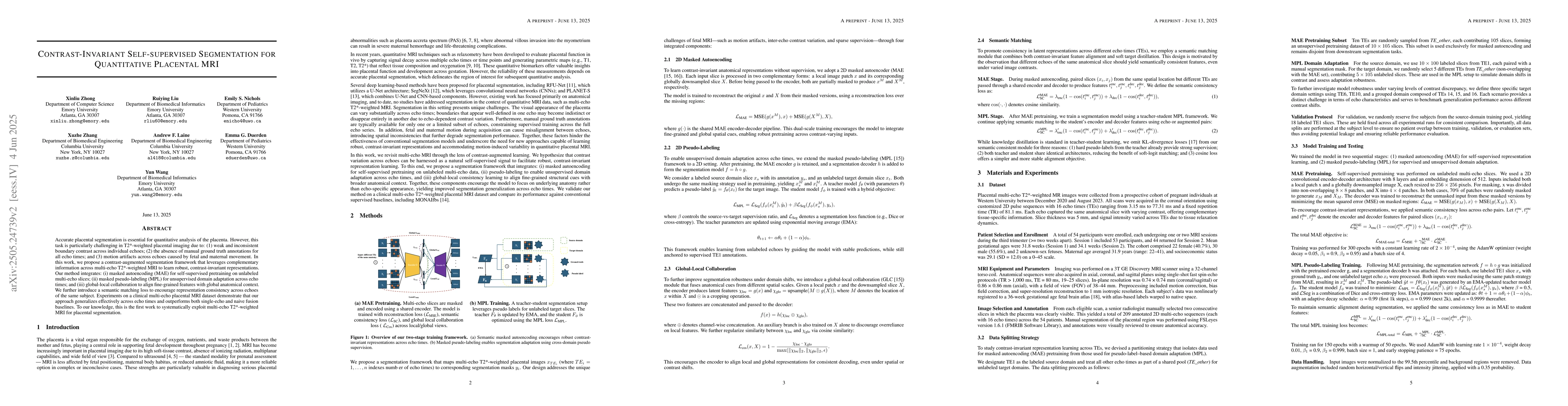
Accurate placental segmentation is essential for quantitative analysis of the placenta. However, this task is particularly challenging in T2*-weighted placental imaging due to: (1) weak and inconsiste...
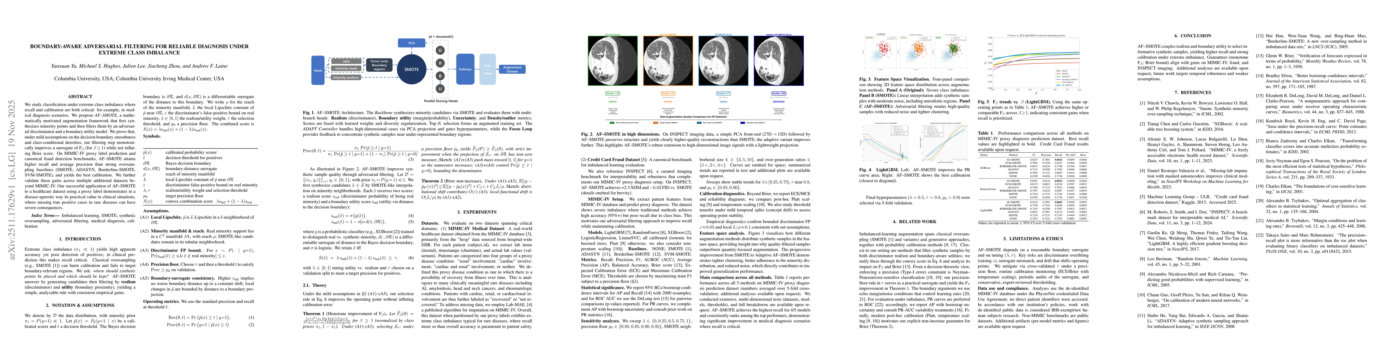
We study classification under extreme class imbalance where recall and calibration are both critical, for example in medical diagnosis scenarios. We propose AF-SMOTE, a mathematically motivated augmen...