Academic Profile
Statistics
Similar Authors
Papers on arXiv
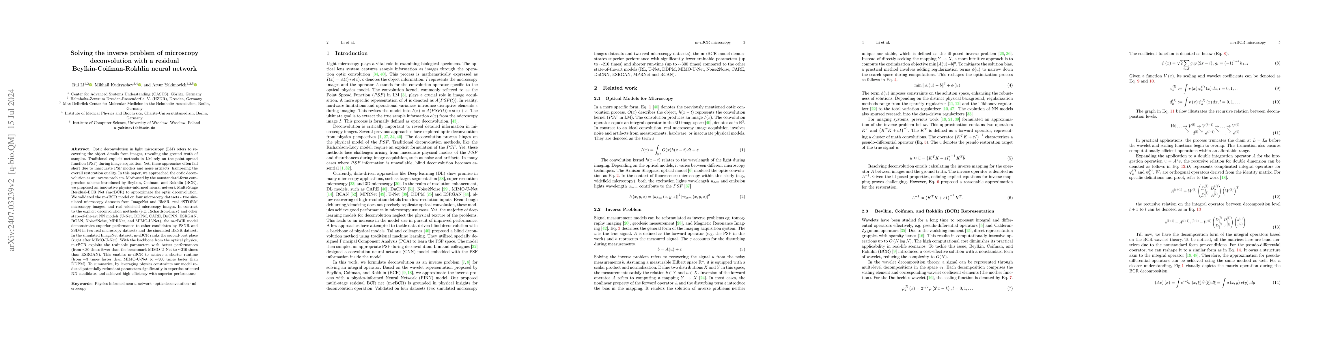
Optic deconvolution in light microscopy (LM) refers to recovering the object details from images, revealing the ground truth of samples. Traditional explicit methods in LM rely on the point spread fun...
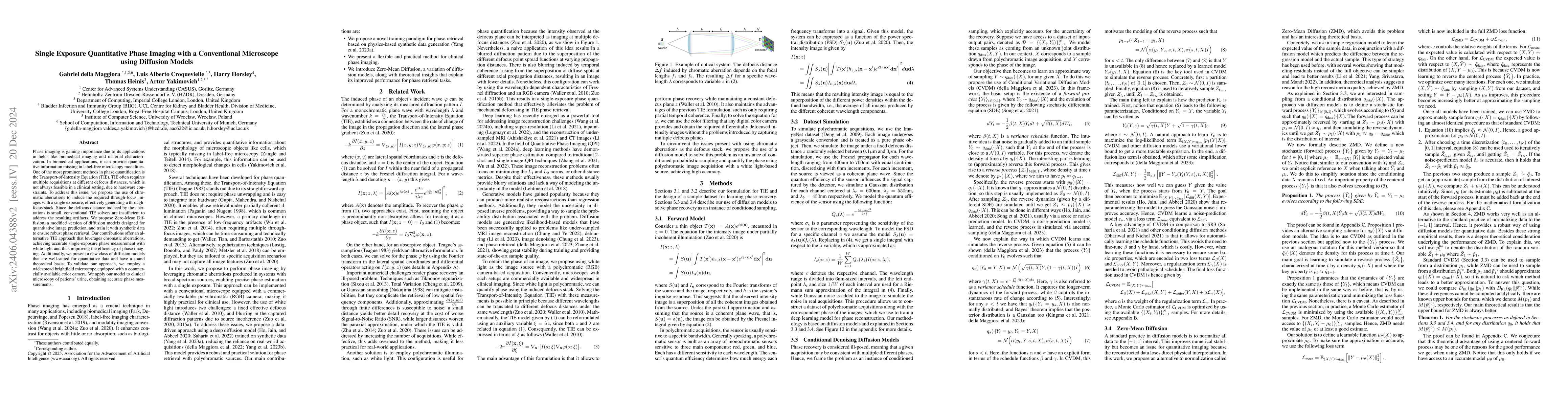
Phase imaging is gaining importance due to its applications in fields like biomedical imaging and material characterization. In biomedical applications, it can provide quantitative information missi...
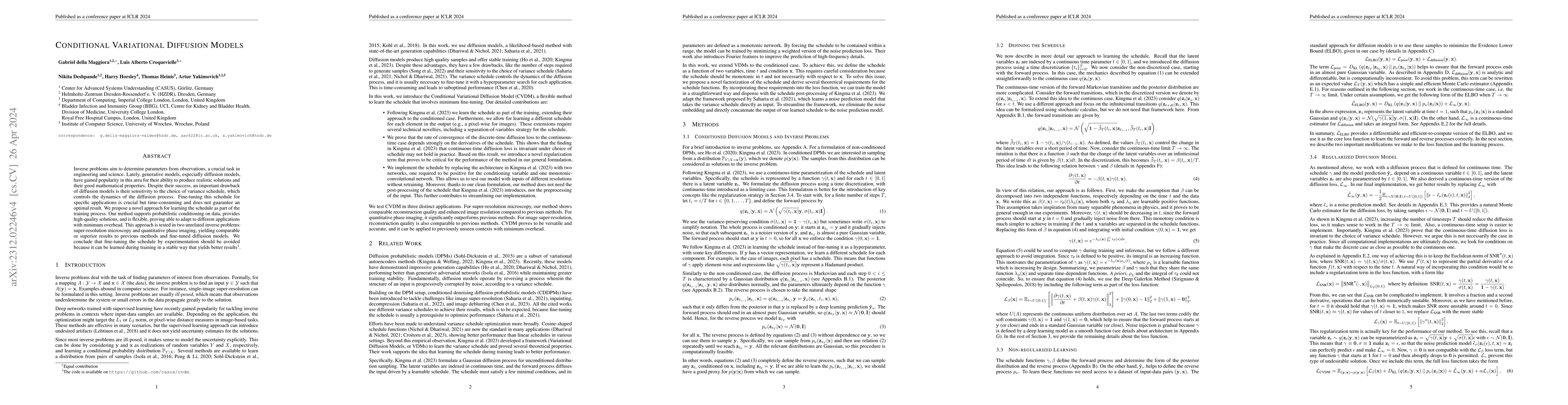
Inverse problems aim to determine parameters from observations, a crucial task in engineering and science. Lately, generative models, especially diffusion models, have gained popularity in this area...
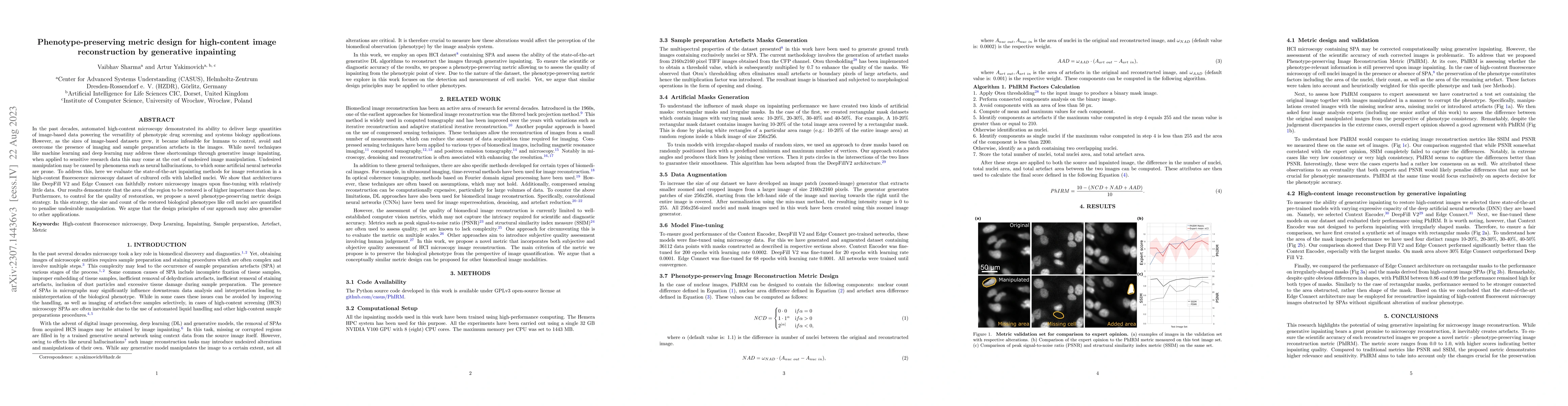
In the past decades, automated high-content microscopy demonstrated its ability to deliver large quantities of image-based data powering the versatility of phenotypic drug screening and systems biol...
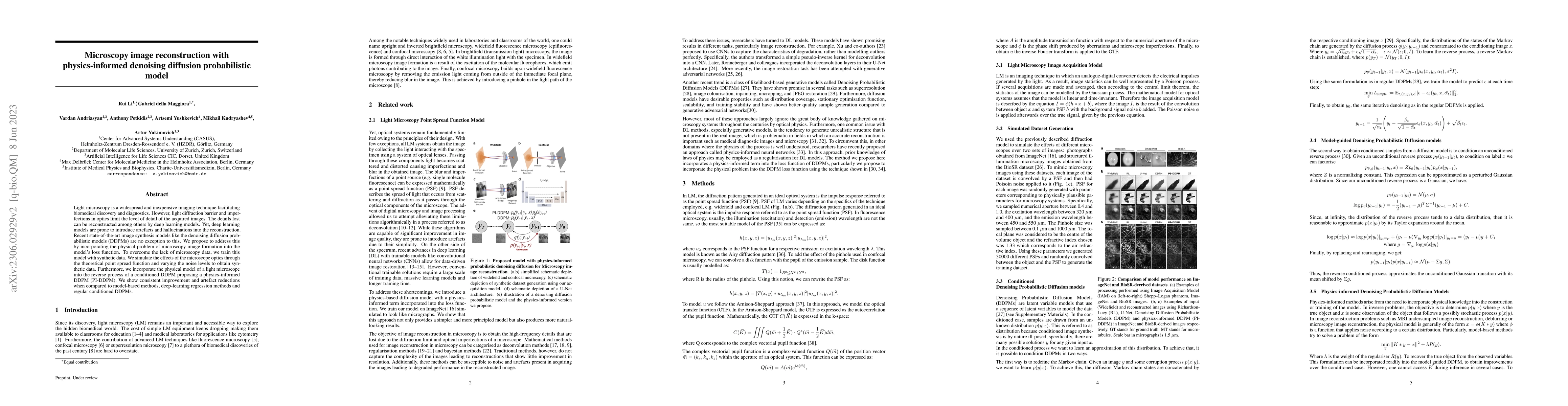
Light microscopy is a widespread and inexpensive imaging technique facilitating biomedical discovery and diagnostics. However, light diffraction barrier and imperfections in optics limit the level o...
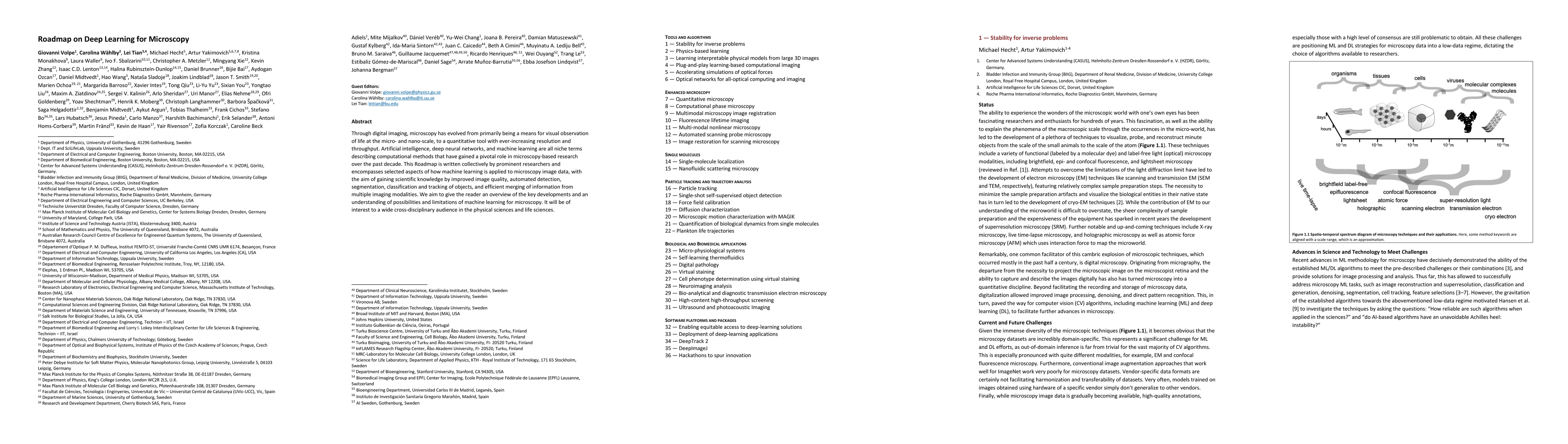
Through digital imaging, microscopy has evolved from primarily being a means for visual observation of life at the micro- and nano-scale, to a quantitative tool with ever-increasing resolution and t...
Progress in our knowledge of tumor mechanisms and complexity led to the understanding of the physical parameters of cancer cells and their microenvironment, including the mechanical, thermal, and elec...
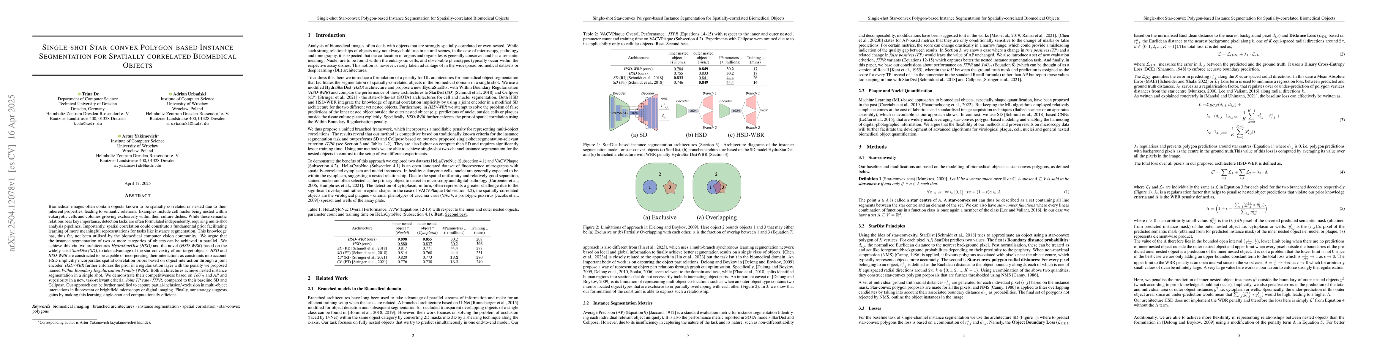
Biomedical images often contain objects known to be spatially correlated or nested due to their inherent properties, leading to semantic relations. Examples include cell nuclei being nested within euk...
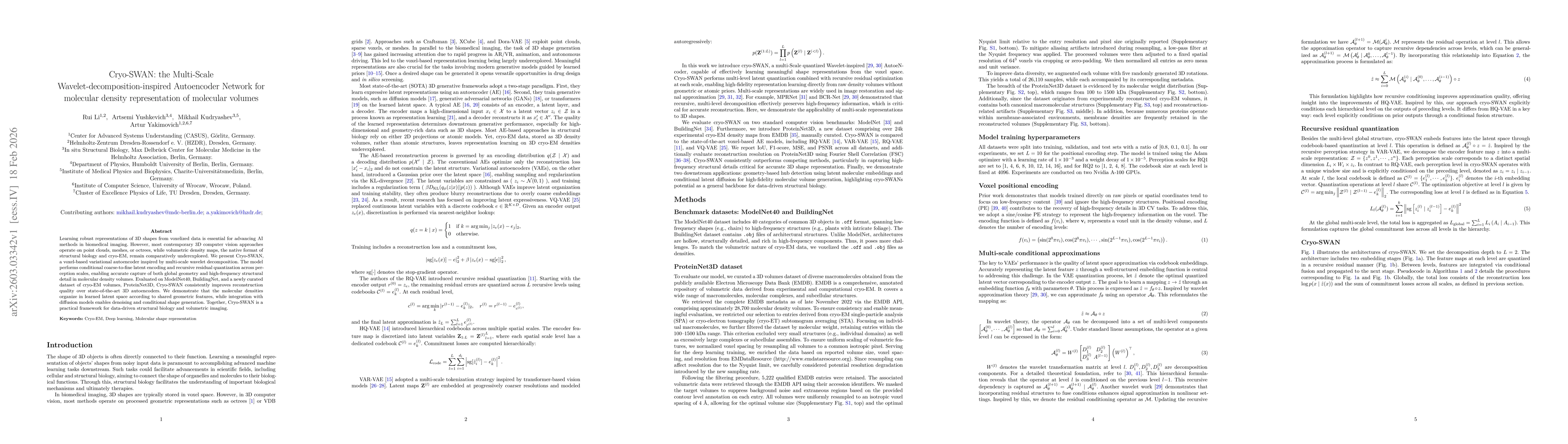
Learning robust representations of 3D shapes from voxelized data is essential for advancing AI methods in biomedical imaging. However, most contemporary 3D computer vision approaches operate on point ...