Academic Profile
Statistics
Similar Authors
Papers on arXiv
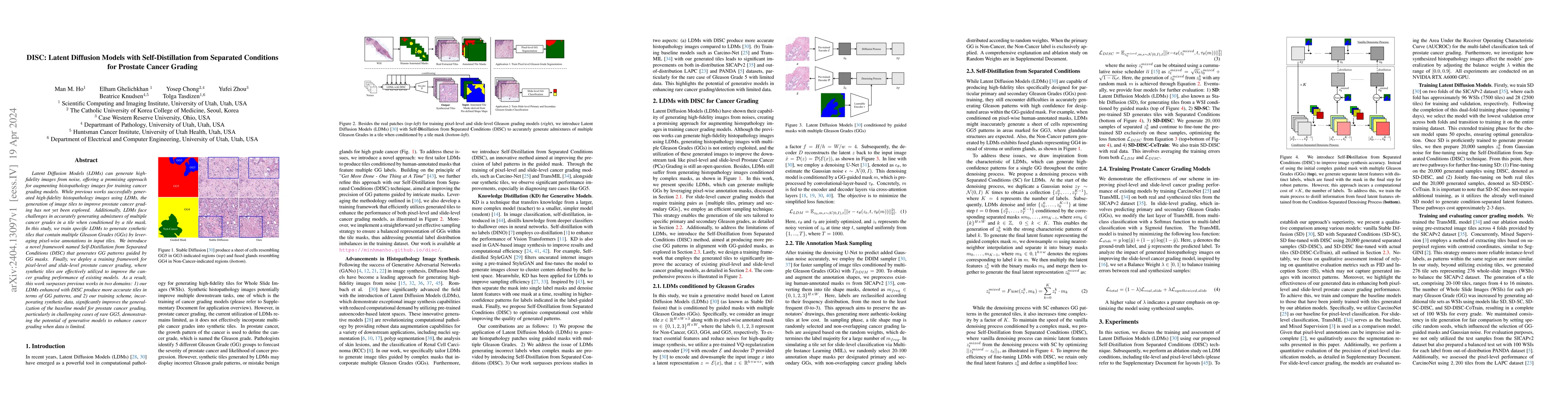
Latent Diffusion Models (LDMs) can generate high-fidelity images from noise, offering a promising approach for augmenting histopathology images for training cancer grading models. While previous wor...
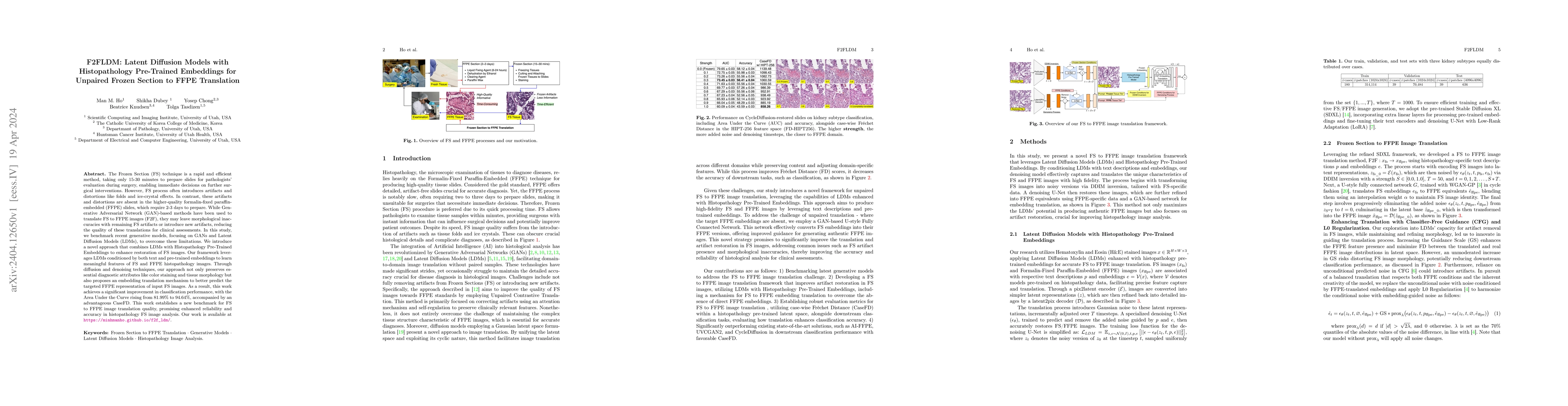
The Frozen Section (FS) technique is a rapid and efficient method, taking only 15-30 minutes to prepare slides for pathologists' evaluation during surgery, enabling immediate decisions on further su...
Hematoxylin and Eosin (H&E) staining is the most commonly used for disease diagnosis and tumor recurrence tracking. Hematoxylin excels at highlighting nuclei, whereas eosin stains the cytoplasm. How...
With the advent of digital scanners and deep learning, diagnostic operations may move from a microscope to a desktop. Hematoxylin and Eosin (H&E) staining is one of the most frequently used stains f...
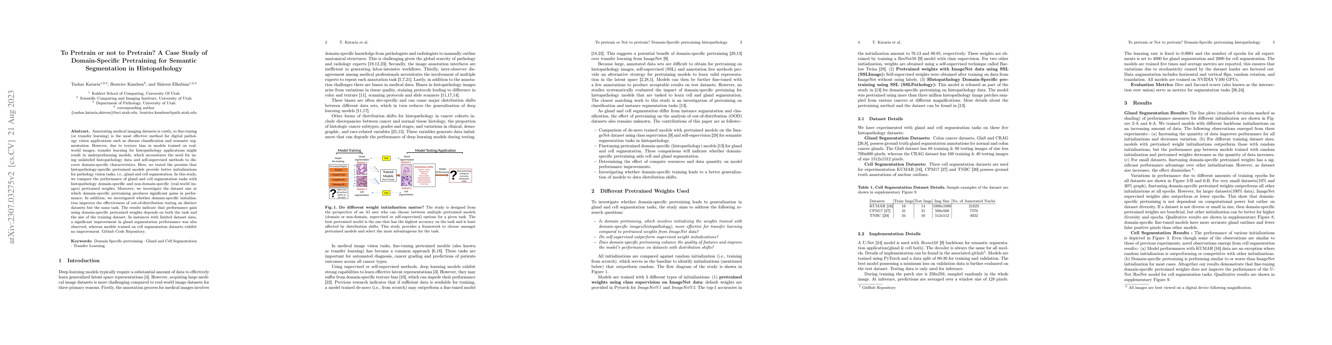
Annotating medical imaging datasets is costly, so fine-tuning (or transfer learning) is the most effective method for digital pathology vision applications such as disease classification and semanti...
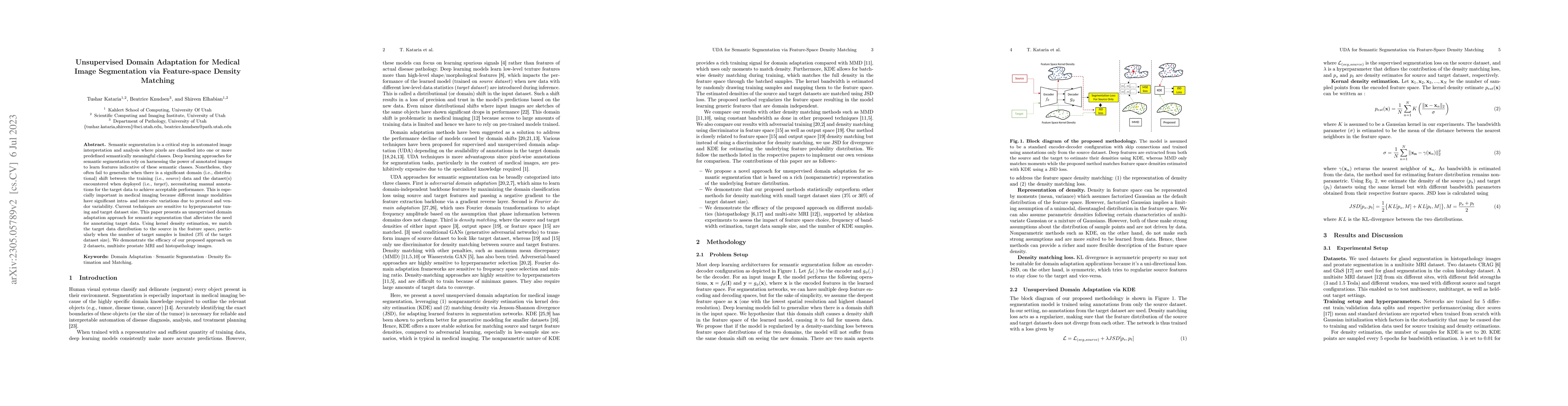
Semantic segmentation is a critical step in automated image interpretation and analysis where pixels are classified into one or more predefined semantically meaningful classes. Deep learning approac...
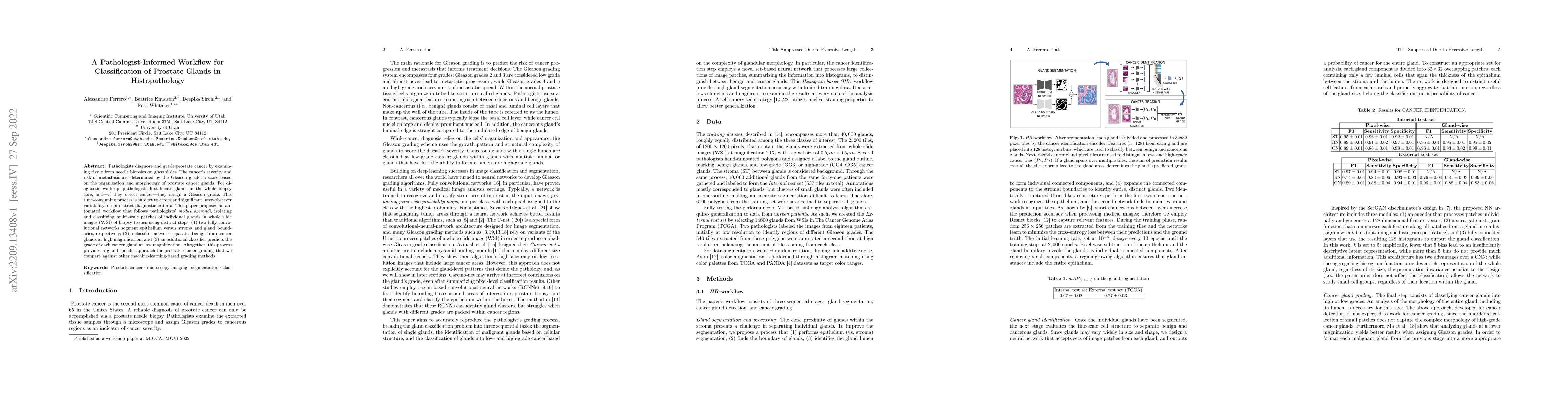
Pathologists diagnose and grade prostate cancer by examining tissue from needle biopsies on glass slides. The cancer's severity and risk of metastasis are determined by the Gleason grade, a score ba...
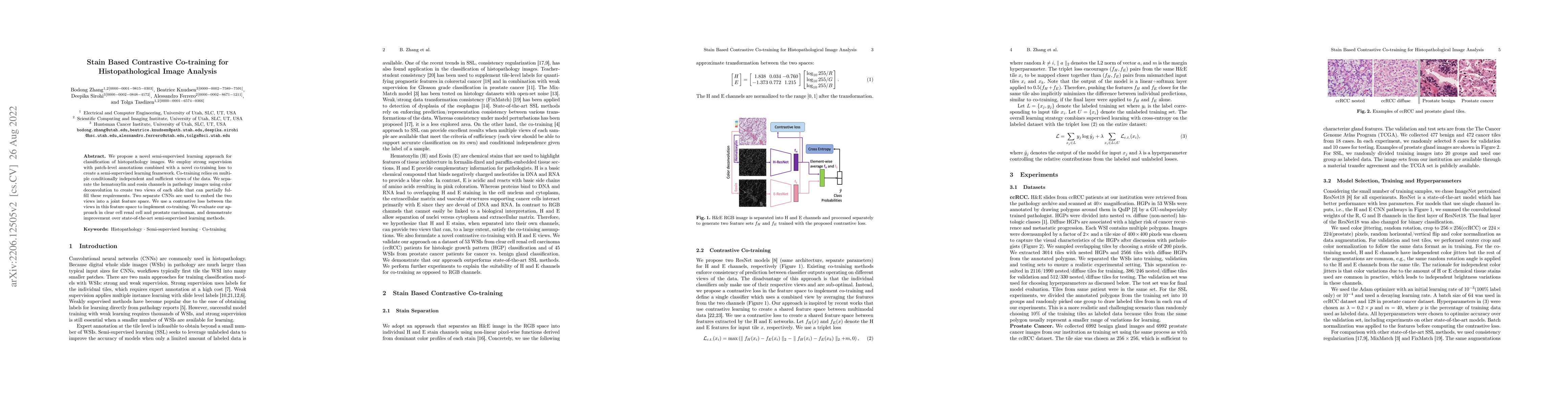
We propose a novel semi-supervised learning approach for classification of histopathology images. We employ strong supervision with patch-level annotations combined with a novel co-training loss to ...
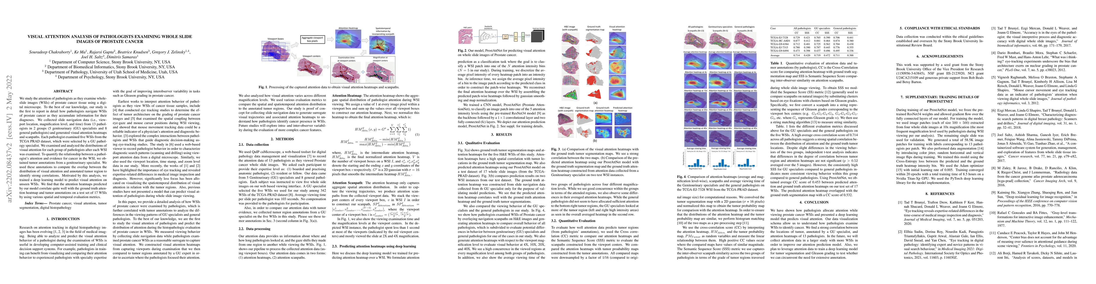
We study the attention of pathologists as they examine whole-slide images (WSIs) of prostate cancer tissue using a digital microscope. To the best of our knowledge, our study is the first to report ...
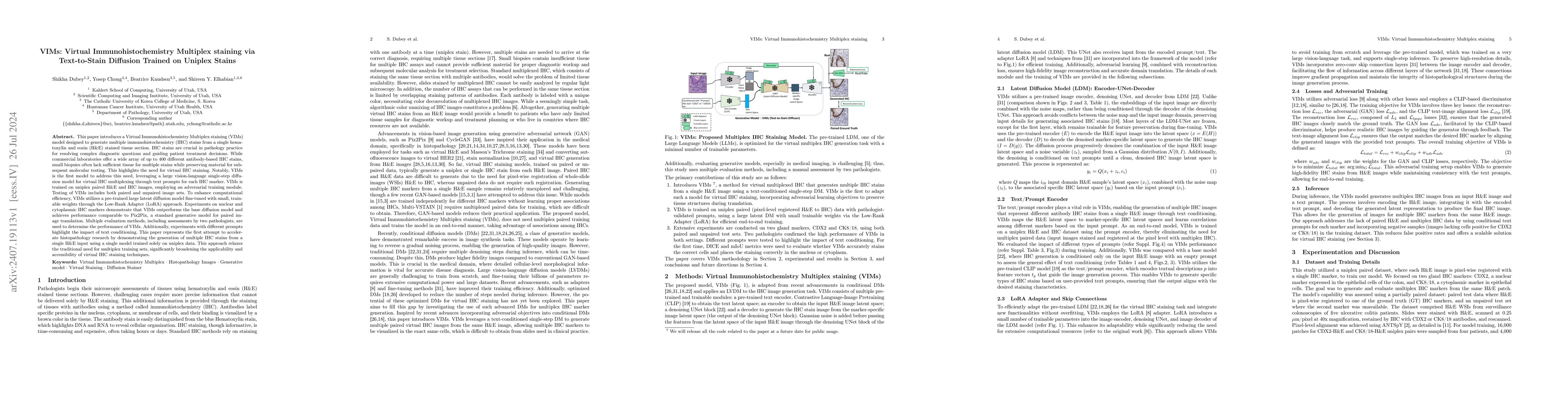
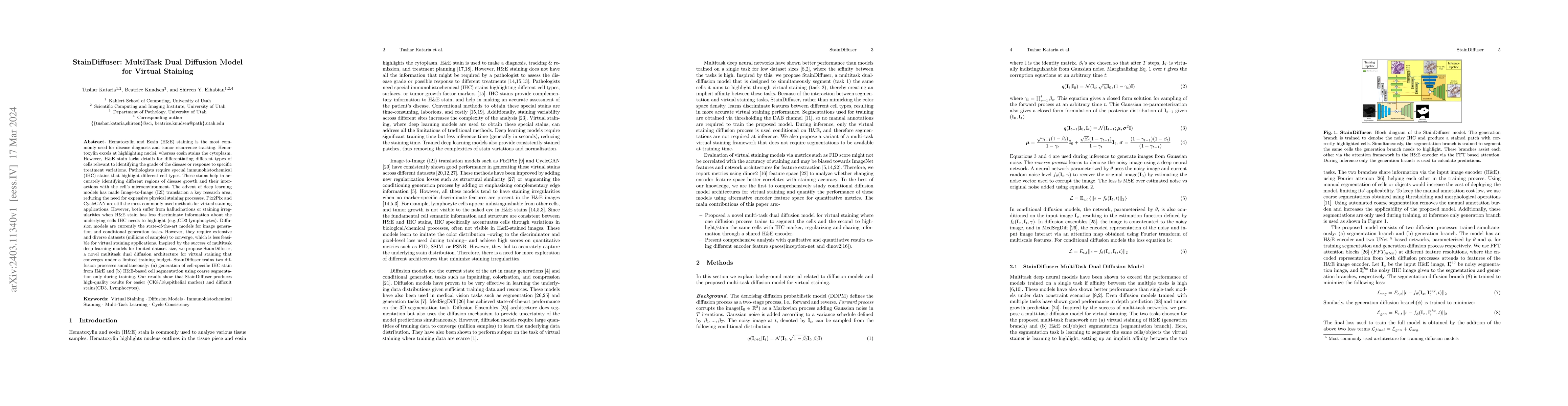
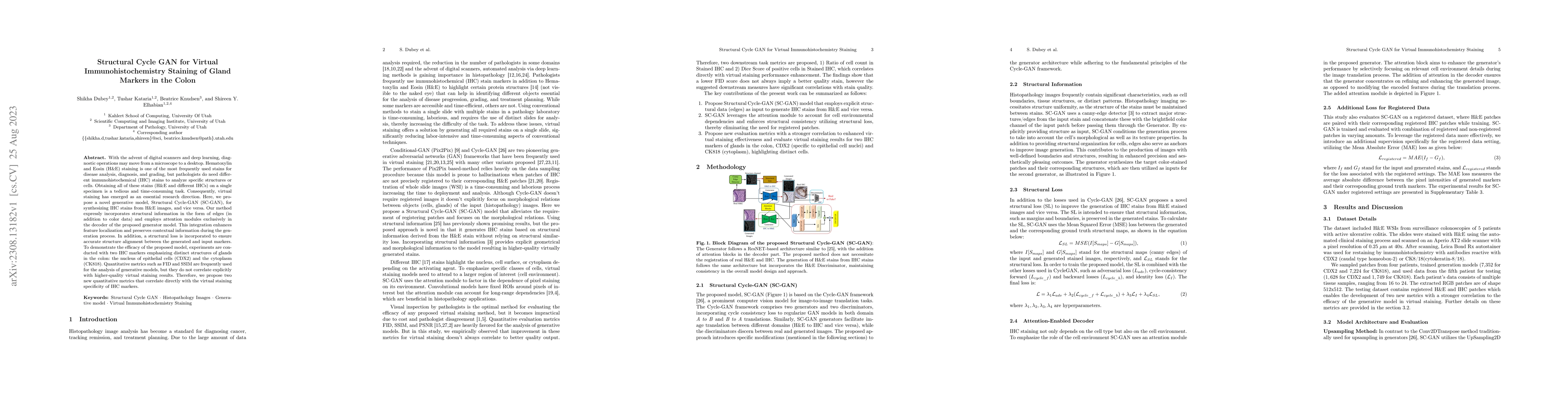
This paper introduces a Virtual Immunohistochemistry Multiplex staining (VIMs) model designed to generate multiple immunohistochemistry (IHC) stains from a single hematoxylin and eosin (H&E) stained t...
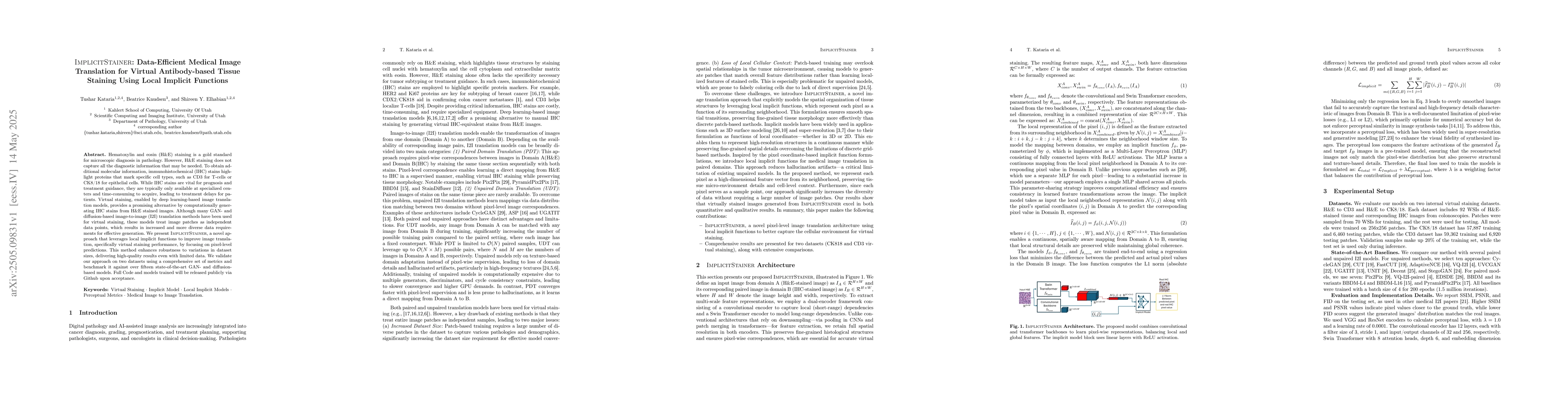
Hematoxylin and eosin (H&E) staining is a gold standard for microscopic diagnosis in pathology. However, H&E staining does not capture all the diagnostic information that may be needed. To obtain addi...
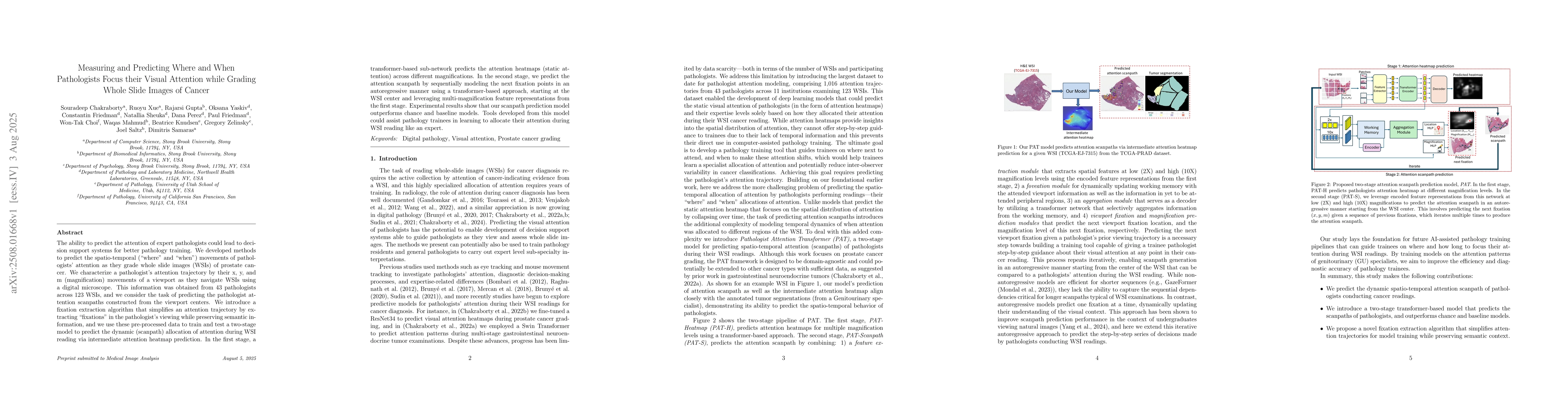
The ability to predict the attention of expert pathologists could lead to decision support systems for better pathology training. We developed methods to predict the spatio-temporal (where and when) m...