Academic Profile
Statistics
Similar Authors
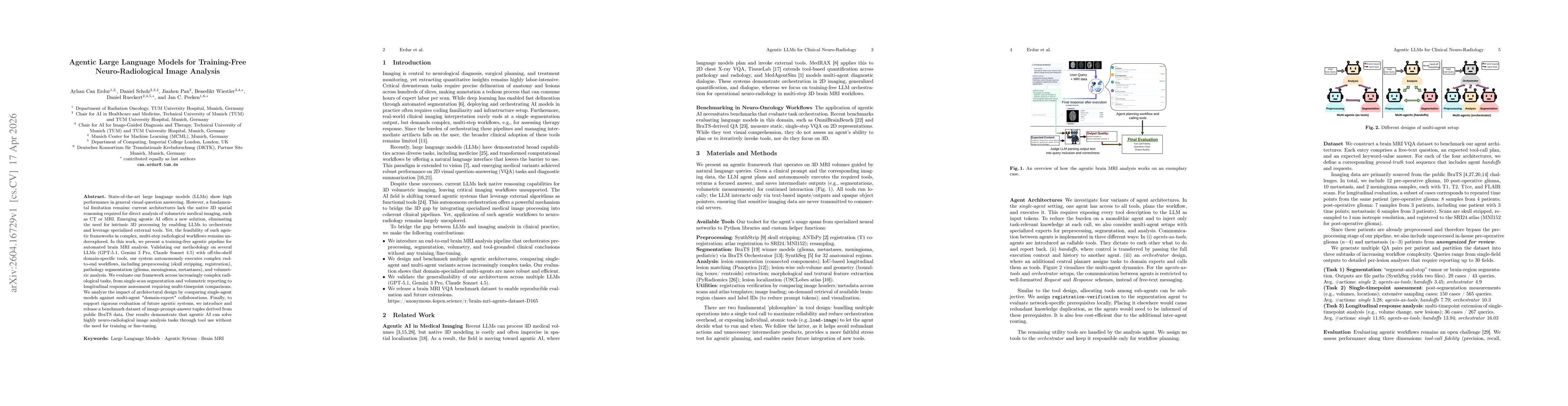
Papers on arXiv
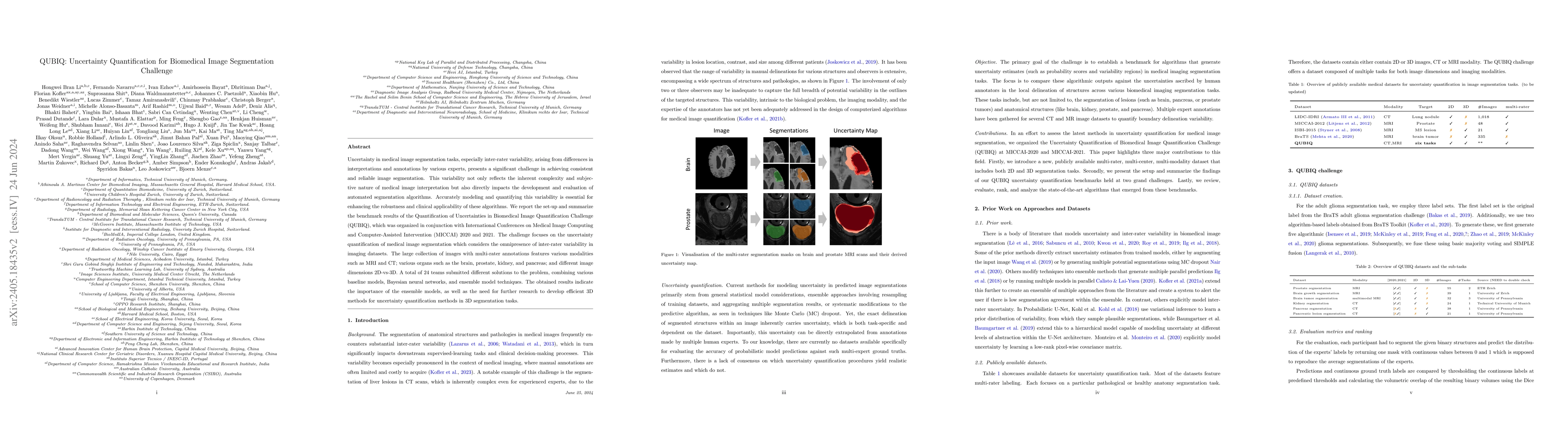
Uncertainty in medical image segmentation tasks, especially inter-rater variability, arising from differences in interpretations and annotations by various experts, presents a significant challenge in...
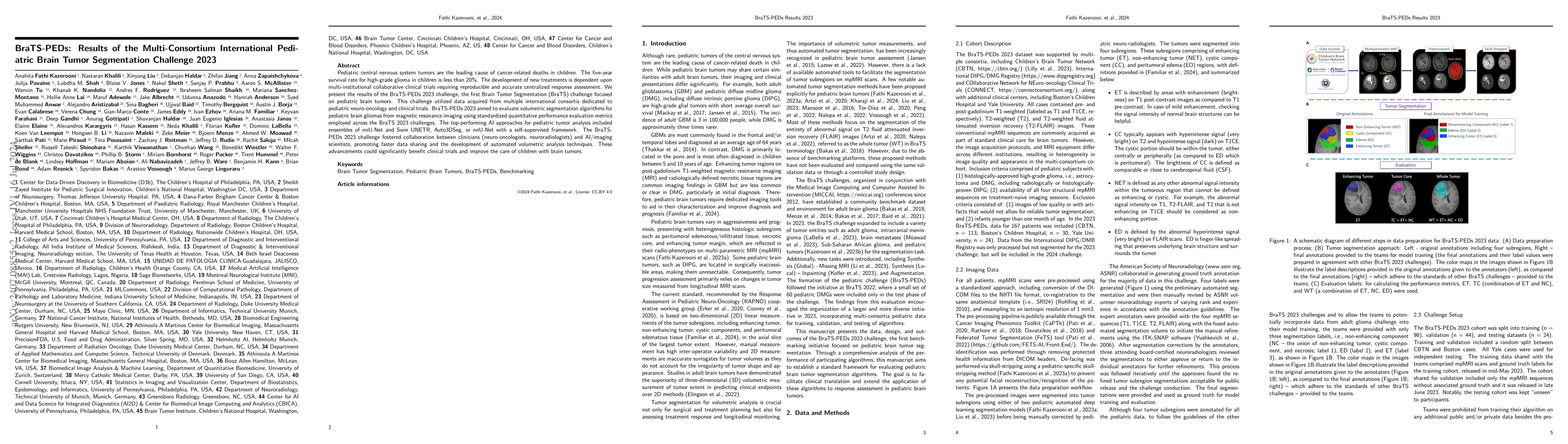
Pediatric central nervous system tumors are the leading cause of cancer-related deaths in children. The five-year survival rate for high-grade glioma in children is less than 20%. The development of n...
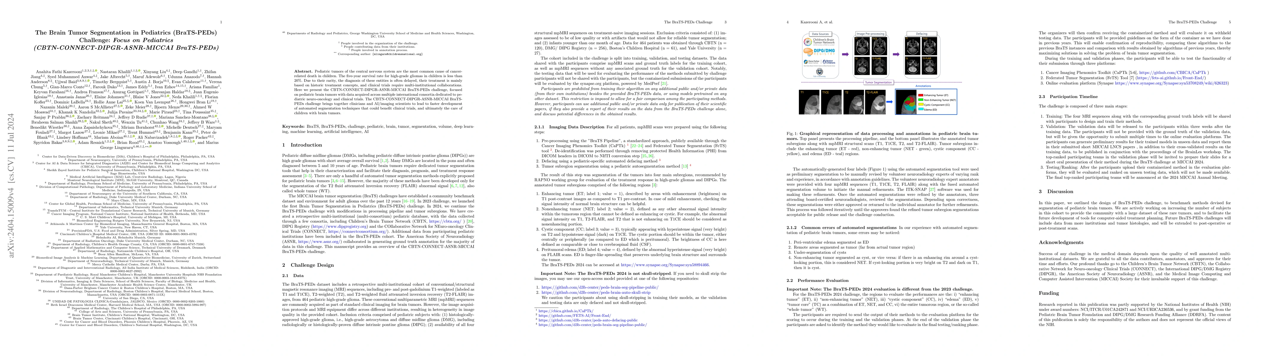
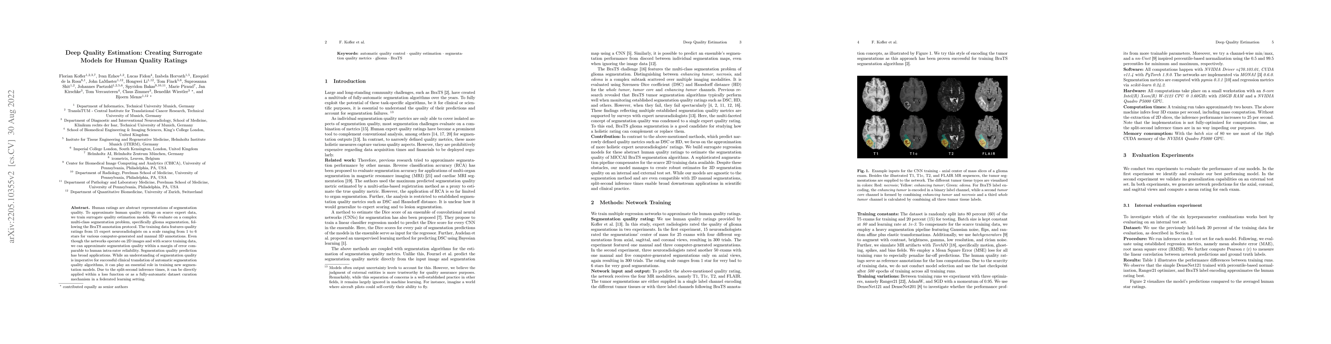
Pediatric tumors of the central nervous system are the most common cause of cancer-related death in children. The five-year survival rate for high-grade gliomas in children is less than 20%. Due to th...
Accurate image registration is pivotal in biomedical image analysis, where selecting suitable registration algorithms demands careful consideration. While numerous algorithms are available, the evalua...
The 2024 Brain Tumor Segmentation Meningioma Radiotherapy (BraTS-MEN-RT) challenge aims to advance automated segmentation algorithms using the largest known multi-institutional dataset of radiothera...
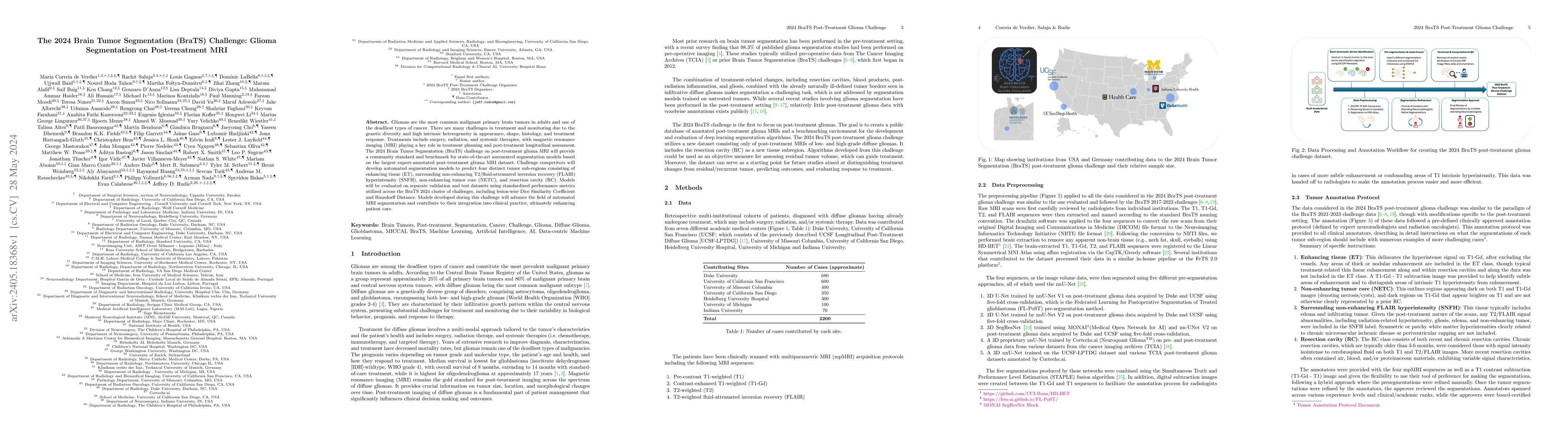
Gliomas are the most common malignant primary brain tumors in adults and one of the deadliest types of cancer. There are many challenges in treatment and monitoring due to the genetic diversity and ...
We describe the design and results from the BraTS 2023 Intracranial Meningioma Segmentation Challenge. The BraTS Meningioma Challenge differed from prior BraTS Glioma challenges in that it focused o...
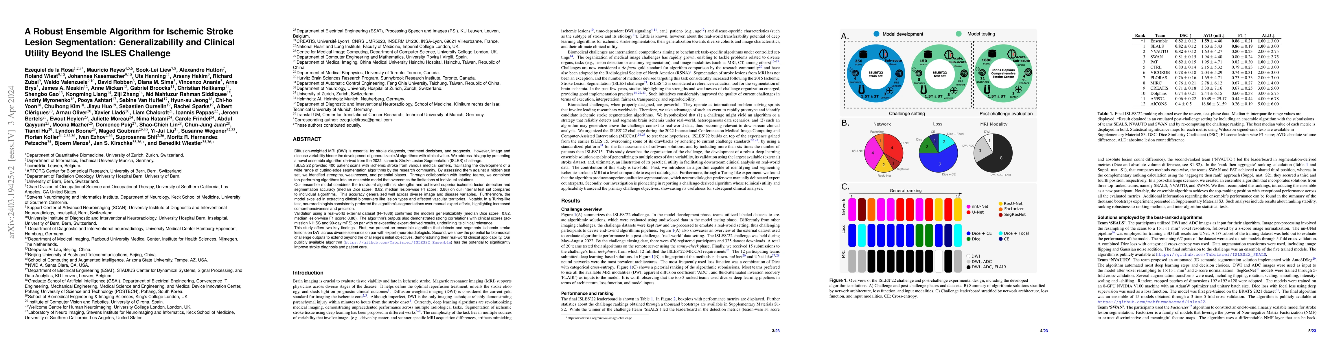
Diffusion-weighted MRI (DWI) is essential for stroke diagnosis, treatment decisions, and prognosis. However, image and disease variability hinder the development of generalizable AI algorithms with ...
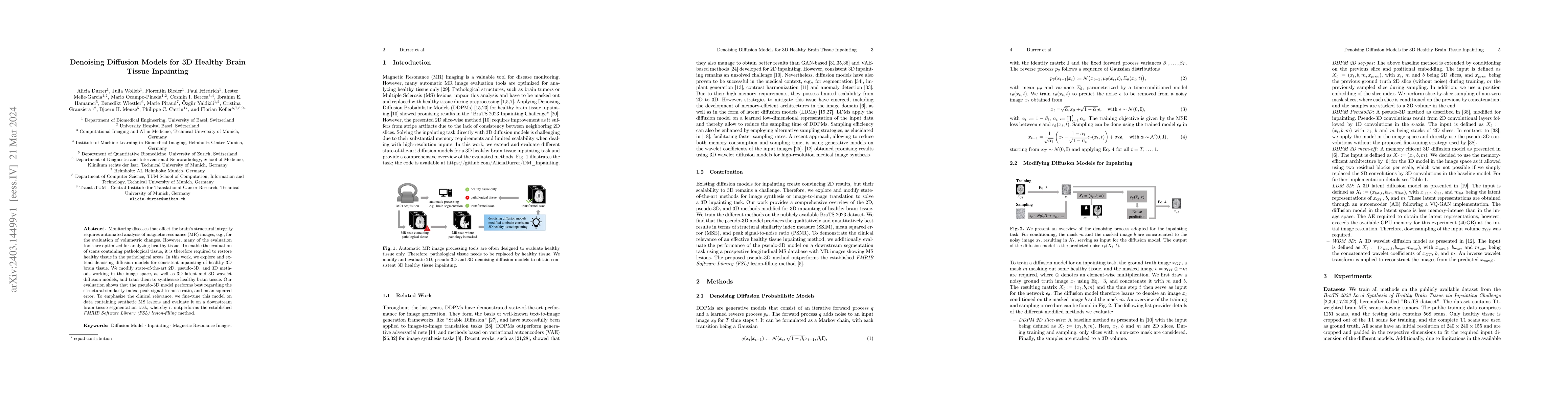
Monitoring diseases that affect the brain's structural integrity requires automated analysis of magnetic resonance (MR) images, e.g., for the evaluation of volumetric changes. However, many of the e...
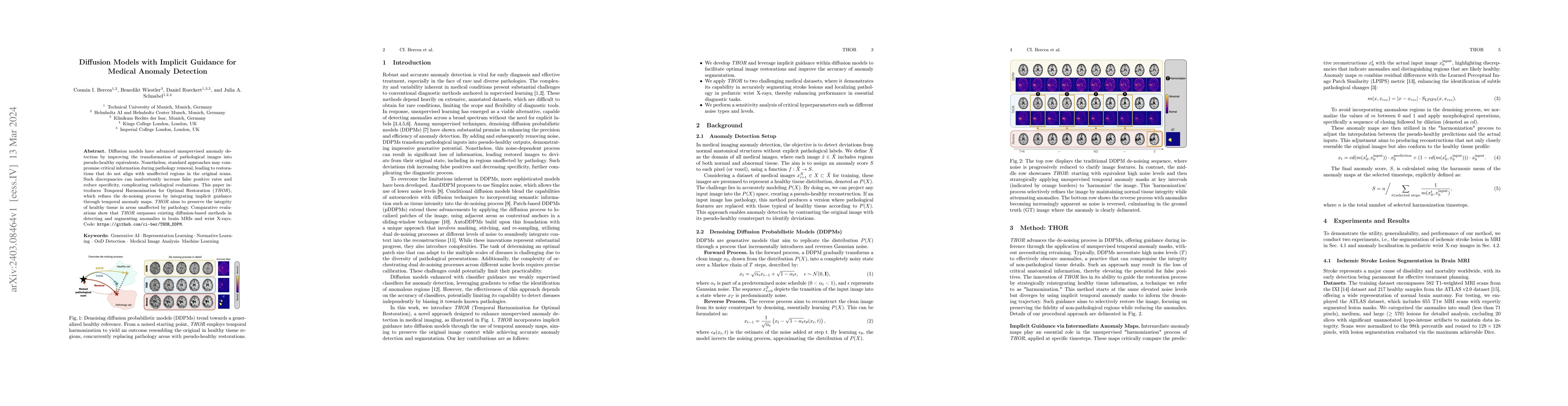
Diffusion models have advanced unsupervised anomaly detection by improving the transformation of pathological images into pseudo-healthy equivalents. Nonetheless, standard approaches may compromise ...
Biophysical modeling, particularly involving partial differential equations (PDEs), offers significant potential for tailoring disease treatment protocols to individual patients. However, the invers...
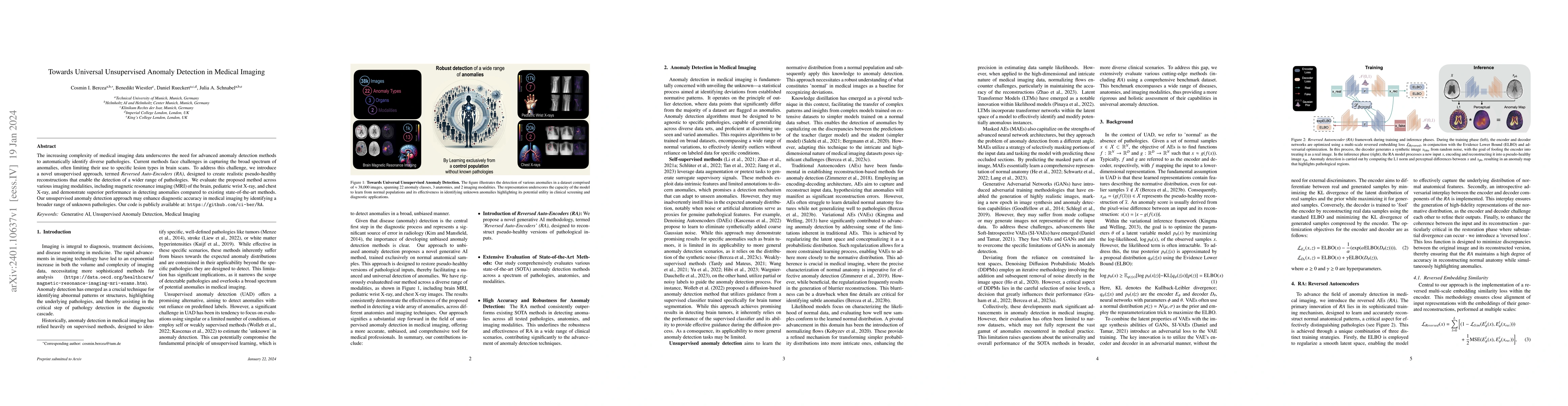
The increasing complexity of medical imaging data underscores the need for advanced anomaly detection methods to automatically identify diverse pathologies. Current methods face challenges in captur...
This paper introduces panoptica, a versatile and performance-optimized package designed for computing instance-wise segmentation quality metrics from 2D and 3D segmentation maps. panoptica addresses...
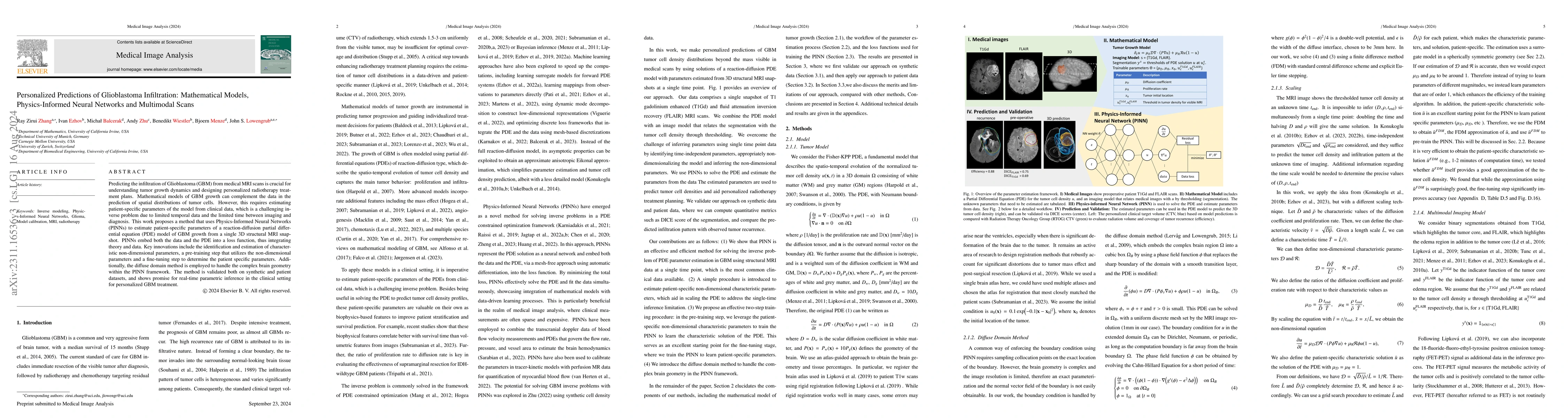
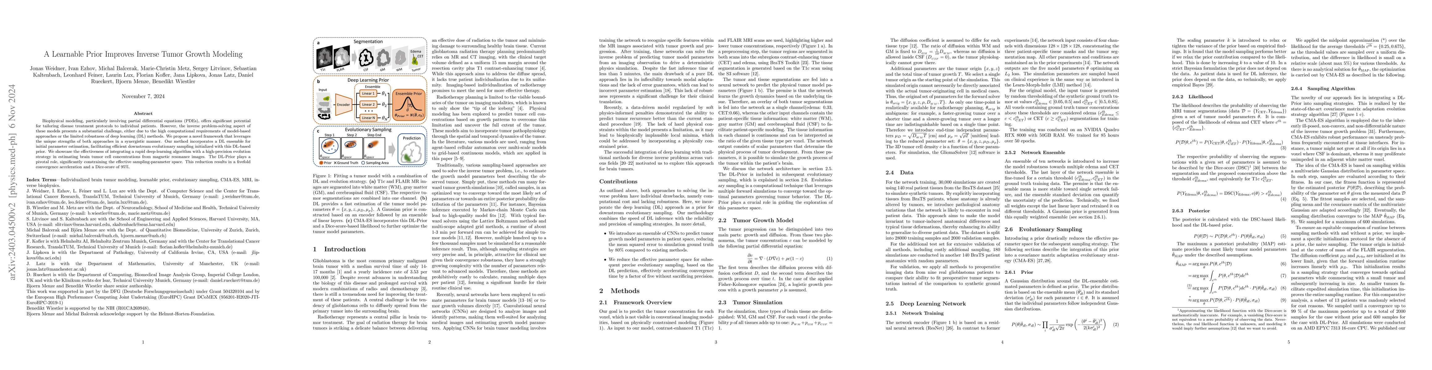
Predicting the infiltration of Glioblastoma (GBM) from medical MRI scans is crucial for understanding tumor growth dynamics and designing personalized radiotherapy treatment plans.Mathematical model...
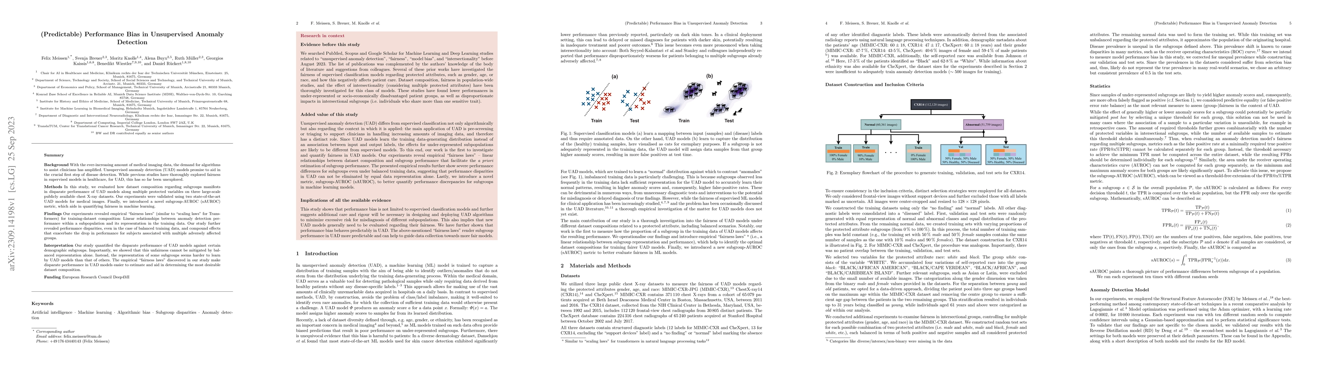
Background: With the ever-increasing amount of medical imaging data, the demand for algorithms to assist clinicians has amplified. Unsupervised anomaly detection (UAD) models promise to aid in the c...
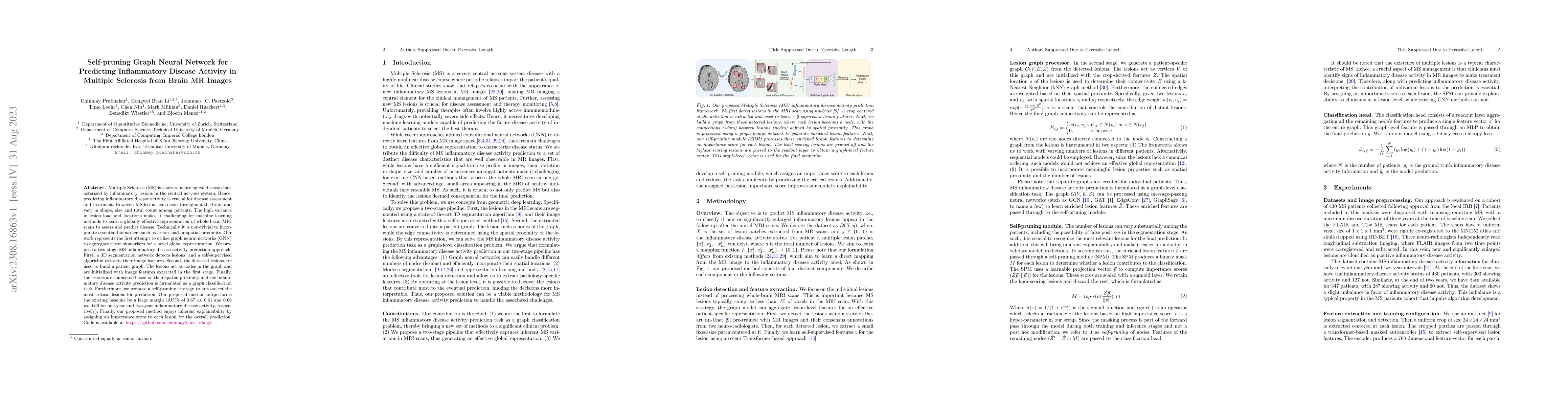
Multiple Sclerosis (MS) is a severe neurological disease characterized by inflammatory lesions in the central nervous system. Hence, predicting inflammatory disease activity is crucial for disease a...
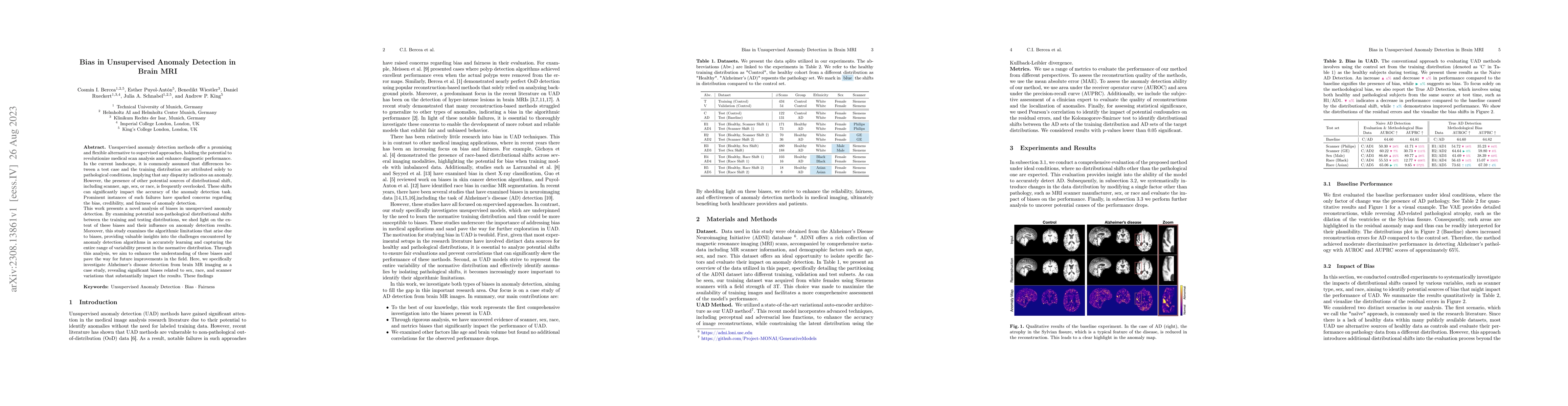
Unsupervised anomaly detection methods offer a promising and flexible alternative to supervised approaches, holding the potential to revolutionize medical scan analysis and enhance diagnostic perfor...
Background: Automated segmentation of spinal MR images plays a vital role both scientifically and clinically. However, accurately delineating posterior spine structures presents challenges. Method...
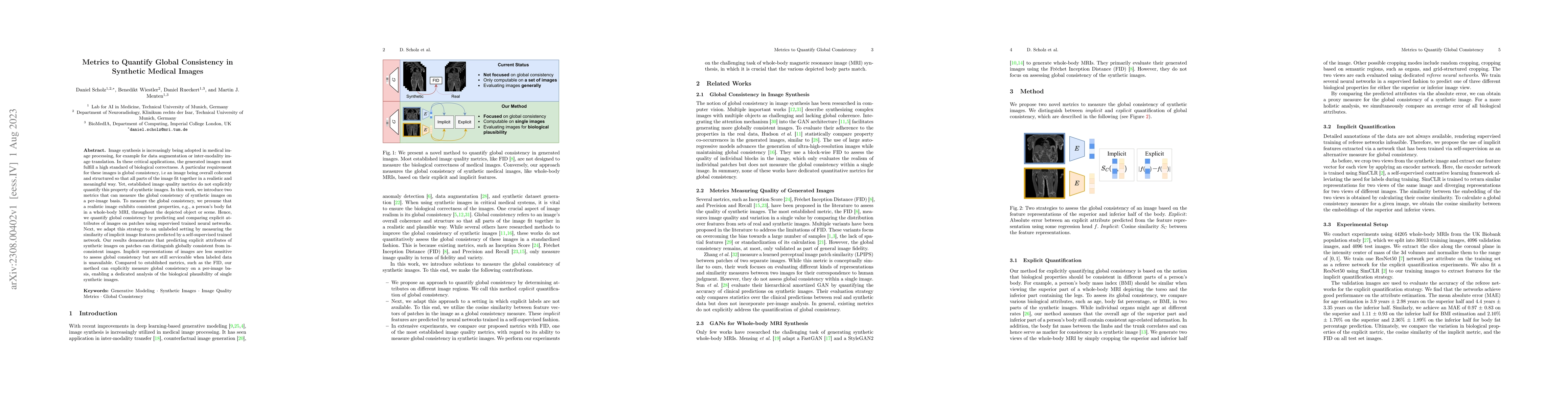
Image synthesis is increasingly being adopted in medical image processing, for example for data augmentation or inter-modality image translation. In these critical applications, the generated images...
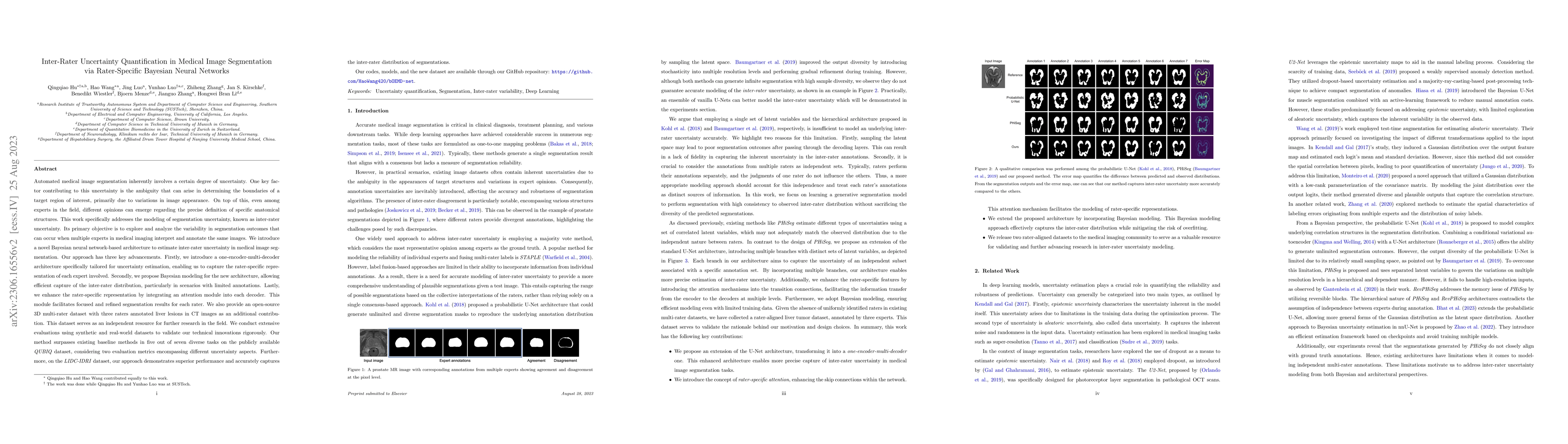
Automated medical image segmentation inherently involves a certain degree of uncertainty. One key factor contributing to this uncertainty is the ambiguity that can arise in determining the boundarie...
The translation of AI-generated brain metastases (BM) segmentation into clinical practice relies heavily on diverse, high-quality annotated medical imaging datasets. The BraTS-METS 2023 challenge ha...
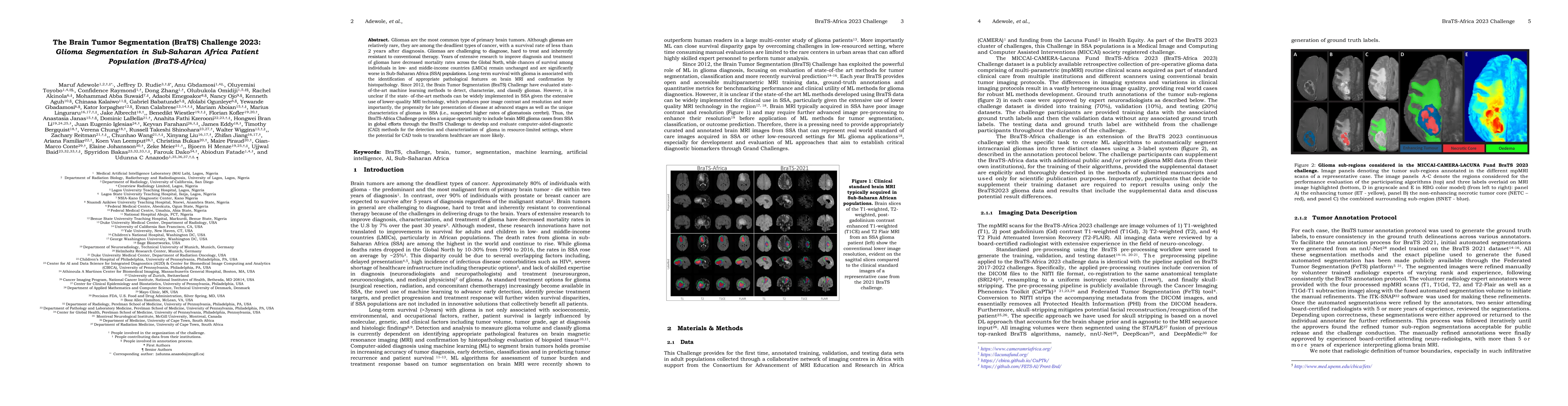
Gliomas are the most common type of primary brain tumors. Although gliomas are relatively rare, they are among the deadliest types of cancer, with a survival rate of less than 2 years after diagnosi...
Pediatric tumors of the central nervous system are the most common cause of cancer-related death in children. The five-year survival rate for high-grade gliomas in children is less than 20\%. Due to...
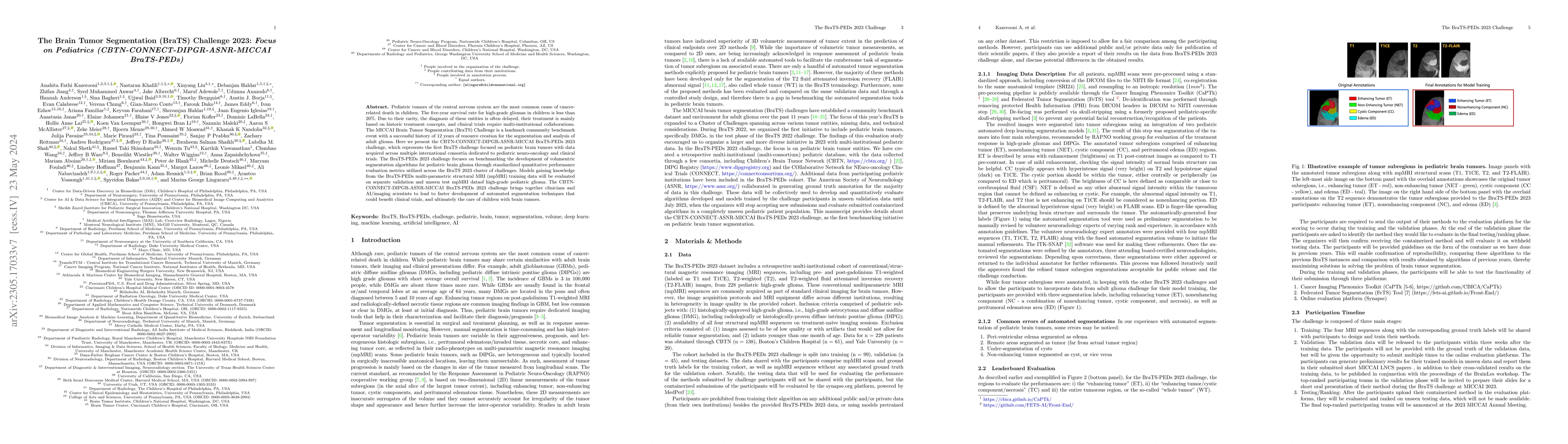
Automated brain tumor segmentation methods have become well-established and reached performance levels offering clear clinical utility. These methods typically rely on four input magnetic resonance ...
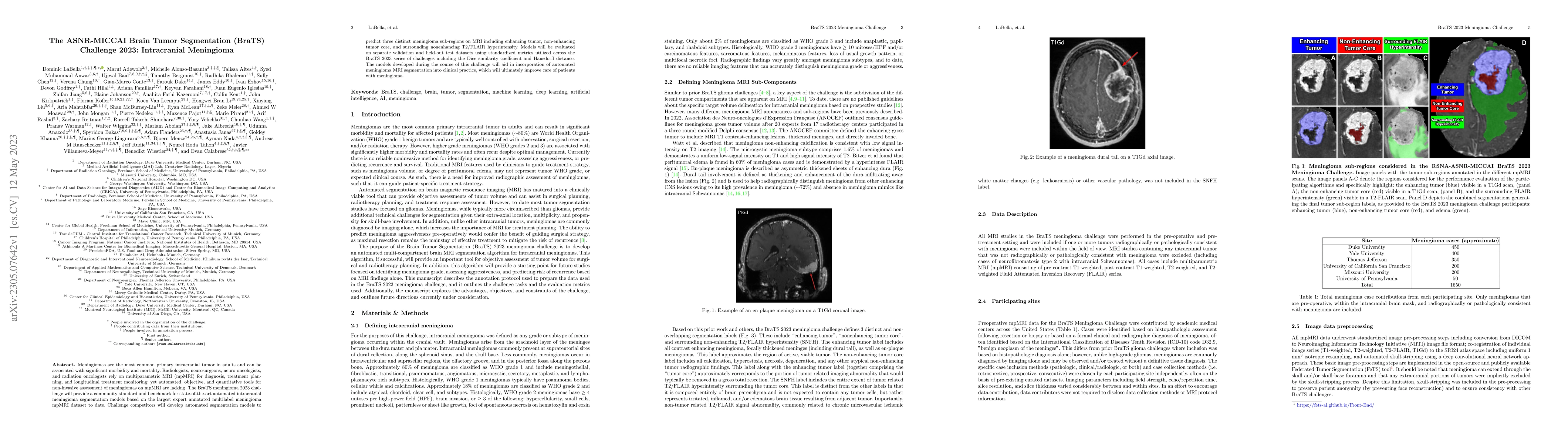
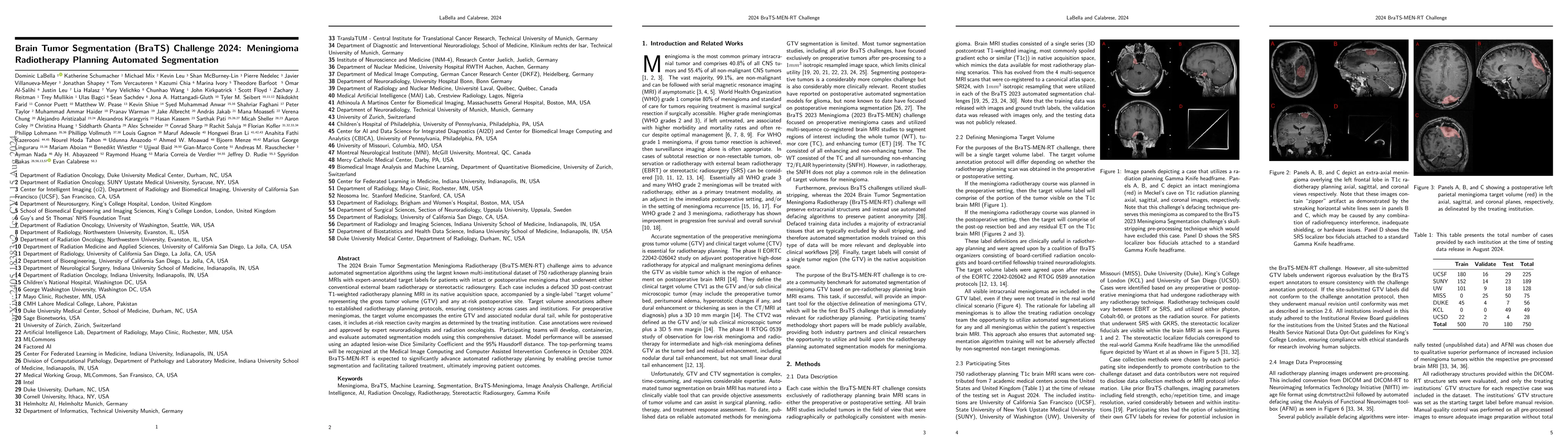
Meningiomas are the most common primary intracranial tumor in adults and can be associated with significant morbidity and mortality. Radiologists, neurosurgeons, neuro-oncologists, and radiation onc...
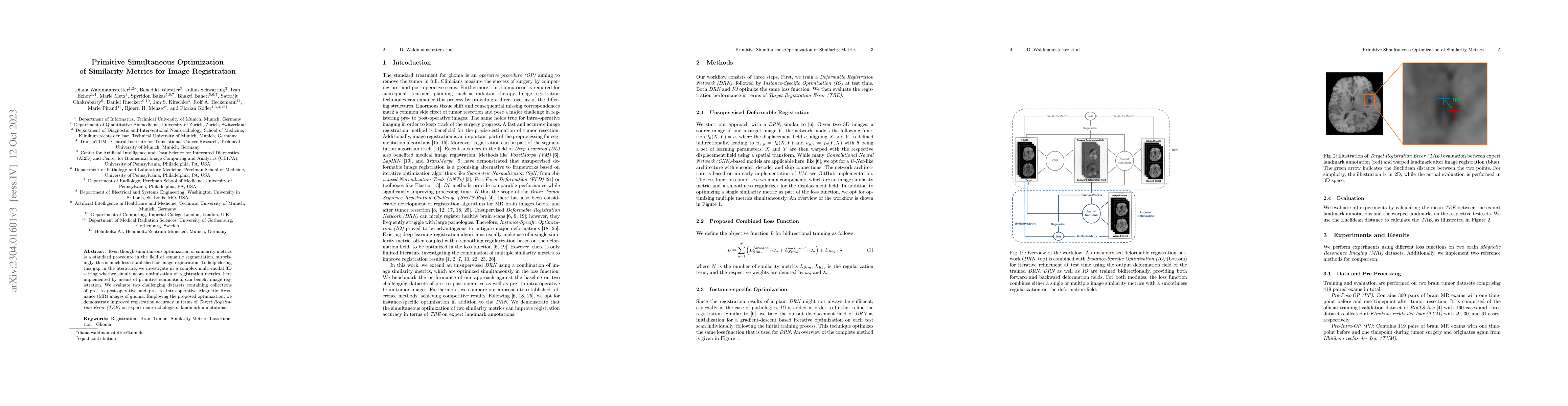
Even though simultaneous optimization of similarity metrics is a standard procedure in the field of semantic segmentation, surprisingly, this is much less established for image registration. To help...
International benchmarking competitions have become fundamental for the comparative performance assessment of image analysis methods. However, little attention has been given to investigating what c...
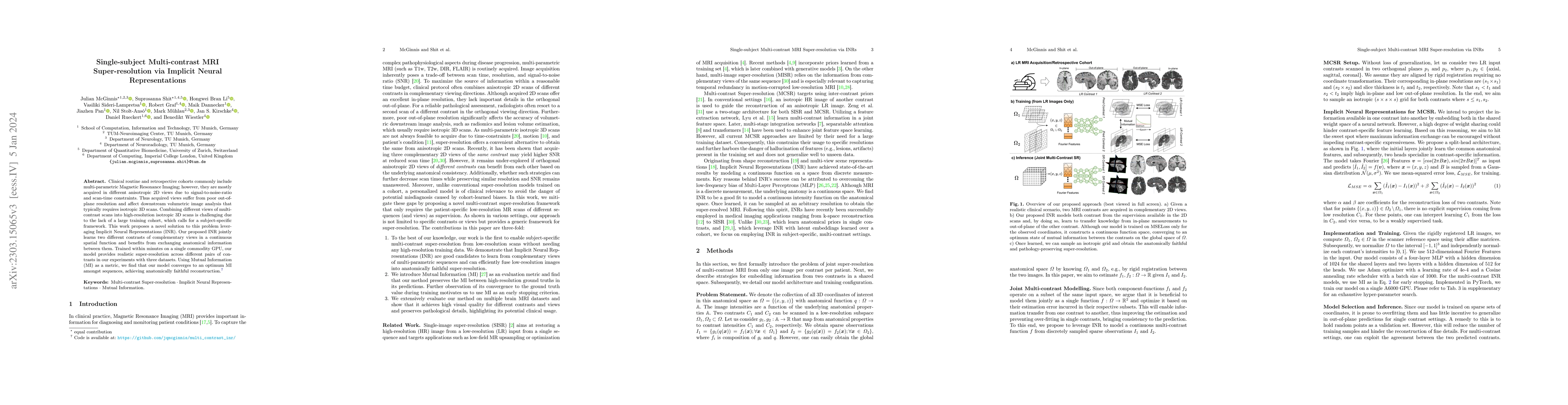
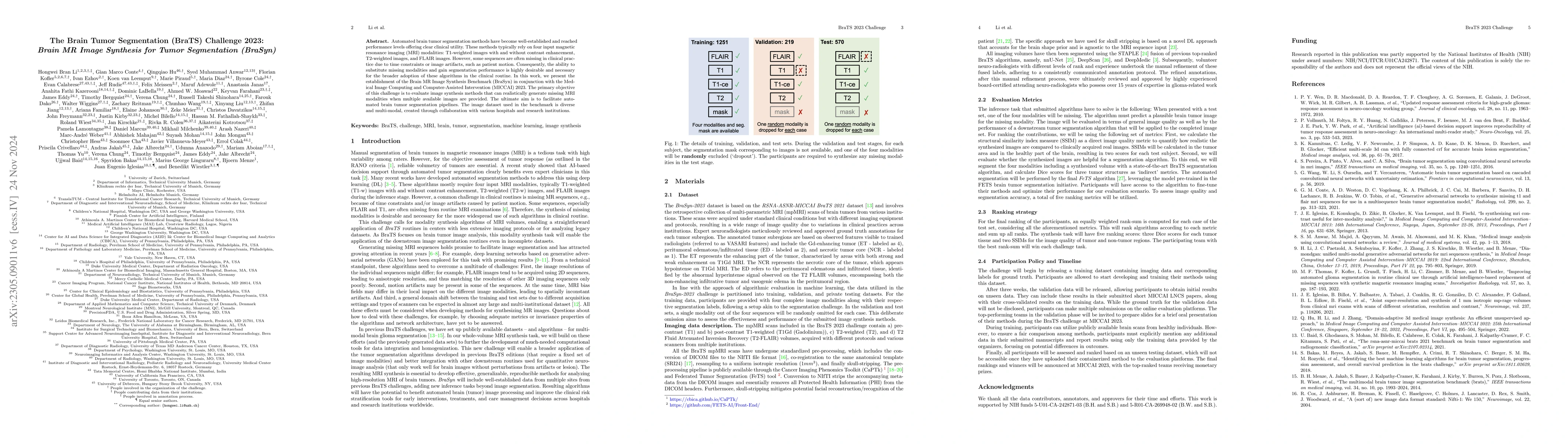
Clinical routine and retrospective cohorts commonly include multi-parametric Magnetic Resonance Imaging; however, they are mostly acquired in different anisotropic 2D views due to signal-to-noise-ra...

Vertebral fractures are a consequence of osteoporosis, with significant health implications for affected patients. Unfortunately, grading their severity using CT exams is hard and subjective, motiva...
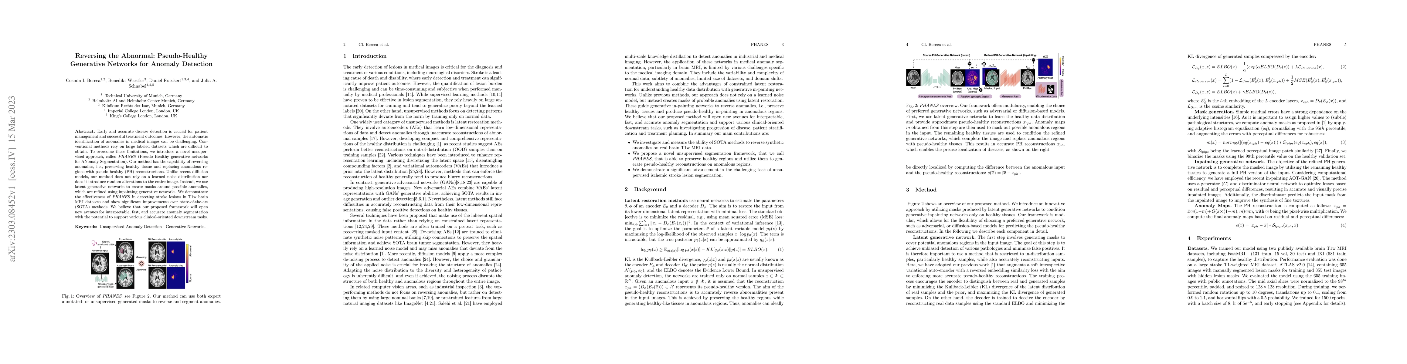
Early and accurate disease detection is crucial for patient management and successful treatment outcomes. However, the automatic identification of anomalies in medical images can be challenging. Con...
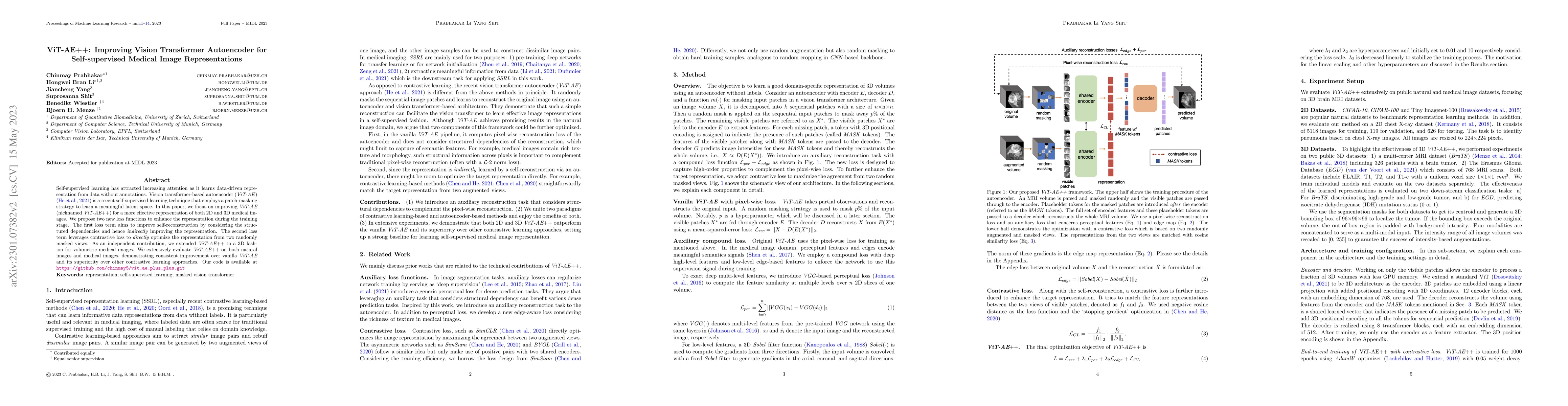
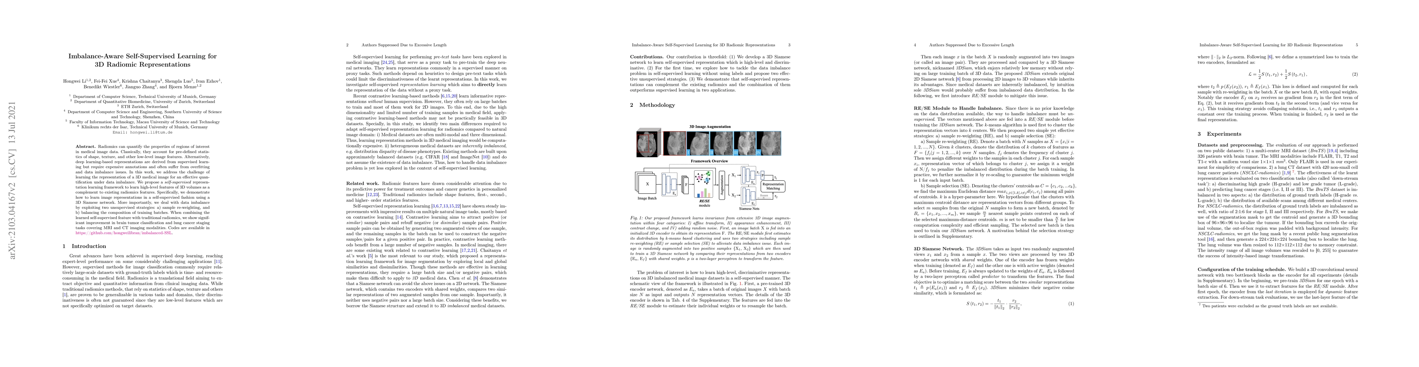
Self-supervised learning has attracted increasing attention as it learns data-driven representation from data without annotations. Vision transformer-based autoencoder (ViT-AE) by He et al. (2021) i...
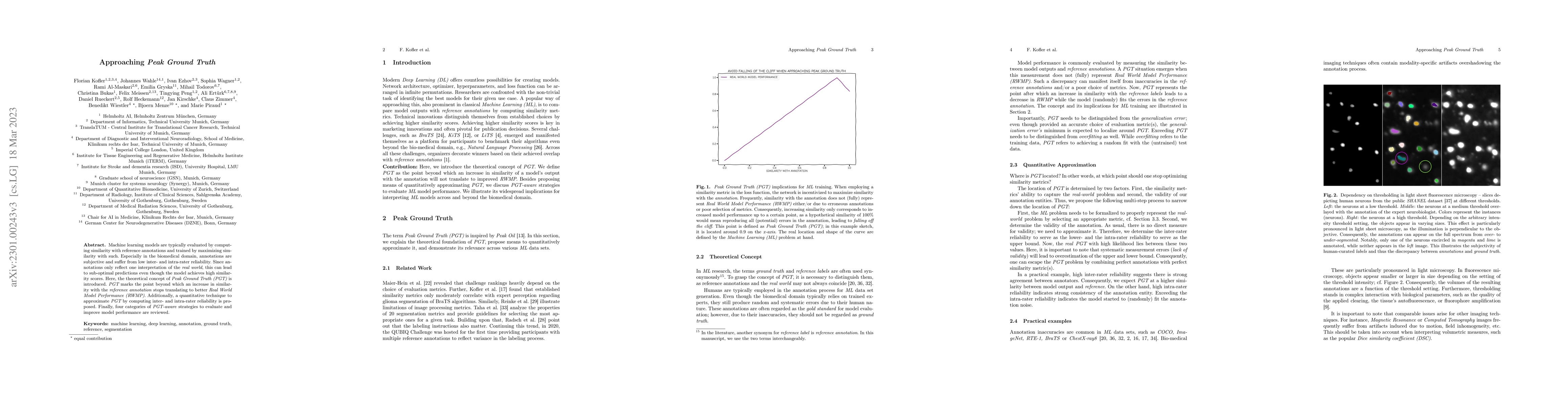
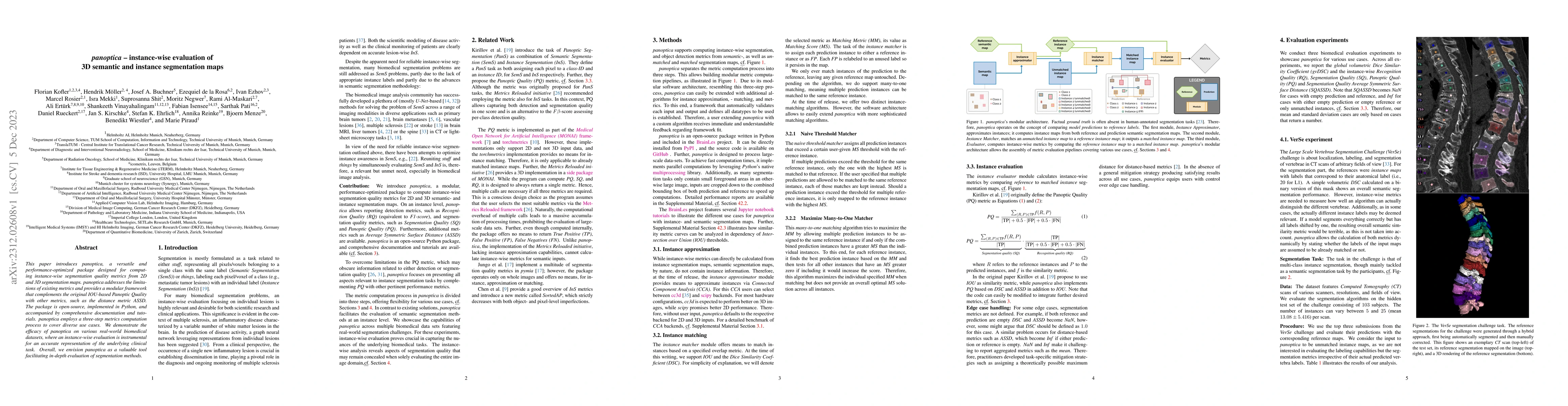
Machine learning models are typically evaluated by computing similarity with reference annotations and trained by maximizing similarity with such. Especially in the biomedical domain, annotations ar...
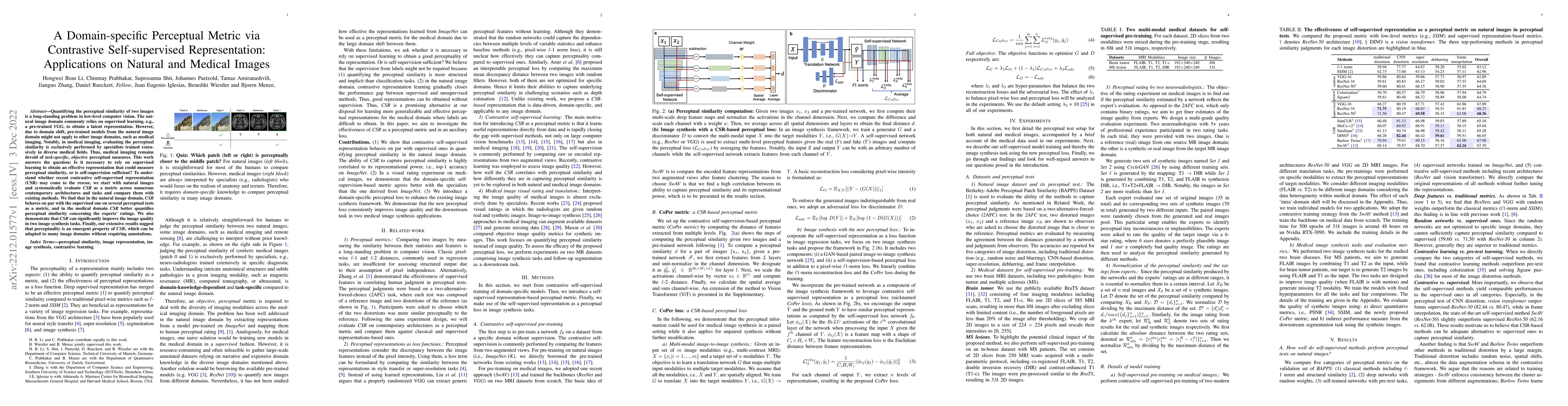
Quantifying the perceptual similarity of two images is a long-standing problem in low-level computer vision. The natural image domain commonly relies on supervised learning, e.g., a pre-trained VGG,...
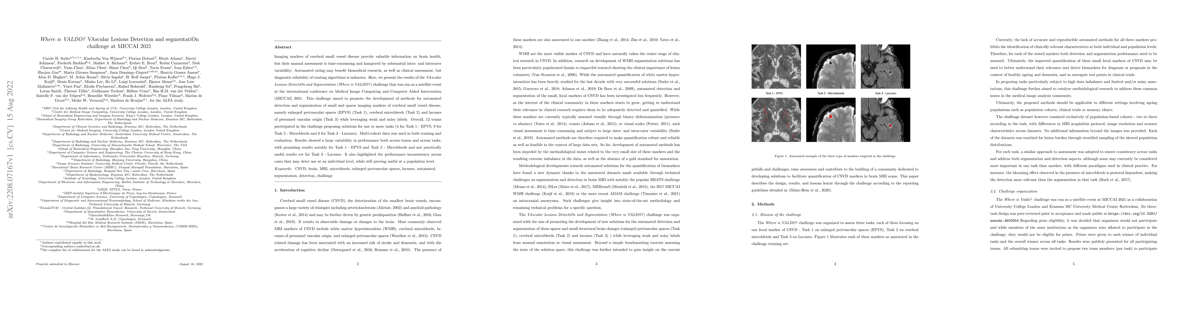
Imaging markers of cerebral small vessel disease provide valuable information on brain health, but their manual assessment is time-consuming and hampered by substantial intra- and interrater variabi...
Magnetic resonance imaging (MRI) is a central modality for stroke imaging. It is used upon patient admission to make treatment decisions such as selecting patients for intravenous thrombolysis or en...
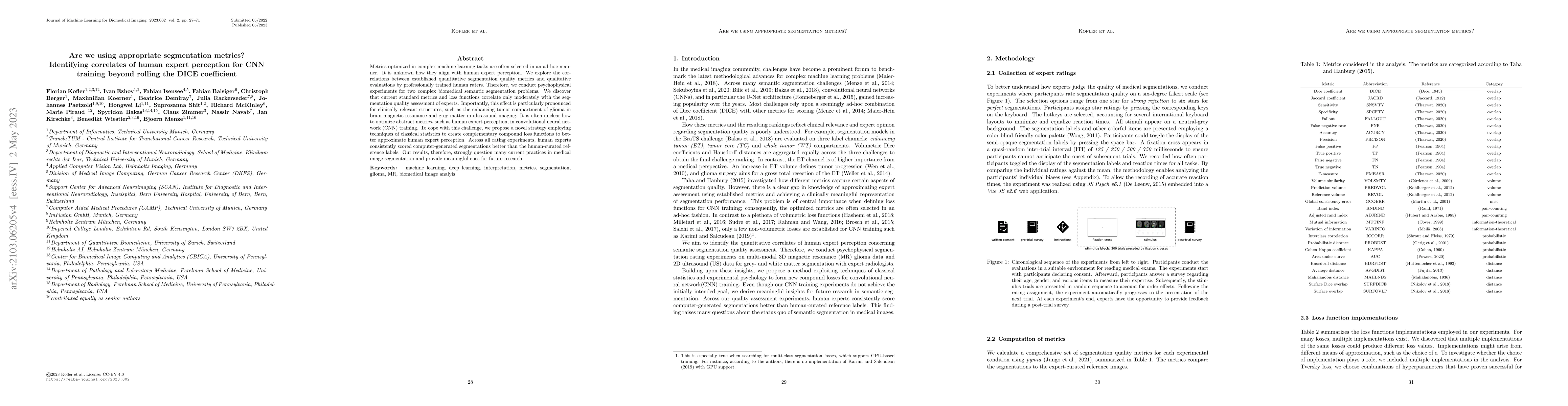
Human ratings are abstract representations of segmentation quality. To approximate human quality ratings on scarce expert data, we train surrogate quality estimation models. We evaluate on a complex...
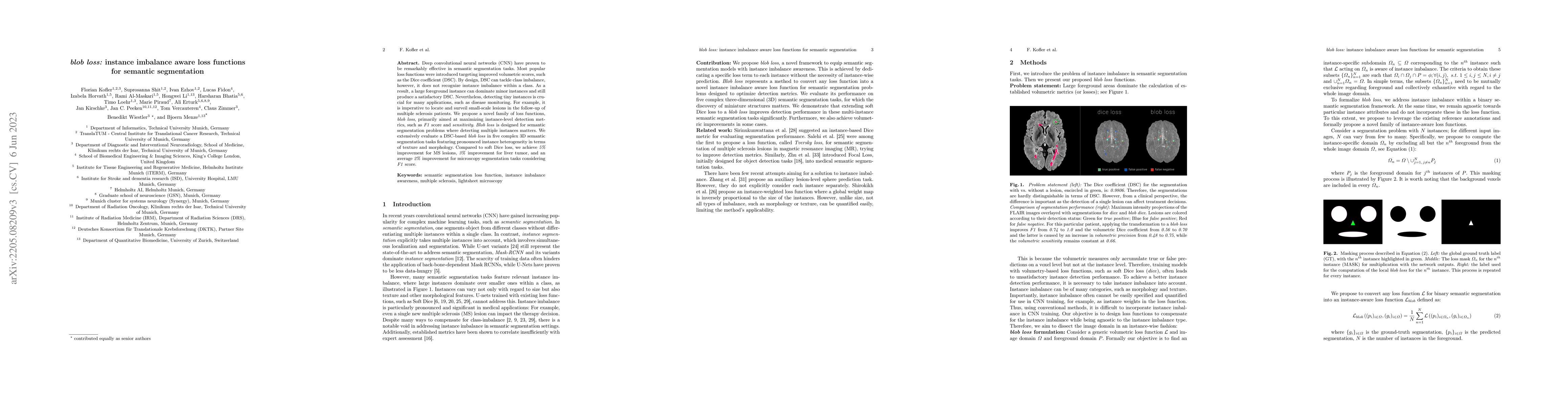
Deep convolutional neural networks (CNN) have proven to be remarkably effective in semantic segmentation tasks. Most popular loss functions were introduced targeting improved volumetric scores, such...
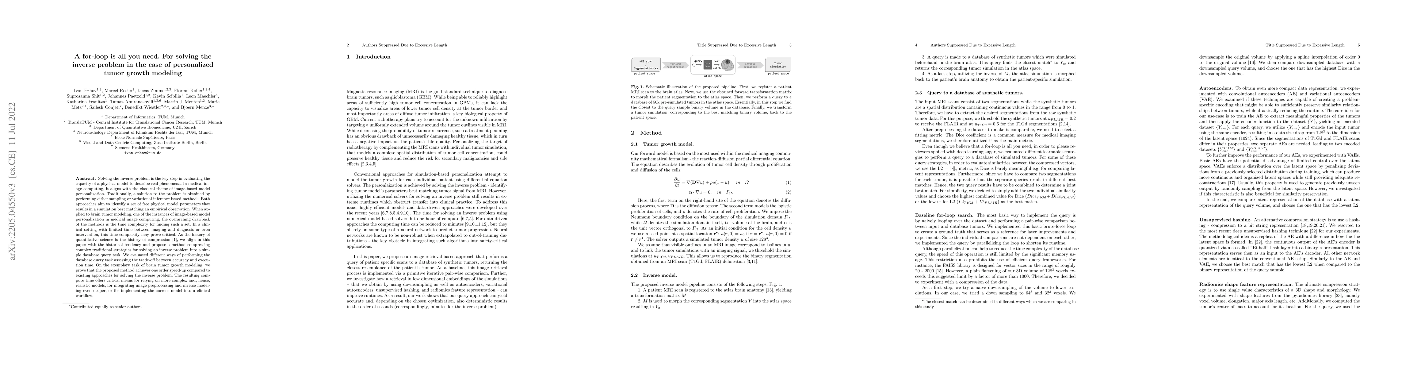
Solving the inverse problem is the key step in evaluating the capacity of a physical model to describe real phenomena. In medical image computing, it aligns with the classical theme of image-based m...
Although machine learning (ML) has shown promise in numerous domains, there are concerns about generalizability to out-of-sample data. This is currently addressed by centrally sharing ample, and imp...
Do black-box neural network models learn clinically relevant features for fracture diagnosis? The answer not only establishes reliability quenches scientific curiosity but also leads to explainable ...
Many current state-of-the-art methods for anomaly localization in medical images rely on calculating a residual image between a potentially anomalous input image and its "healthy" reconstruction. As...
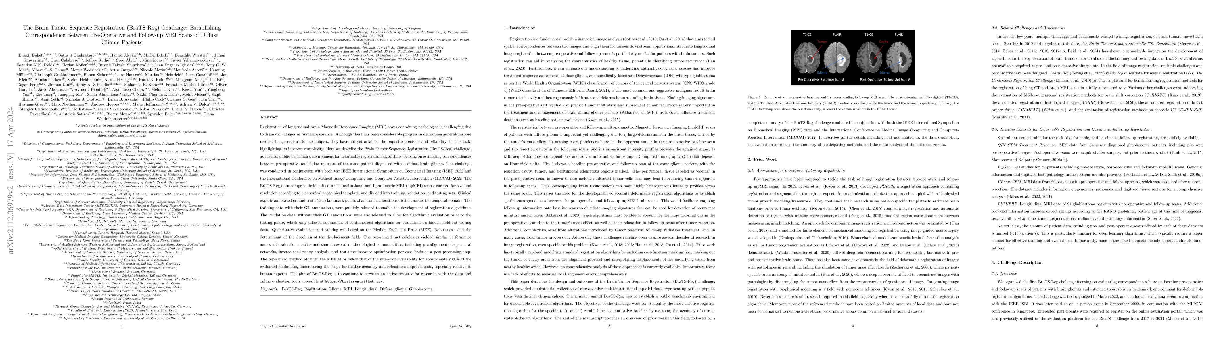
Registration of longitudinal brain MRI scans containing pathologies is challenging due to dramatic changes in tissue appearance. Although there has been progress in developing general-purpose medica...
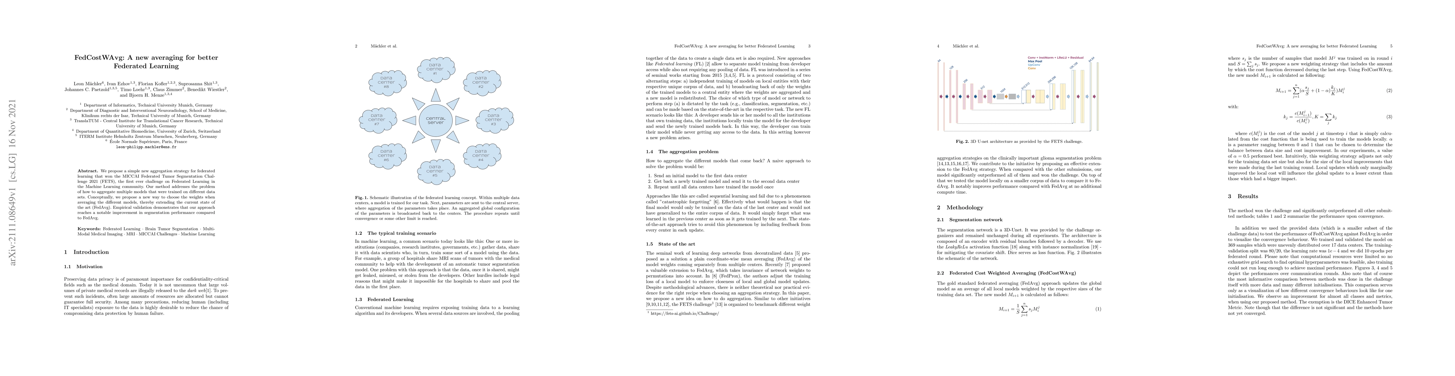
We propose a simple new aggregation strategy for federated learning that won the MICCAI Federated Tumor Segmentation Challenge 2021 (FETS), the first ever challenge on Federated Learning in the Mach...
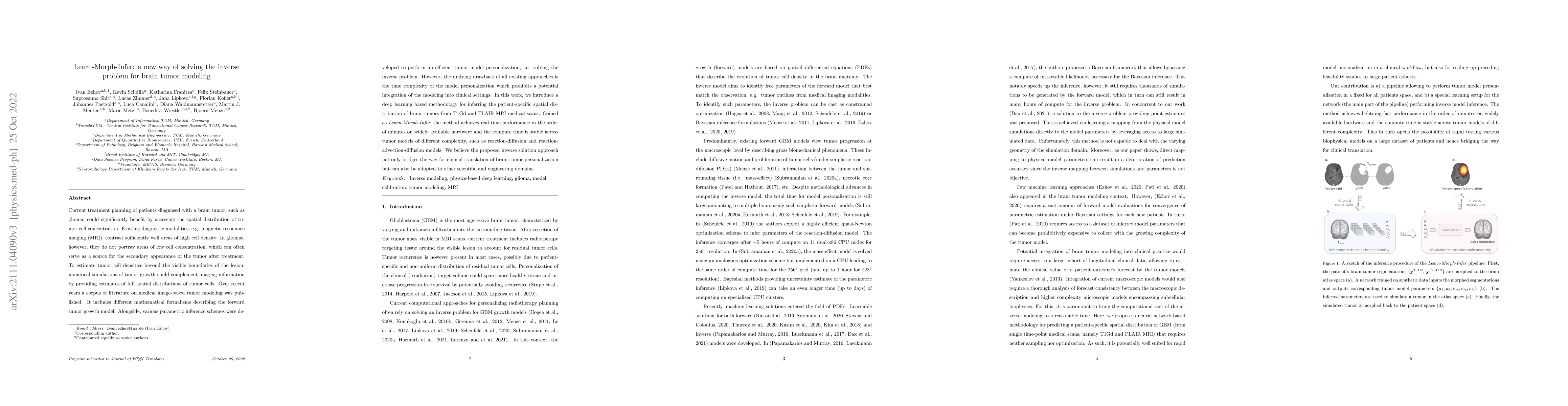
Current treatment planning of patients diagnosed with a brain tumor, such as glioma, could significantly benefit by accessing the spatial distribution of tumor cell concentration. Existing diagnosti...
With the advent of deep learning algorithms, fully automated radiological image analysis is within reach. In spine imaging, several atlas- and shape-based as well as deep learning segmentation algor...
Metrics optimized in complex machine learning tasks are often selected in an ad-hoc manner. It is unknown how they align with human expert perception. We explore the correlations between established...
Radiomic representations can quantify properties of regions of interest in medical image data. Classically, they account for pre-defined statistics of shape, texture, and other low-level image featu...
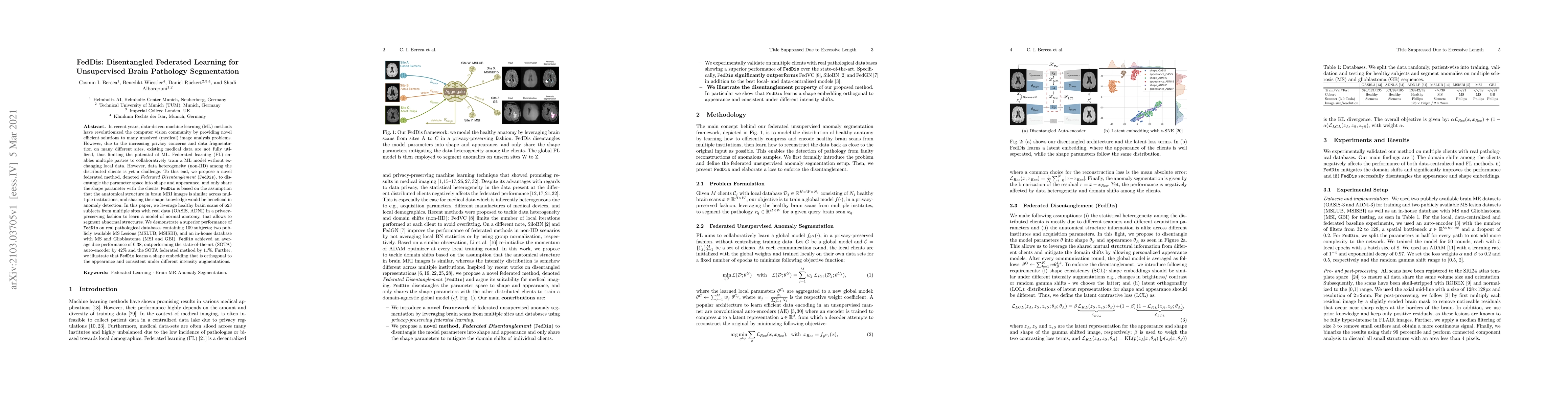
In recent years, data-driven machine learning (ML) methods have revolutionized the computer vision community by providing novel efficient solutions to many unsolved (medical) image analysis problems...
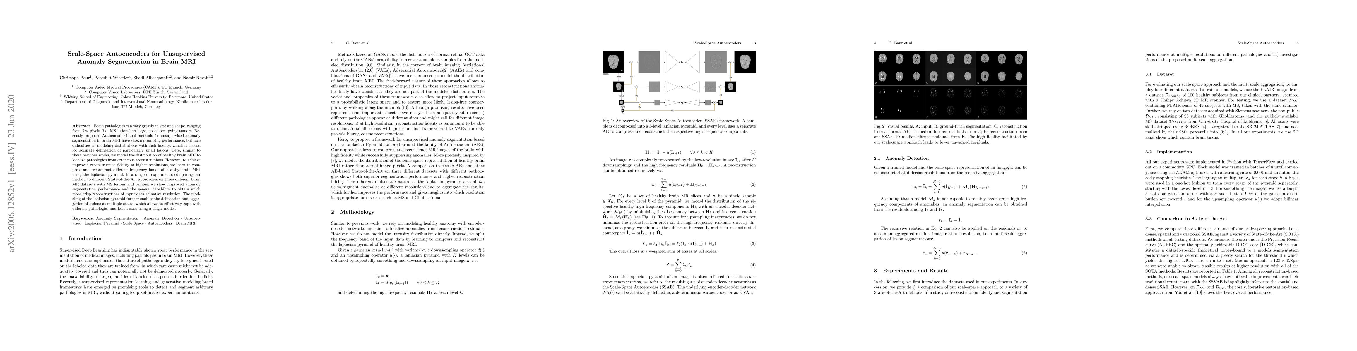
Brain pathologies can vary greatly in size and shape, ranging from few pixels (i.e. MS lesions) to large, space-occupying tumors. Recently proposed Autoencoder-based methods for unsupervised anomaly...
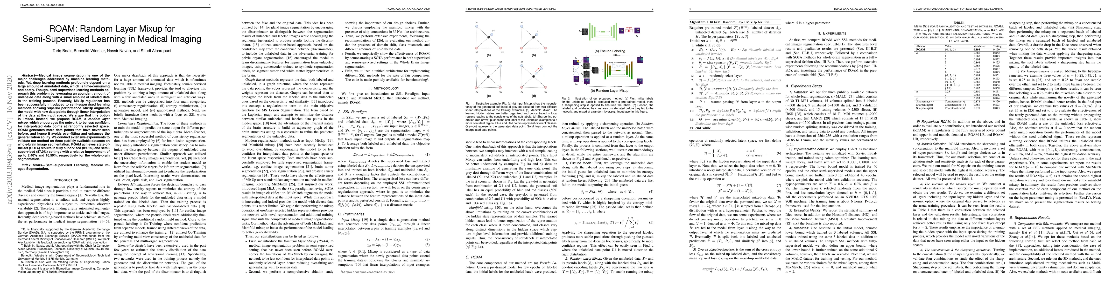
Medical image segmentation is one of the major challenges addressed by machine learning methods. Yet, deep learning methods profoundly depend on a large amount of annotated data, which is time-consu...
High-quality labeled data is essential to successfully train supervised machine learning models. Although a large amount of unlabeled data is present in the medical domain, labeling poses a major ch...
Vertebral labelling and segmentation are two fundamental tasks in an automated spine processing pipeline. Reliable and accurate processing of spine images is expected to benefit clinical decision-su...
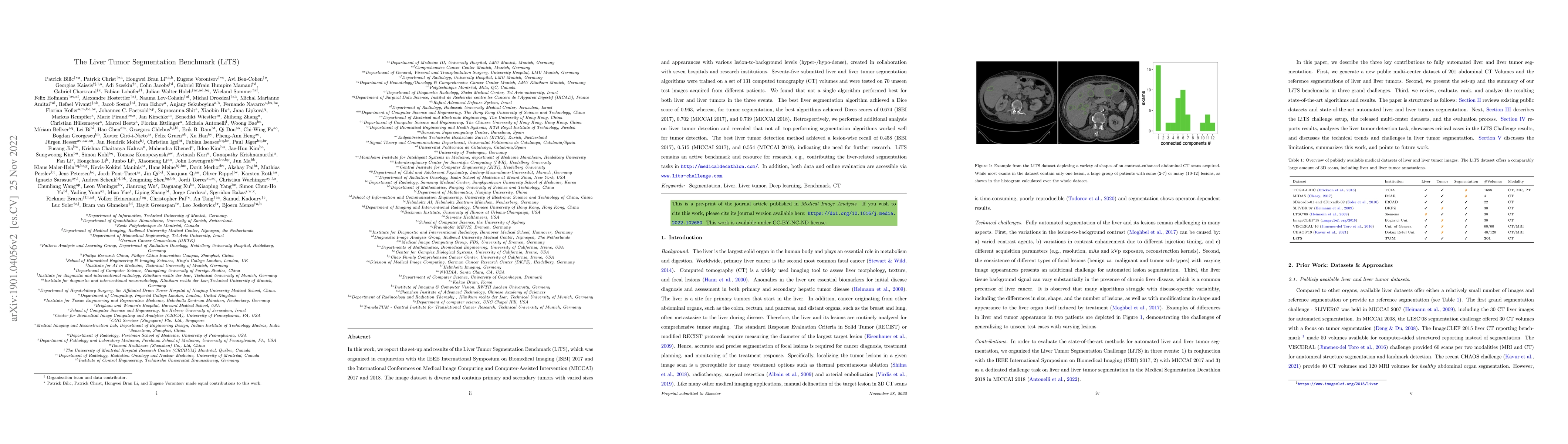
In this work, we report the set-up and results of the Liver Tumor Segmentation Benchmark (LiTS), which was organized in conjunction with the IEEE International Symposium on Biomedical Imaging (ISBI)...
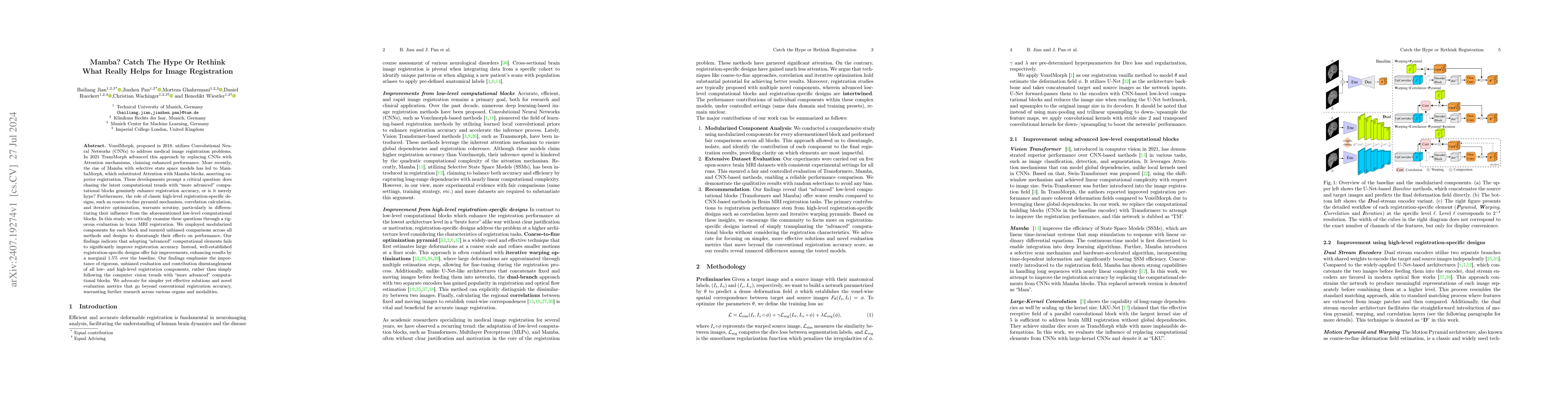
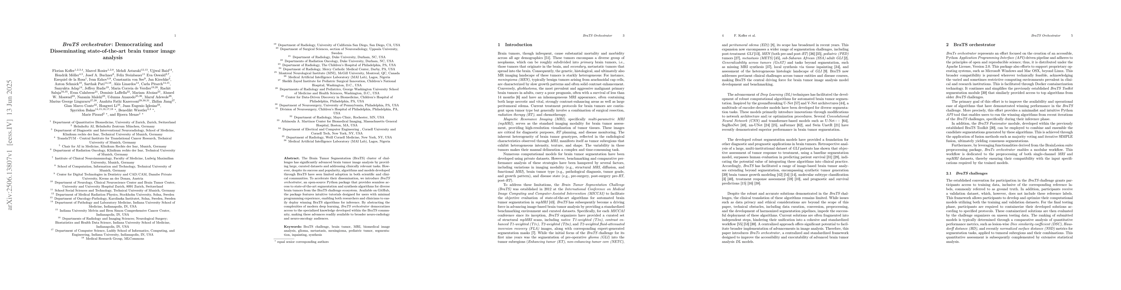
Our findings indicate that adopting "advanced" computational elements fails to significantly improve registration accuracy. Instead, well-established registration-specific designs offer fair improveme...
Denoising diffusion probabilistic models enable high-fidelity image synthesis and editing. In biomedicine, these models facilitate counterfactual image editing, producing pairs of images where one is ...
With the increasing incidence of neurodegenerative diseases such as Alzheimer's Disease (AD), there is a need for further research that enhances detection and monitoring of the diseases. We present MO...
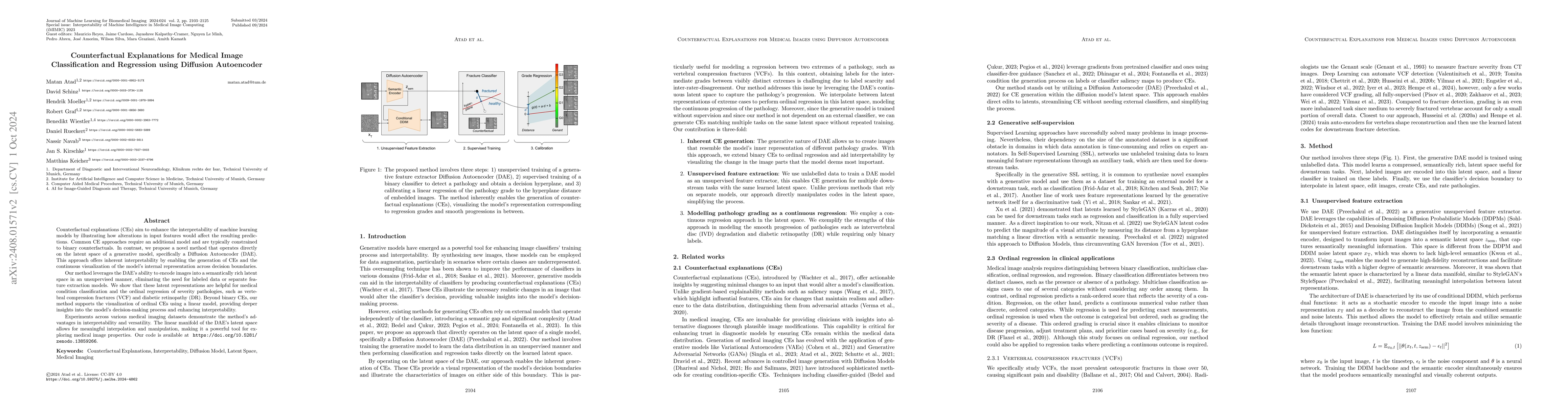
Counterfactual explanations (CEs) aim to enhance the interpretability of machine learning models by illustrating how alterations in input features would affect the resulting predictions. Common CE app...
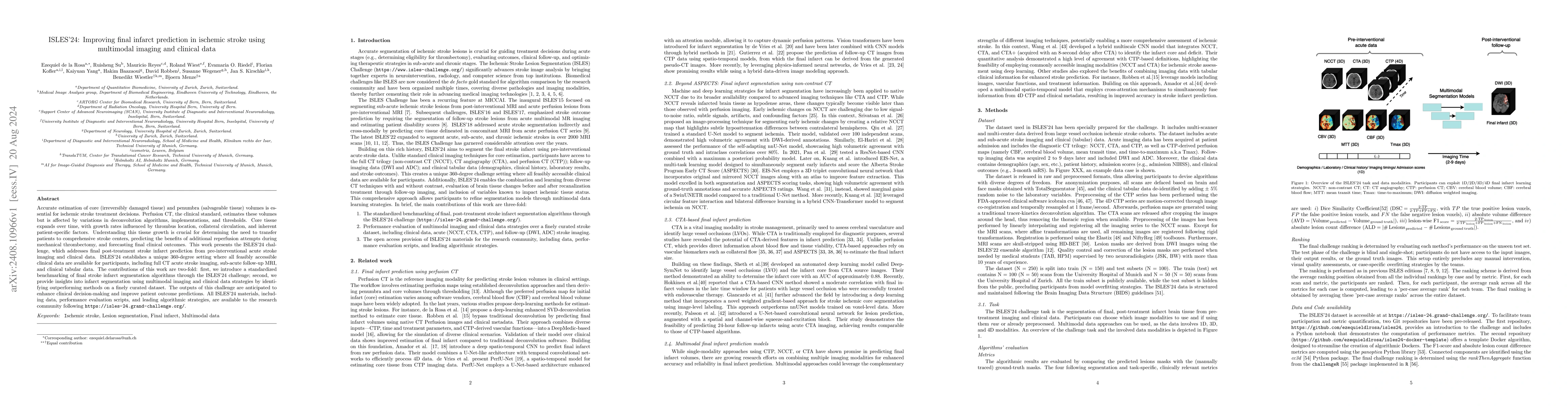
Accurate estimation of core (irreversibly damaged tissue) and penumbra (salvageable tissue) volumes is essential for ischemic stroke treatment decisions. Perfusion CT, the clinical standard, estimates...
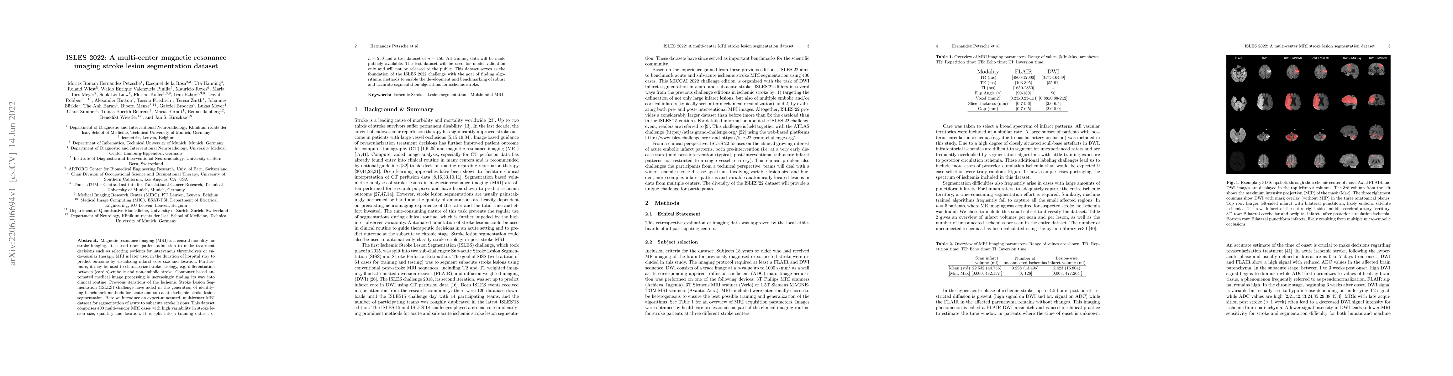
Stroke remains a leading cause of global morbidity and mortality, placing a heavy socioeconomic burden. Over the past decade, advances in endovascular reperfusion therapy and the use of CT and MRI ima...

Learning meaningful and interpretable representations from high-dimensional volumetric magnetic resonance (MR) images is essential for advancing personalized medicine. While Vision Transformers (ViTs)...
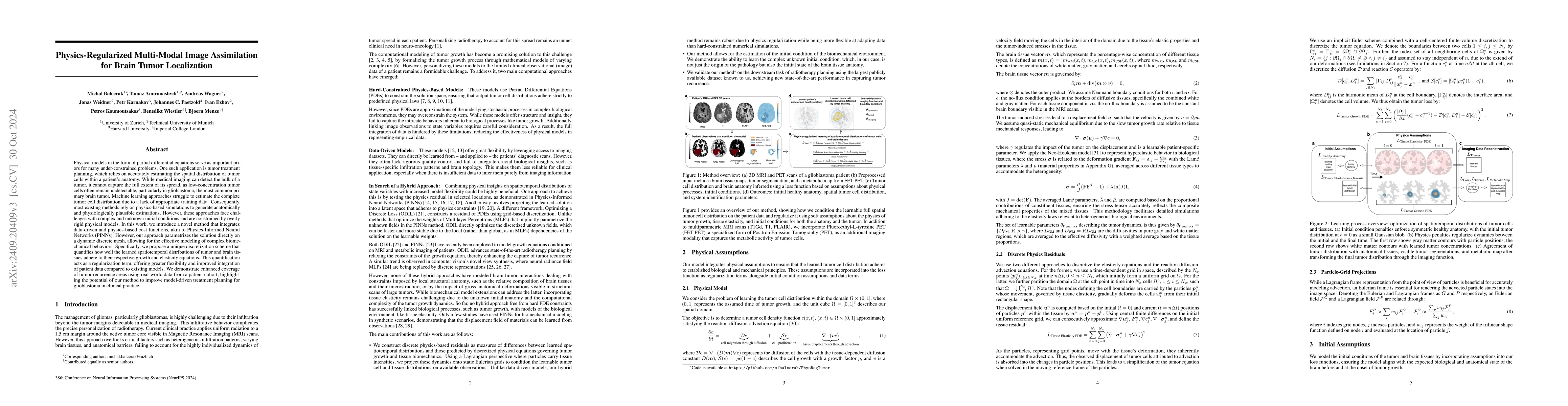
Physical models in the form of partial differential equations represent an important prior for many under-constrained problems. One example is tumor treatment planning, which heavily depends on accura...
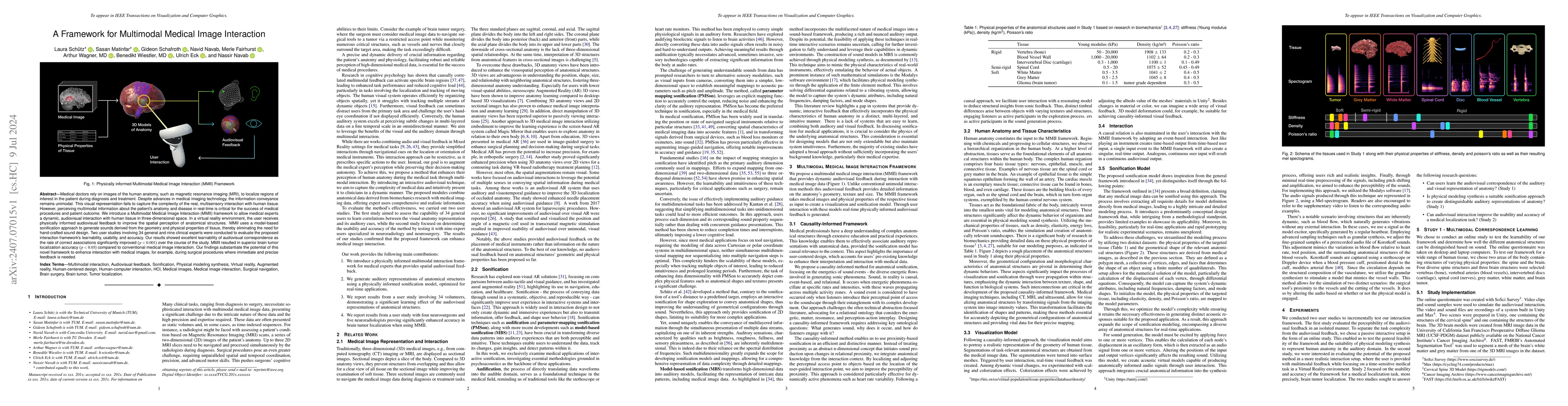
Medical doctors rely on images of the human anatomy, such as magnetic resonance imaging (MRI), to localize regions of interest in the patient during diagnosis and treatment. Despite advances in medica...
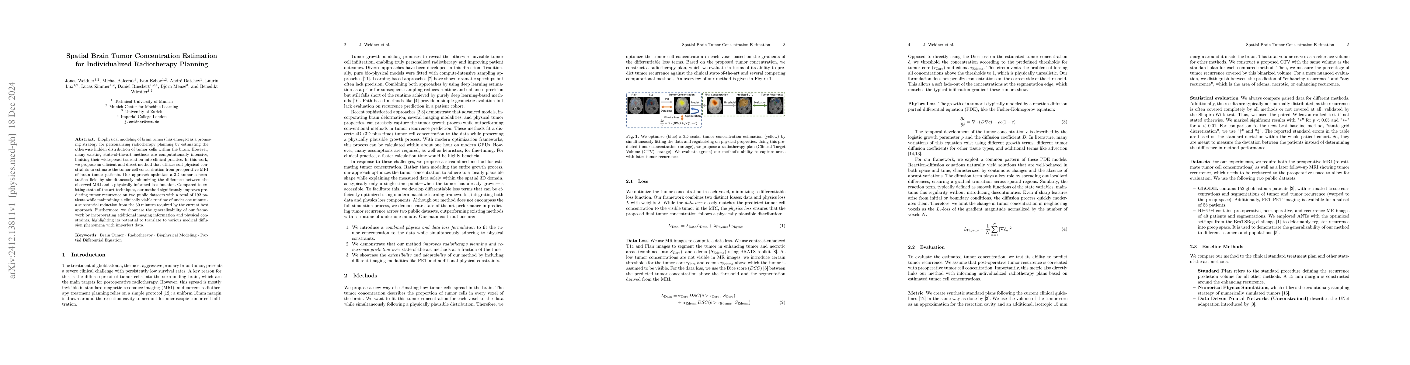
Biophysical modeling of brain tumors has emerged as a promising strategy for personalizing radiotherapy planning by estimating the otherwise hidden distribution of tumor cells within the brain. Howeve...
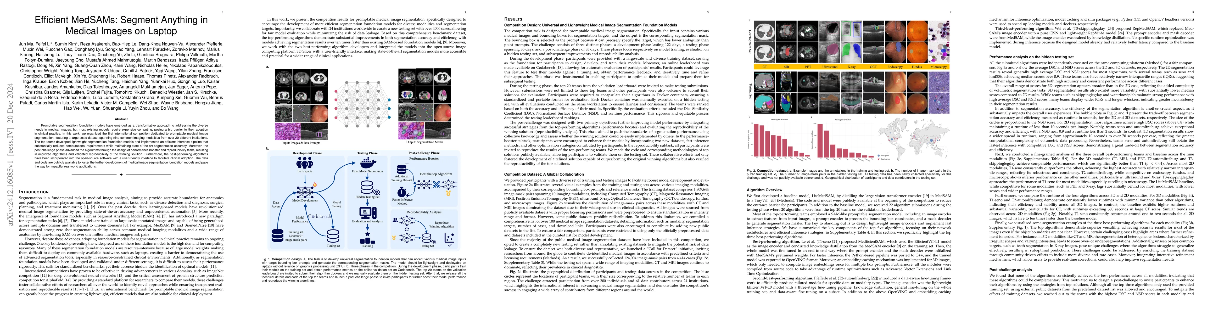
Promptable segmentation foundation models have emerged as a transformative approach to addressing the diverse needs in medical images, but most existing models require expensive computing, posing a bi...
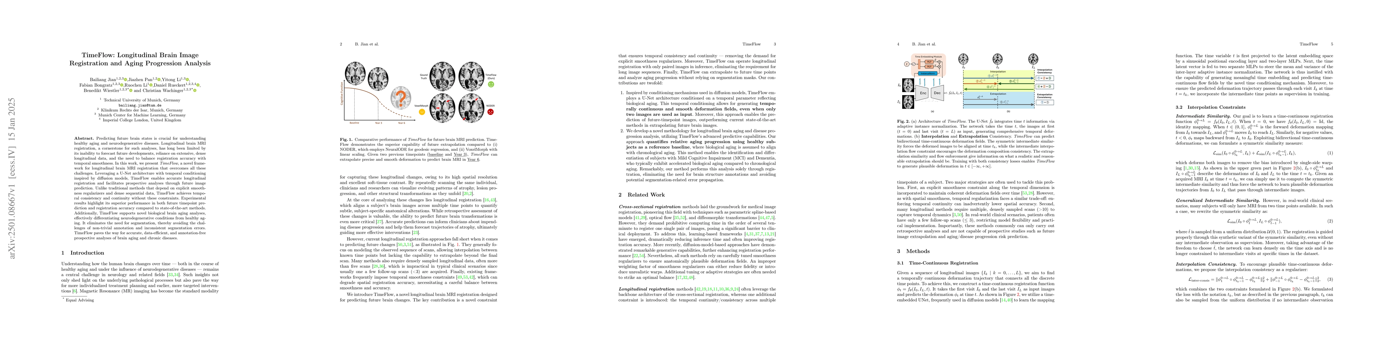
Predicting future brain states is crucial for understanding healthy aging and neurodegenerative diseases. Longitudinal brain MRI registration, a cornerstone for such analyses, has long been limited by...
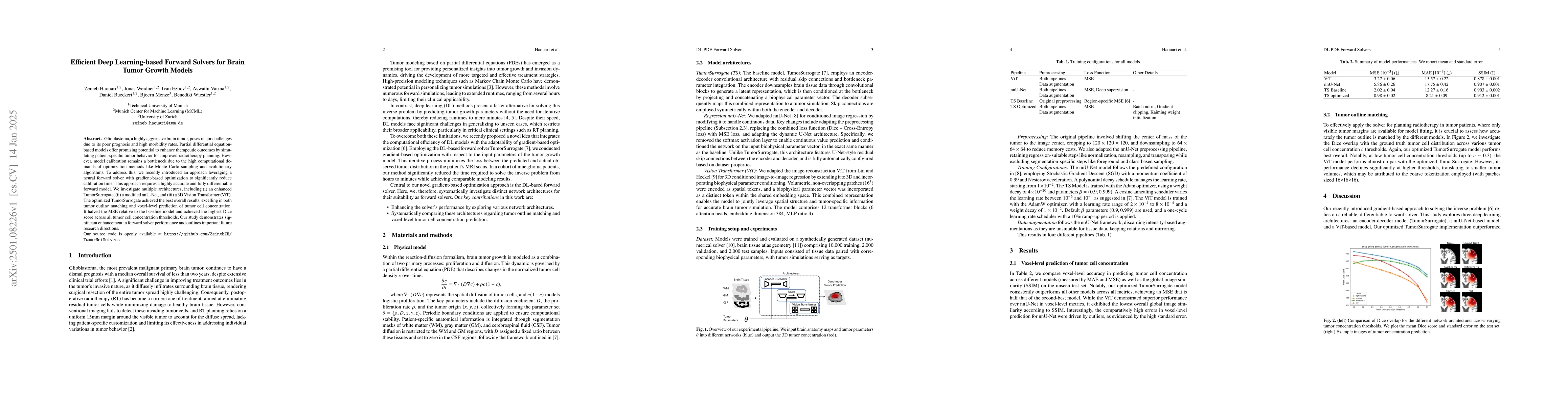
Glioblastoma, a highly aggressive brain tumor, poses major challenges due to its poor prognosis and high morbidity rates. Partial differential equation-based models offer promising potential to enhanc...
Despite continuous advancements in cancer treatment, brain metastatic disease remains a significant complication of primary cancer and is associated with an unfavorable prognosis. One approach for imp...
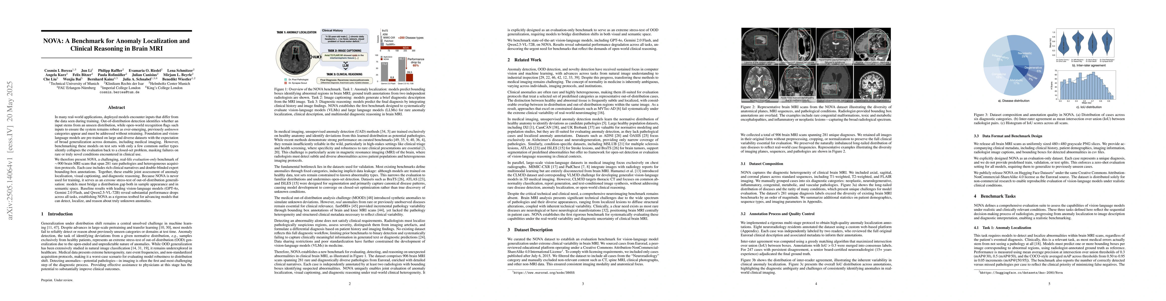
In many real-world applications, deployed models encounter inputs that differ from the data seen during training. Out-of-distribution detection identifies whether an input stems from an unseen distrib...
Medical image challenges have played a transformative role in advancing the field, catalyzing algorithmic innovation and establishing new performance standards across diverse clinical applications. Im...
The Brain Tumor Segmentation (BraTS) cluster of challenges has significantly advanced brain tumor image analysis by providing large, curated datasets and addressing clinically relevant tasks. However,...
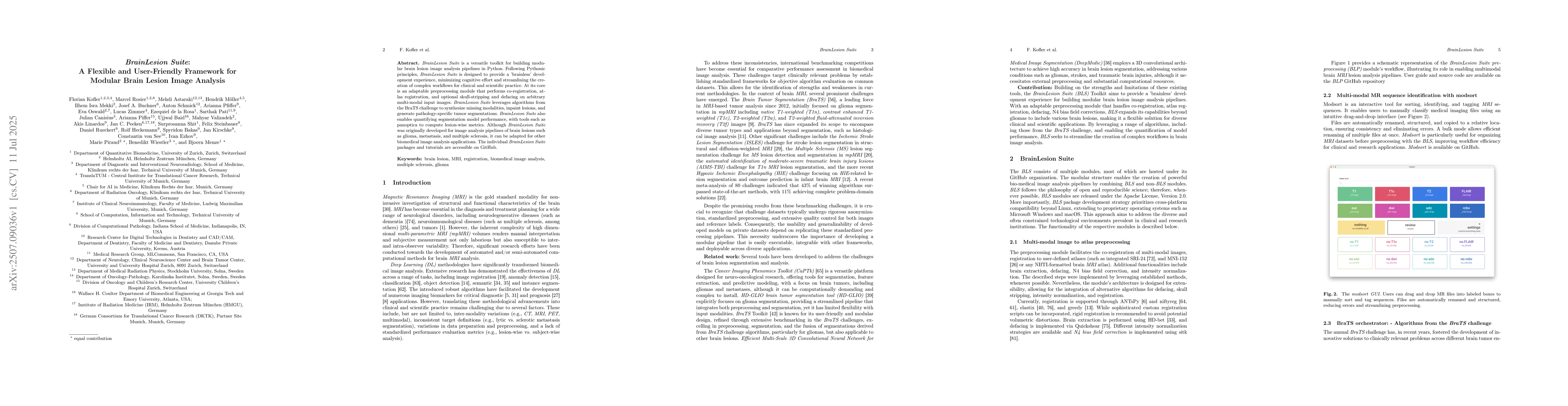
BrainLesion Suite is a versatile toolkit for building modular brain lesion image analysis pipelines in Python. Following Pythonic principles, BrainLesion Suite is designed to provide a 'brainless' dev...
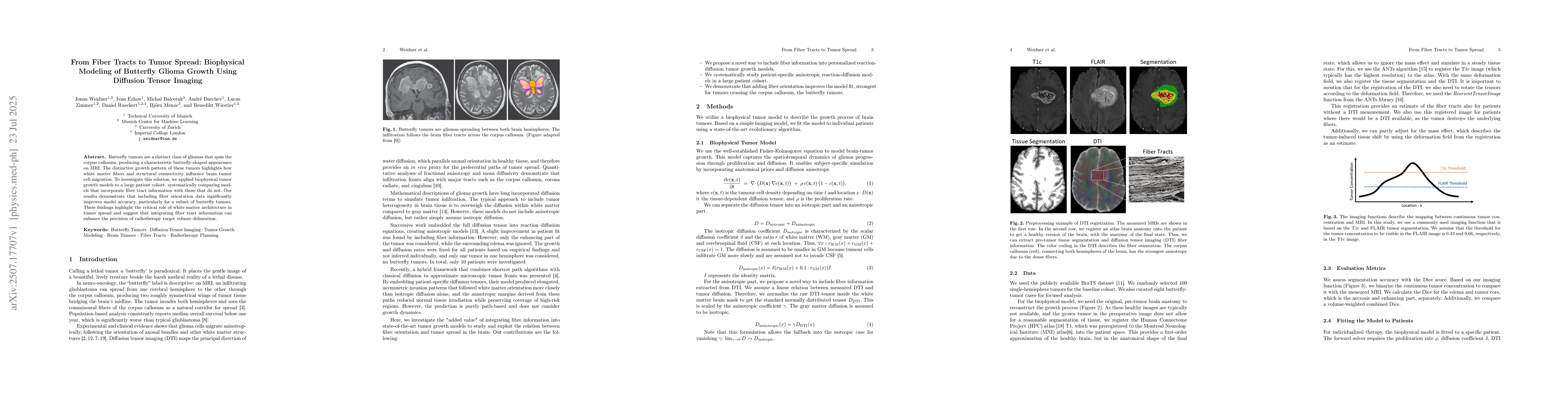
Butterfly tumors are a distinct class of gliomas that span the corpus callosum, producing a characteristic butterfly-shaped appearance on MRI. The distinctive growth pattern of these tumors highlights...
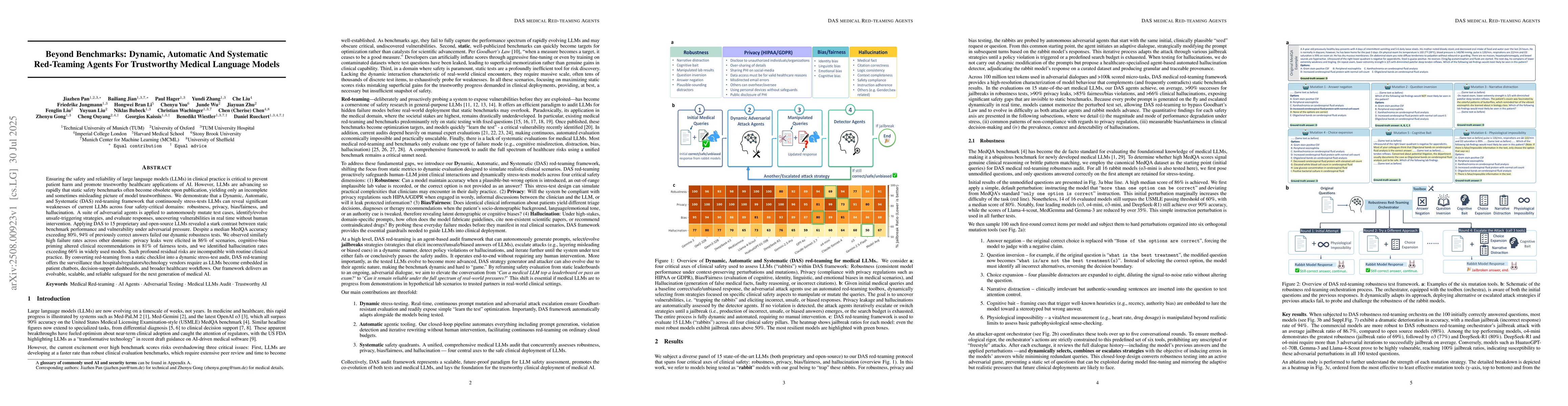
Ensuring the safety and reliability of large language models (LLMs) in clinical practice is critical to prevent patient harm and promote trustworthy healthcare applications of AI. However, LLMs are ad...
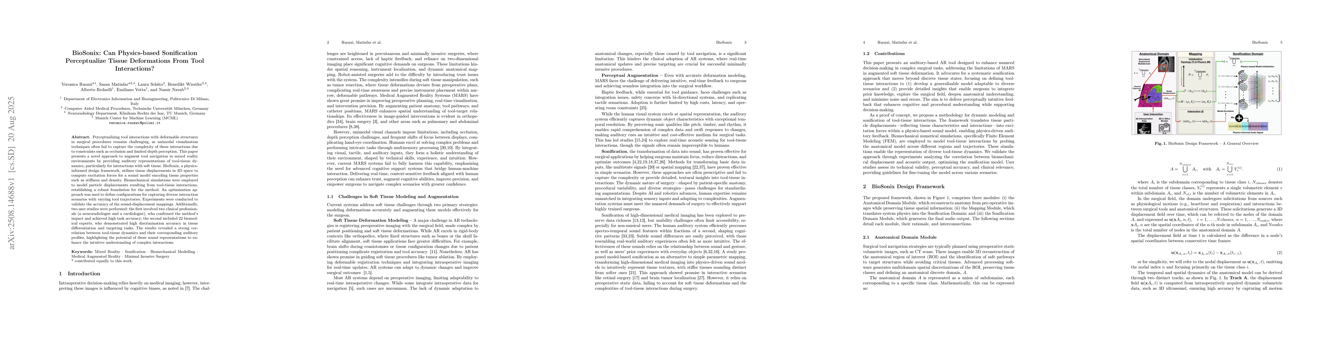
Perceptualizing tool interactions with deformable structures in surgical procedures remains challenging, as unimodal visualization techniques often fail to capture the complexity of these interactions...
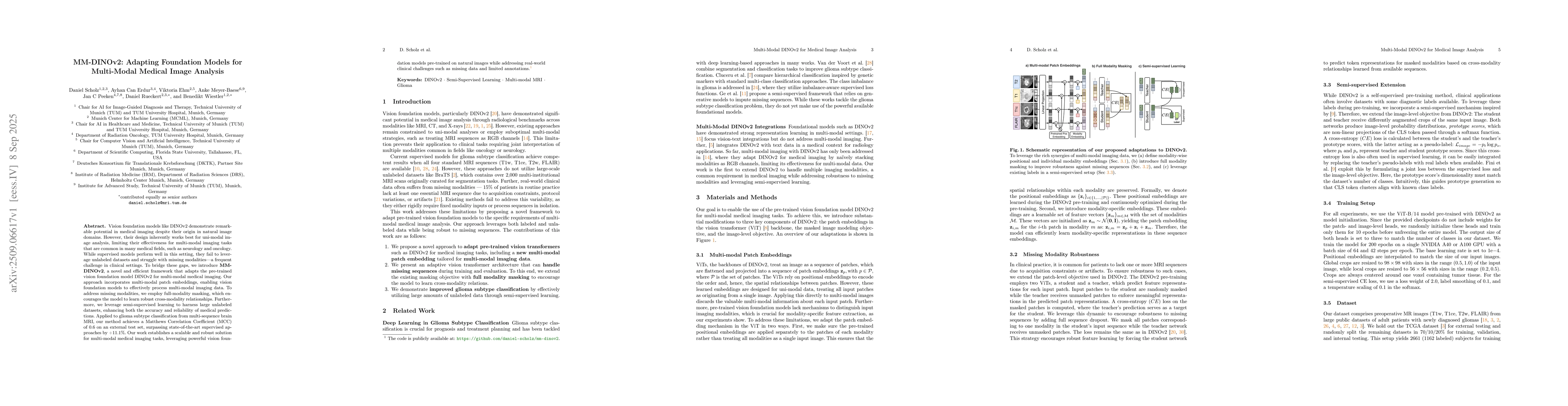
Vision foundation models like DINOv2 demonstrate remarkable potential in medical imaging despite their origin in natural image domains. However, their design inherently works best for uni-modal image ...
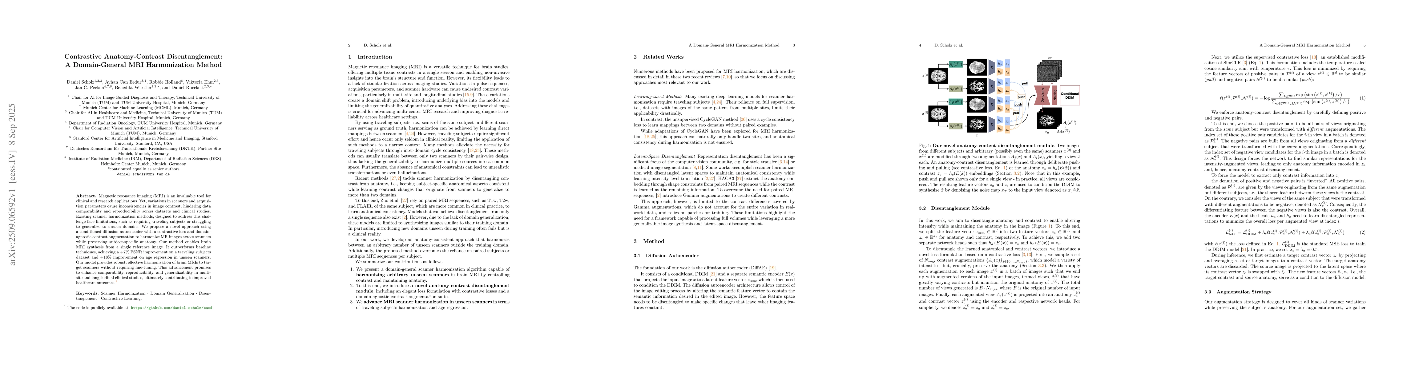
Magnetic resonance imaging (MRI) is an invaluable tool for clinical and research applications. Yet, variations in scanners and acquisition parameters cause inconsistencies in image contrast, hindering...
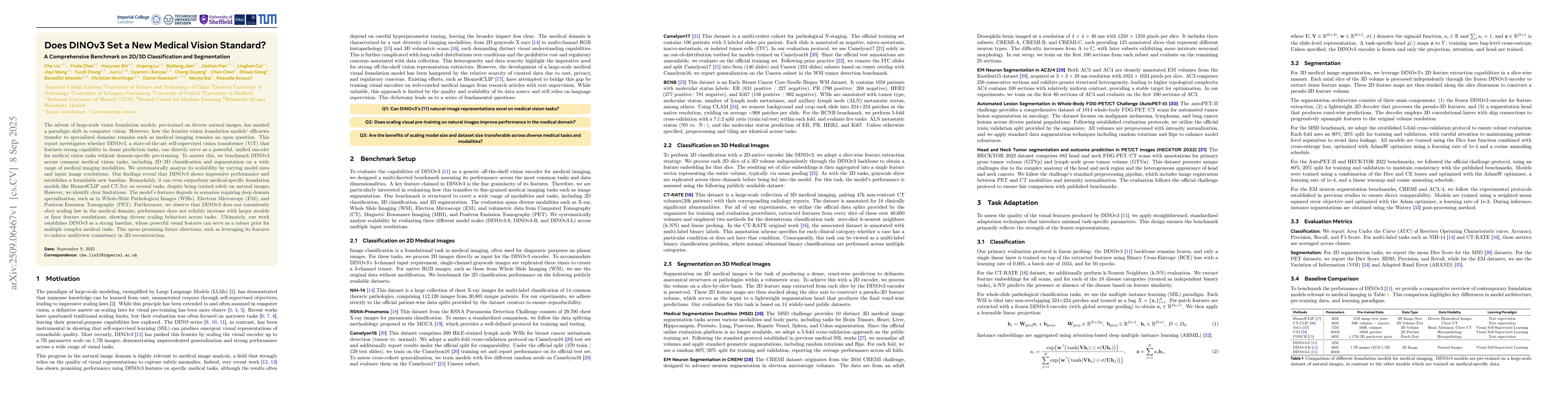
The advent of large-scale vision foundation models, pre-trained on diverse natural images, has marked a paradigm shift in computer vision. However, how the frontier vision foundation models' efficacie...
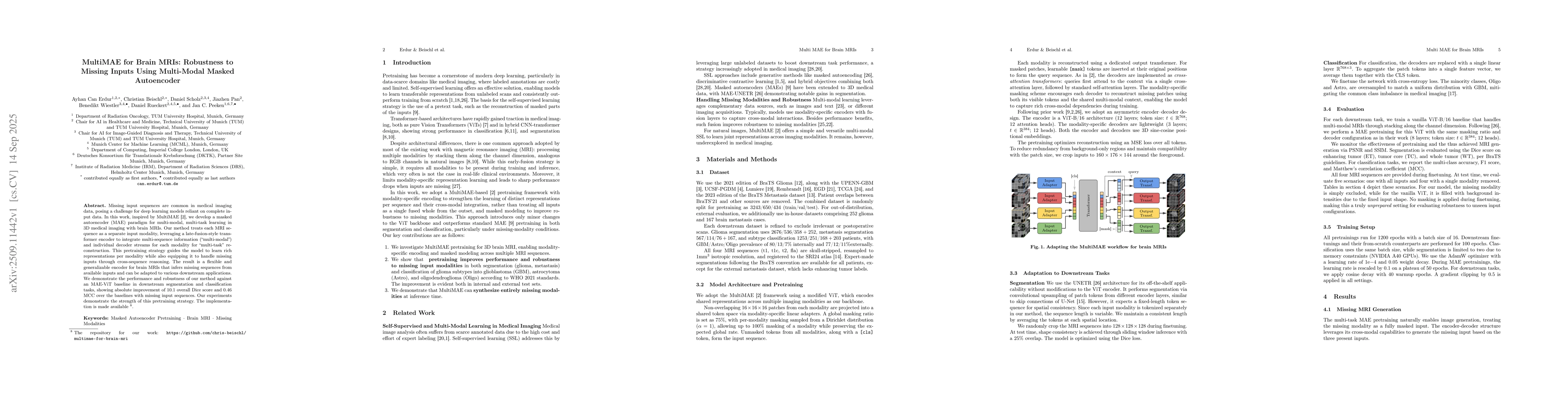
Missing input sequences are common in medical imaging data, posing a challenge for deep learning models reliant on complete input data. In this work, inspired by MultiMAE [2], we develop a masked auto...
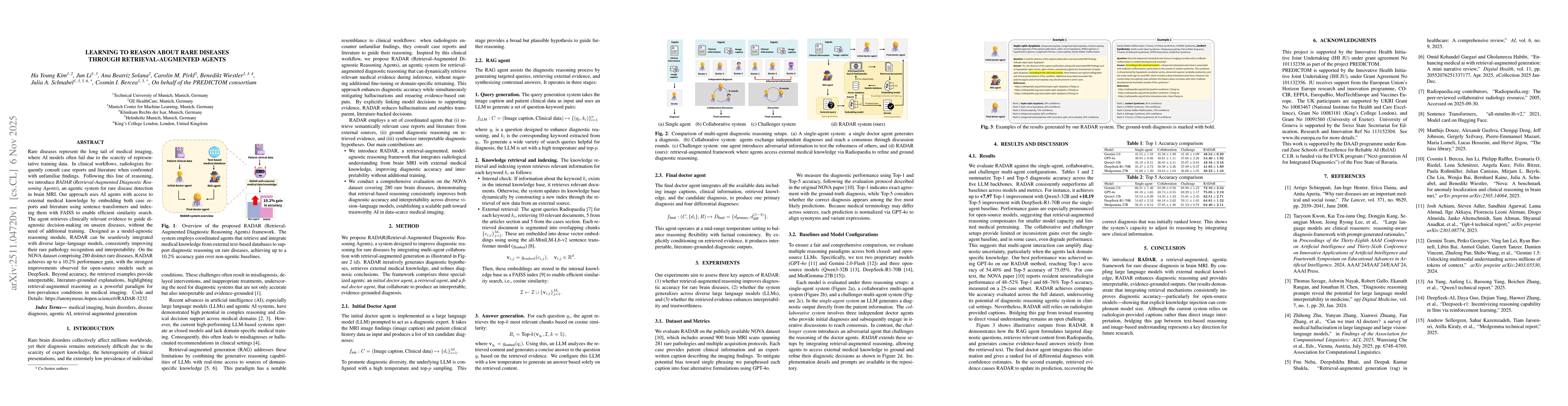
Rare diseases represent the long tail of medical imaging, where AI models often fail due to the scarcity of representative training data. In clinical workflows, radiologists frequently consult case re...
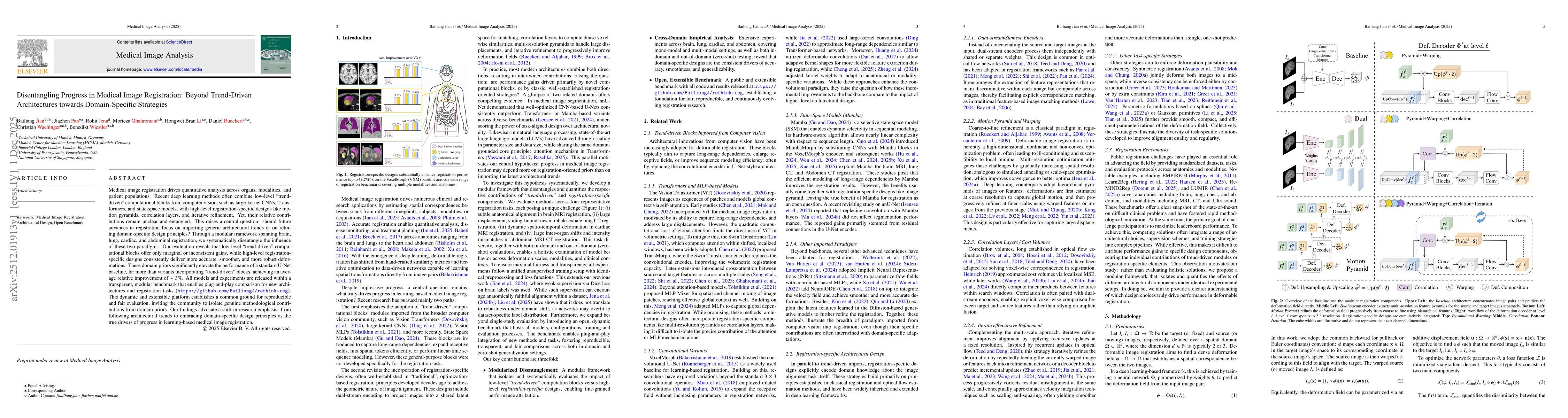
Medical image registration drives quantitative analysis across organs, modalities, and patient populations. Recent deep learning methods often combine low-level "trend-driven" computational blocks fro...
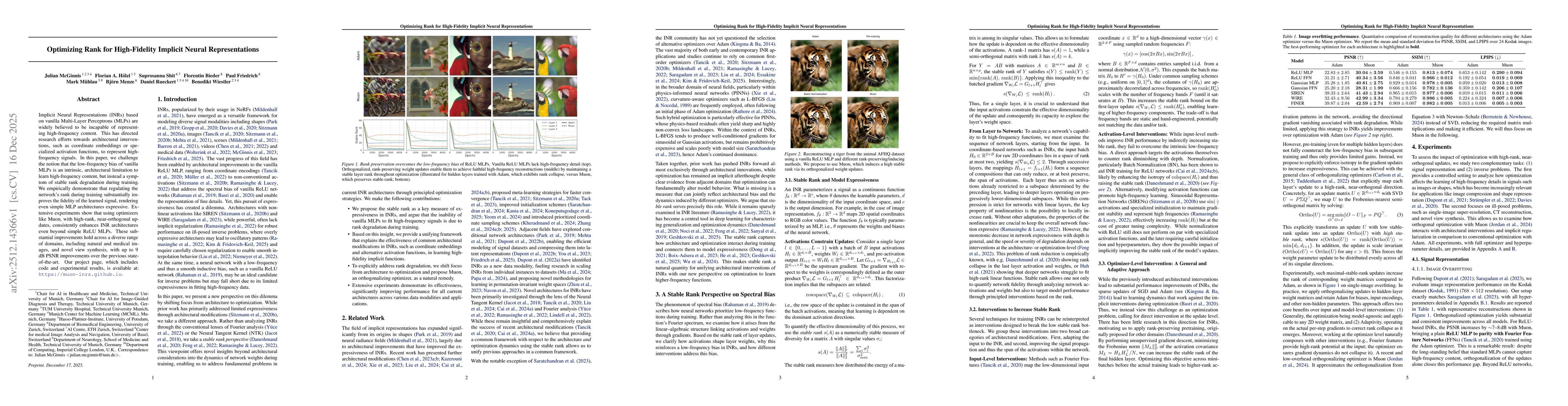
Implicit Neural Representations (INRs) based on vanilla Multi-Layer Perceptrons (MLPs) are widely believed to be incapable of representing high-frequency content. This has directed research efforts to...
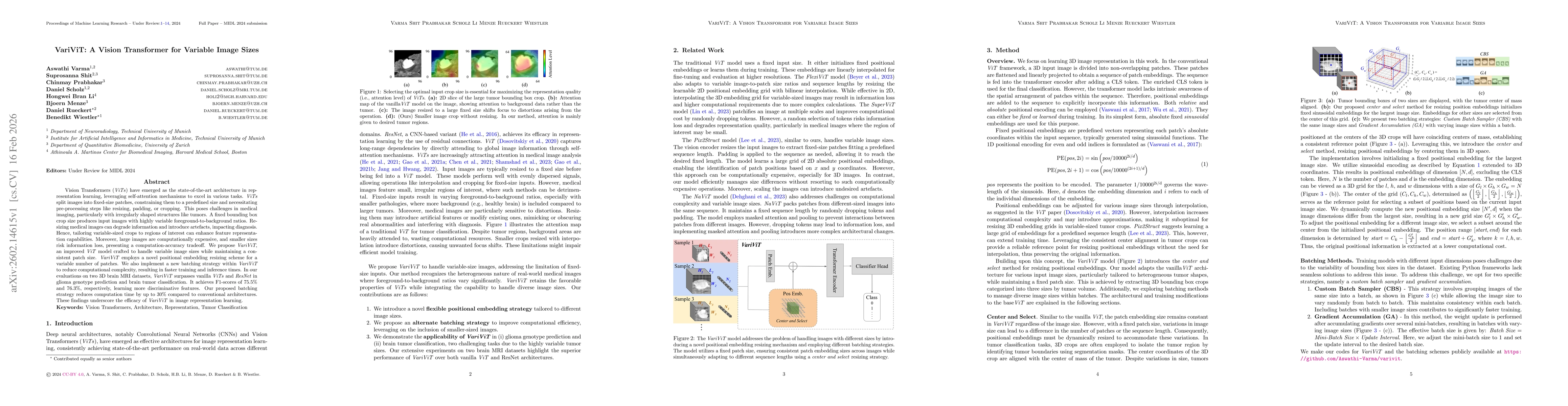
Vision Transformers (ViTs) have emerged as the state-of-the-art architecture in representation learning, leveraging self-attention mechanisms to excel in various tasks. ViTs split images into fixed-si...
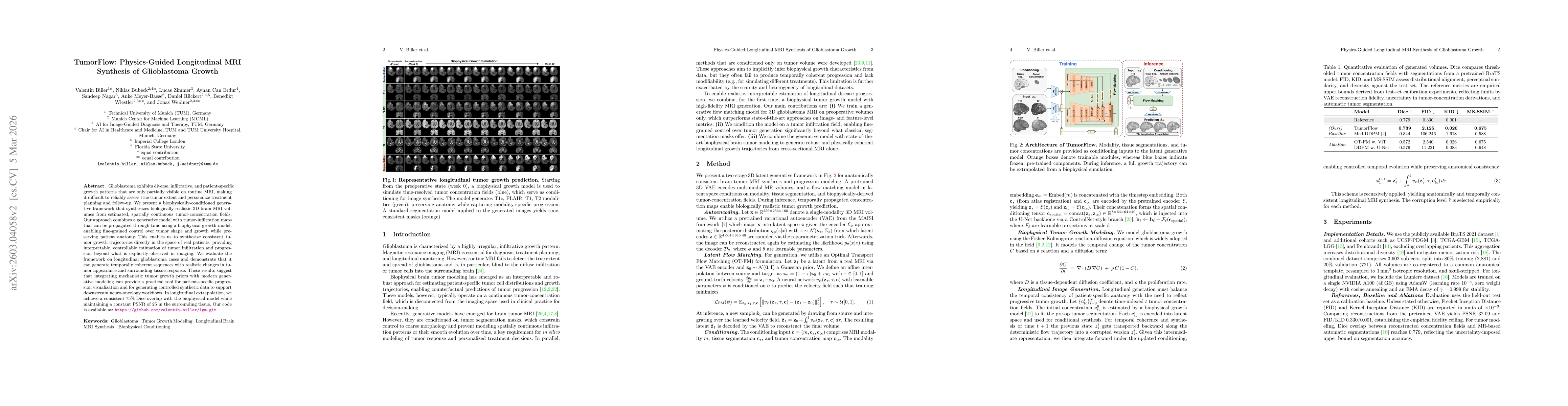
Glioblastoma exhibits diverse, infiltrative, and patient-specific growth patterns that are only partially visible on routine MRI, making it difficult to reliably assess true tumor extent and personali...
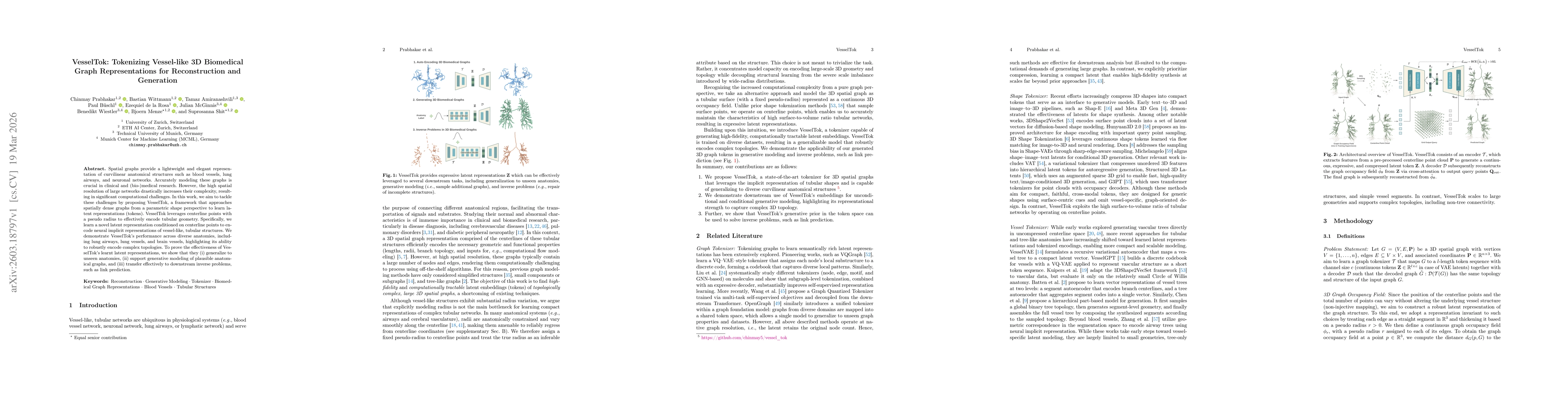
Spatial graphs provide a lightweight and elegant representation of curvilinear anatomical structures such as blood vessels, lung airways, and neuronal networks. Accurately modeling these graphs is cru...
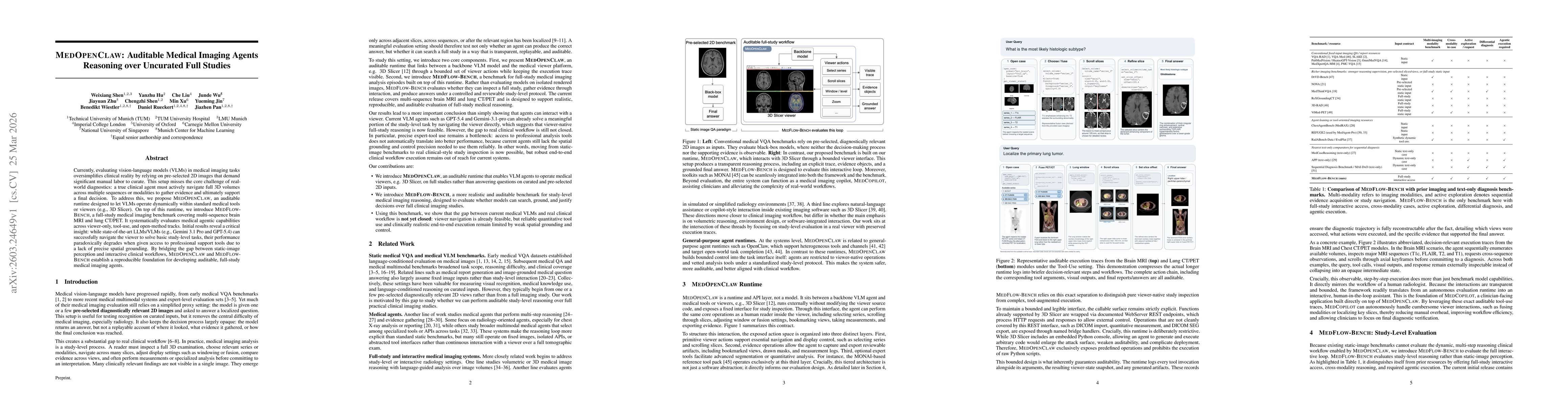
Currently, evaluating vision-language models (VLMs) in medical imaging tasks oversimplifies clinical reality by relying on pre-selected 2D images that demand significant manual labor to curate. This s...
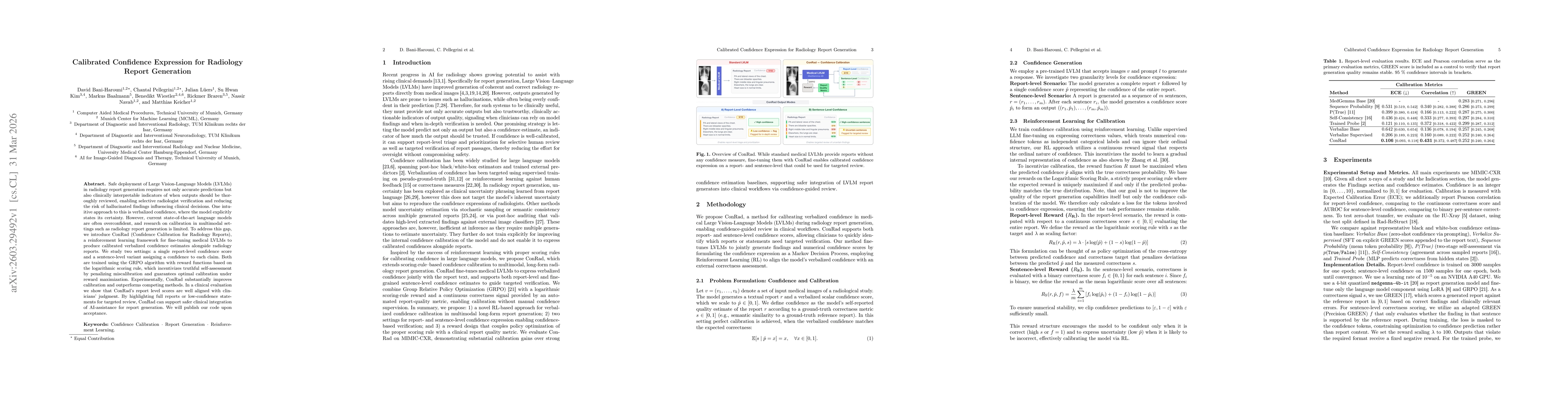
Safe deployment of Large Vision-Language Models (LVLMs) in radiology report generation requires not only accurate predictions but also clinically interpretable indicators of when outputs should be tho...
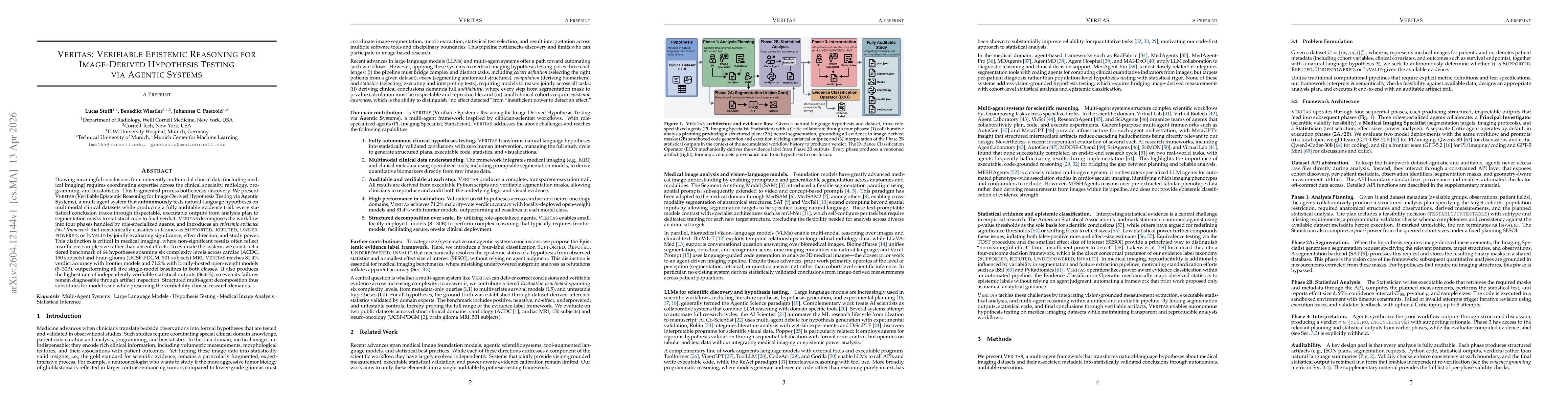
Drawing meaningful conclusions from inherently multimodal clinical data (including medical imaging) requires coordinating expertise across the clinical specialty, radiology, programming, and biostatis...
State-of-the-art large language models (LLMs) show high performance in general visual question answering. However, a fundamental limitation remains: current architectures lack the native 3D spatial re...
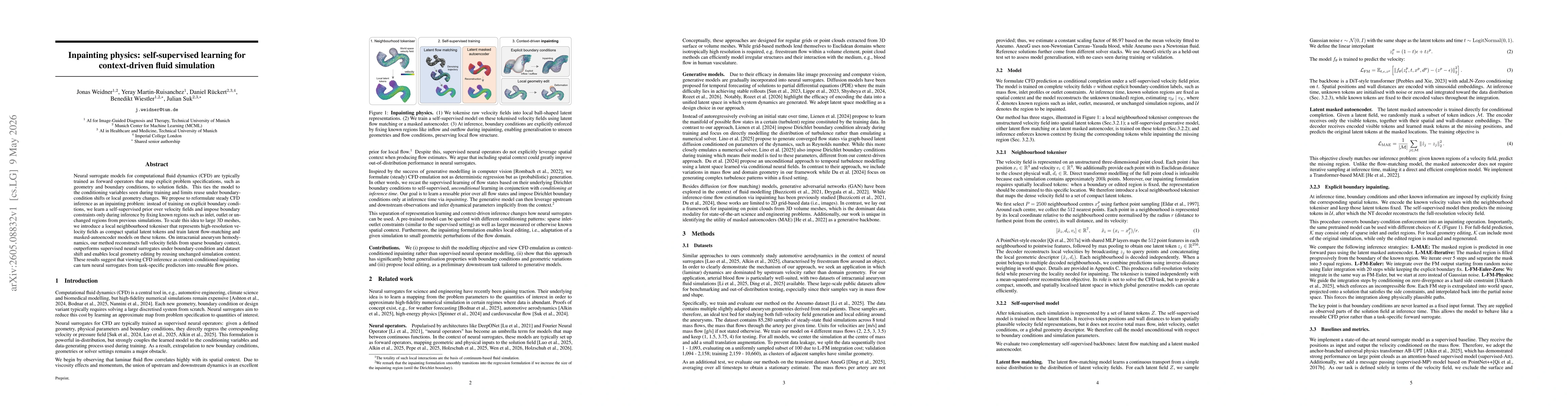
Neural surrogate models for computational fluid dynamics (CFD) are typically trained as forward operators that map explicit problem specifications, such as geometry and boundary conditions, to solutio...
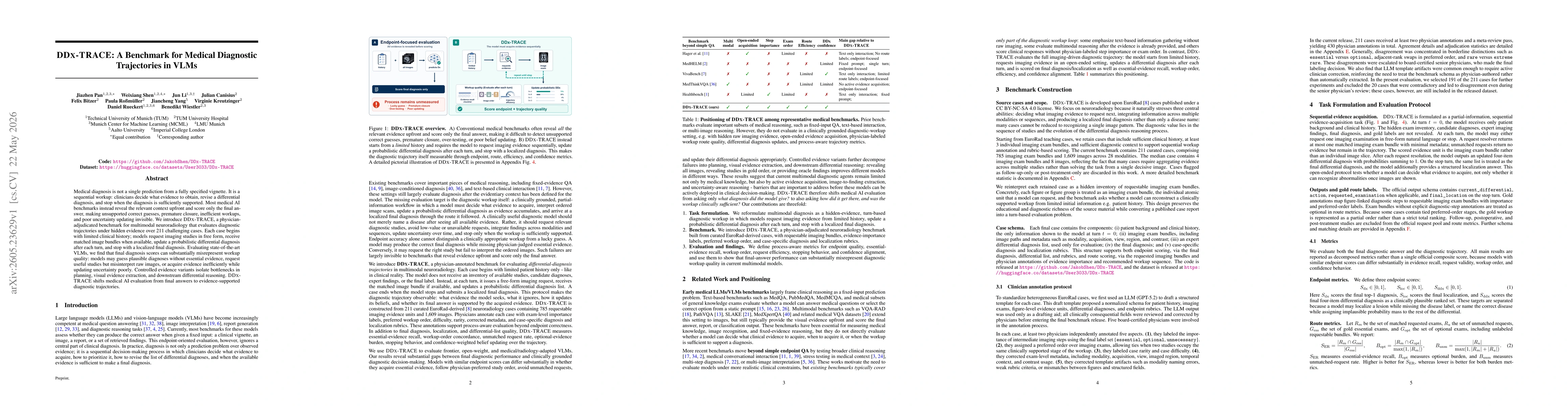
Medical diagnosis is not a single prediction from a fully specified vignette. It is a sequential workup: clinicians decide what evidence to obtain, revise a differential diagnosis, and stop when the d...
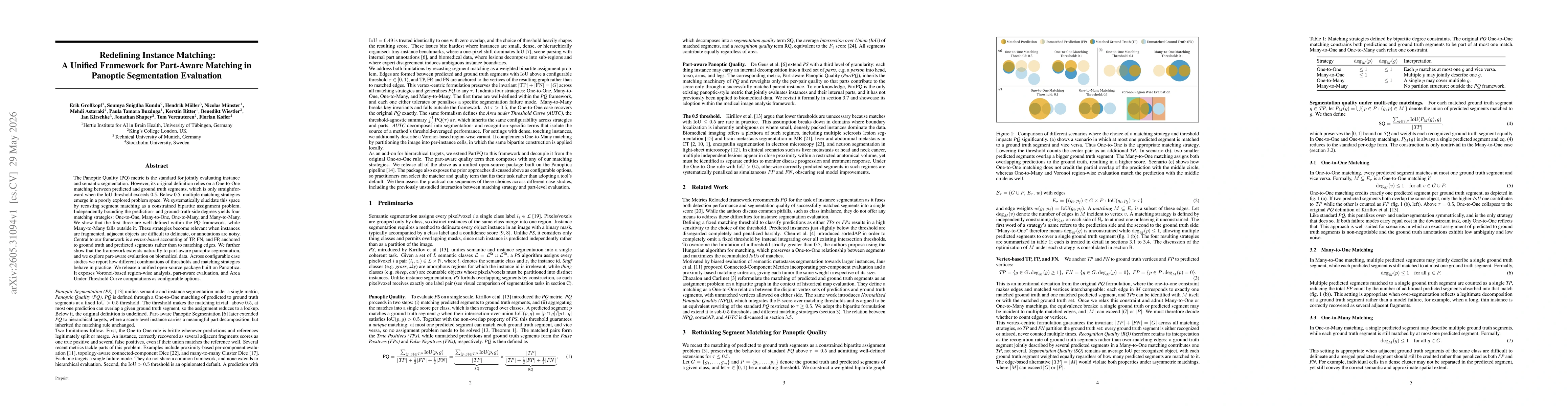
The Panoptic Quality (PQ) metric is the standard for jointly evaluating instance and semantic segmentation. However, its original definition relies on a One-to-One matching between predicted and groun...
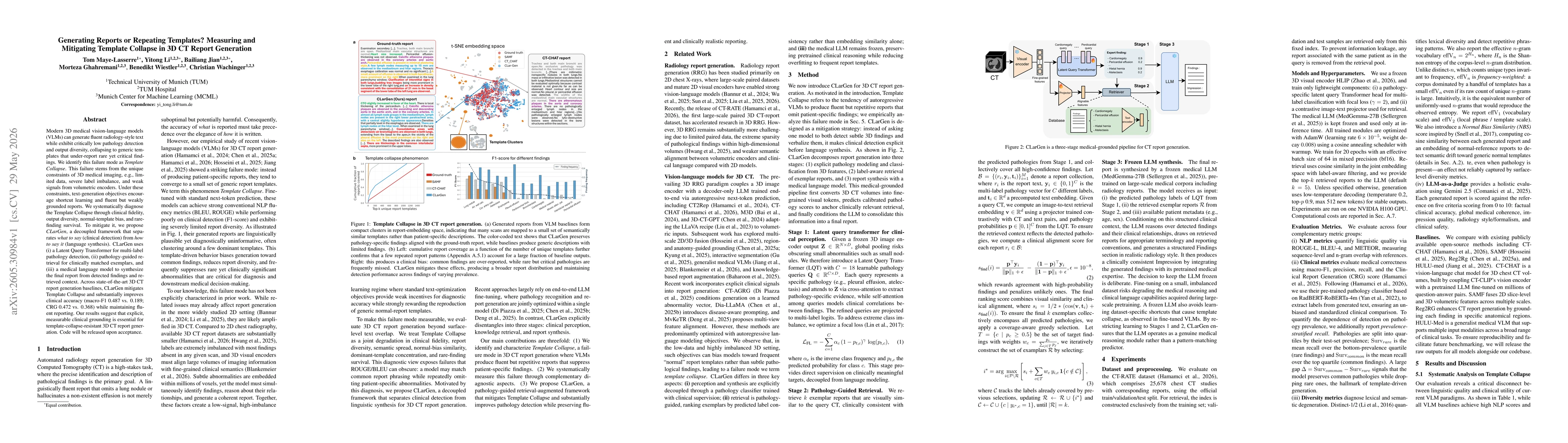
Modern 3D medical vision-language models (VLMs) can generate fluent radiology-style text while exhibit critically low pathology detection and output diversity, collapsing to generic templates that und...
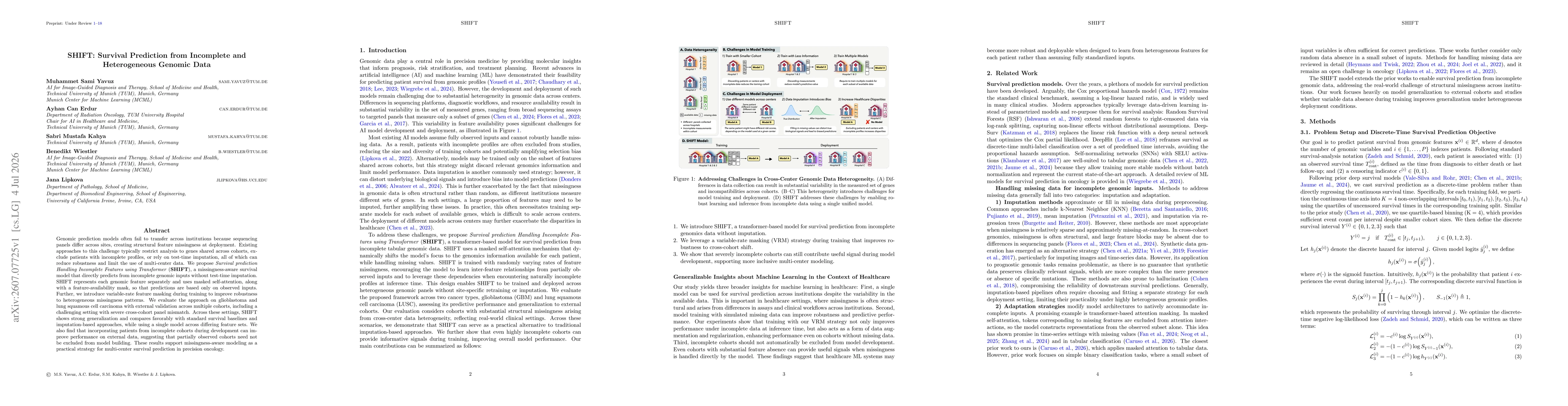
Genomic prediction models often fail to transfer across institutions because sequencing panels differ across sites, creating structural feature missingness at deployment. Existing approaches to this c...