Academic Profile
Statistics
Similar Authors
Papers on arXiv
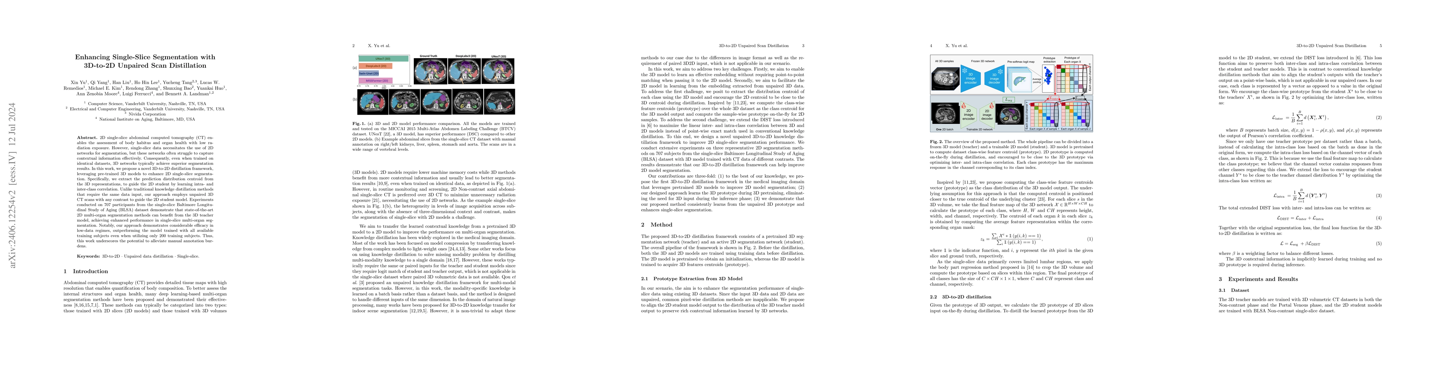
2D single-slice abdominal computed tomography (CT) enables the assessment of body habitus and organ health with low radiation exposure. However, single-slice data necessitates the use of 2D networks f...
Statistical models for predicting lung cancer have the potential to facilitate earlier diagnosis of malignancy and avoid invasive workup of benign disease. Many models have been published, but compa...
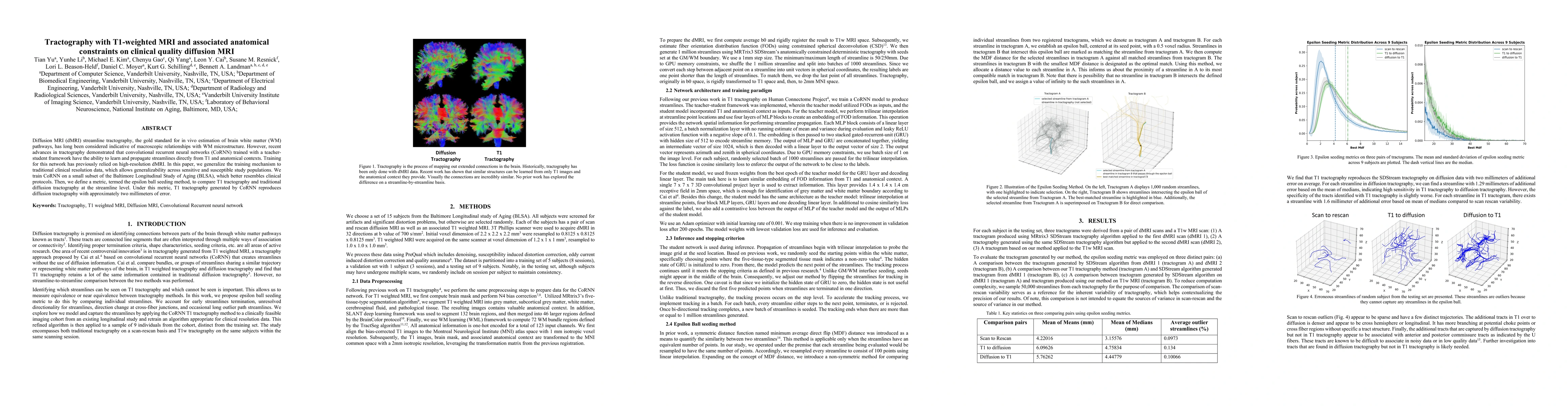
Diffusion MRI (dMRI) streamline tractography, the gold standard for in vivo estimation of brain white matter (WM) pathways, has long been considered indicative of macroscopic relationships with WM m...
Insufficiently precise diagnosis of clinical disease is likely responsible for many treatment failures, even for common conditions and treatments. With a large enough dataset, it may be possible to ...
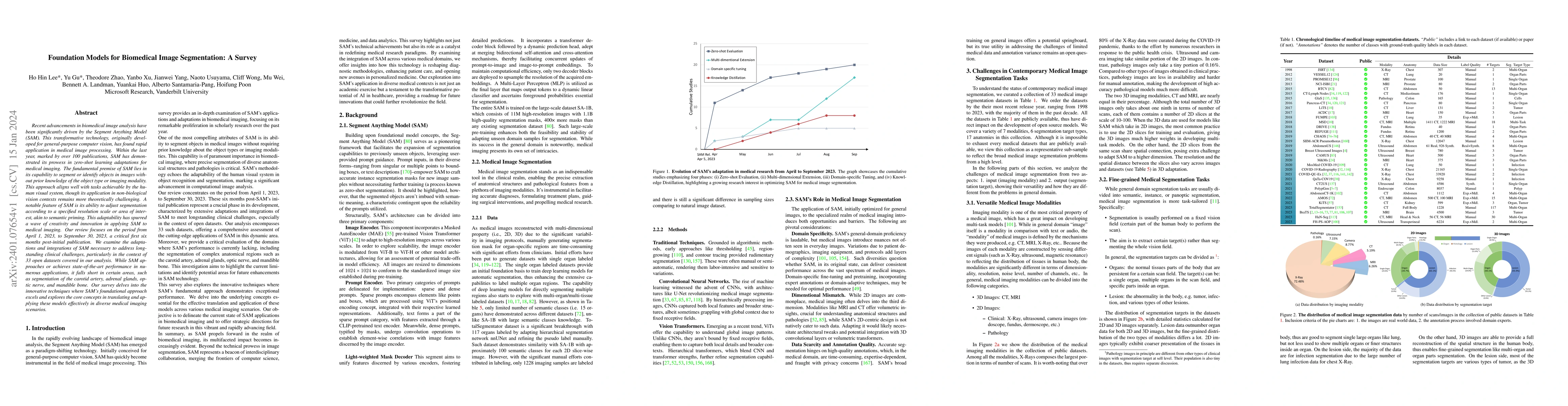
Recent advancements in biomedical image analysis have been significantly driven by the Segment Anything Model (SAM). This transformative technology, originally developed for general-purpose computer...
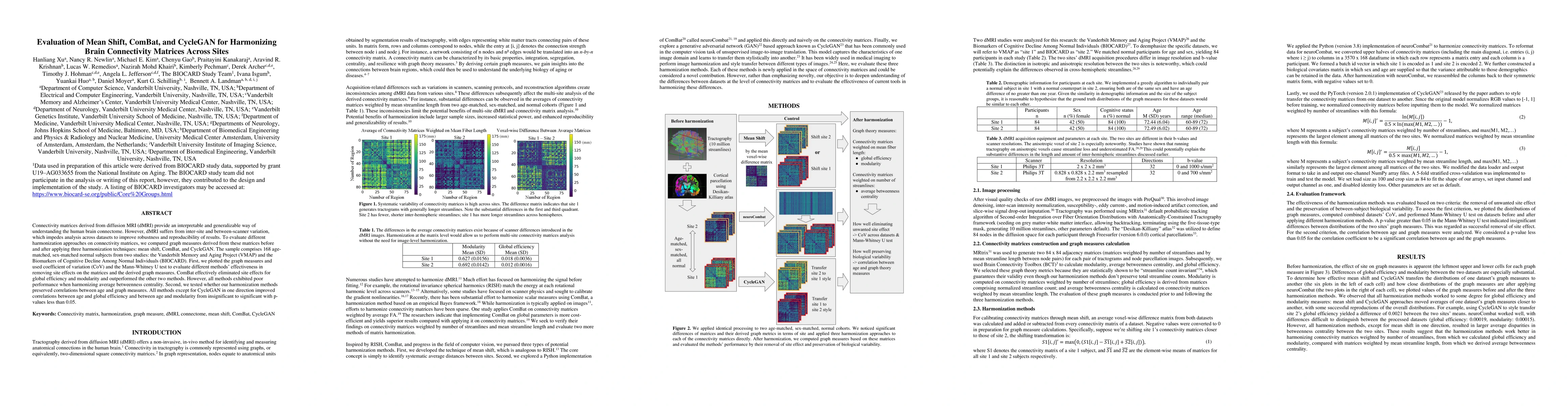
Connectivity matrices derived from diffusion MRI (dMRI) provide an interpretable and generalizable way of understanding the human brain connectome. However, dMRI suffers from inter-site and between-...
Understanding the way cells communicate, co-locate, and interrelate is essential to understanding human physiology. Hematoxylin and eosin (H&E) staining is ubiquitously available both for clinical s...
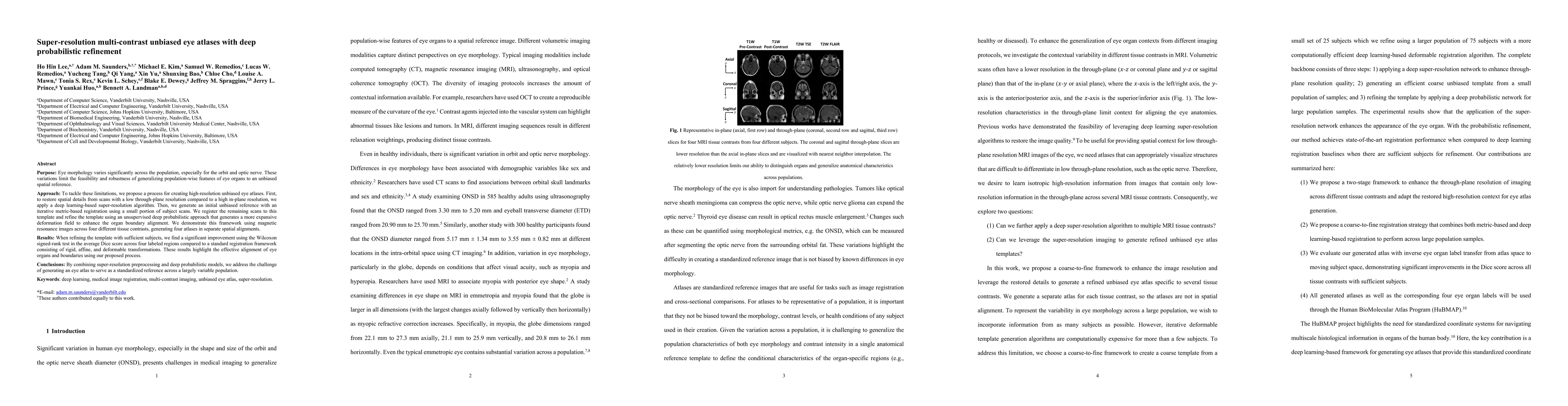
Purpose: Eye morphology varies significantly across the population, especially for the orbit and optic nerve. These variations limit the feasibility and robustness of generalizing population-wise fe...
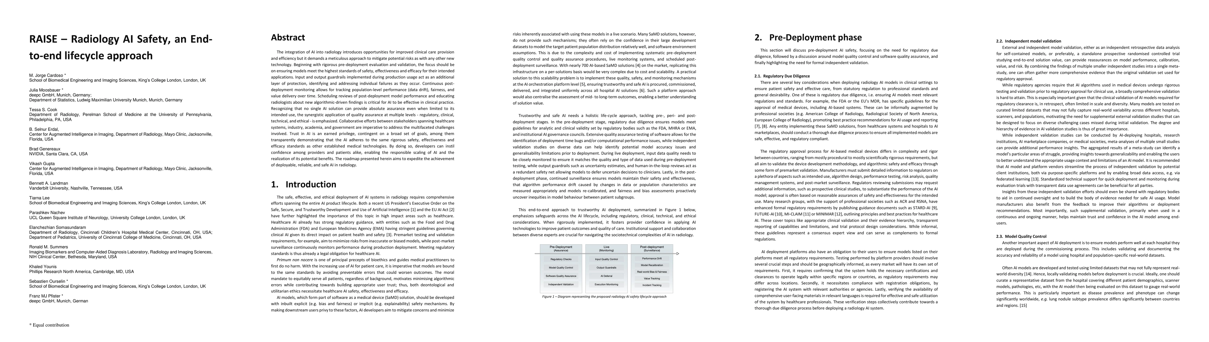
The integration of AI into radiology introduces opportunities for improved clinical care provision and efficiency but it demands a meticulous approach to mitigate potential risks as with any other n...
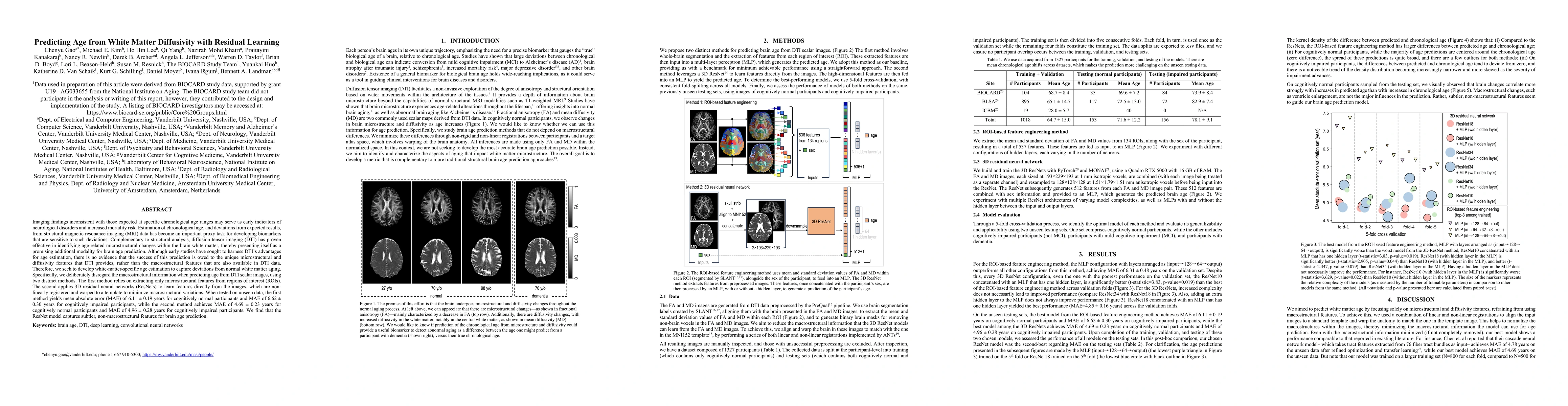
Imaging findings inconsistent with those expected at specific chronological age ranges may serve as early indicators of neurological disorders and increased mortality risk. Estimation of chronologic...
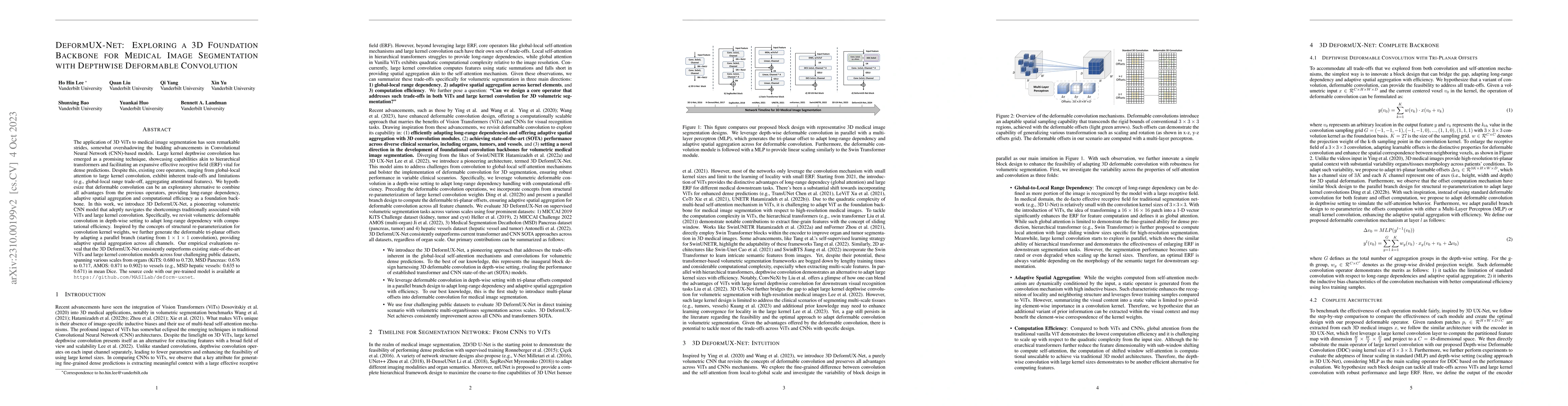
The application of 3D ViTs to medical image segmentation has seen remarkable strides, somewhat overshadowing the budding advancements in Convolutional Neural Network (CNN)-based models. Large kernel...
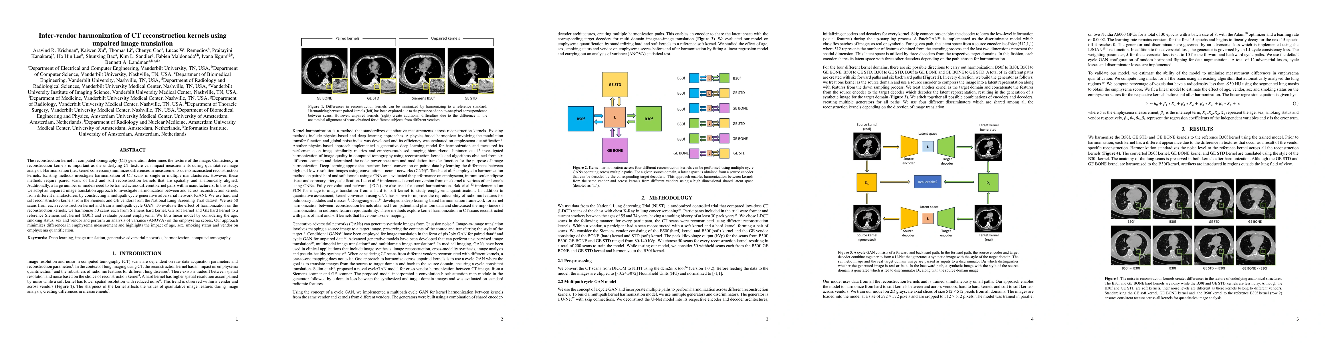
The reconstruction kernel in computed tomography (CT) generation determines the texture of the image. Consistency in reconstruction kernels is important as the underlying CT texture can impact measu...
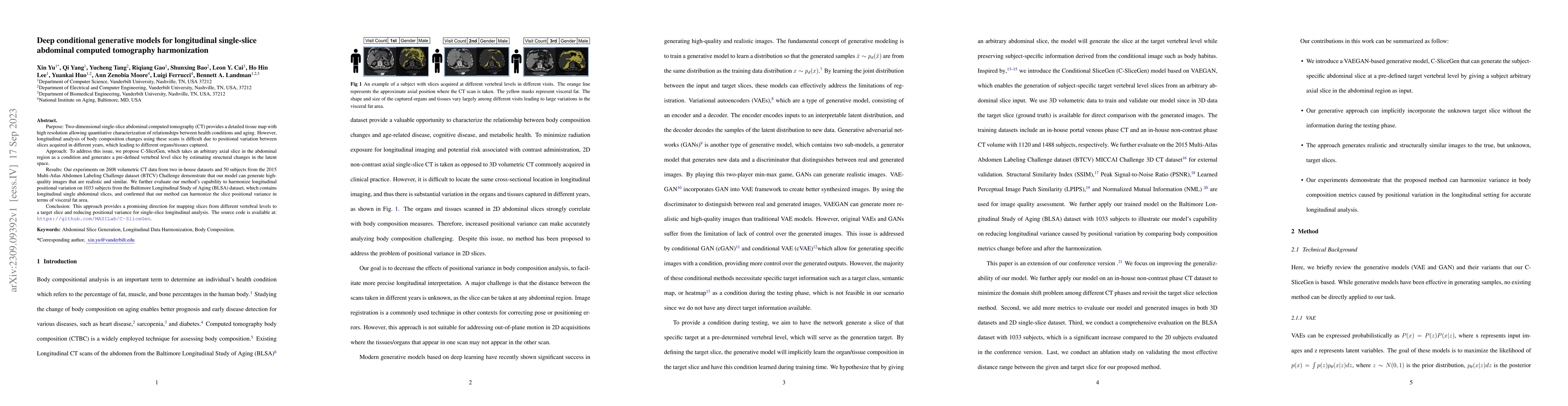
Two-dimensional single-slice abdominal computed tomography (CT) provides a detailed tissue map with high resolution allowing quantitative characterization of relationships between health conditions ...
Whole brain segmentation with magnetic resonance imaging (MRI) enables the non-invasive measurement of brain regions, including total intracranial volume (TICV) and posterior fossa volume (PFV). Enh...
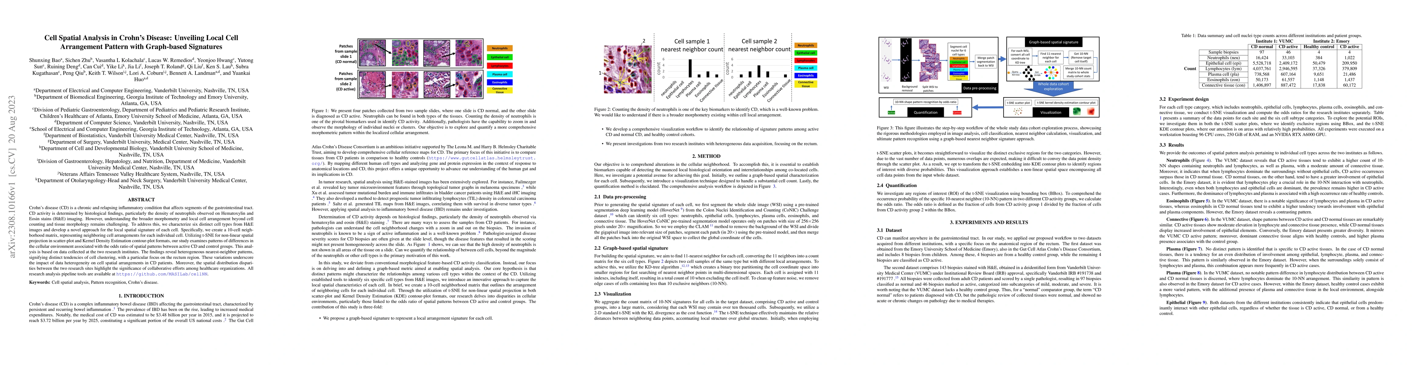
Crohn's disease (CD) is a chronic and relapsing inflammatory condition that affects segments of the gastrointestinal tract. CD activity is determined by histological findings, particularly the densi...
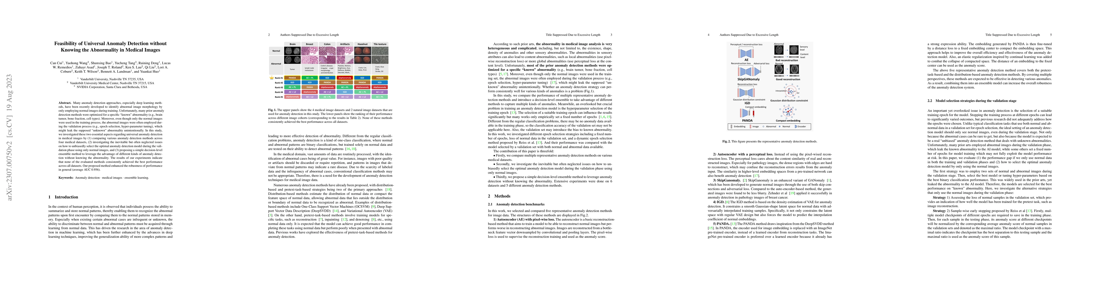
Many anomaly detection approaches, especially deep learning methods, have been recently developed to identify abnormal image morphology by only employing normal images during training. Unfortunately...
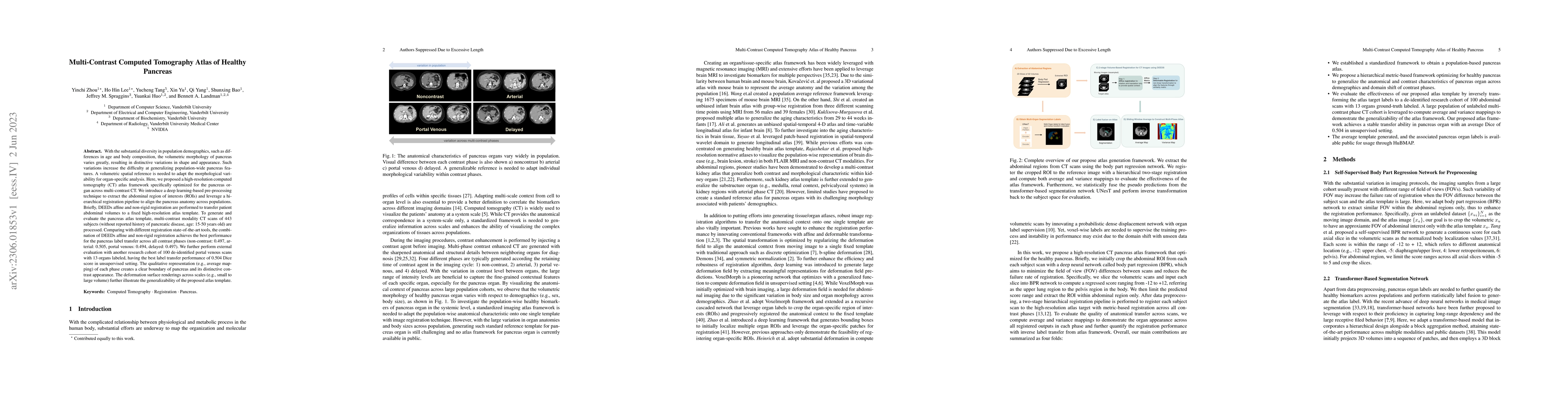
With the substantial diversity in population demographics, such as differences in age and body composition, the volumetric morphology of pancreas varies greatly, resulting in distinctive variations ...
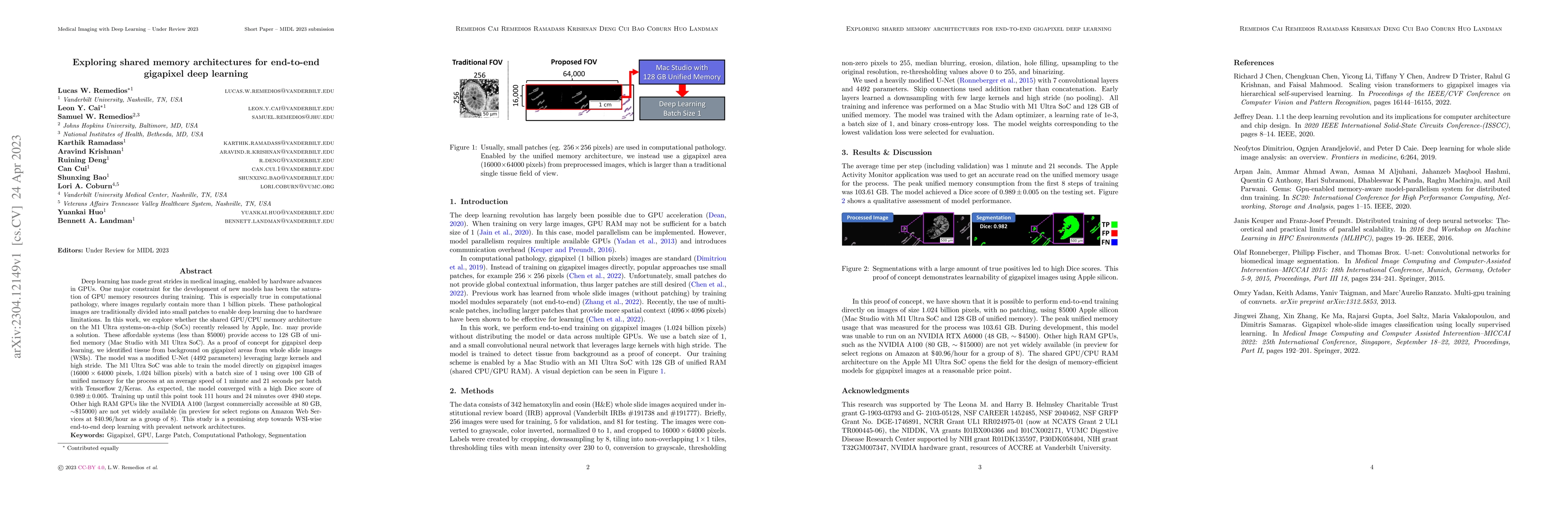
Deep learning has made great strides in medical imaging, enabled by hardware advances in GPUs. One major constraint for the development of new models has been the saturation of GPU memory resources ...
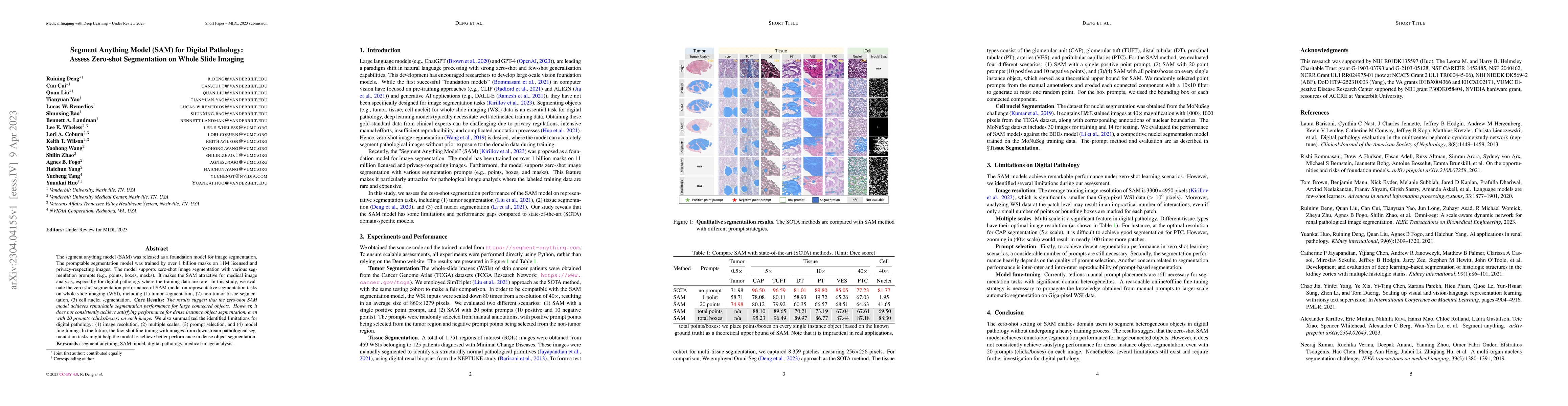
The segment anything model (SAM) was released as a foundation model for image segmentation. The promptable segmentation model was trained by over 1 billion masks on 11M licensed and privacy-respecti...
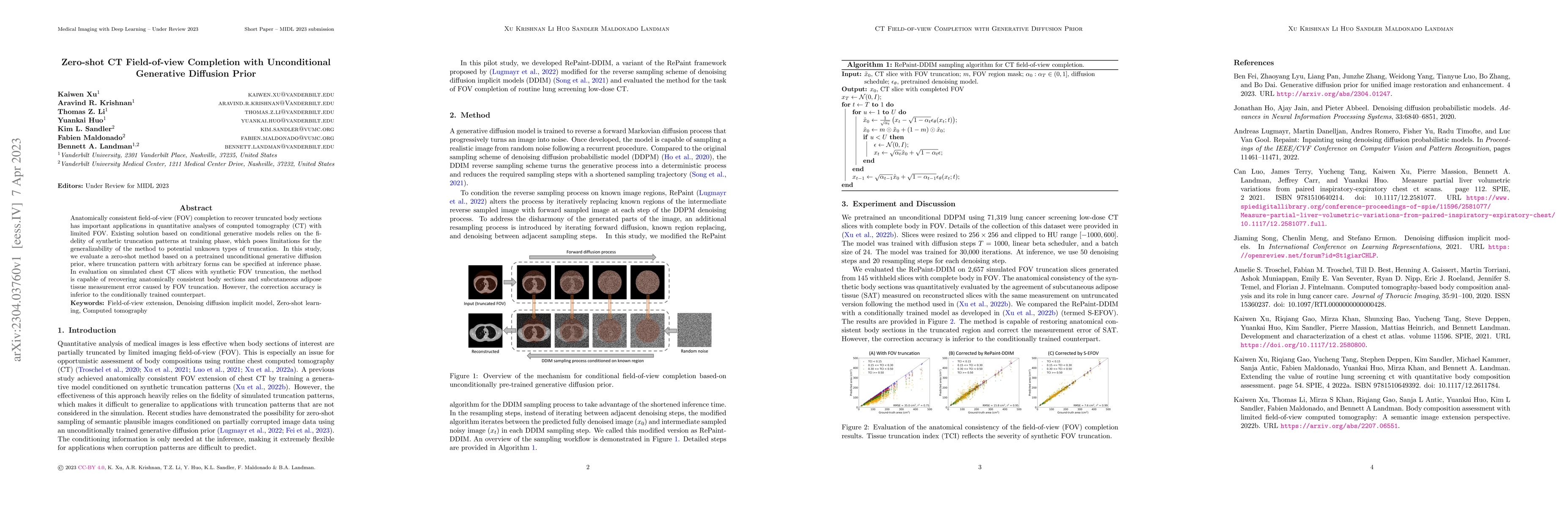
Anatomically consistent field-of-view (FOV) completion to recover truncated body sections has important applications in quantitative analyses of computed tomography (CT) with limited FOV. Existing s...
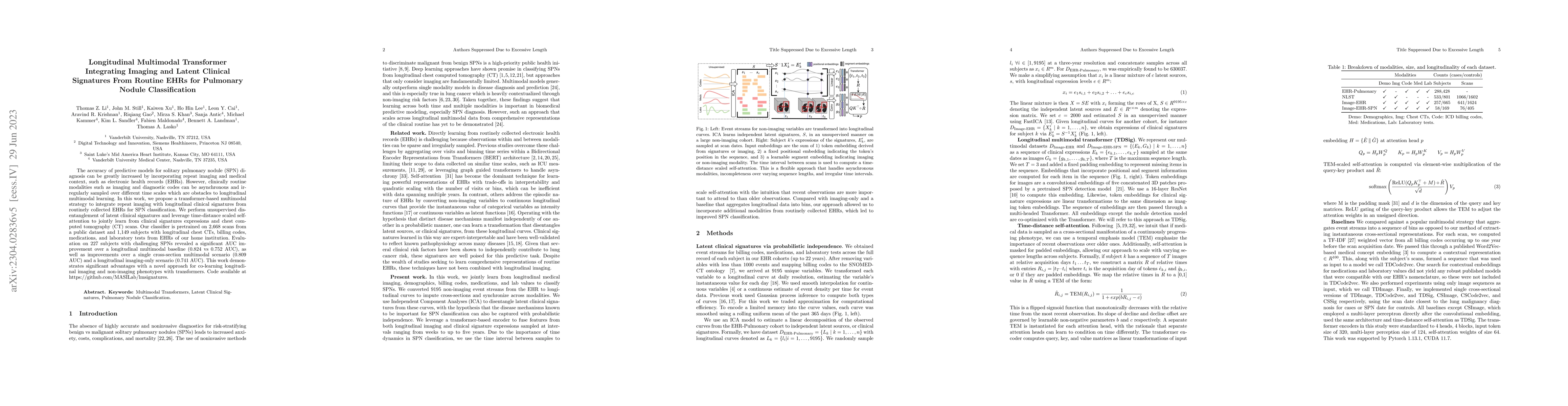
The accuracy of predictive models for solitary pulmonary nodule (SPN) diagnosis can be greatly increased by incorporating repeat imaging and medical context, such as electronic health records (EHRs)...
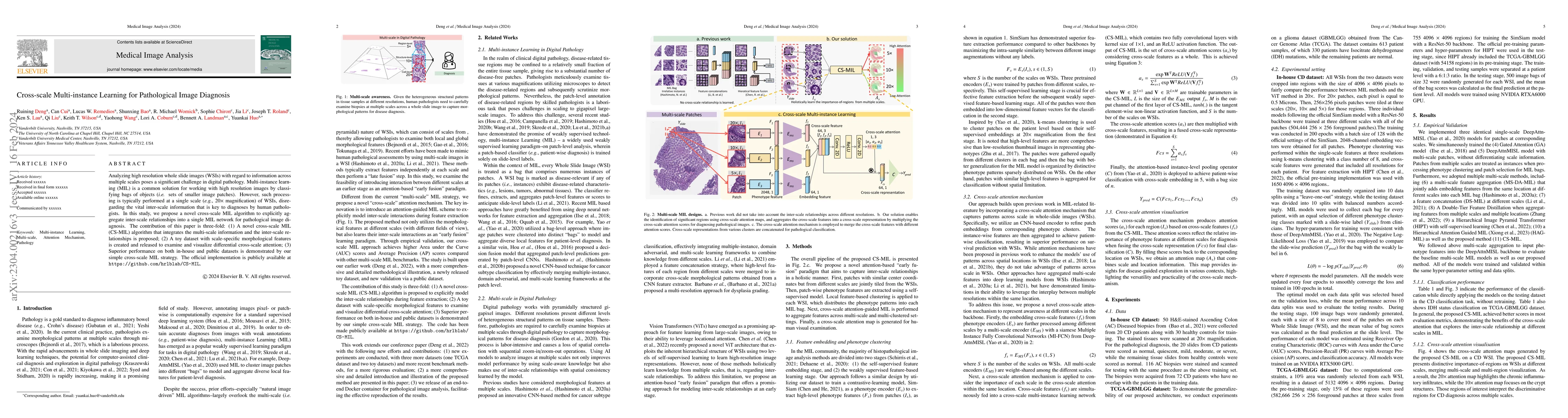
Analyzing high resolution whole slide images (WSIs) with regard to information across multiple scales poses a significant challenge in digital pathology. Multi-instance learning (MIL) is a common so...
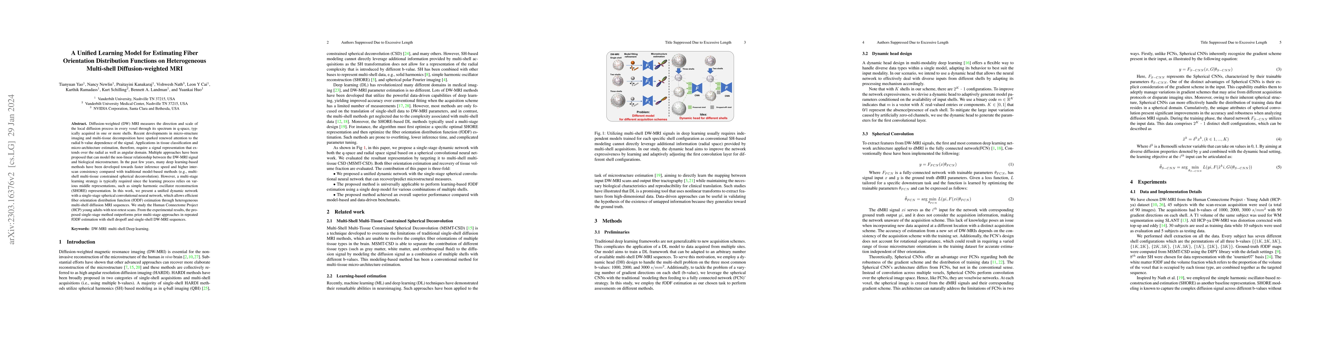
Diffusion-weighted (DW) MRI measures the direction and scale of the local diffusion process in every voxel through its spectrum in q-space, typically acquired in one or more shells. Recent developme...
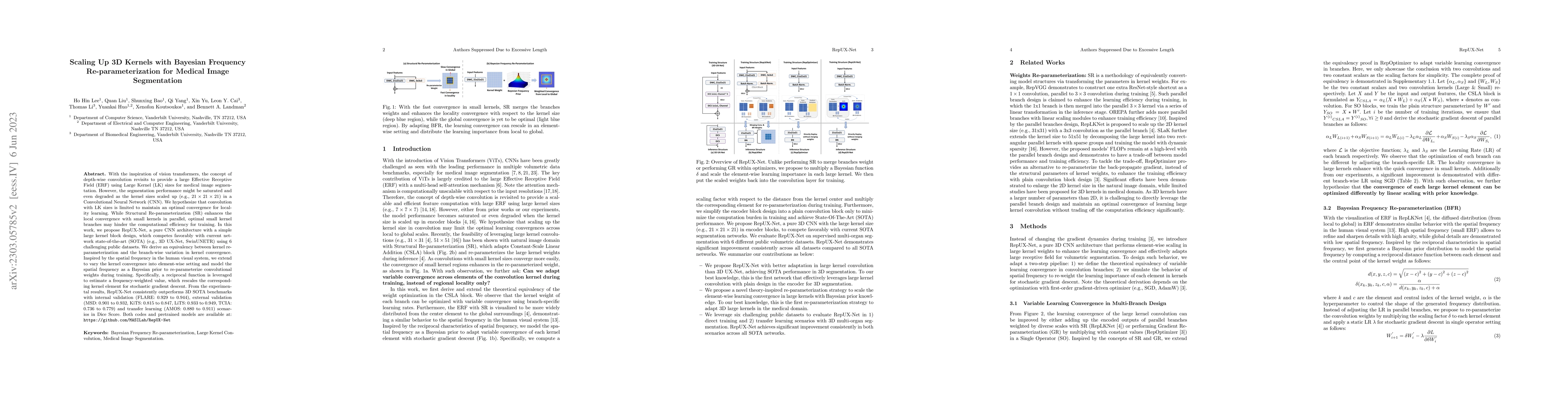
With the inspiration of vision transformers, the concept of depth-wise convolution revisits to provide a large Effective Receptive Field (ERF) using Large Kernel (LK) sizes for medical image segment...
Validation metrics are key for the reliable tracking of scientific progress and for bridging the current chasm between artificial intelligence (AI) research and its translation into practice. Howeve...
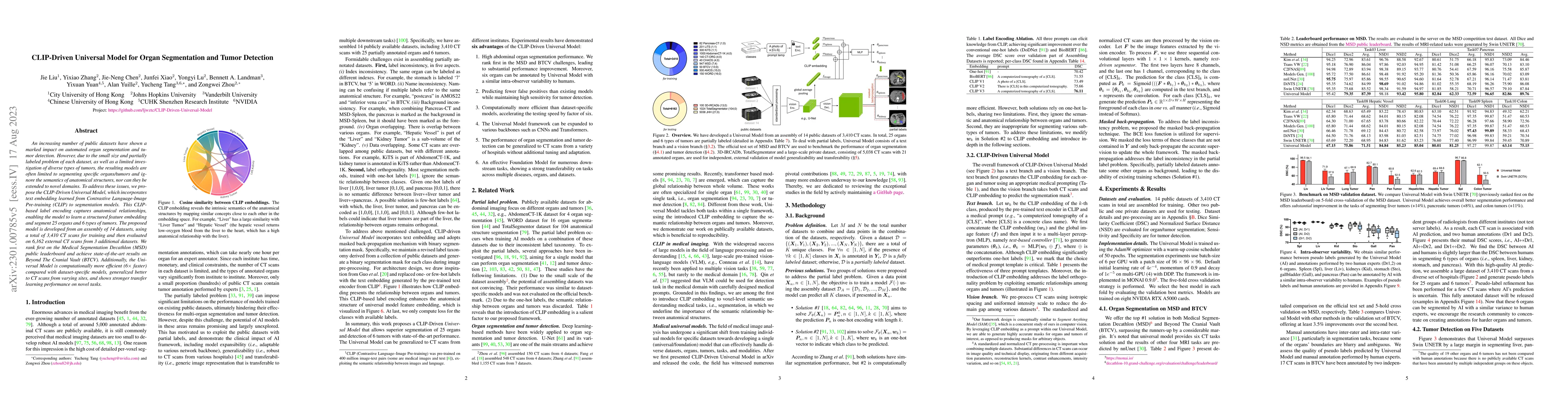
An increasing number of public datasets have shown a marked impact on automated organ segmentation and tumor detection. However, due to the small size and partially labeled problem of each dataset, ...
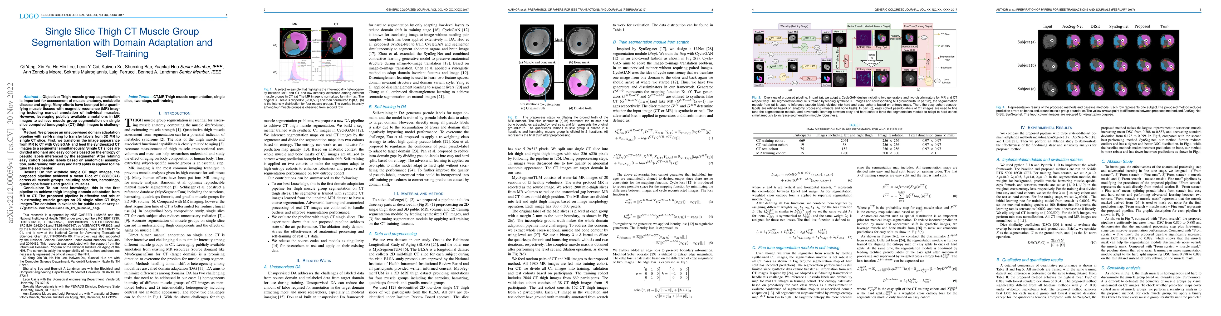
Objective: Thigh muscle group segmentation is important for assessment of muscle anatomy, metabolic disease and aging. Many efforts have been put into quantifying muscle tissues with magnetic resona...
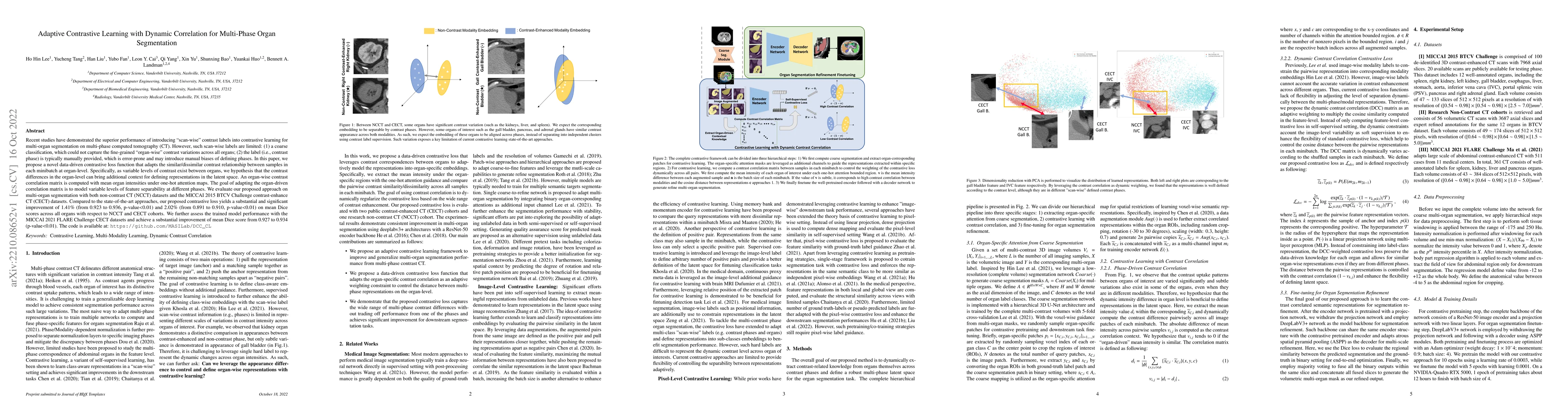
Recent studies have demonstrated the superior performance of introducing ``scan-wise" contrast labels into contrastive learning for multi-organ segmentation on multi-phase computed tomography (CT). ...
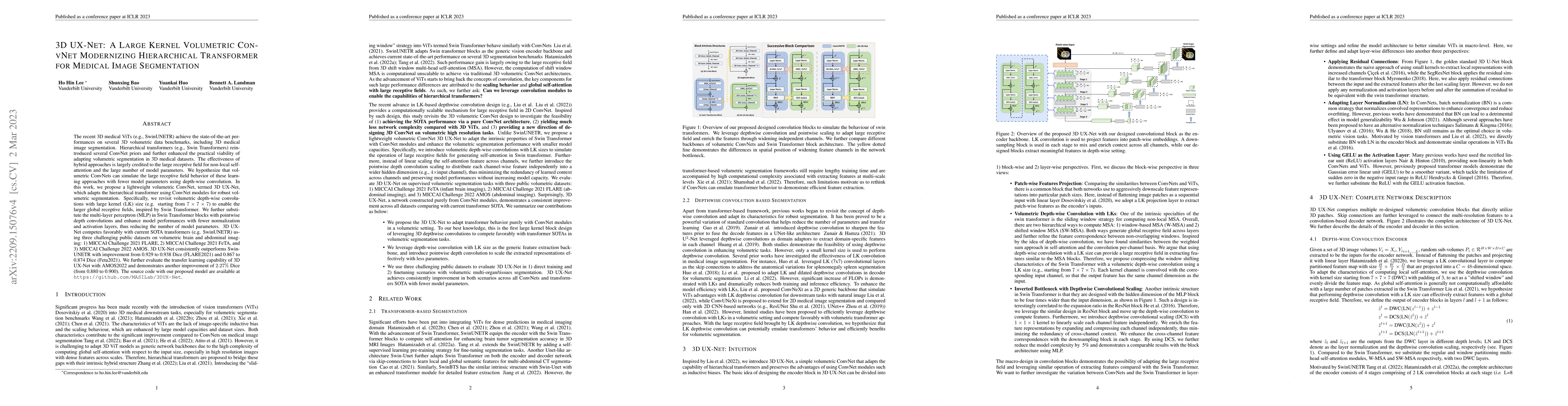
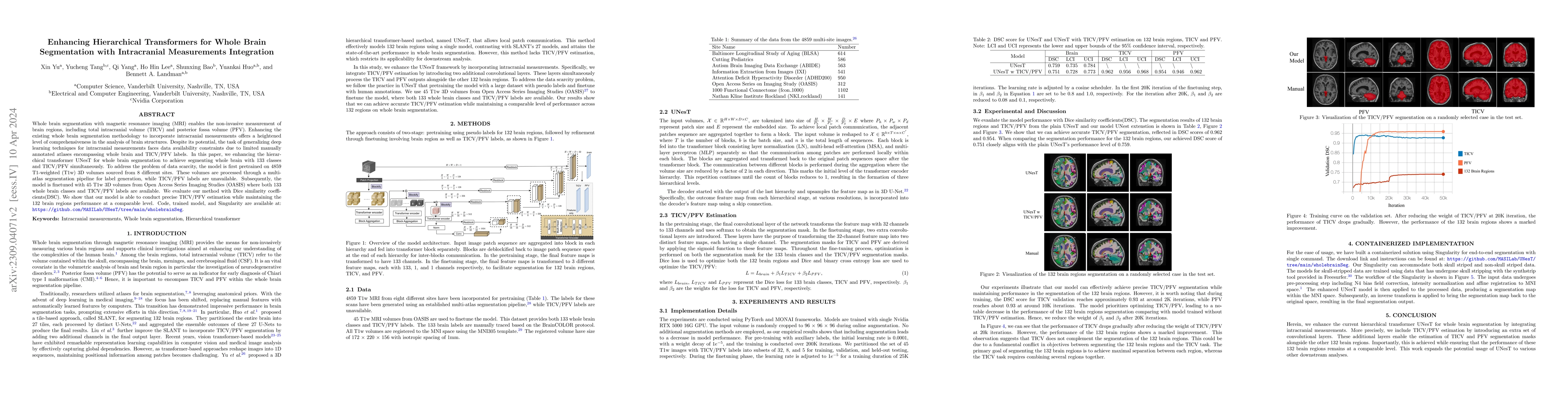
The recent 3D medical ViTs (e.g., SwinUNETR) achieve the state-of-the-art performances on several 3D volumetric data benchmarks, including 3D medical image segmentation. Hierarchical transformers (e...
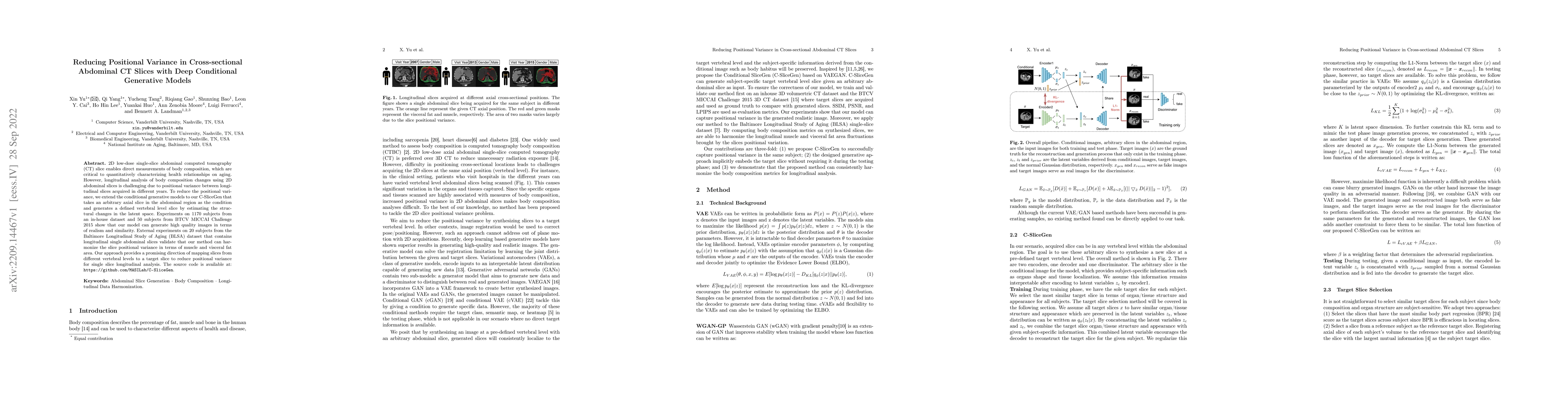
2D low-dose single-slice abdominal computed tomography (CT) slice enables direct measurements of body composition, which are critical to quantitatively characterizing health relationships on aging. ...
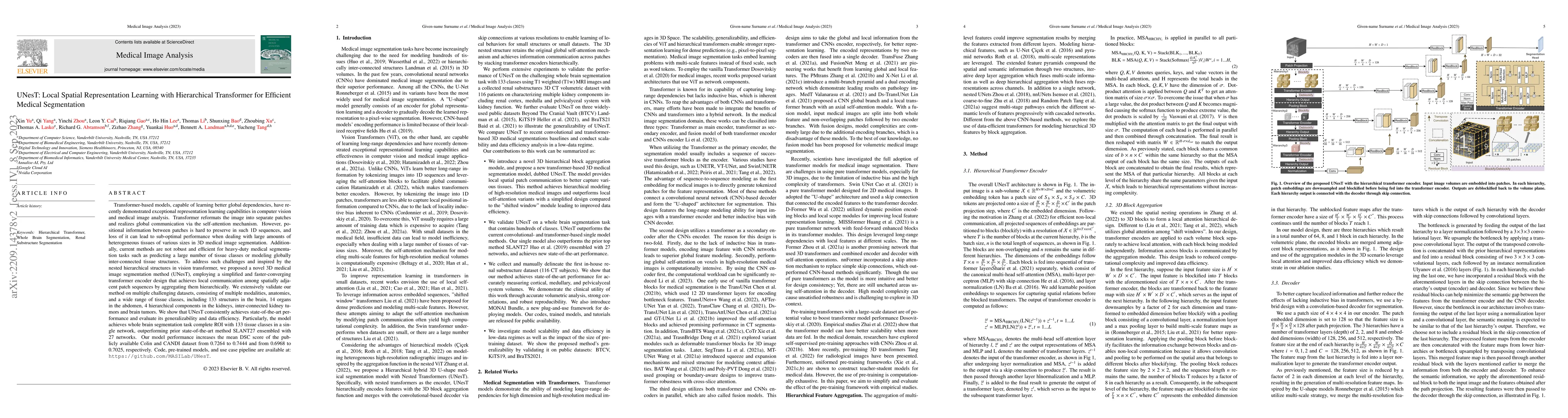
Transformer-based models, capable of learning better global dependencies, have recently demonstrated exceptional representation learning capabilities in computer vision and medical image analysis. T...
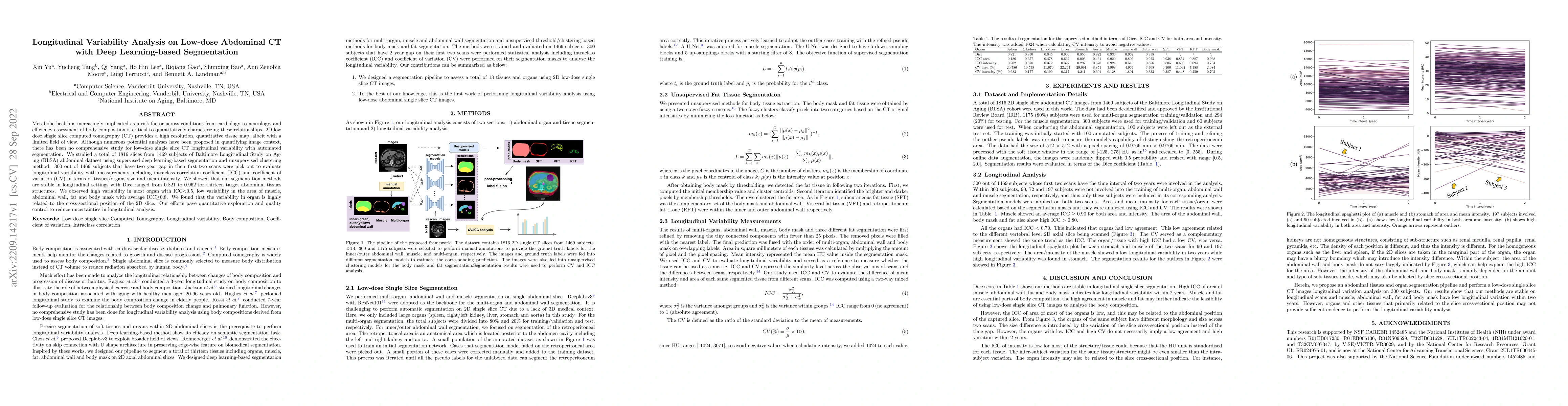
Metabolic health is increasingly implicated as a risk factor across conditions from cardiology to neurology, and efficiency assessment of body composition is critical to quantitatively characterizin...
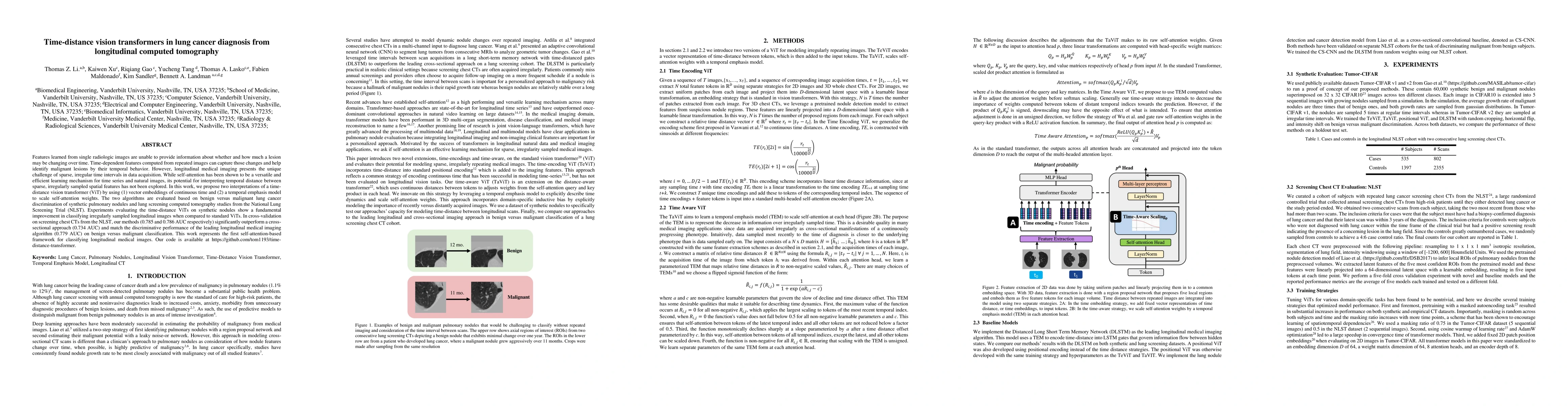
Features learned from single radiologic images are unable to provide information about whether and how much a lesion may be changing over time. Time-dependent features computed from repeated images ...
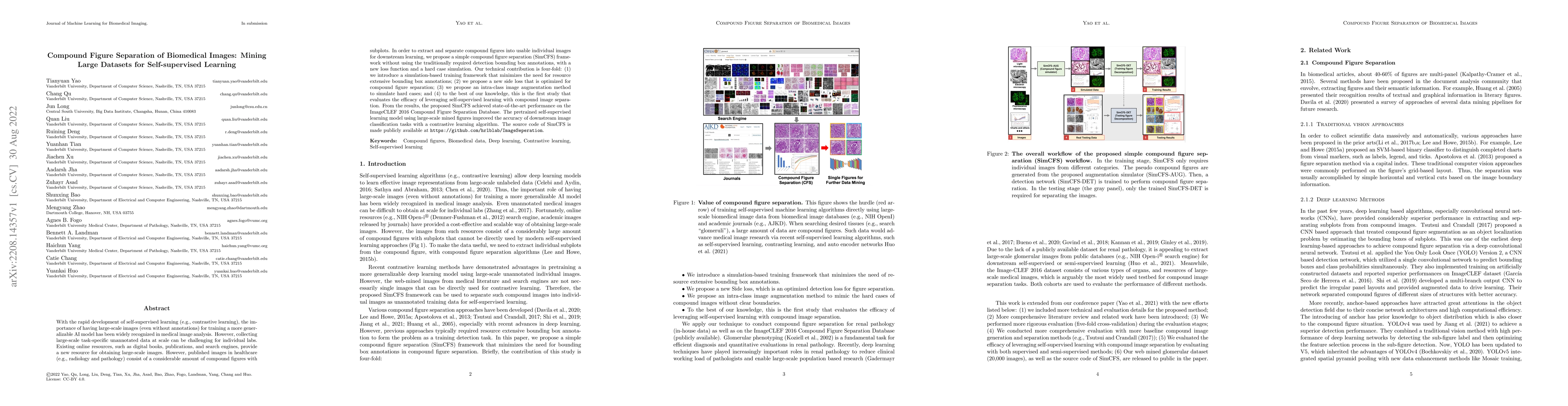
With the rapid development of self-supervised learning (e.g., contrastive learning), the importance of having large-scale images (even without annotations) for training a more generalizable AI model...
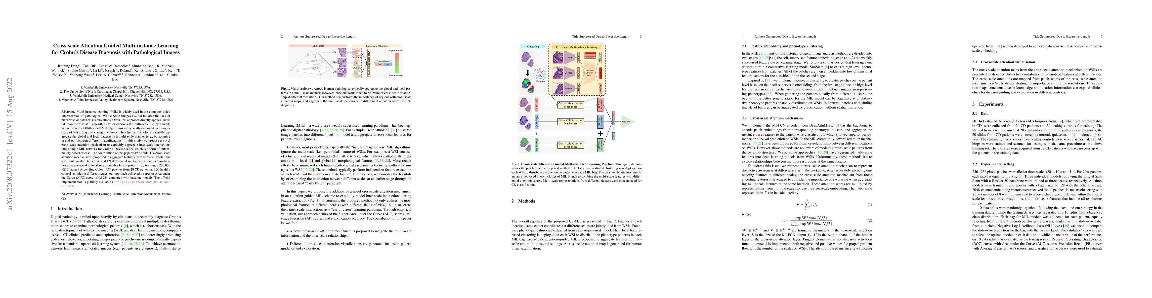
Multi-instance learning (MIL) is widely used in the computer-aided interpretation of pathological Whole Slide Images (WSIs) to solve the lack of pixel-wise or patch-wise annotations. Often, this app...
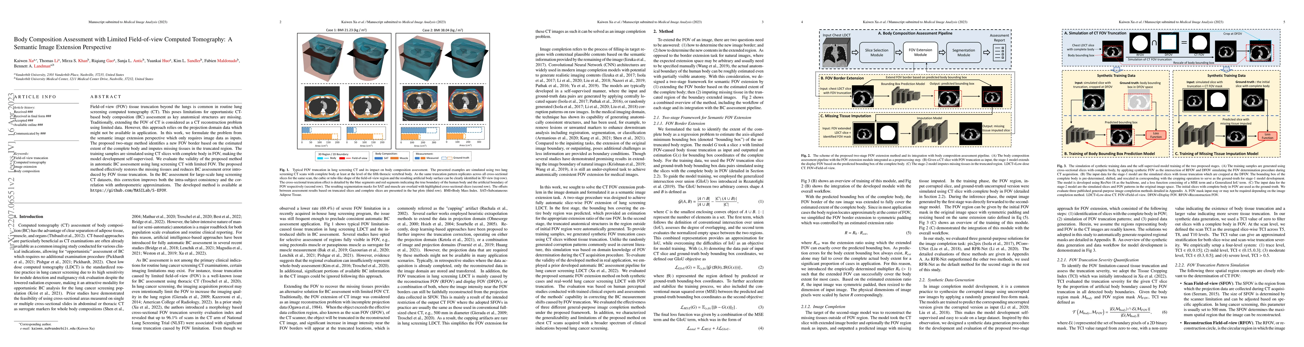
Field-of-view (FOV) tissue truncation beyond the lungs is common in routine lung screening computed tomography (CT). This poses limitations for opportunistic CT- based body composition (BC) assessme...
Increasing evidence shows that flaws in machine learning (ML) algorithm validation are an underestimated global problem. Particularly in automatic biomedical image analysis, chosen performance metri...
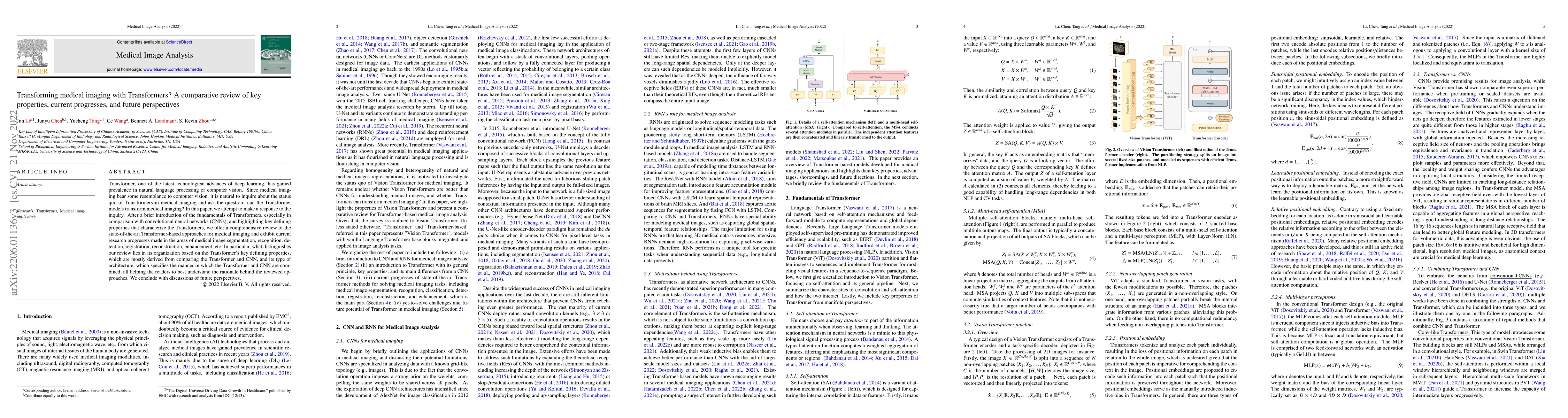
Transformer, the latest technological advance of deep learning, has gained prevalence in natural language processing or computer vision. Since medical imaging bear some resemblance to computer visio...
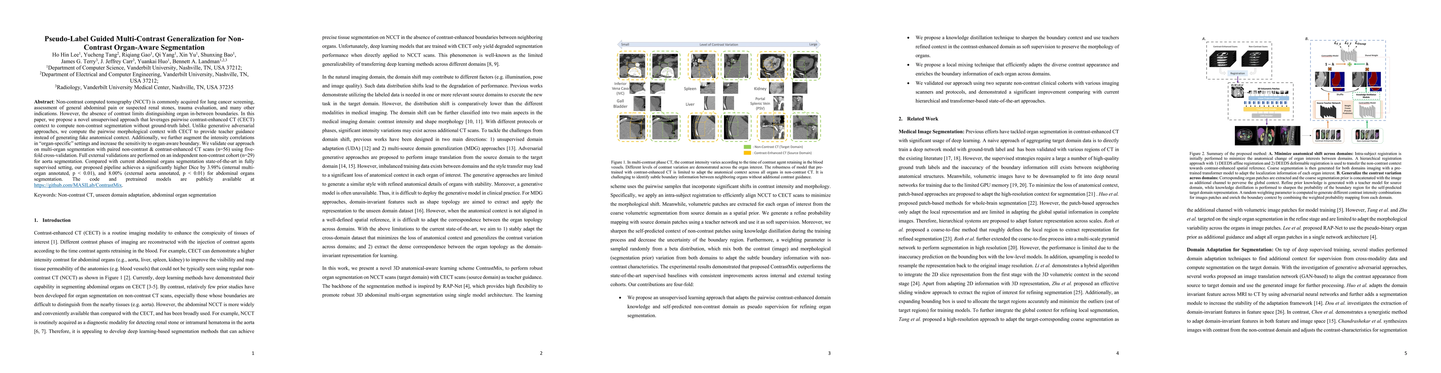
Non-contrast computed tomography (NCCT) is commonly acquired for lung cancer screening, assessment of general abdominal pain or suspected renal stones, trauma evaluation, and many other indications....
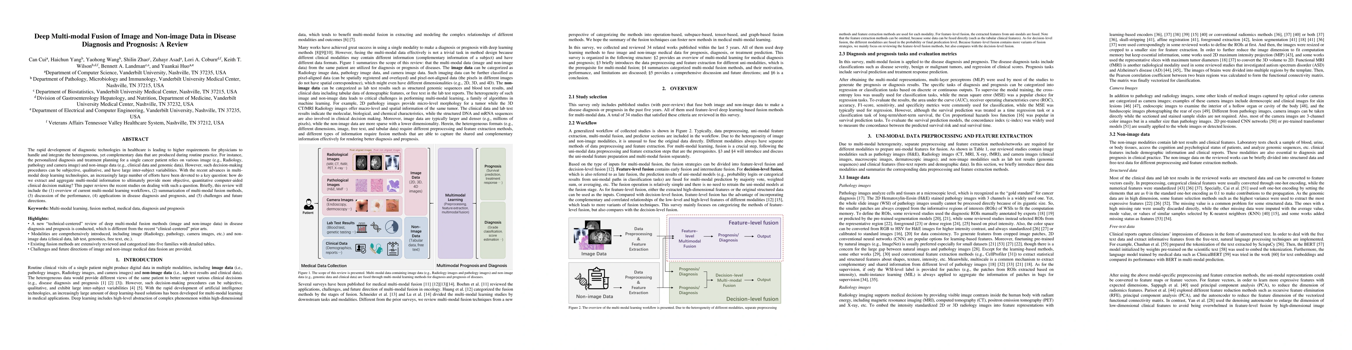
The rapid development of diagnostic technologies in healthcare is leading to higher requirements for physicians to handle and integrate the heterogeneous, yet complementary data that are produced du...

Integrating cross-department multi-modal data (e.g., radiological, pathological, genomic, and clinical data) is ubiquitous in brain cancer diagnosis and survival prediction. To date, such an integra...
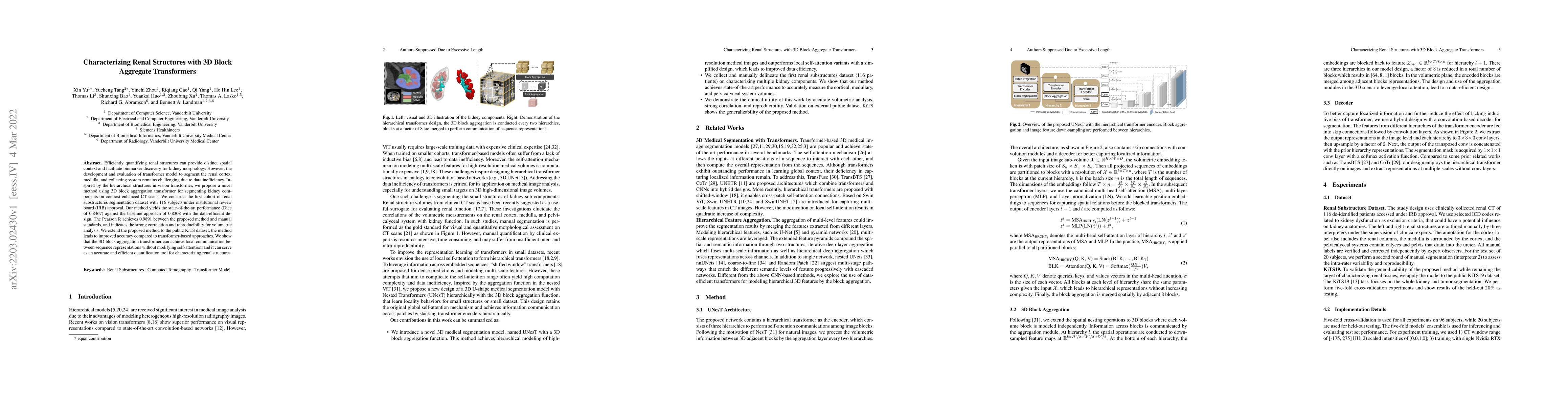
Efficiently quantifying renal structures can provide distinct spatial context and facilitate biomarker discovery for kidney morphology. However, the development and evaluation of the transformer mod...
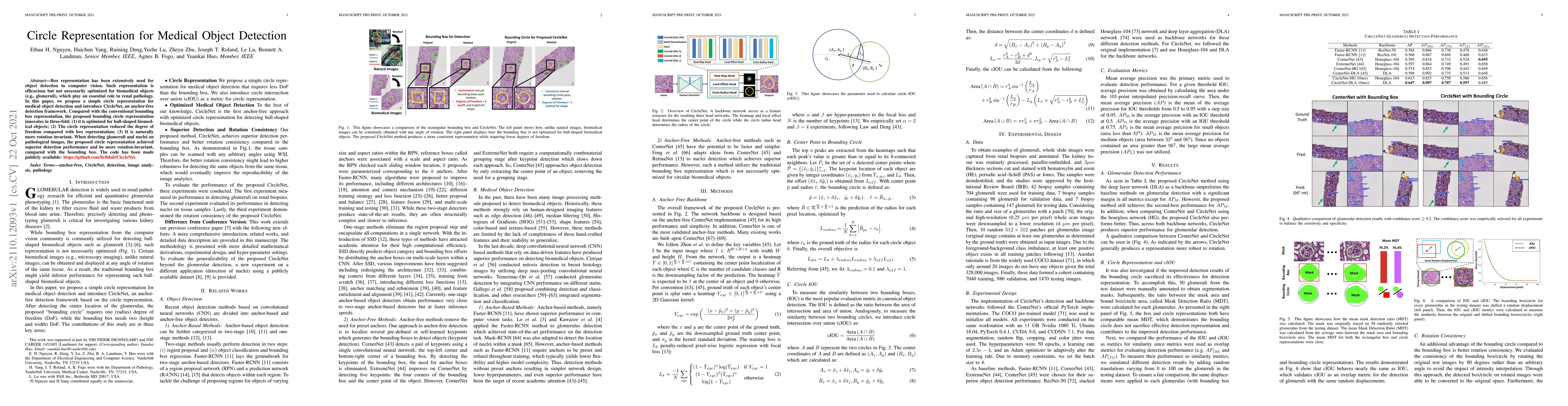
Box representation has been extensively used for object detection in computer vision. Such representation is efficacious but not necessarily optimized for biomedical objects (e.g., glomeruli), which...
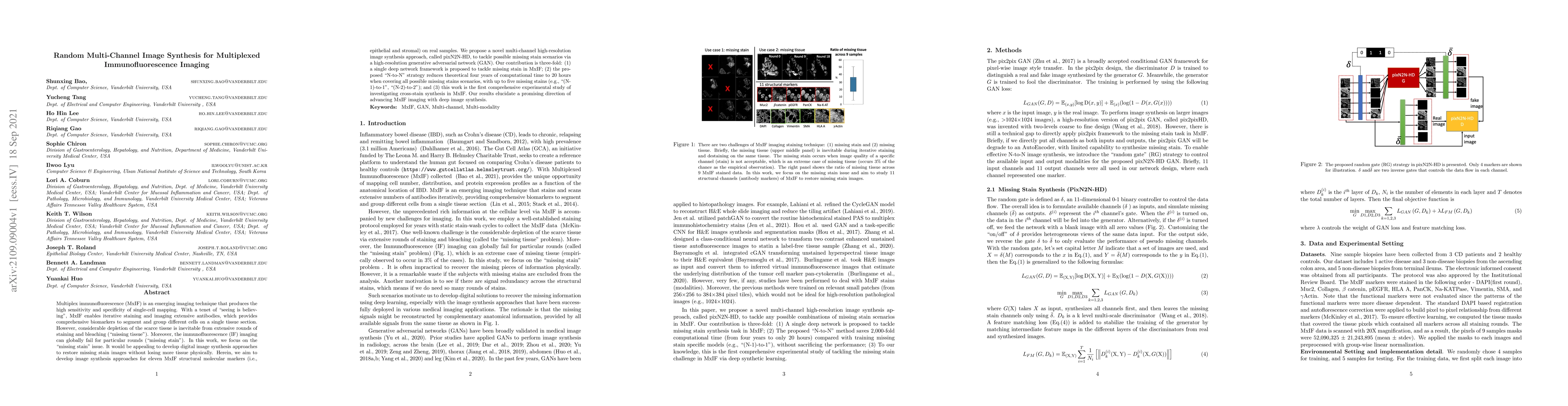
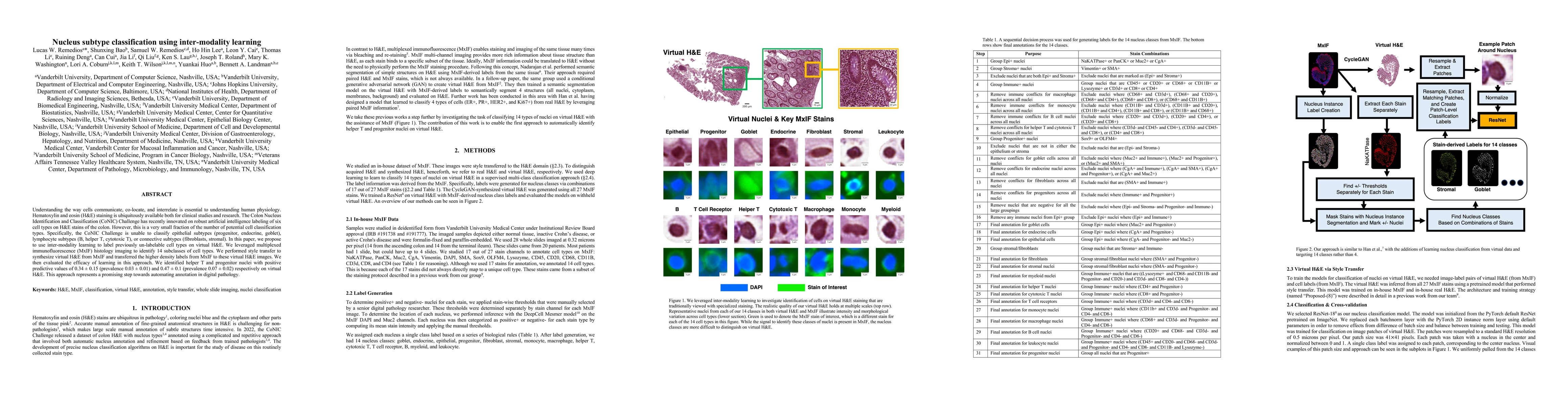
Multiplex immunofluorescence (MxIF) is an emerging imaging technique that produces the high sensitivity and specificity of single-cell mapping. With a tenet of 'seeing is believing', MxIF enables it...
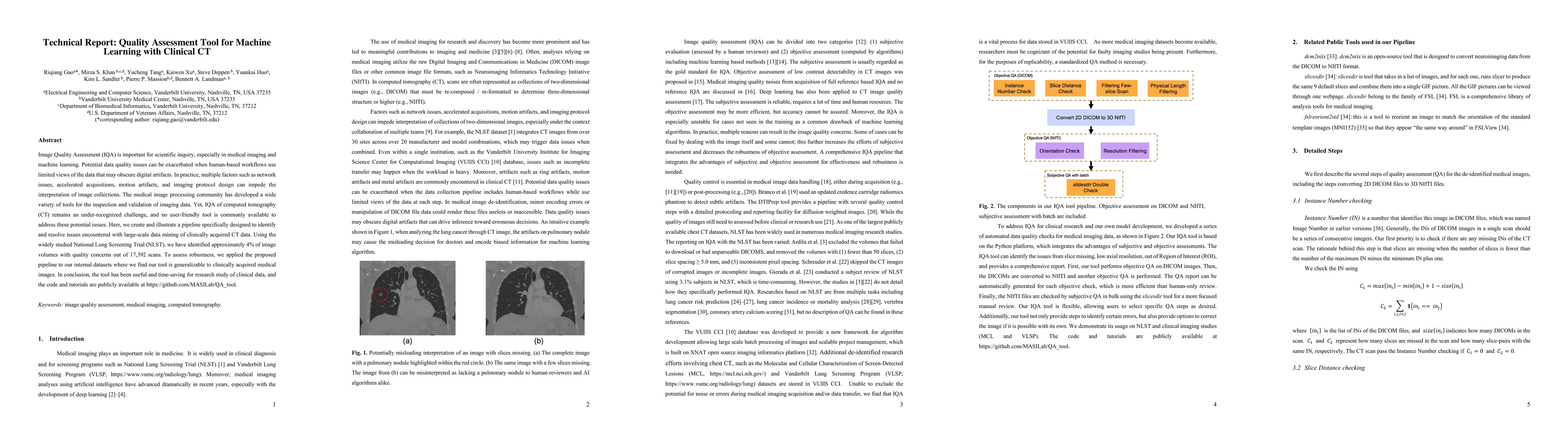
Image Quality Assessment (IQA) is important for scientific inquiry, especially in medical imaging and machine learning. Potential data quality issues can be exacerbated when human-based workflows us...
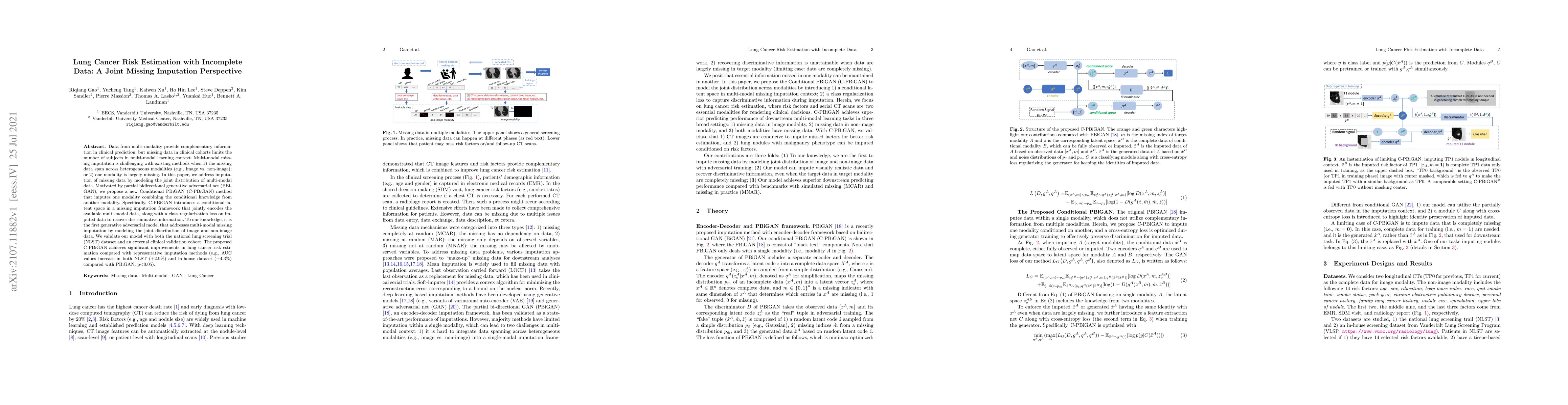
Data from multi-modality provide complementary information in clinical prediction, but missing data in clinical cohorts limits the number of subjects in multi-modal learning context. Multi-modal mis...
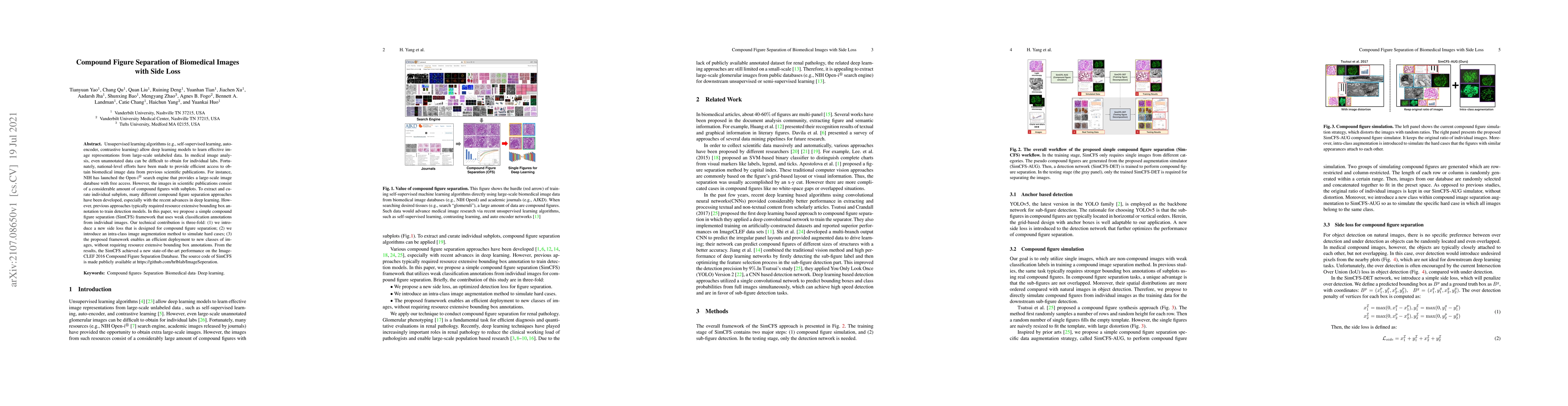
Unsupervised learning algorithms (e.g., self-supervised learning, auto-encoder, contrastive learning) allow deep learning models to learn effective image representations from large-scale unlabeled d...
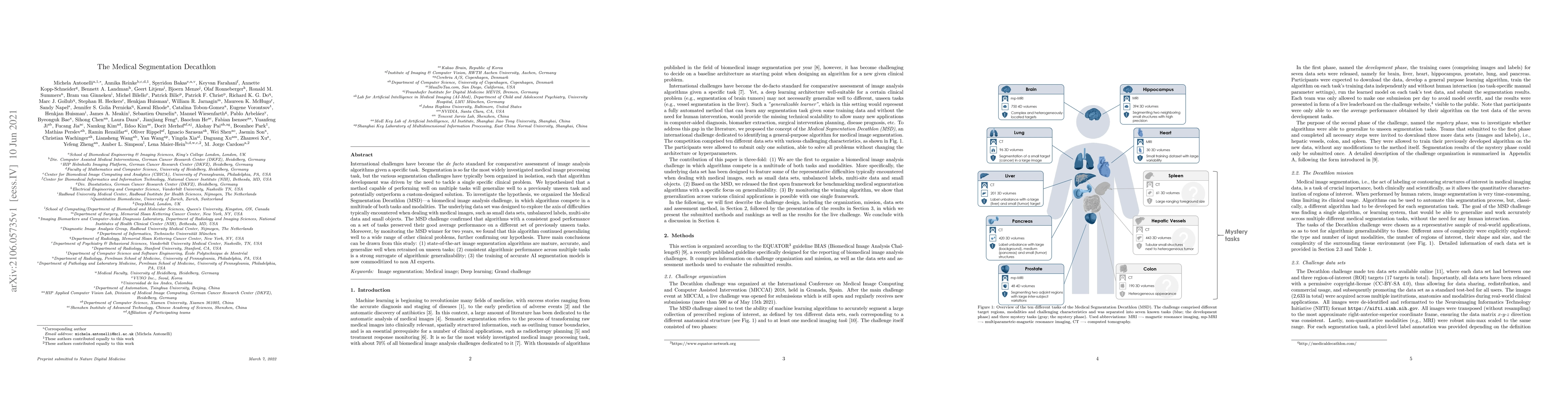
International challenges have become the de facto standard for comparative assessment of image analysis algorithms given a specific task. Segmentation is so far the most widely investigated medical ...

Medical image segmentation, or computing voxelwise semantic masks, is a fundamental yet challenging task to compute a voxel-level semantic mask. To increase the ability of encoder-decoder neural net...
While the importance of automatic image analysis is continuously increasing, recent meta-research revealed major flaws with respect to algorithm validation. Performance metrics are particularly key ...
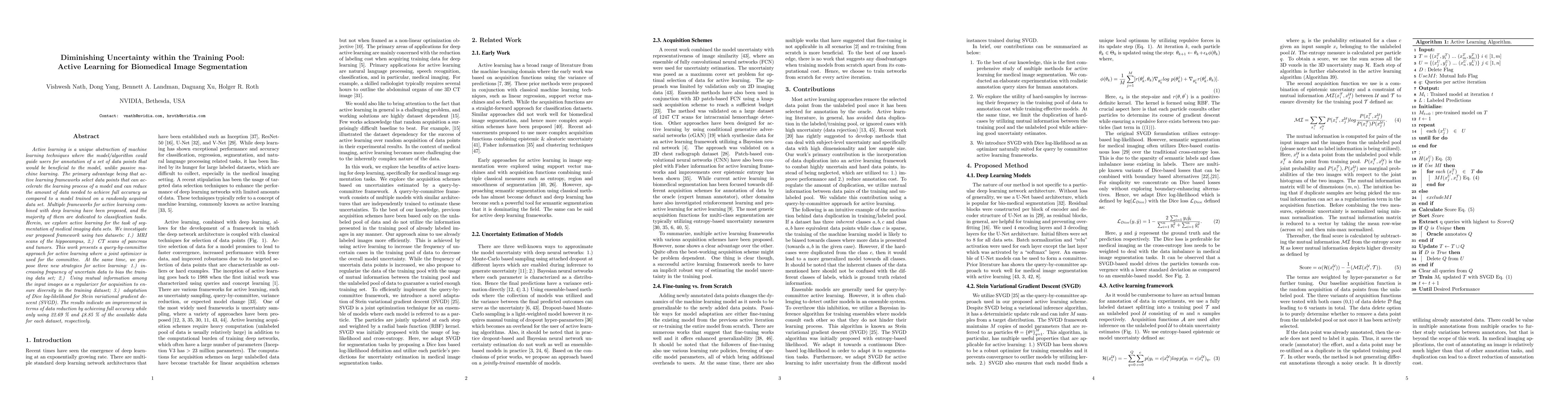
Active learning is a unique abstraction of machine learning techniques where the model/algorithm could guide users for annotation of a set of data points that would be beneficial to the model, unlik...
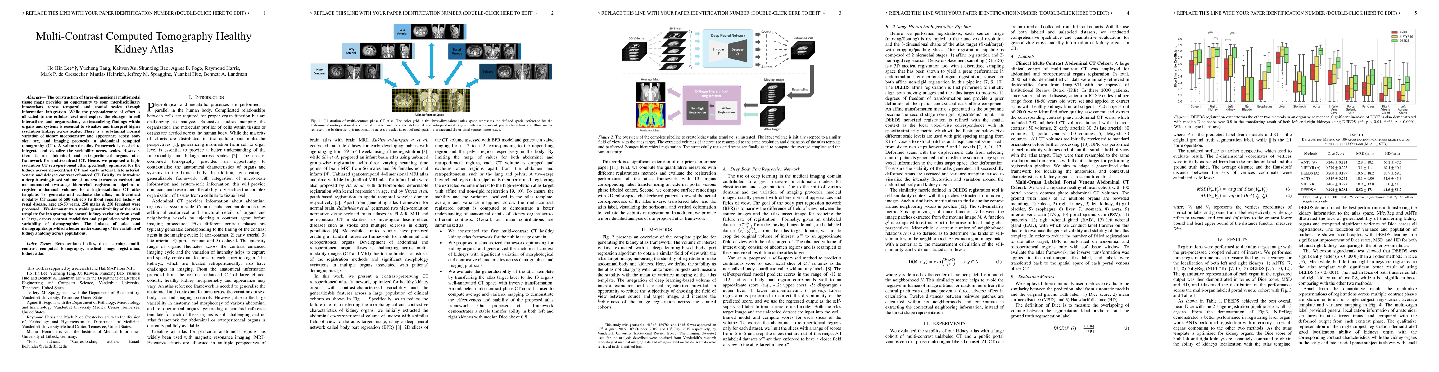
The construction of three-dimensional multi-modal tissue maps provides an opportunity to spur interdisciplinary innovations across temporal and spatial scales through information integration. While ...
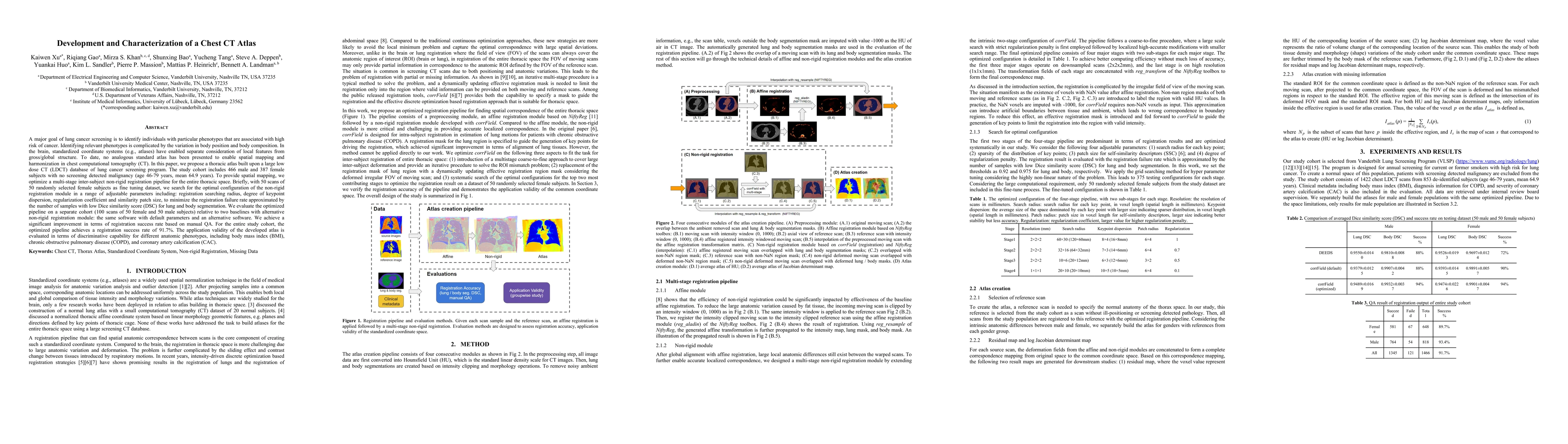
A major goal of lung cancer screening is to identify individuals with particular phenotypes that are associated with high risk of cancer. Identifying relevant phenotypes is complicated by the variat...
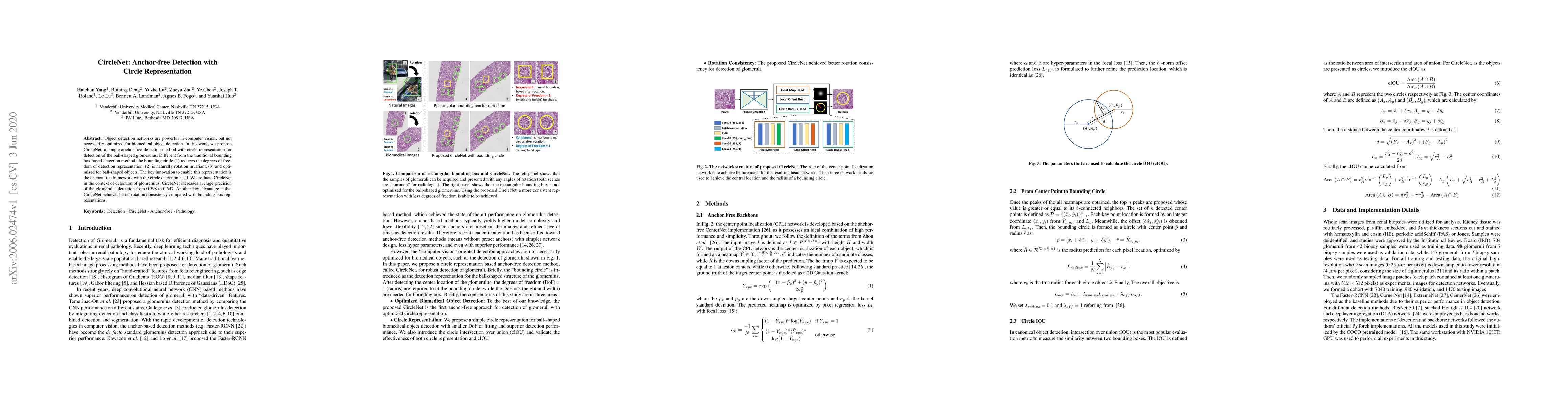
Object detection networks are powerful in computer vision, but not necessarily optimized for biomedical object detection. In this work, we propose CircleNet, a simple anchor-free detection method wi...
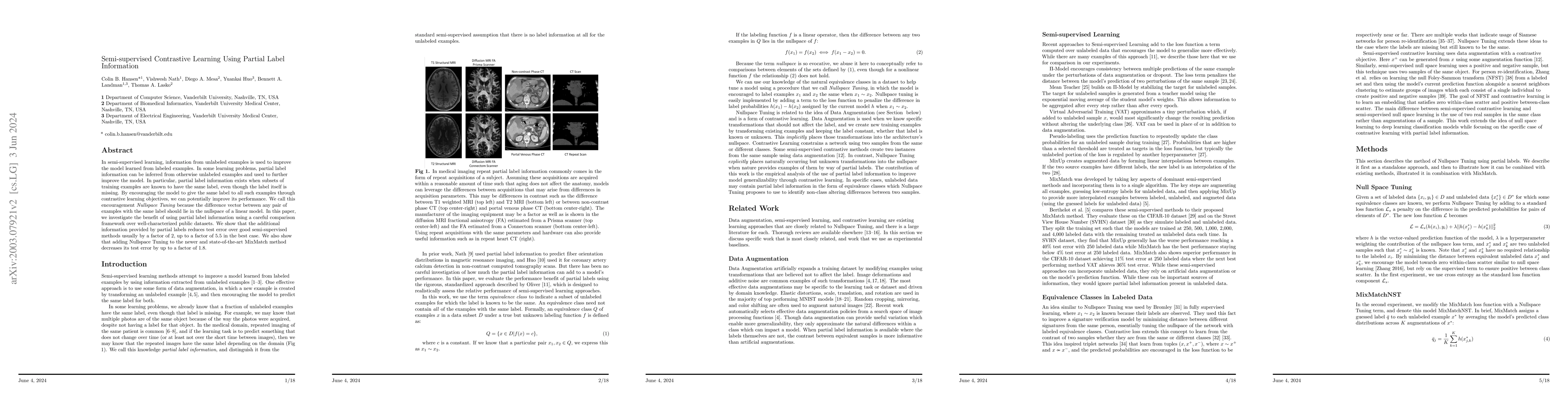
In semi-supervised learning, information from unlabeled examples is used to improve the model learned from labeled examples. In some learning problems, partial label information can be inferred from...
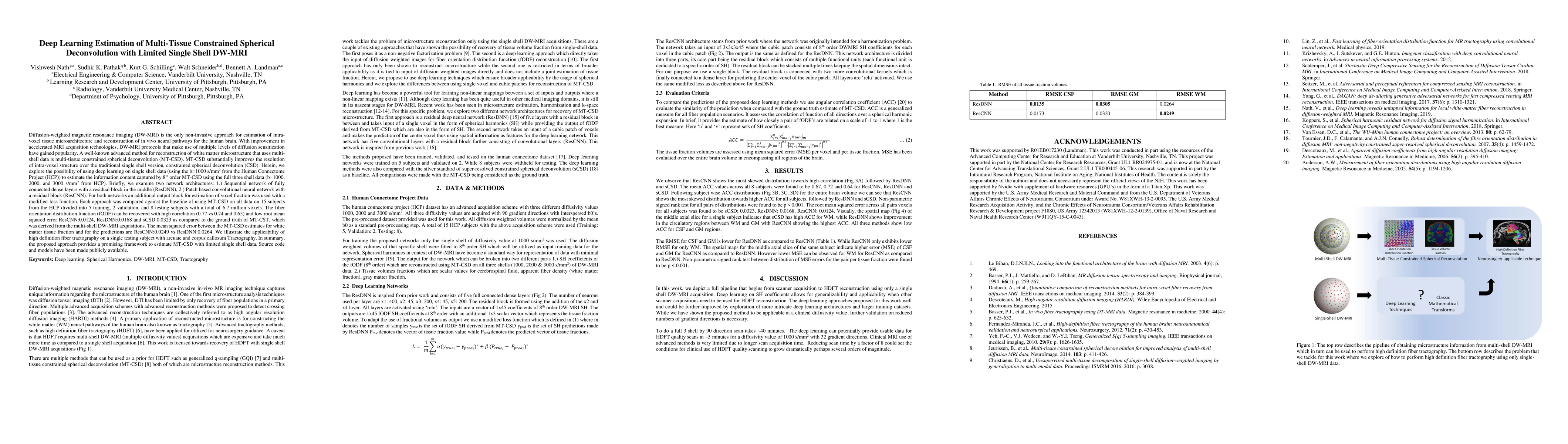
Diffusion-weighted magnetic resonance imaging (DW-MRI) is the only non-invasive approach for estimation of intra-voxel tissue microarchitecture and reconstruction of in vivo neural pathways for the ...
Segmentation of abdominal computed tomography(CT) provides spatial context, morphological properties, and a framework for tissue-specific radiomics to guide quantitative Radiological assessment. A 2...
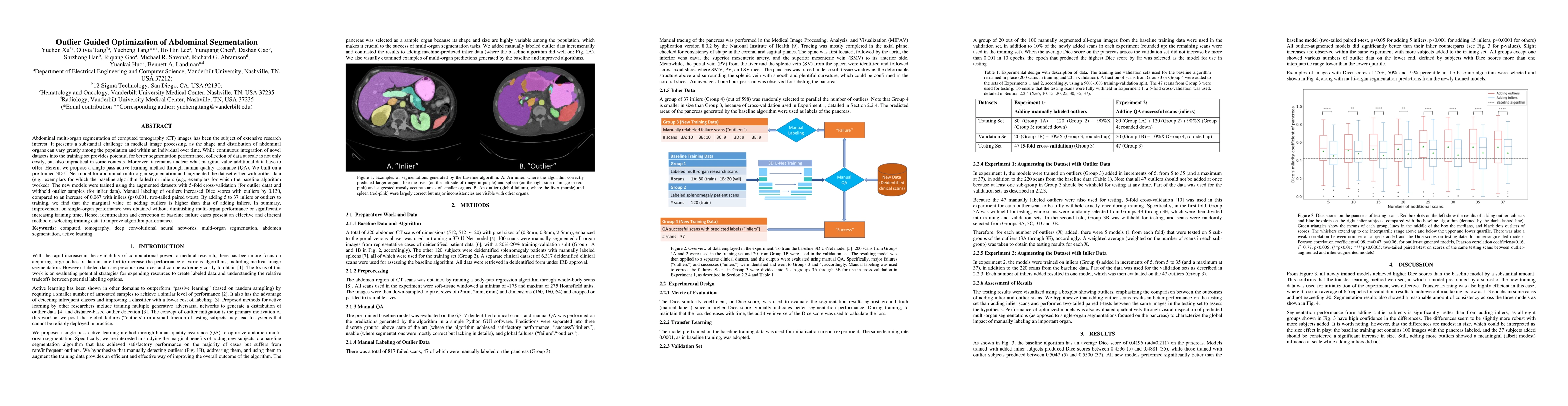
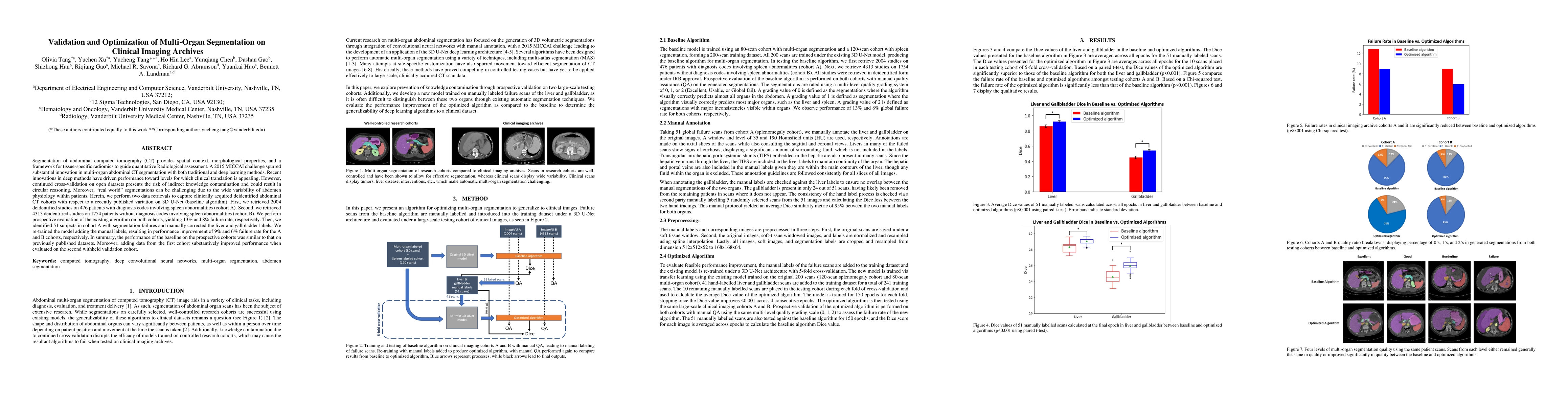
Abdominal multi-organ segmentation of computed tomography (CT) images has been the subject of extensive research interest. It presents a substantial challenge in medical image processing, as the sha...
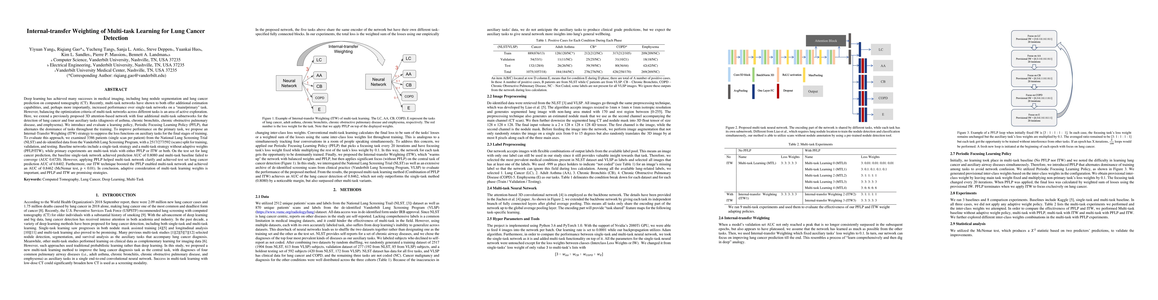
Recently, multi-task networks have shown to both offer additional estimation capabilities, and, perhaps more importantly, increased performance over single-task networks on a "main/primary" task. Ho...
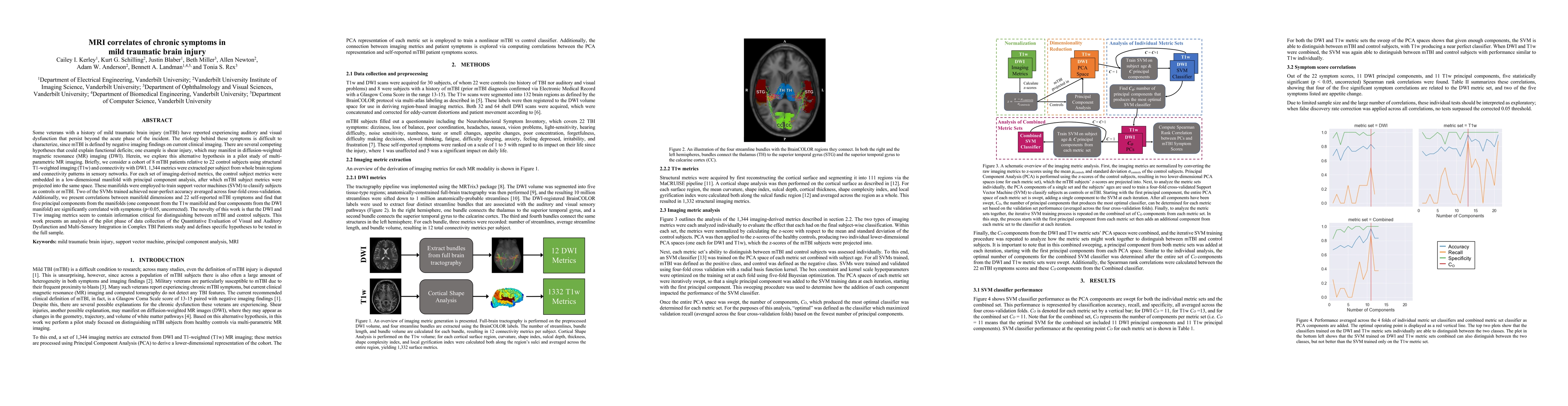
Veterans with mild traumatic brain injury (mTBI) have reported auditory and visual dysfunction that persists beyond the acute incident. The etiology behind these symptoms is difficult to characteriz...
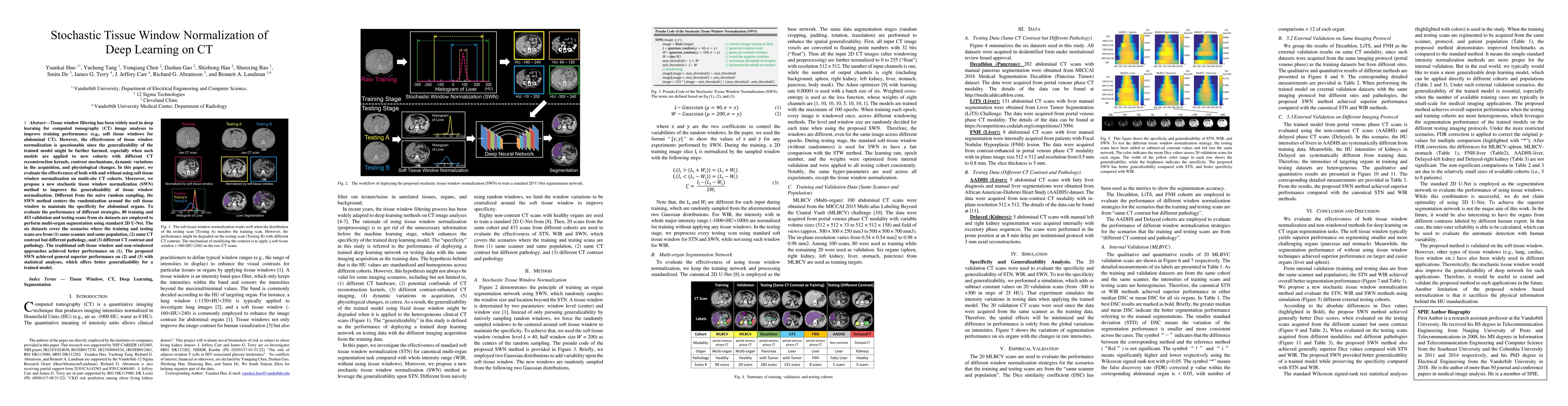
Tissue window filtering has been widely used in deep learning for computed tomography (CT) image analyses to improve training performance (e.g., soft tissue windows for abdominal CT). However, the e...
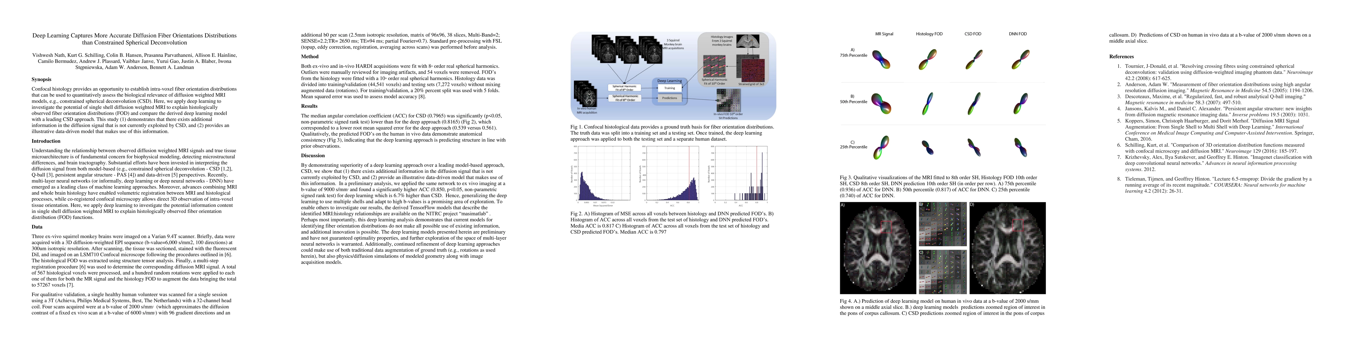
Confocal histology provides an opportunity to establish intra-voxel fiber orientation distributions that can be used to quantitatively assess the biological relevance of diffusion weighted MRI model...
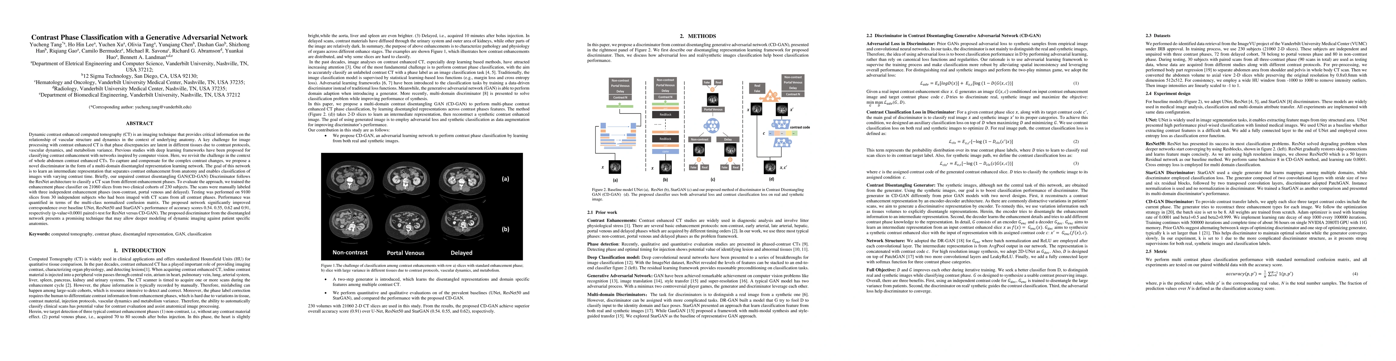
Dynamic contrast enhanced computed tomography (CT) is an imaging technique that provides critical information on the relationship of vascular structure and dynamics in the context of underlying anat...
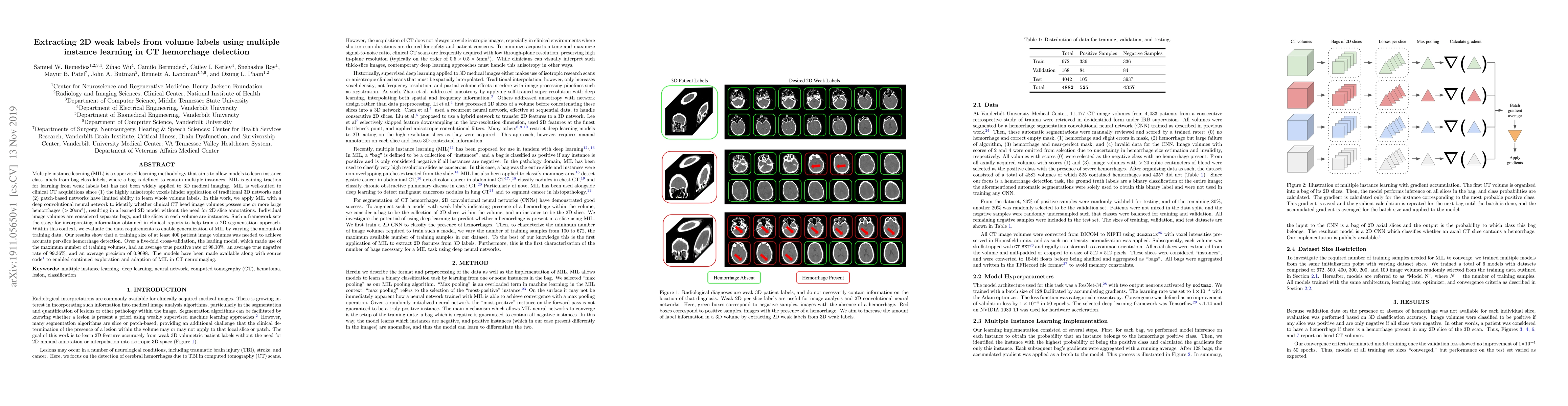
Multiple instance learning (MIL) is a supervised learning methodology that aims to allow models to learn instance class labels from bag class labels, where a bag is defined to contain multiple insta...
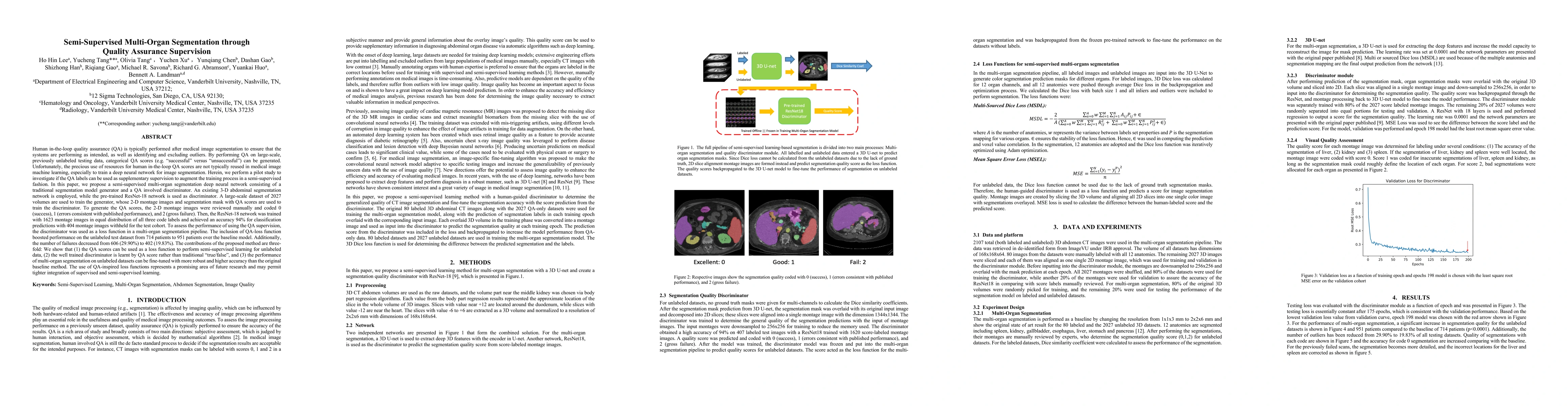
Human in-the-loop quality assurance (QA) is typically performed after medical image segmentation to ensure that the systems are performing as intended, as well as identifying and excluding outliers....
The field of lung nodule detection and cancer prediction has been rapidly developing with the support of large public data archives. Previous studies have largely focused on cross-sectional (single)...
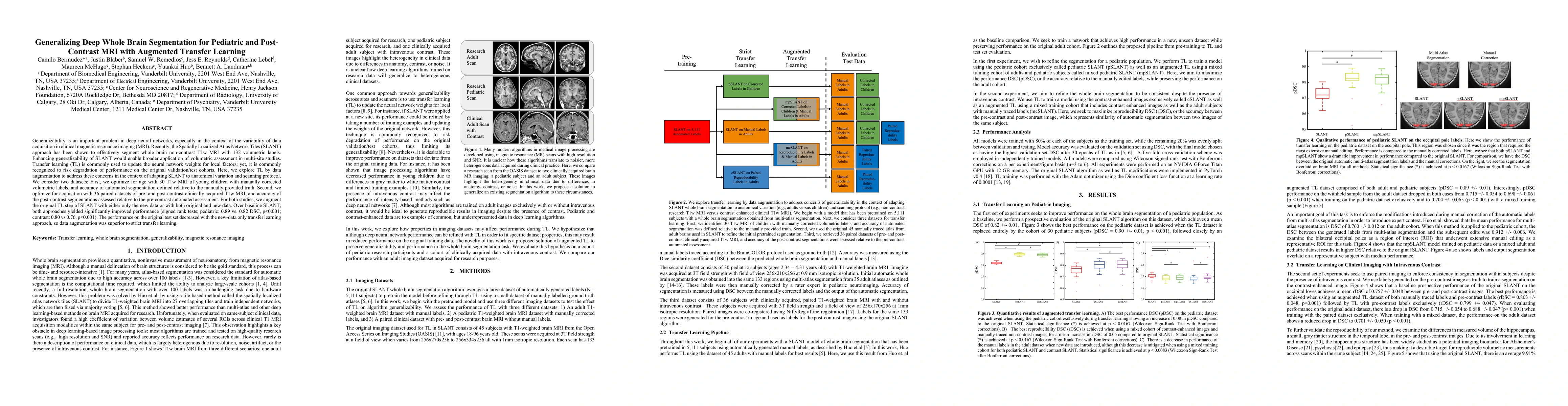
Generalizability is an important problem in deep neural networks, especially in the context of the variability of data acquisition in clinical magnetic resonance imaging (MRI). Recently, the Spatial...
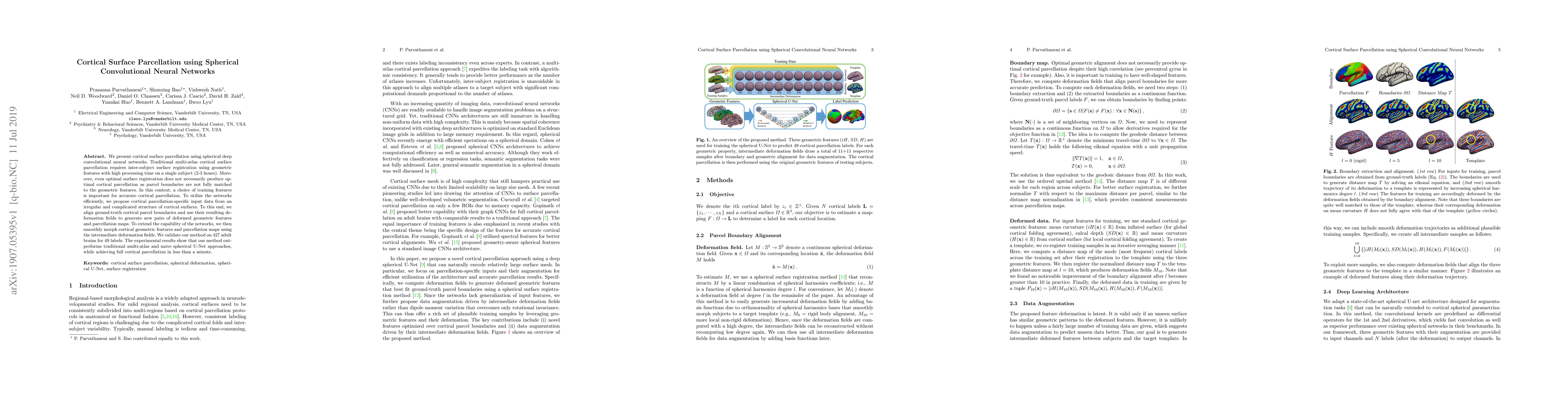
We present cortical surface parcellation using spherical deep convolutional neural networks. Traditional multi-atlas cortical surface parcellation requires inter-subject surface registration using g...
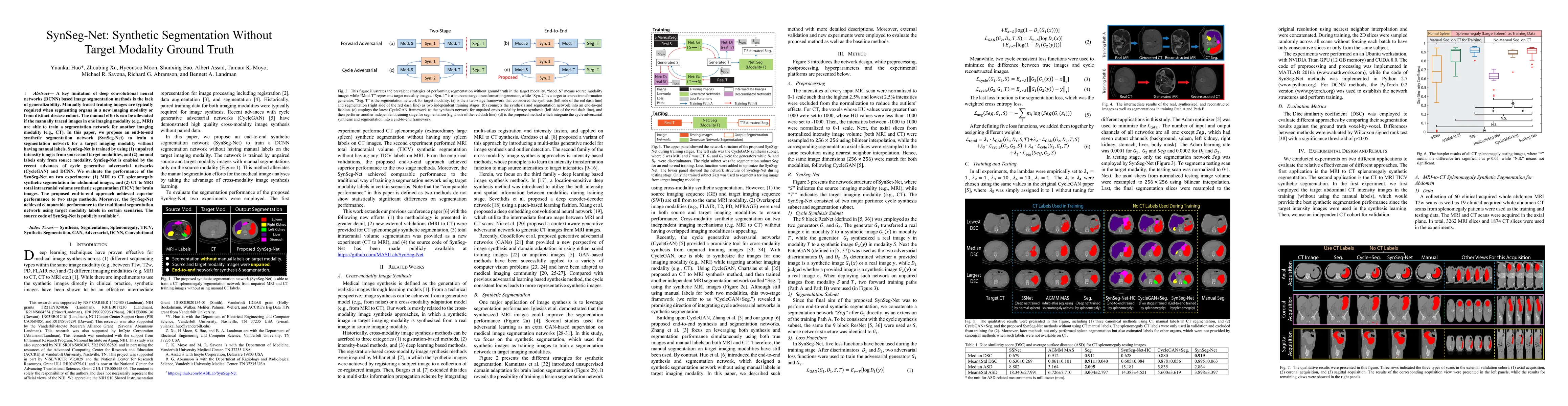
A key limitation of deep convolutional neural networks (DCNN) based image segmentation methods is the lack of generalizability. Manually traced training images are typically required when segmenting...
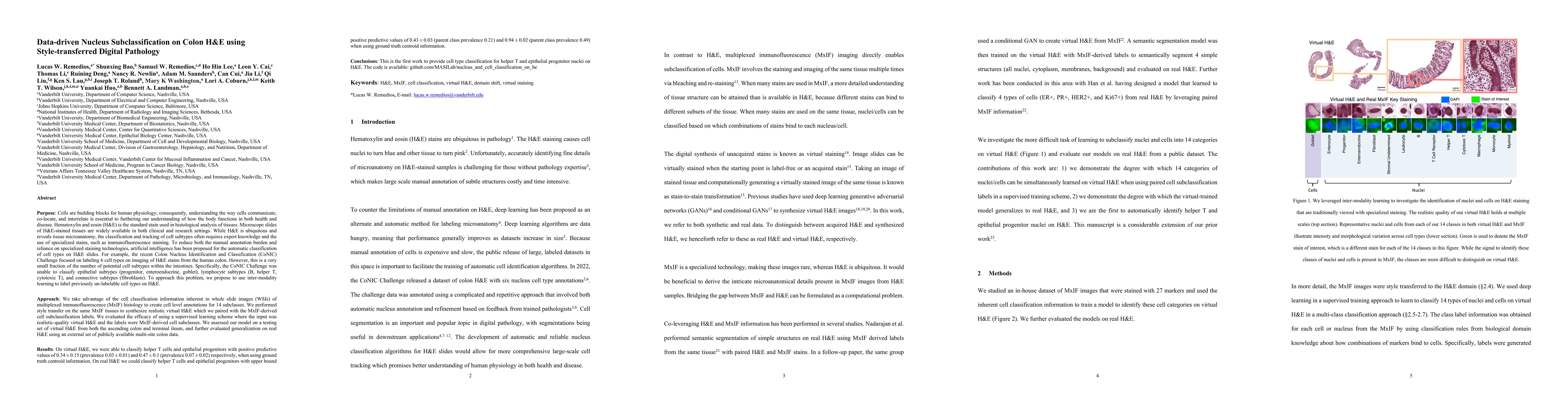
Understanding the way cells communicate, co-locate, and interrelate is essential to furthering our understanding of how the body functions. H&E is widely available, however, cell subtyping often requi...
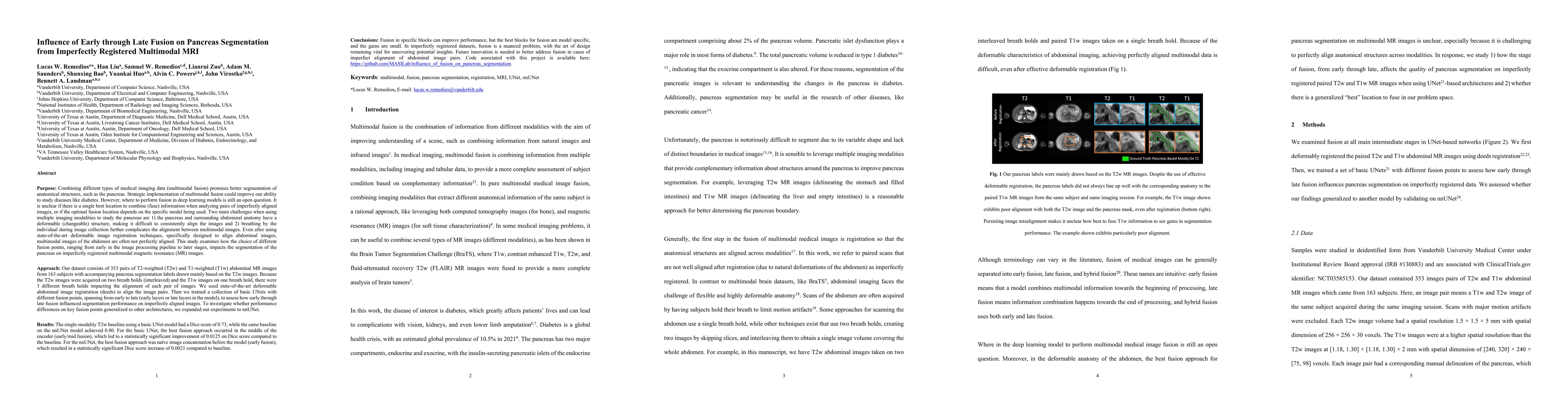
Multimodal fusion promises better pancreas segmentation. However, where to perform fusion in models is still an open question. It is unclear if there is a best location to fuse information when analyz...
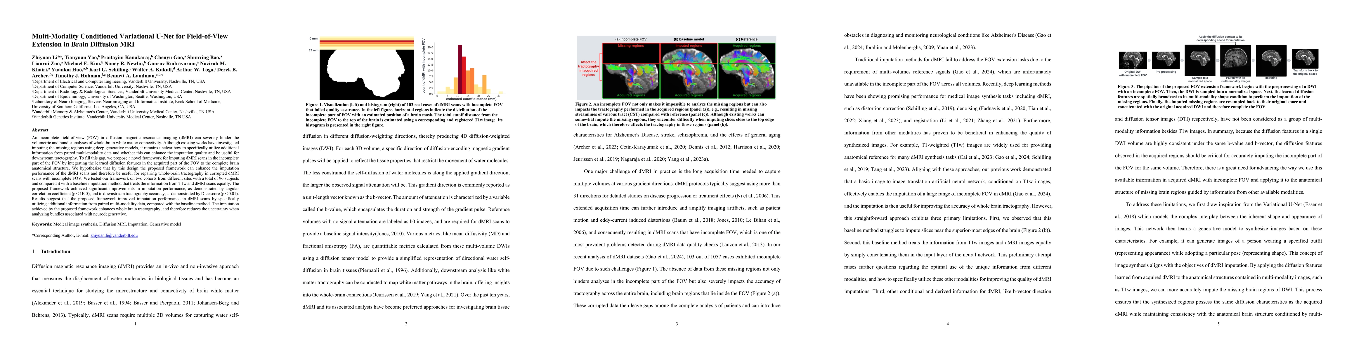
An incomplete field-of-view (FOV) in diffusion magnetic resonance imaging (dMRI) can severely hinder the volumetric and bundle analyses of whole-brain white matter connectivity. Although existing work...
Estimated brain age from magnetic resonance image (MRI) and its deviation from chronological age can provide early insights into potential neurodegenerative diseases, supporting early detection and im...
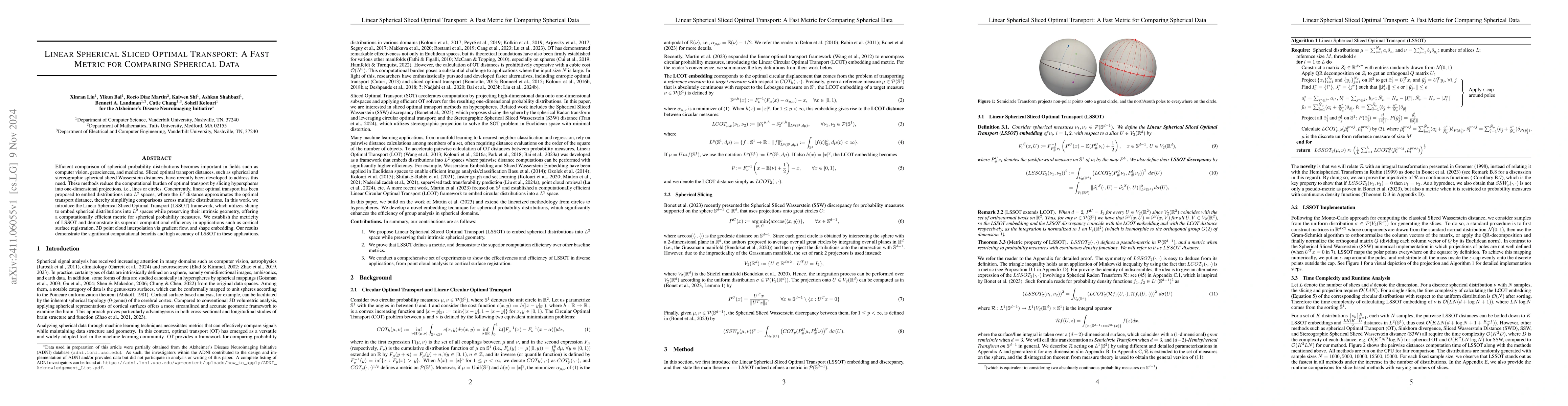
Efficient comparison of spherical probability distributions becomes important in fields such as computer vision, geosciences, and medicine. Sliced optimal transport distances, such as spherical and st...
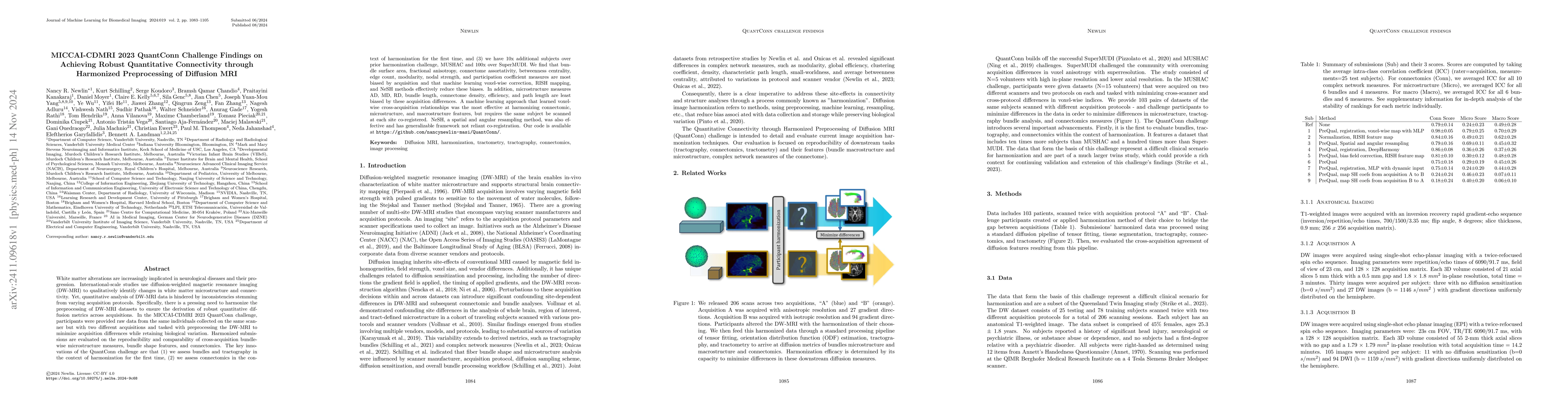
White matter alterations are increasingly implicated in neurological diseases and their progression. International-scale studies use diffusion-weighted magnetic resonance imaging (DW-MRI) to qualitati...
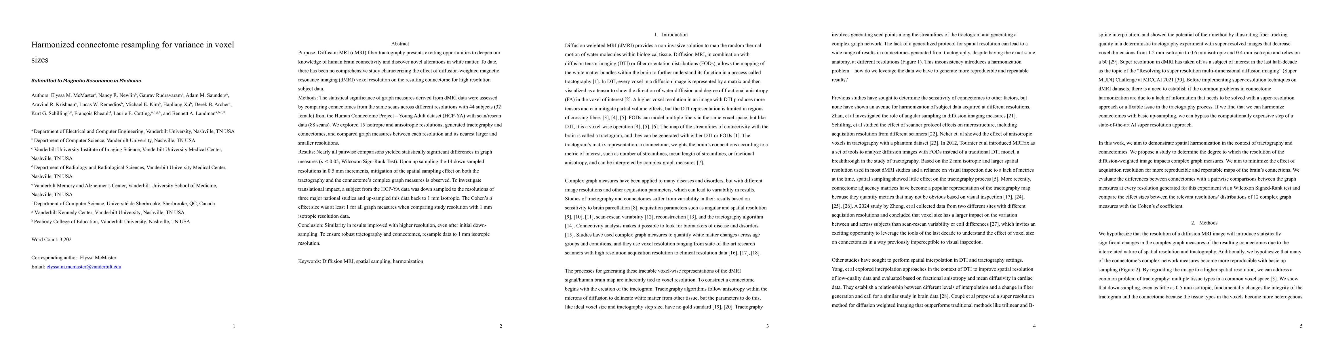
To date, there has been no comprehensive study characterizing the effect of diffusion-weighted magnetic resonance imaging voxel resolution on the resulting connectome for high resolution subject data....
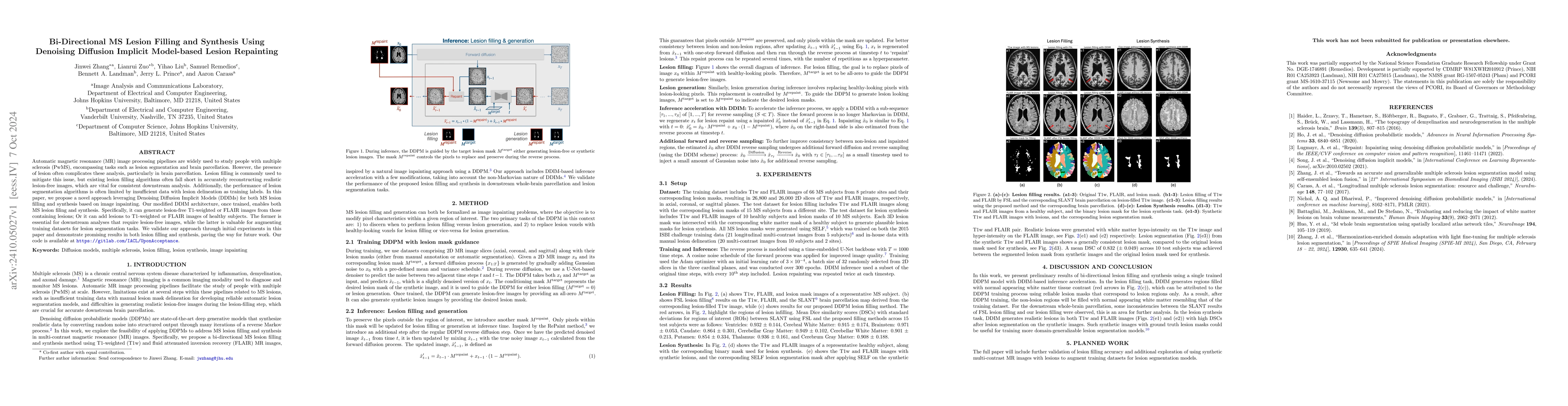
Automatic magnetic resonance (MR) image processing pipelines are widely used to study people with multiple sclerosis (PwMS), encompassing tasks such as lesion segmentation and brain parcellation. Howe...
While typical qualitative T1-weighted magnetic resonance images reflect scanner and protocol differences, quantitative T1 mapping aims to measure T1 independent of these effects. Changes in T1 in the ...
Purpose: Diffusion weighted MRI (dMRI) and its models of neural structure provide insight into human brain organization and variations in white matter. A recent study by McMaster, et al. showed that c...
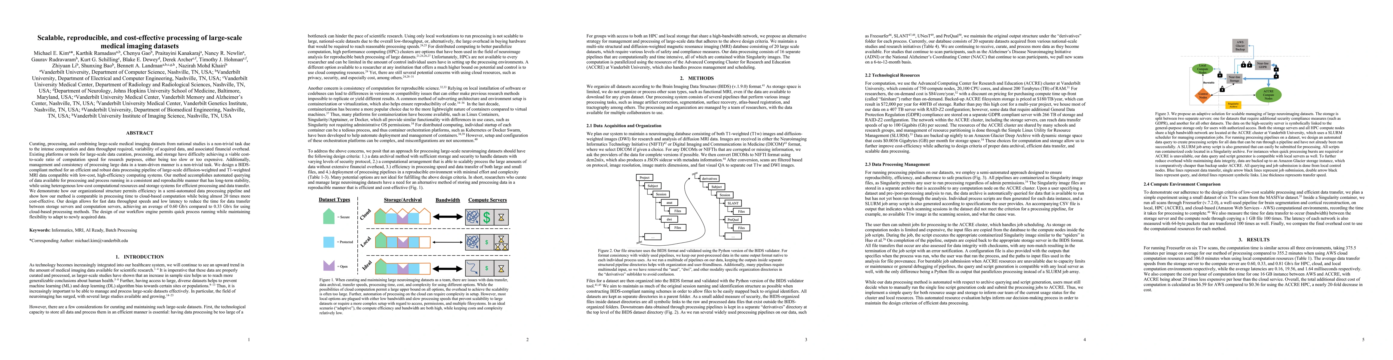
Curating, processing, and combining large-scale medical imaging datasets from national studies is a non-trivial task due to the intense computation and data throughput required, variability of acquire...
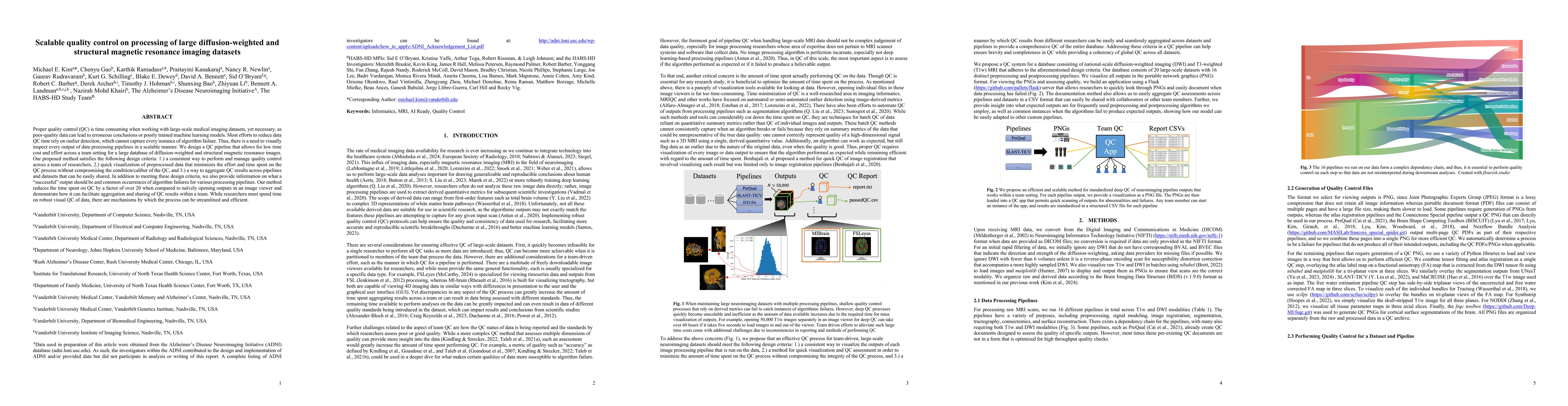
Proper quality control (QC) is time consuming when working with large-scale medical imaging datasets, yet necessary, as poor-quality data can lead to erroneous conclusions or poorly trained machine le...
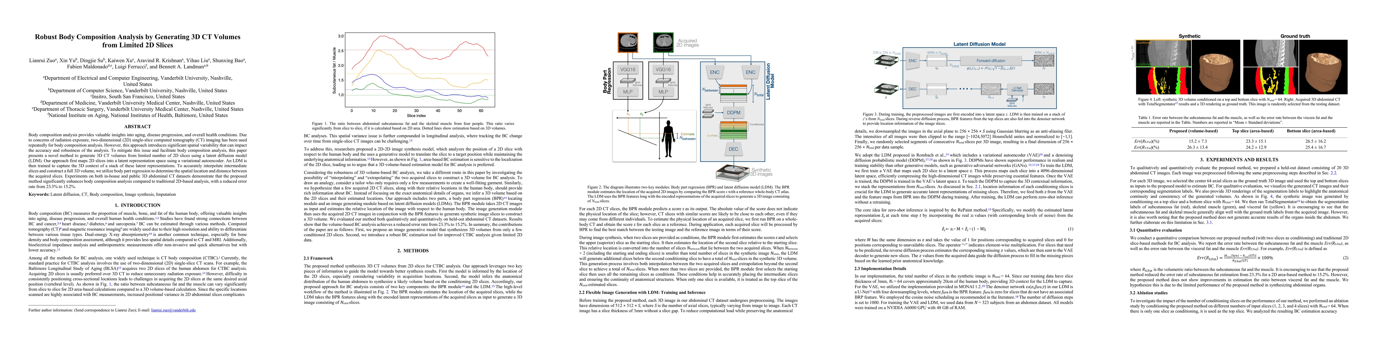
Body composition analysis provides valuable insights into aging, disease progression, and overall health conditions. Due to concerns of radiation exposure, two-dimensional (2D) single-slice computed t...
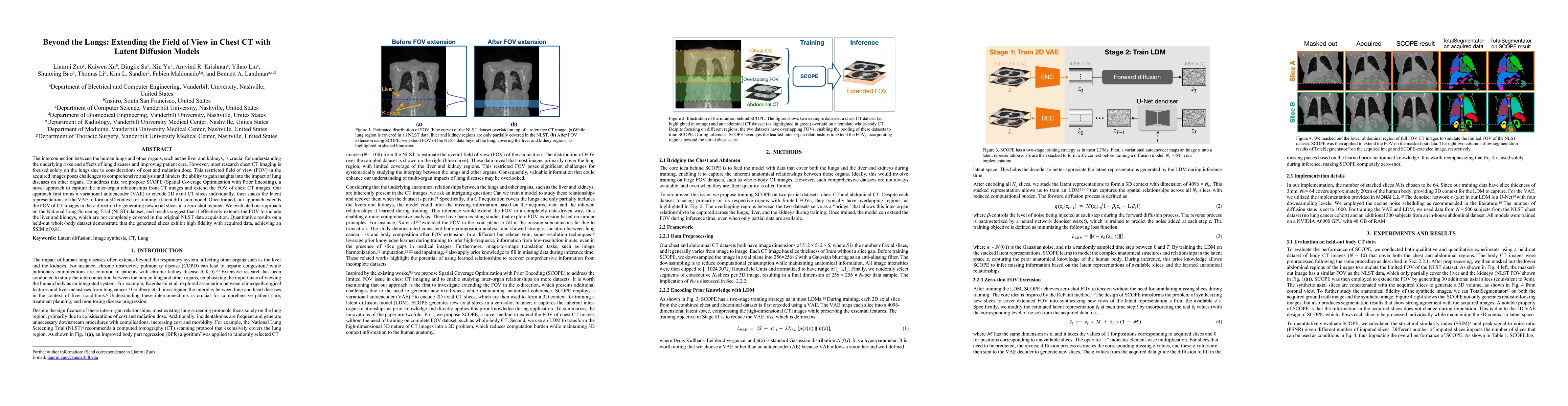
The interconnection between the human lungs and other organs, such as the liver and kidneys, is crucial for understanding the underlying risks and effects of lung diseases and improving patient care. ...
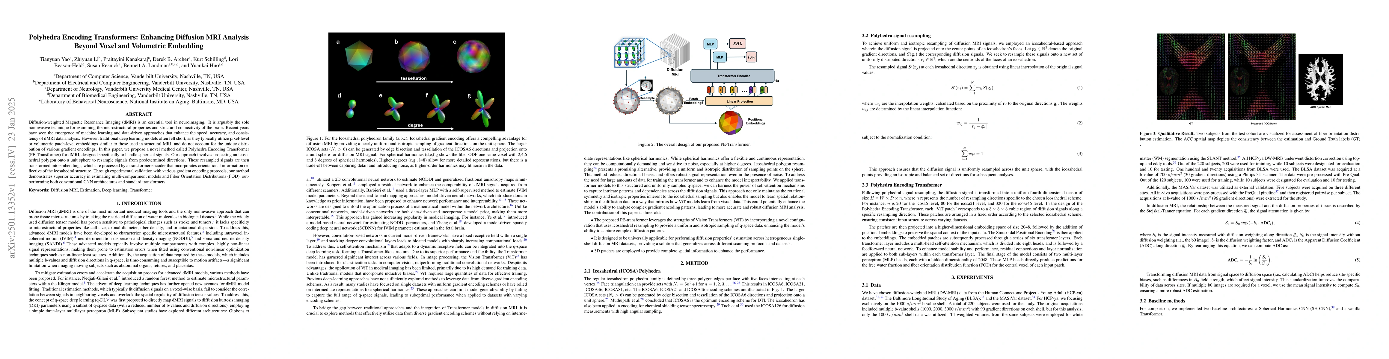
Diffusion-weighted Magnetic Resonance Imaging (dMRI) is an essential tool in neuroimaging. It is arguably the sole noninvasive technique for examining the microstructural properties and structural con...
Quantizing deep neural networks ,reducing the precision (bit-width) of their computations, can remarkably decrease memory usage and accelerate processing, making these models more suitable for large-s...
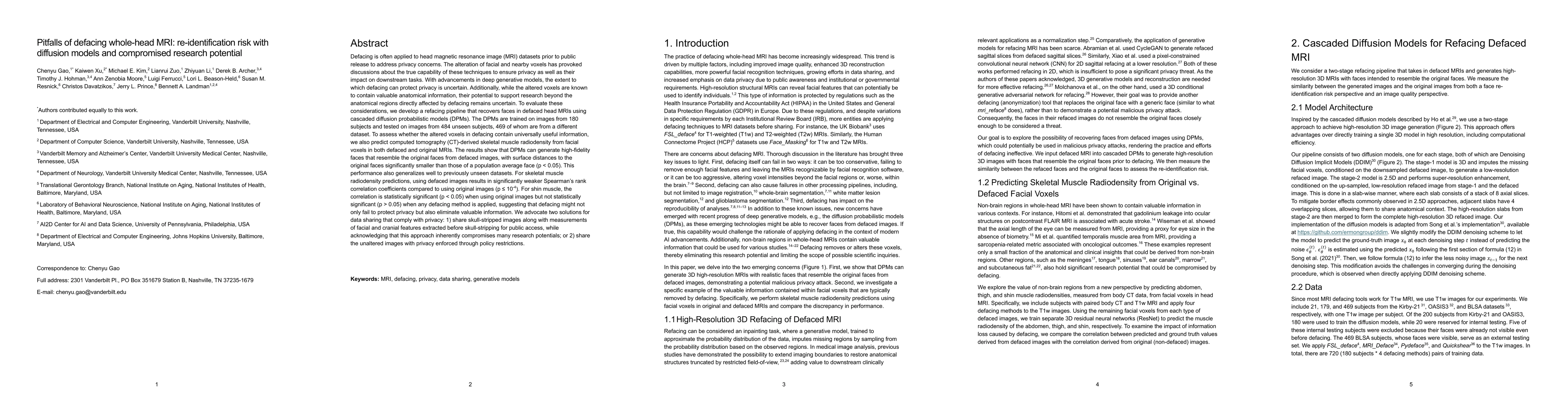
Defacing is often applied to head magnetic resonance image (MRI) datasets prior to public release to address privacy concerns. The alteration of facial and nearby voxels has provoked discussions about...
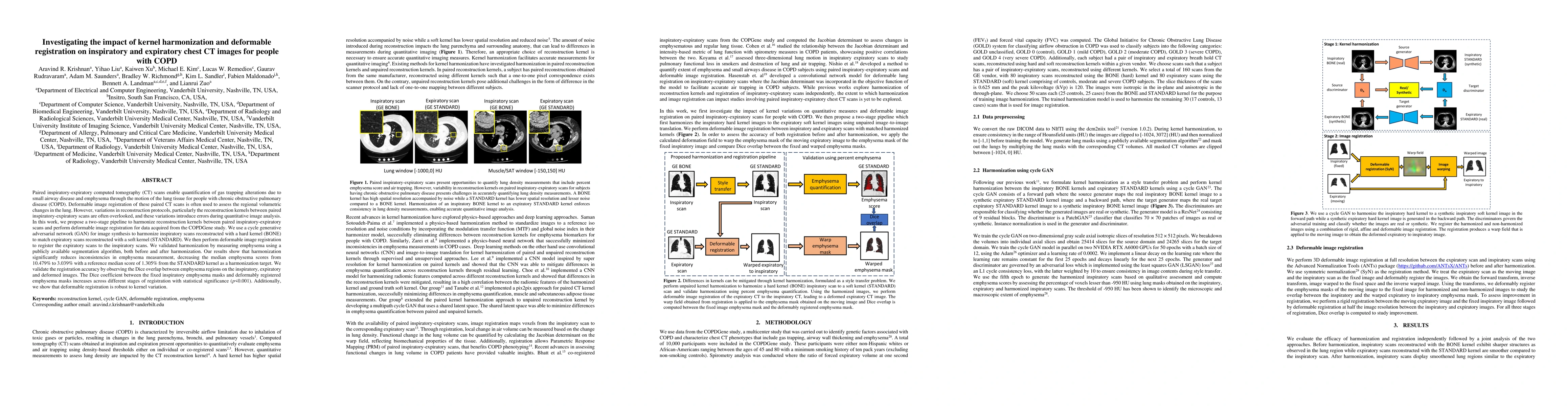
Paired inspiratory-expiratory CT scans enable the quantification of gas trapping due to small airway disease and emphysema by analyzing lung tissue motion in COPD patients. Deformable image registrati...
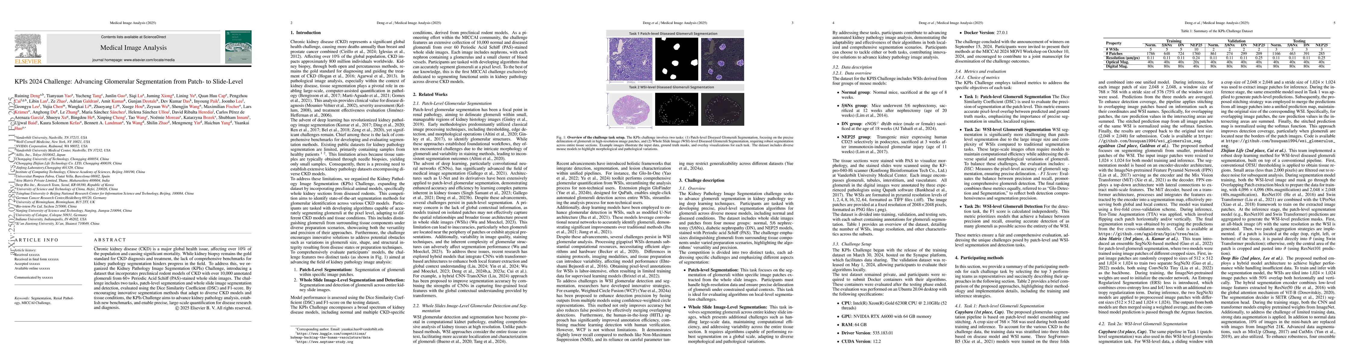
Chronic kidney disease (CKD) is a major global health issue, affecting over 10% of the population and causing significant mortality. While kidney biopsy remains the gold standard for CKD diagnosis and...
Reconstruction kernels in computed tomography (CT) affect spatial resolution and noise characteristics, introducing systematic variability in quantitative imaging measurements such as emphysema quanti...
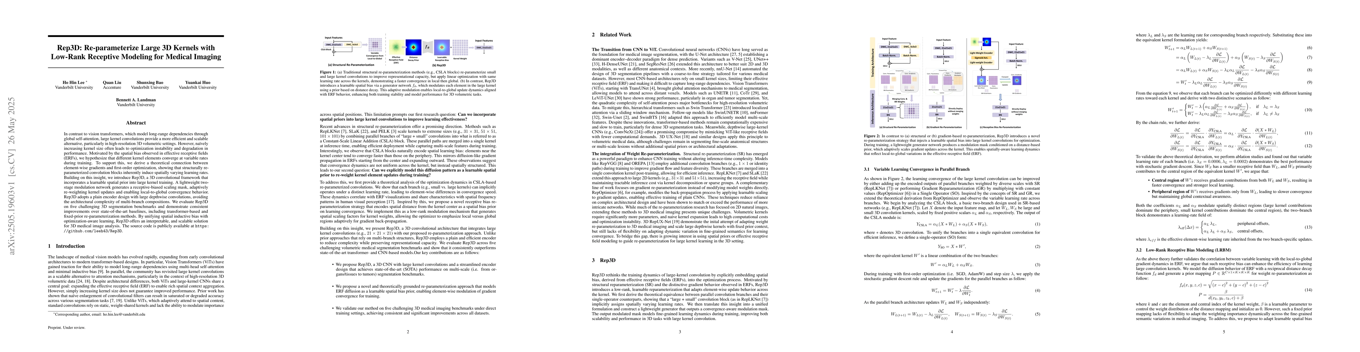
In contrast to vision transformers, which model long-range dependencies through global self-attention, large kernel convolutions provide a more efficient and scalable alternative, particularly in high...
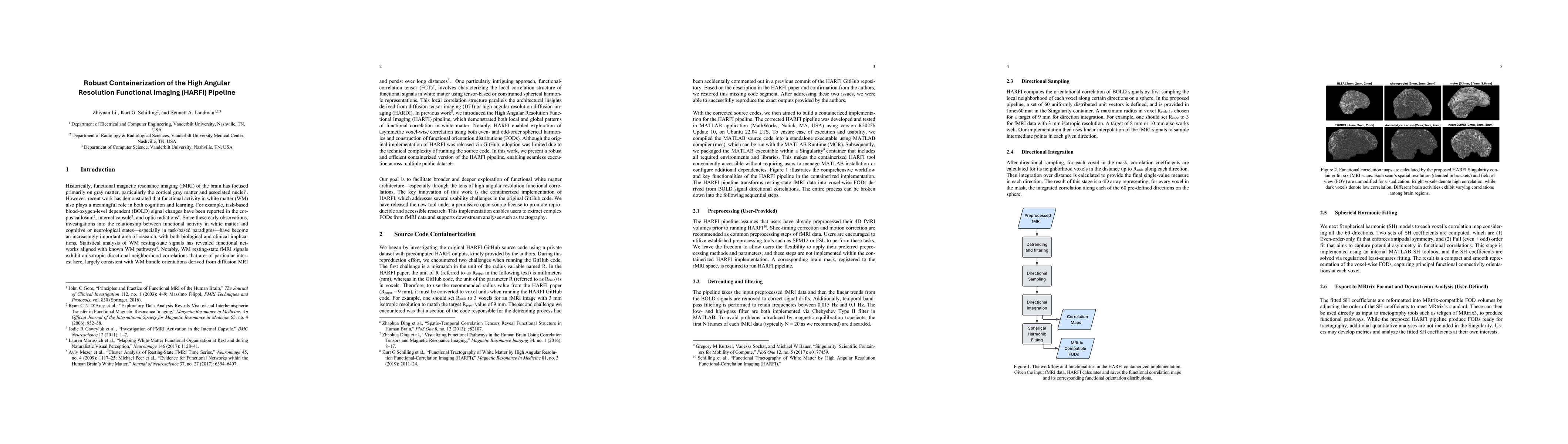
Historically, functional magnetic resonance imaging (fMRI) of the brain has focused primarily on gray matter, particularly the cortical gray matter and associated nuclei. However, recent work has demo...
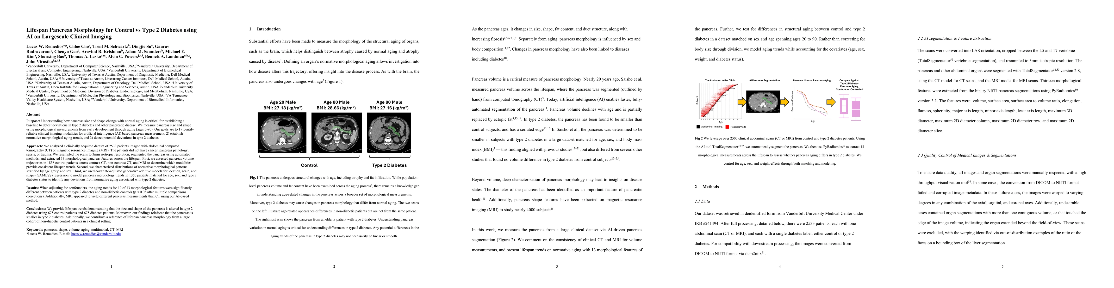
Purpose: Understanding how the pancreas changes is critical for detecting deviations in type 2 diabetes and other pancreatic disease. We measure pancreas size and shape using morphological measurement...
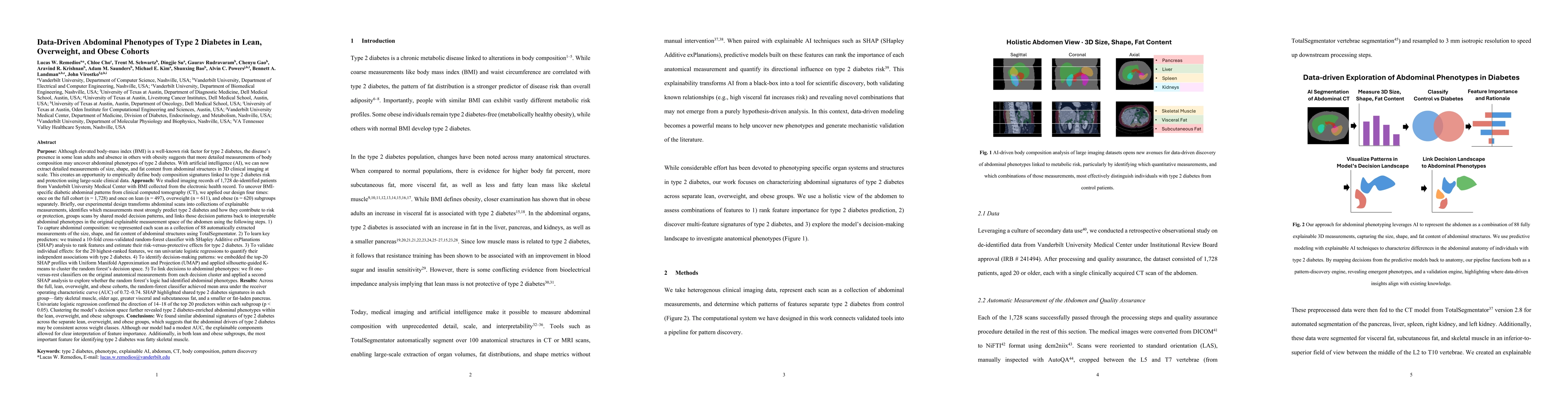
Purpose: Although elevated BMI is a well-known risk factor for type 2 diabetes, the disease's presence in some lean adults and absence in others with obesity suggests that detailed body composition ma...
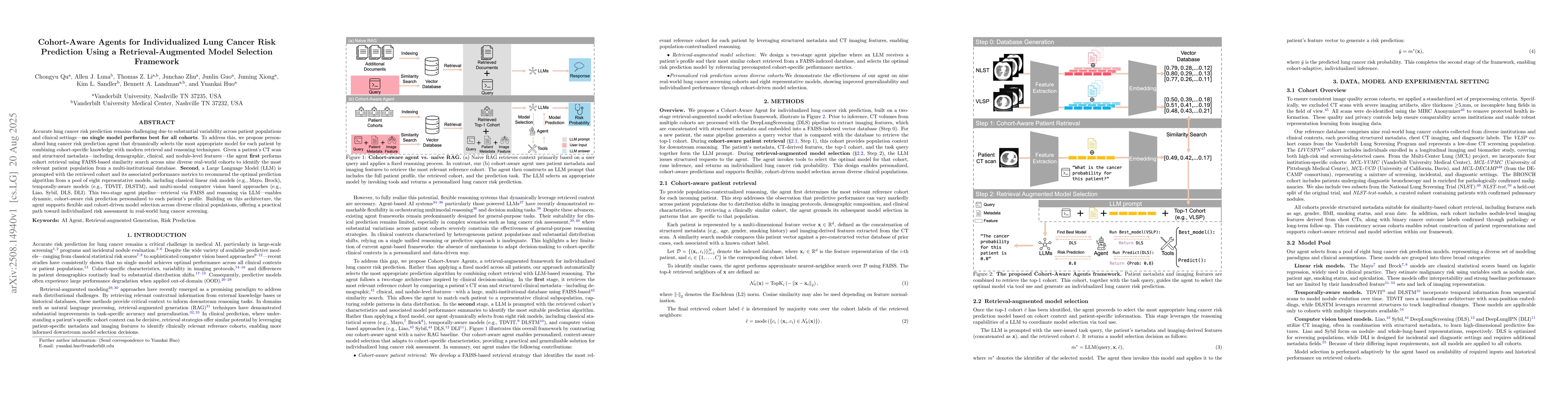
Accurate lung cancer risk prediction remains challenging due to substantial variability across patient populations and clinical settings -- no single model performs best for all cohorts. To address th...
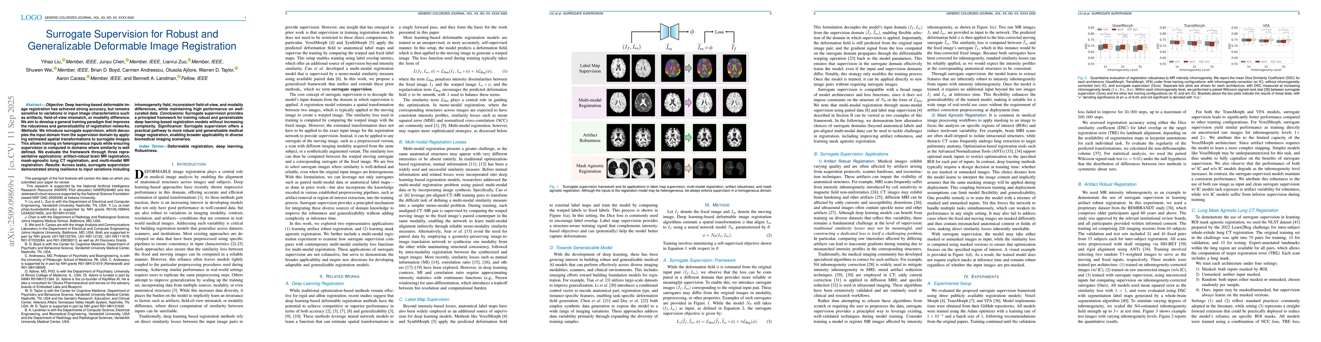
Objective: Deep learning-based deformable image registration has achieved strong accuracy, but remains sensitive to variations in input image characteristics such as artifacts, field-of-view mismatch,...
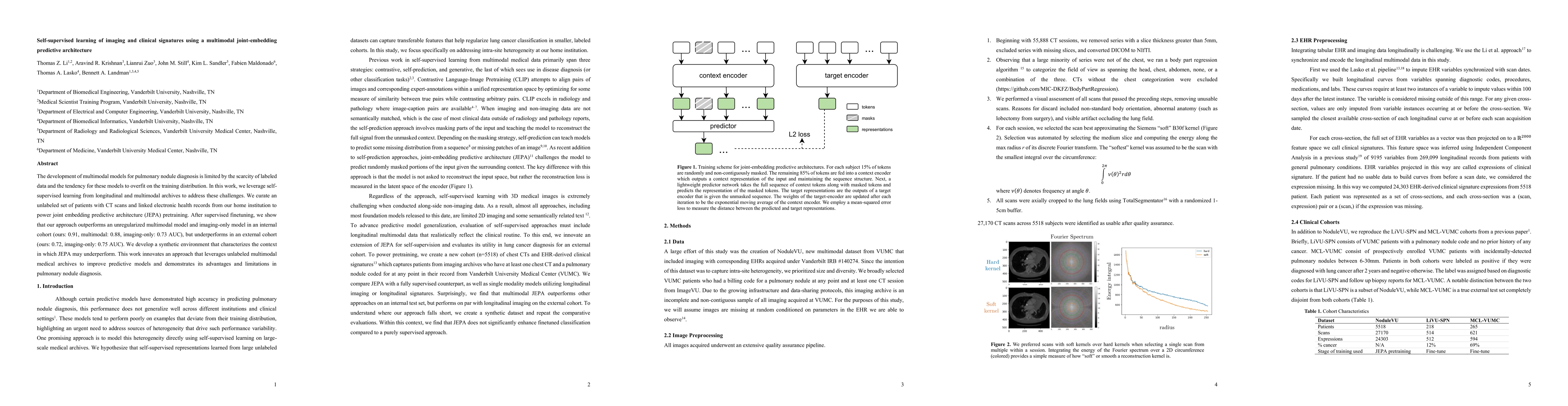
The development of multimodal models for pulmonary nodule diagnosis is limited by the scarcity of labeled data and the tendency for these models to overfit on the training distribution. In this work, ...
In multiple sclerosis, lesions interfere with automated magnetic resonance imaging analyses such as brain parcellation and deformable registration, while lesion segmentation models are hindered by the...
Diffusion MRI (dMRI) provides a distinctive means to probe the microstructural architecture of living tissue, facilitating applications such as brain connectivity analysis, modeling across multiple co...
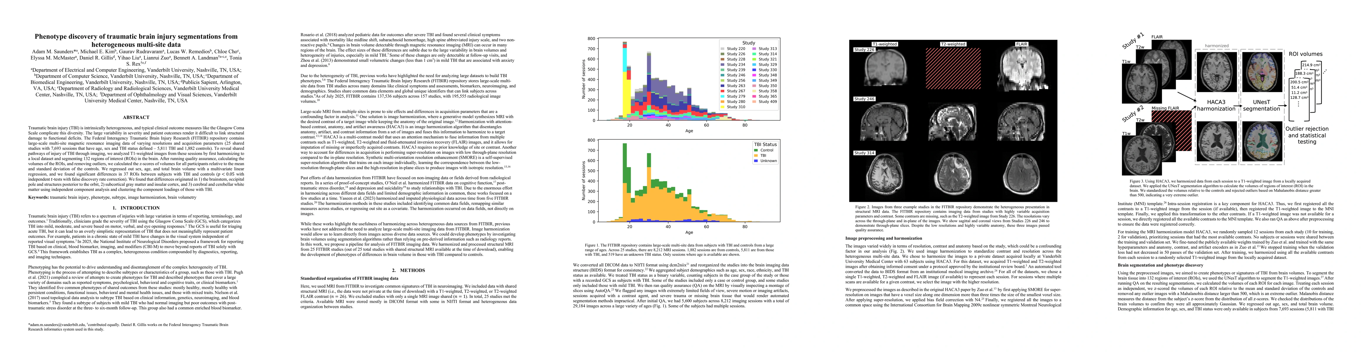
Traumatic brain injury (TBI) is intrinsically heterogeneous, and typical clinical outcome measures like the Glasgow Coma Scale complicate this diversity. The large variability in severity and patient ...
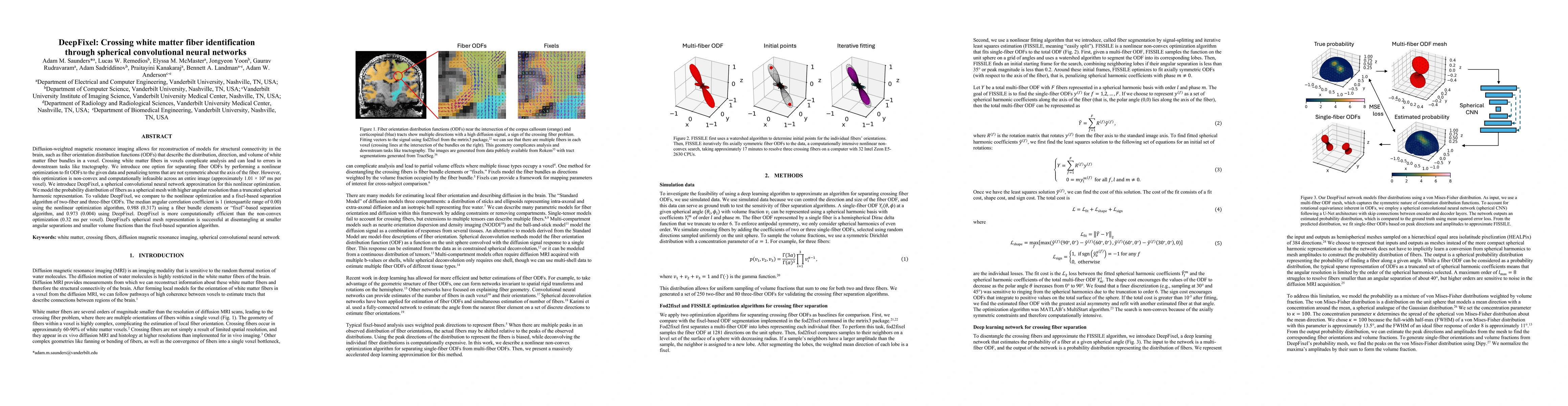
Diffusion-weighted magnetic resonance imaging allows for reconstruction of models for structural connectivity in the brain, such as fiber orientation distribution functions (ODFs) that describe the di...
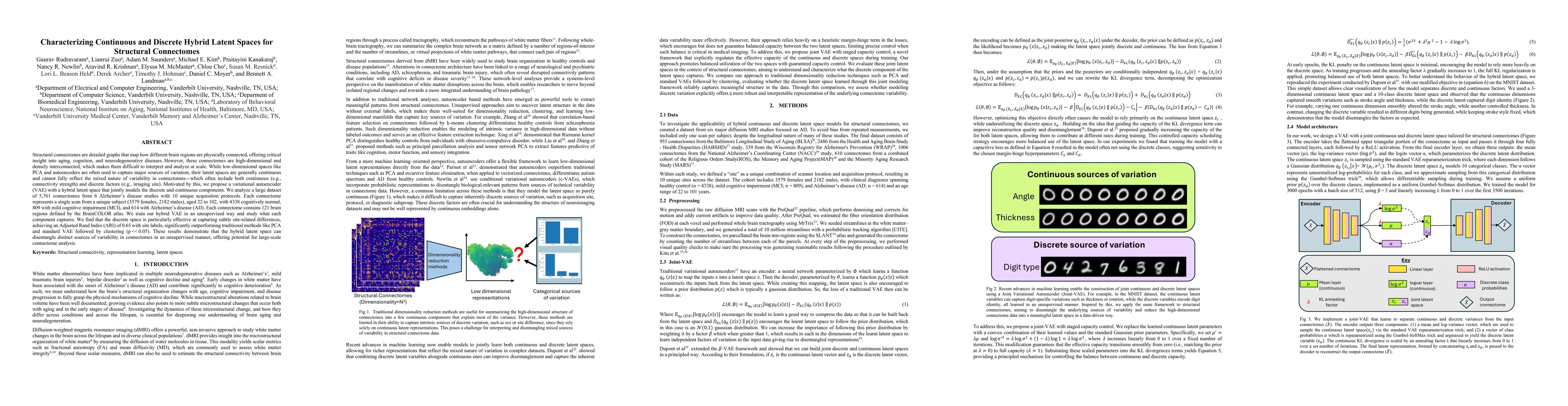
Structural connectomes are detailed graphs that map how different brain regions are physically connected, offering critical insight into aging, cognition, and neurodegenerative diseases. However, thes...
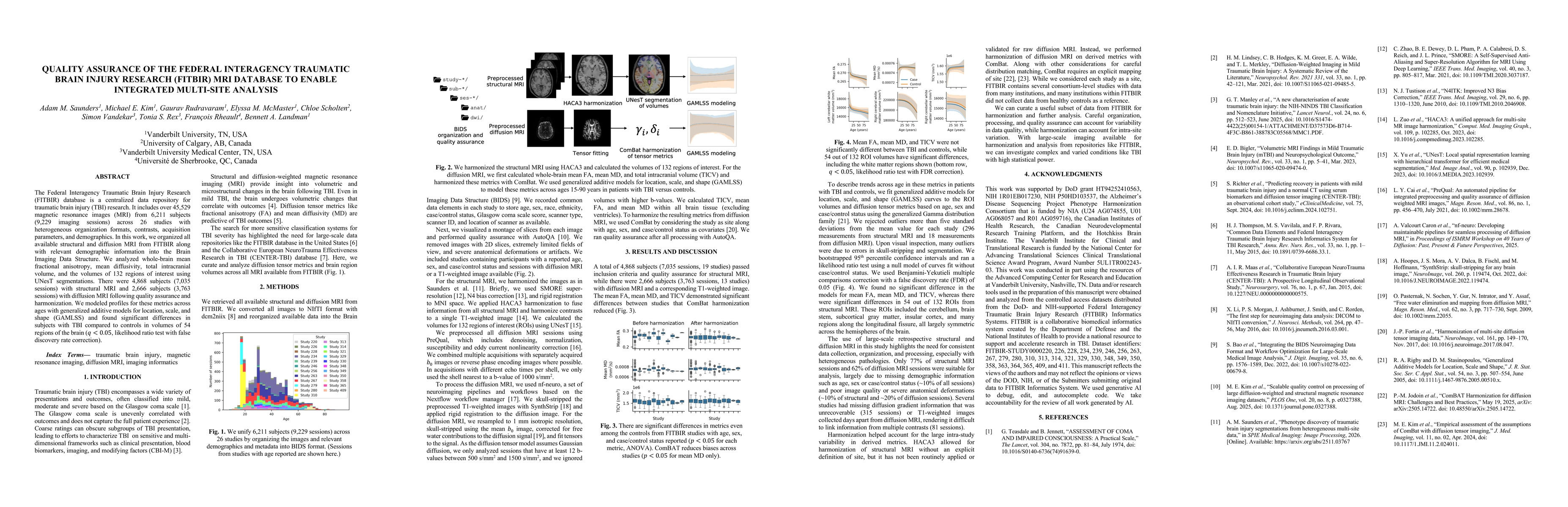
The Federal Interagency Traumatic Brain Injury Research (FITBIR) database is a centralized data repository for traumatic brain injury (TBI) research. It includes over 45,529 magnetic resonance images ...
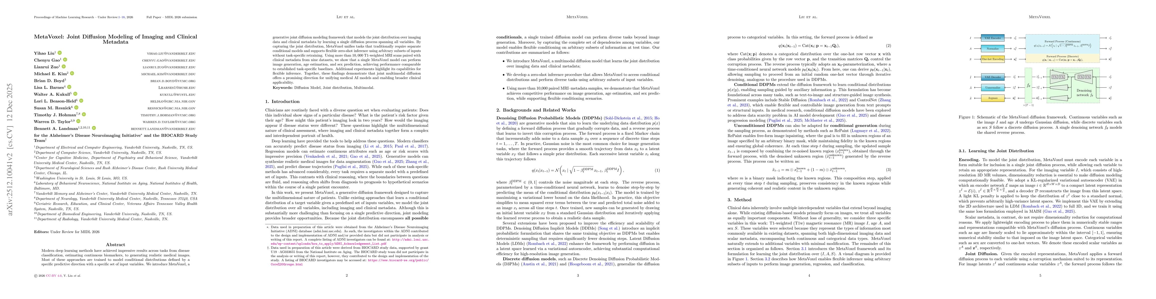
Modern deep learning methods have achieved impressive results across tasks from disease classification, estimating continuous biomarkers, to generating realistic medical images. Most of these approach...
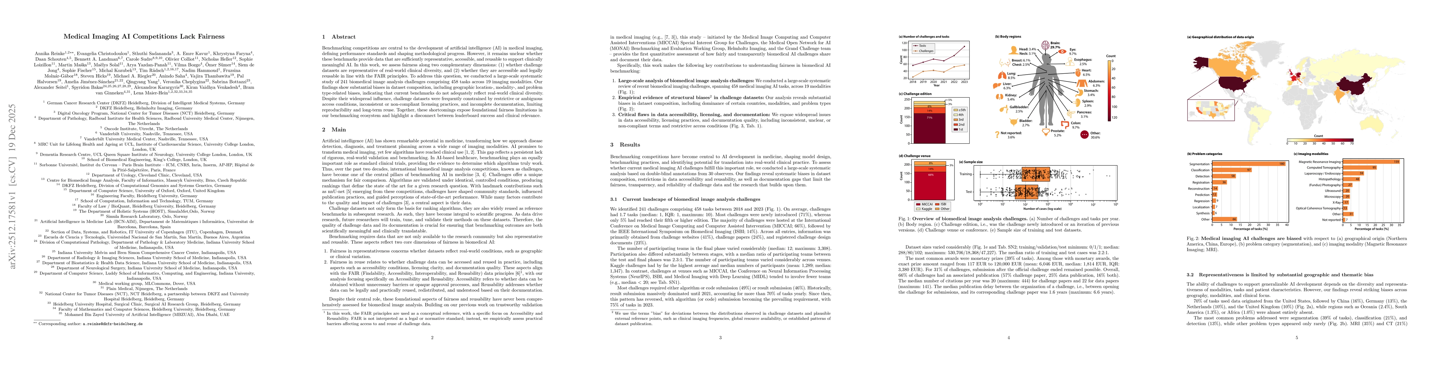
Benchmarking competitions are central to the development of artificial intelligence (AI) in medical imaging, defining performance standards and shaping methodological progress. However, it remains unc...
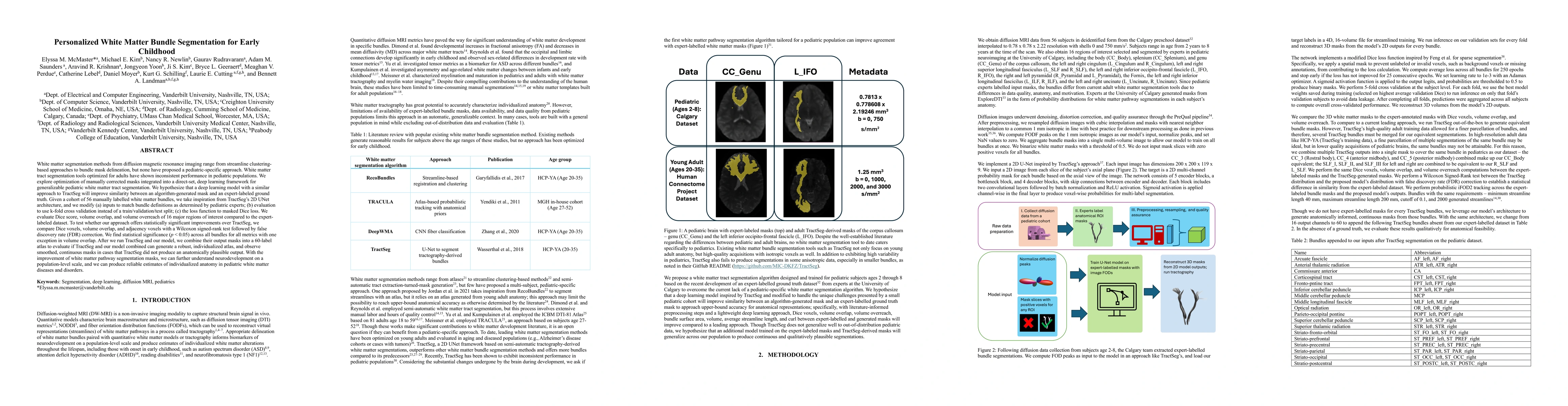
White matter segmentation methods from diffusion magnetic resonance imaging range from streamline clustering-based approaches to bundle mask delineation, but none have proposed a pediatric-specific ap...
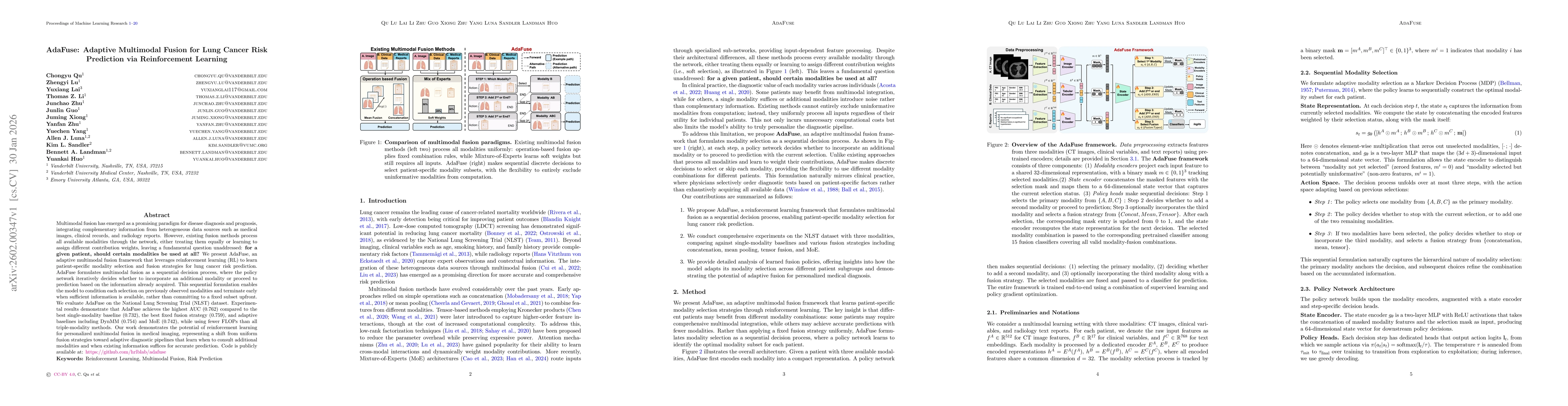
Multimodal fusion has emerged as a promising paradigm for disease diagnosis and prognosis, integrating complementary information from heterogeneous data sources such as medical images, clinical record...
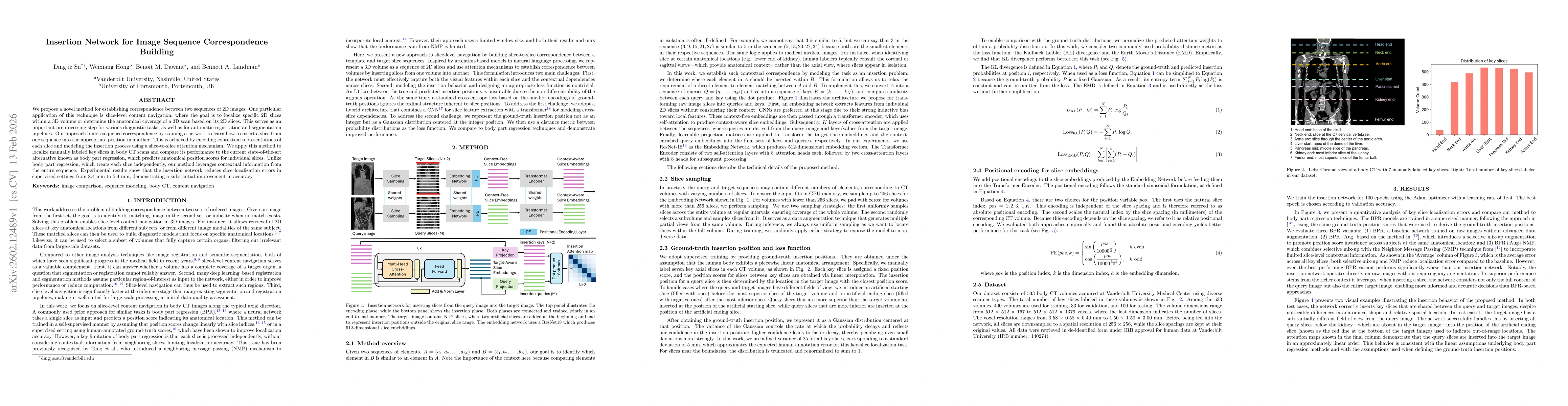
We propose a novel method for establishing correspondence between two sequences of 2D images. One particular application of this technique is slice-level content navigation, where the goal is to local...
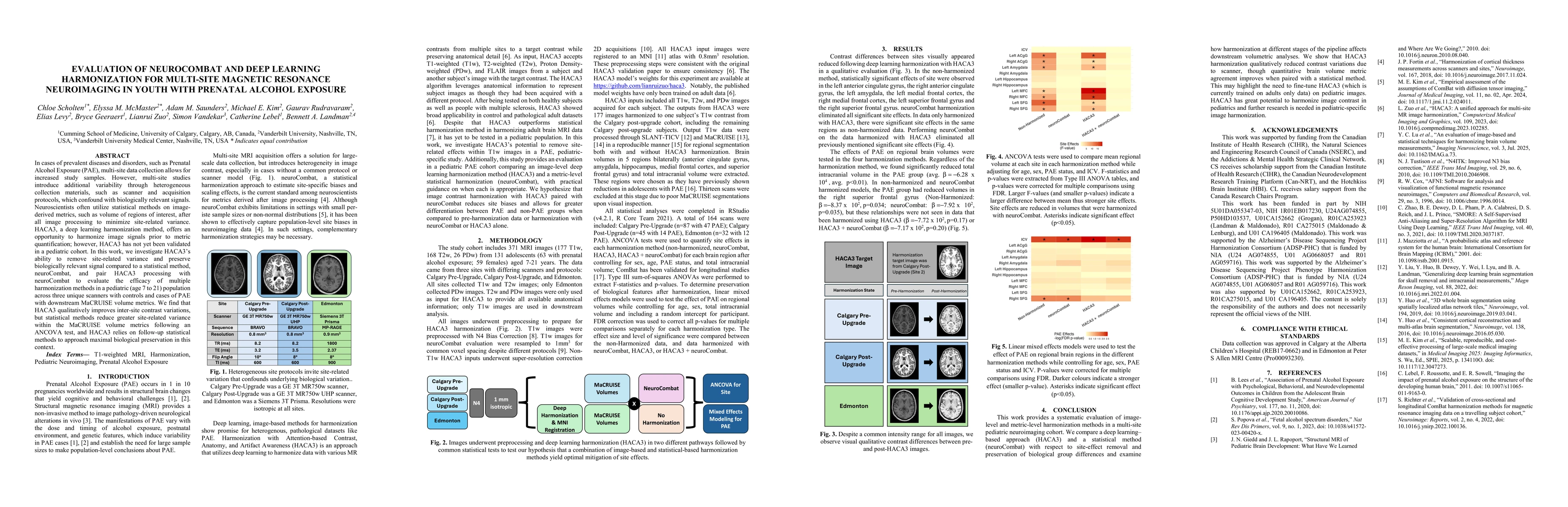
In cases of prevalent diseases and disorders, such as Prenatal Alcohol Exposure (PAE), multi-site data collection allows for increased study samples. However, multi-site studies introduce additional v...
The HEALthy Brain and Childhood Development (HBCD) Study is an ongoing longitudinal initiative to understand population-level brain maturation; however, large-scale studies must overcome site-related ...
Acquisition differences across sites, scanners, and protocols in dMRI introduce variability that complicates structural connectome analysis. This motivates deep learning models that can represent high...
Purpose: Diffusion MRI (dMRI) provides a diverse set of quantitative measures and derived datatypes to assess white matter microstructure and macrostructure. Coupled with the increasing size of imagin...