Academic Profile
Statistics
Similar Authors
Papers on arXiv
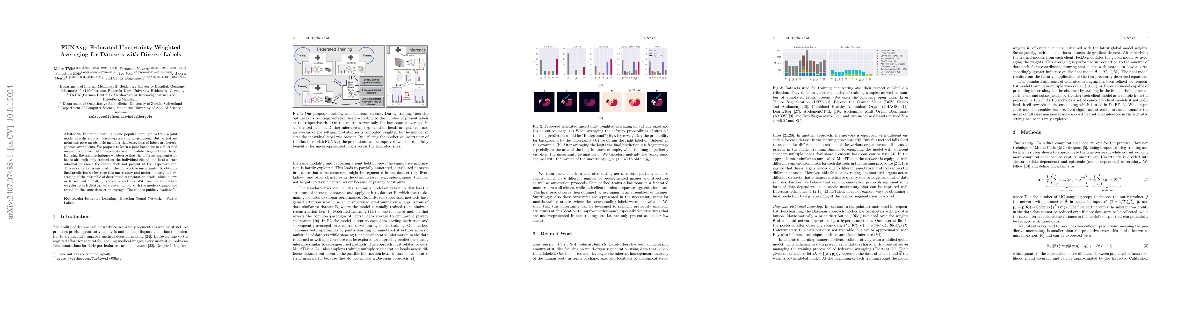
Federated learning is one popular paradigm to train a joint model in a distributed, privacy-preserving environment. But partial annotations pose an obstacle meaning that categories of labels are heter...
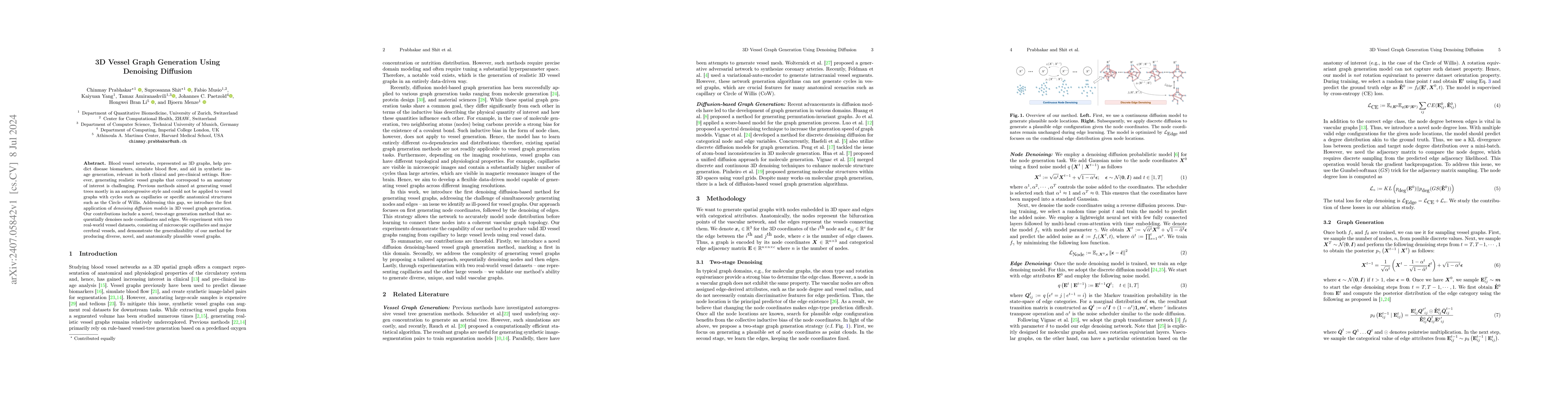
Blood vessel networks, represented as 3D graphs, help predict disease biomarkers, simulate blood flow, and aid in synthetic image generation, relevant in both clinical and pre-clinical settings. Howev...
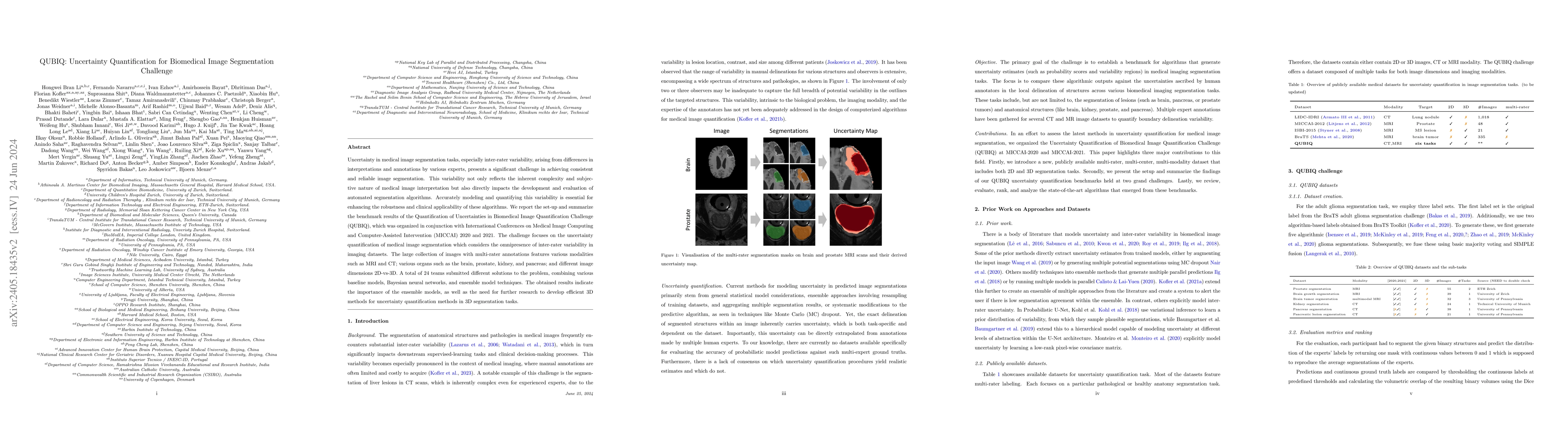
Uncertainty in medical image segmentation tasks, especially inter-rater variability, arising from differences in interpretations and annotations by various experts, presents a significant challenge in...
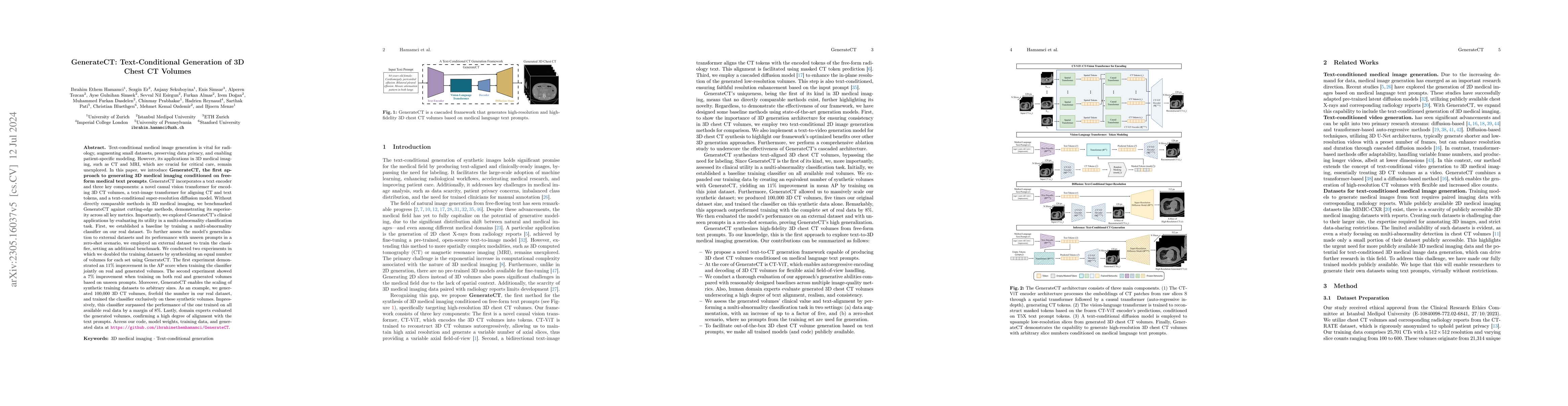
GenerateCT, the first approach to generating 3D medical imaging conditioned on free-form medical text prompts, incorporates a text encoder and three key components: a novel causal vision transformer f...
Pediatric central nervous system tumors are the leading cause of cancer-related deaths in children. The five-year survival rate for high-grade glioma in children is less than 20%. The development of n...
Pediatric tumors of the central nervous system are the most common cause of cancer-related death in children. The five-year survival rate for high-grade gliomas in children is less than 20%. Due to th...
The 2024 Brain Tumor Segmentation Meningioma Radiotherapy (BraTS-MEN-RT) challenge aims to advance automated segmentation algorithms using the largest known multi-institutional dataset of radiothera...
Gliomas are the most common malignant primary brain tumors in adults and one of the deadliest types of cancer. There are many challenges in treatment and monitoring due to the genetic diversity and ...
Self-supervised contrastive learning has predominantly adopted deterministic methods, which are not suited for environments characterized by uncertainty and noise. This paper introduces a new perspe...
We describe the design and results from the BraTS 2023 Intracranial Meningioma Segmentation Challenge. The BraTS Meningioma Challenge differed from prior BraTS Glioma challenges in that it focused o...
Vision-language pre-training for chest X-rays has made significant strides, primarily by utilizing paired radiographs and radiology reports. However, existing approaches often face challenges in enc...
Diffusion-weighted MRI (DWI) is essential for stroke diagnosis, treatment decisions, and prognosis. However, image and disease variability hinder the development of generalizable AI algorithms with ...
A major challenge in computational research in 3D medical imaging is the lack of comprehensive datasets. Addressing this issue, our study introduces CT-RATE, the first 3D medical imaging dataset tha...
Segmentation of blood vessels in murine cerebral 3D OCTA images is foundational for in vivo quantitative analysis of the effects of neurovascular disorders, such as stroke or Alzheimer's, on the vas...
Medical imaging plays a crucial role in diagnosis, with radiology reports serving as vital documentation. Automating report generation has emerged as a critical need to alleviate the workload of rad...
Biophysical modeling, particularly involving partial differential equations (PDEs), offers significant potential for tailoring disease treatment protocols to individual patients. However, the invers...
Purpose. To present SPINEPS, an open-source deep learning approach for semantic and instance segmentation of 14 spinal structures (ten vertebra substructures, intervertebral discs, spinal cord, spin...
We introduce MultiMedEval, an open-source toolkit for fair and reproducible evaluation of large, medical vision-language models (VLM). MultiMedEval comprehensively assesses the models' performance o...
The Circle of Willis (CoW) is an important network of arteries connecting major circulations of the brain. Its vascular architecture is believed to affect the risk, severity, and clinical outcome of...
Brain tumor growth is unique to each glioma patient and extends beyond what is visible in imaging scans, infiltrating surrounding brain tissue. Understanding these hidden patient-specific progressio...
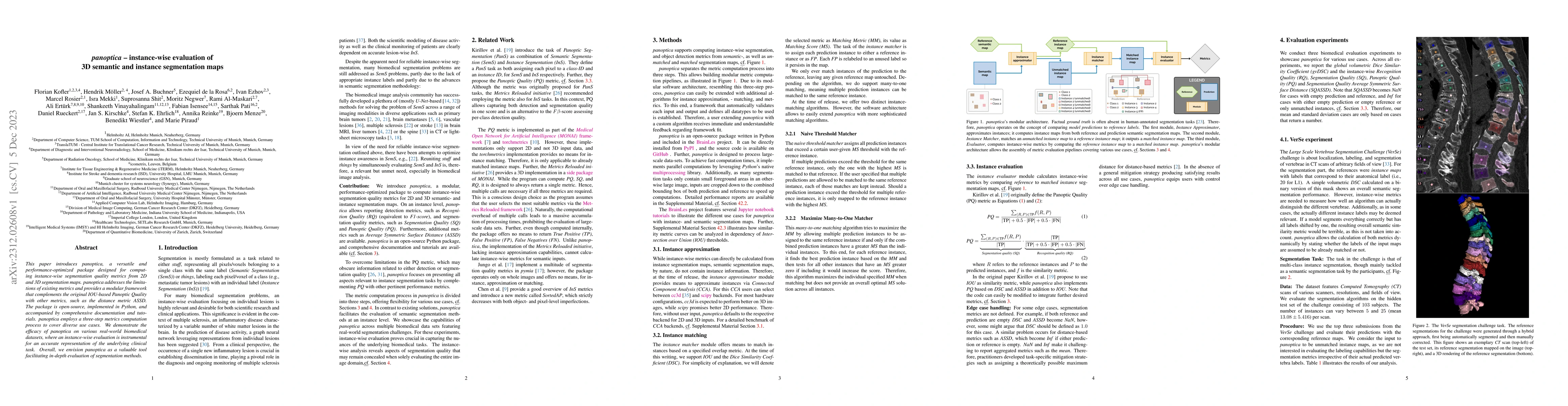
This paper introduces panoptica, a versatile and performance-optimized package designed for computing instance-wise segmentation quality metrics from 2D and 3D segmentation maps. panoptica addresses...
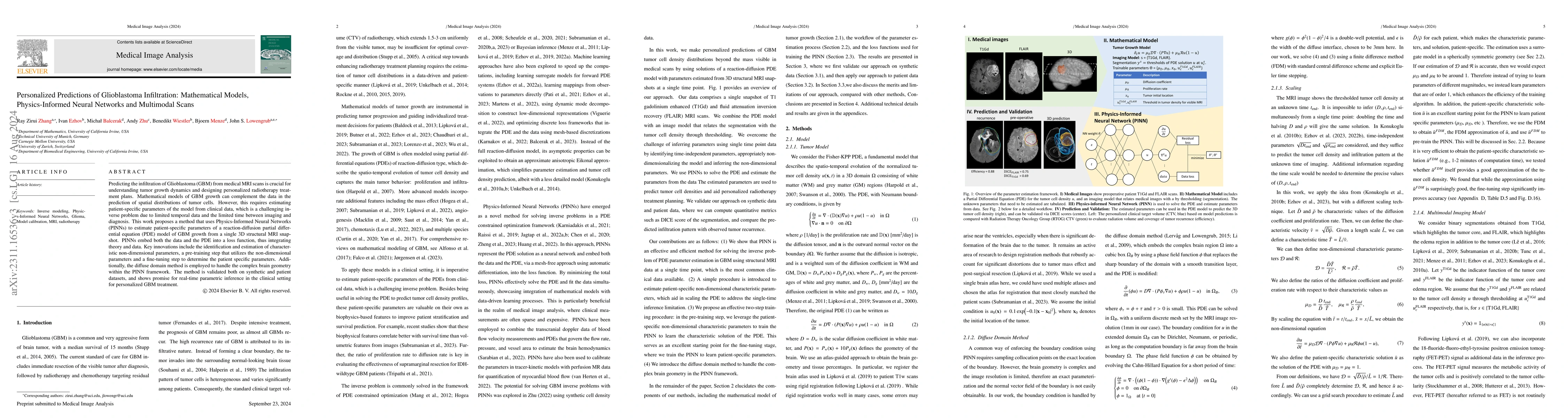
Predicting the infiltration of Glioblastoma (GBM) from medical MRI scans is crucial for understanding tumor growth dynamics and designing personalized radiotherapy treatment plans.Mathematical model...
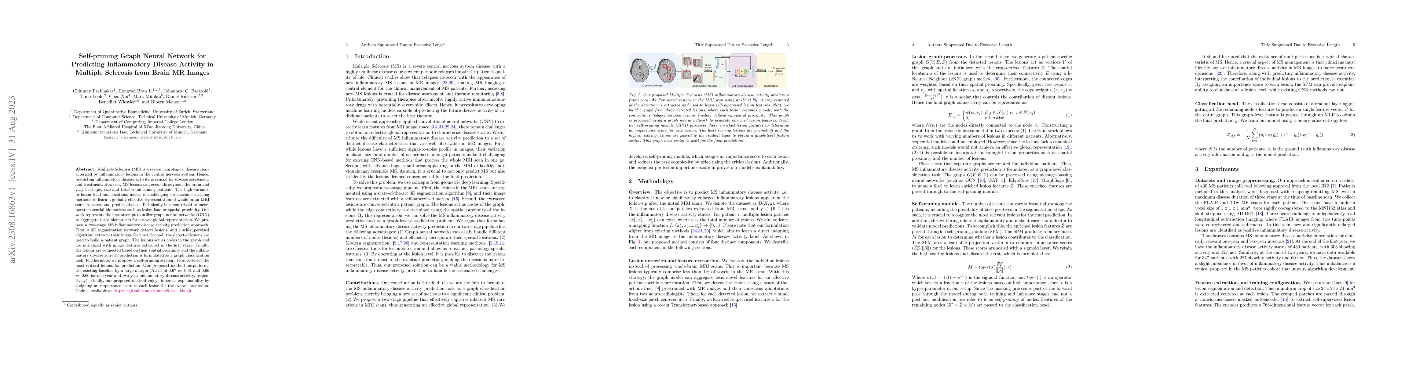
Multiple Sclerosis (MS) is a severe neurological disease characterized by inflammatory lesions in the central nervous system. Hence, predicting inflammatory disease activity is crucial for disease a...
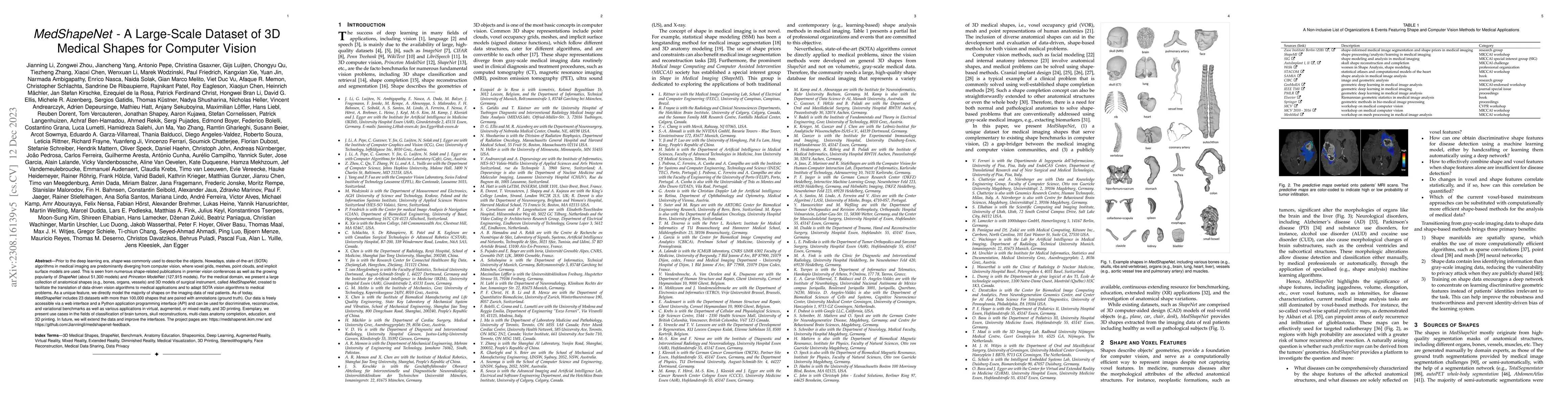
Prior to the deep learning era, shape was commonly used to describe the objects. Nowadays, state-of-the-art (SOTA) algorithms in medical imaging are predominantly diverging from computer vision, whe...
Background: Automated segmentation of spinal MR images plays a vital role both scientifically and clinically. However, accurately delineating posterior spine structures presents challenges. Method...
Automated medical image segmentation inherently involves a certain degree of uncertainty. One key factor contributing to this uncertainty is the ambiguity that can arise in determining the boundarie...
Panoramic X-rays are frequently used in dentistry for treatment planning, but their interpretation can be both time-consuming and prone to error. Artificial intelligence (AI) has the potential to ai...
Recent trends in Video Instance Segmentation (VIS) have seen a growing reliance on online methods to model complex and lengthy video sequences. However, the degradation of representation and noise a...
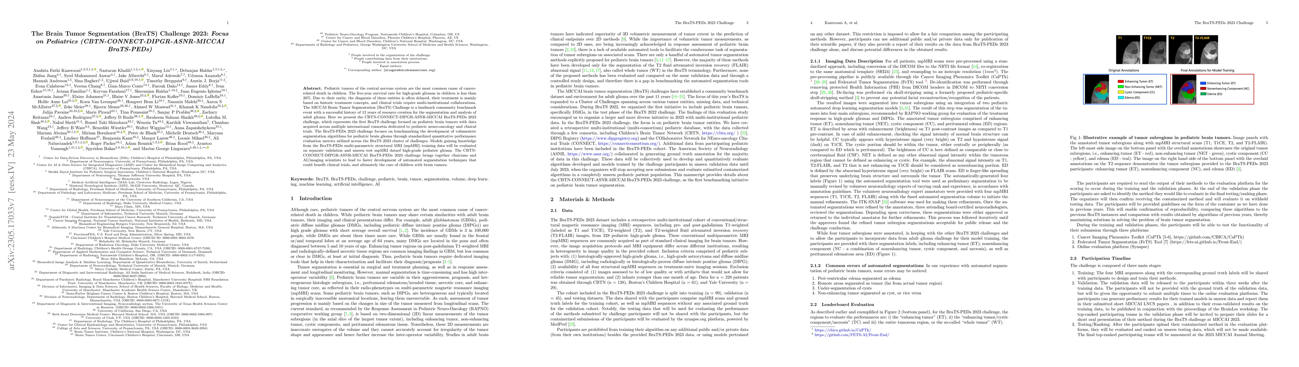
Pediatric tumors of the central nervous system are the most common cause of cancer-related death in children. The five-year survival rate for high-grade gliomas in children is less than 20\%. Due to...
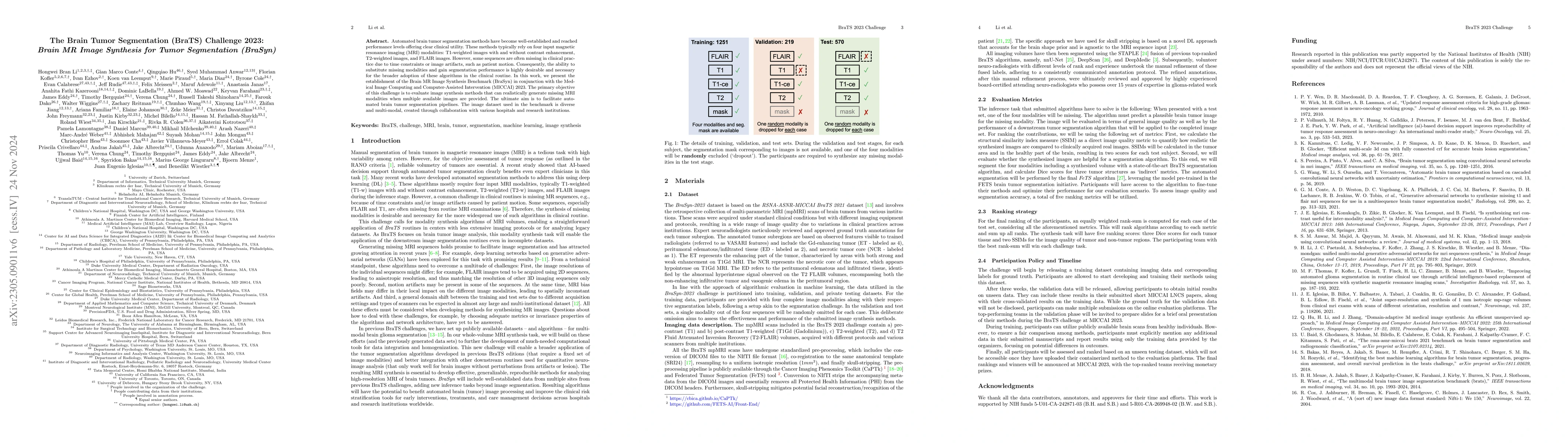
Automated brain tumor segmentation methods have become well-established and reached performance levels offering clear clinical utility. These methods typically rely on four input magnetic resonance ...
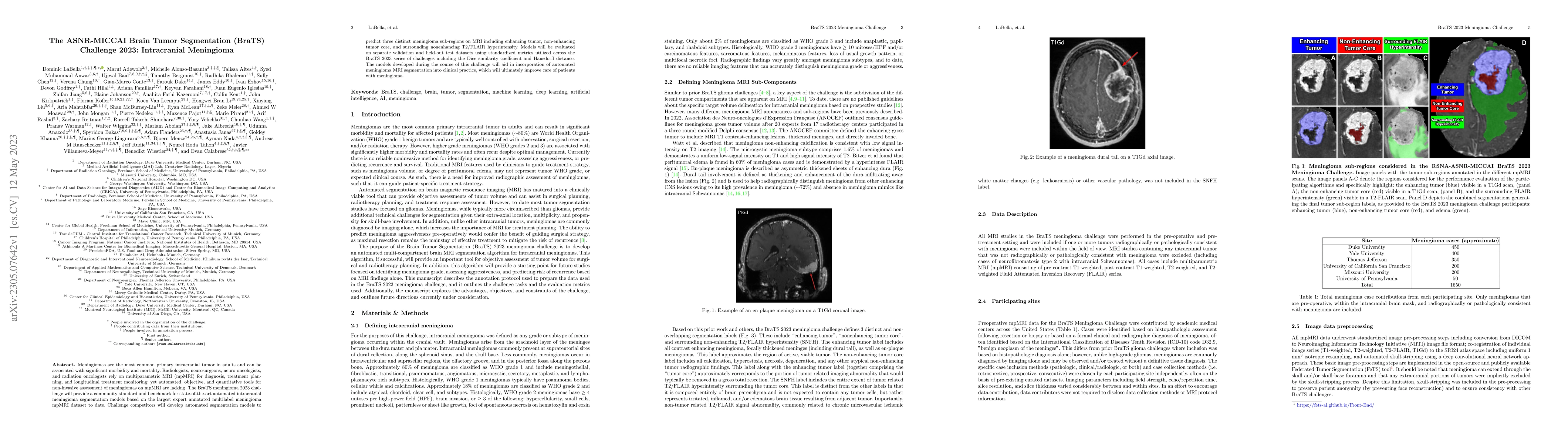
Meningiomas are the most common primary intracranial tumor in adults and can be associated with significant morbidity and mortality. Radiologists, neurosurgeons, neuro-oncologists, and radiation onc...
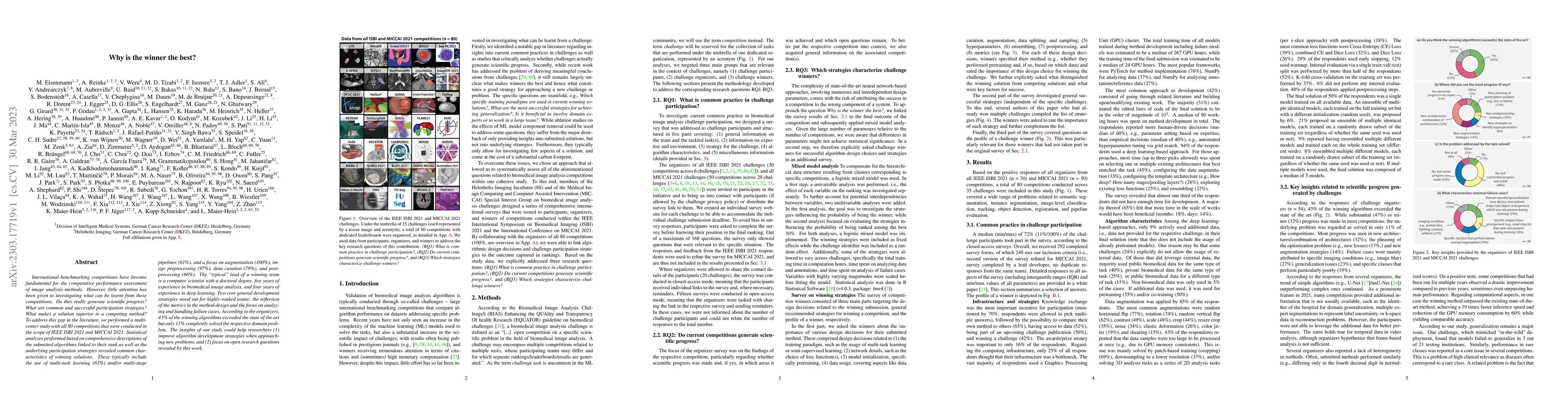
International benchmarking competitions have become fundamental for the comparative performance assessment of image analysis methods. However, little attention has been given to investigating what c...
Link prediction algorithms aim to infer the existence of connections (or links) between nodes in network-structured data and are typically applied to refine the connectivity among nodes. In this wor...
Due to the necessity for precise treatment planning, the use of panoramic X-rays to identify different dental diseases has tremendously increased. Although numerous ML models have been developed for...
Validation metrics are key for the reliable tracking of scientific progress and for bridging the current chasm between artificial intelligence (AI) research and its translation into practice. Howeve...
Self-supervised learning has attracted increasing attention as it learns data-driven representation from data without annotations. Vision transformer-based autoencoder (ViT-AE) by He et al. (2021) i...
Hyperspectral imaging (HSI) is an optical technique that processes the electromagnetic spectrum at a multitude of monochromatic, adjacent frequency bands. The wide-bandwidth spectral signature of a ...
Machine learning models are typically evaluated by computing similarity with reference annotations and trained by maximizing similarity with such. Especially in the biomedical domain, annotations ar...
The number of international benchmarking competitions is steadily increasing in various fields of machine learning (ML) research and practice. So far, however, little is known about the common pract...
Quantifying the perceptual similarity of two images is a long-standing problem in low-level computer vision. The natural image domain commonly relies on supervised learning, e.g., a pre-trained VGG,...
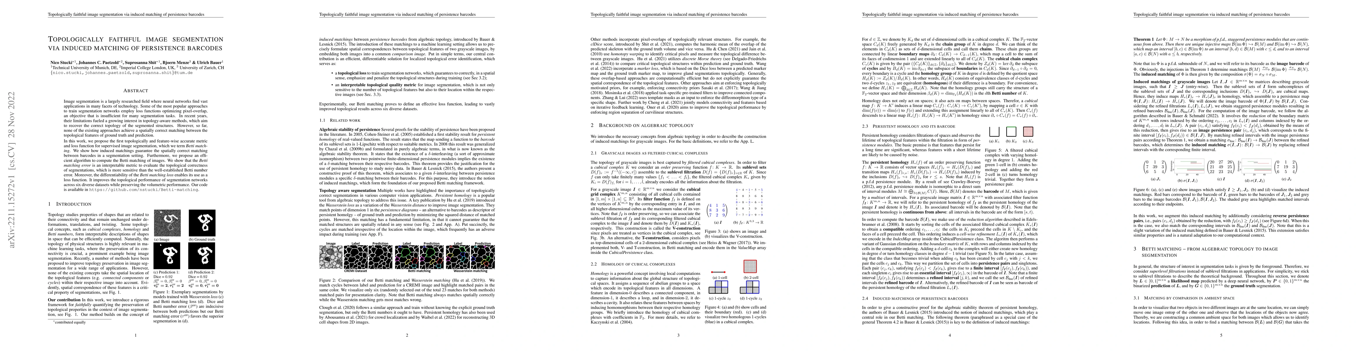
Image segmentation is a largely researched field where neural networks find vast applications in many facets of technology. Some of the most popular approaches to train segmentation networks employ ...
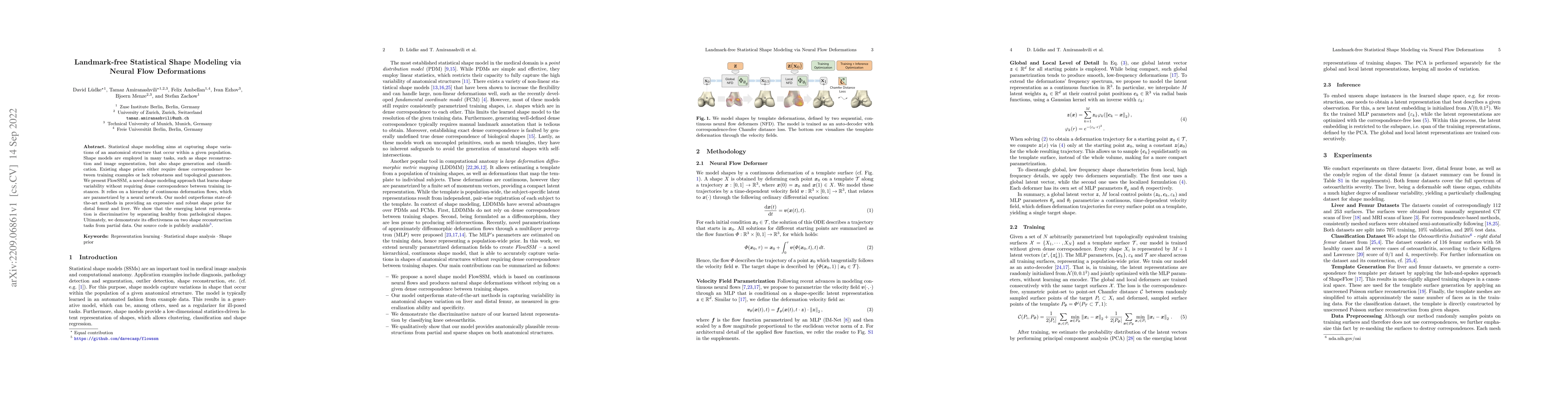
Statistical shape modeling aims at capturing shape variations of an anatomical structure that occur within a given population. Shape models are employed in many tasks, such as shape reconstruction a...
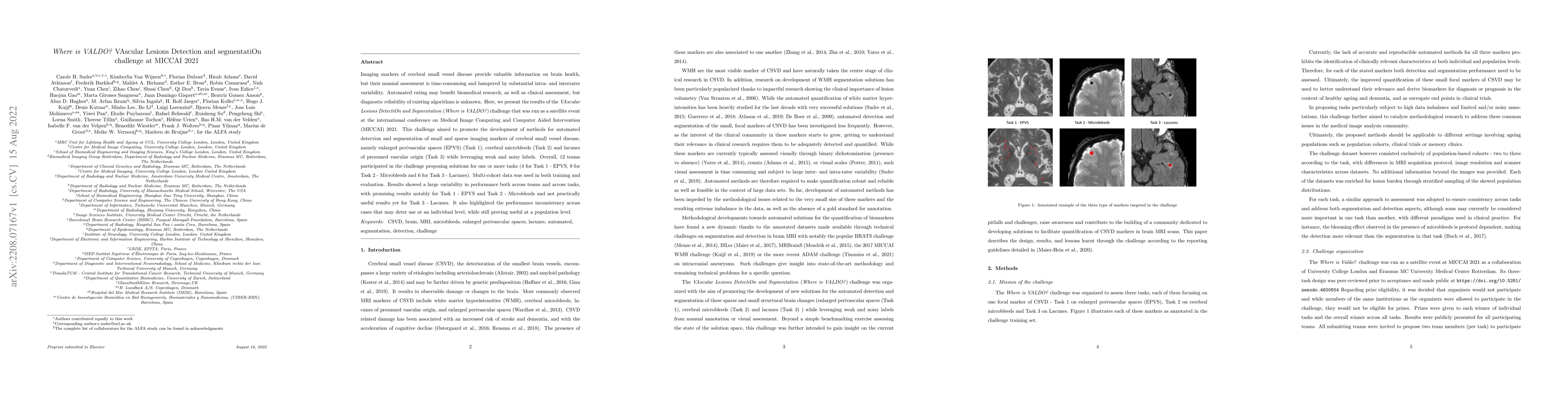
Imaging markers of cerebral small vessel disease provide valuable information on brain health, but their manual assessment is time-consuming and hampered by substantial intra- and interrater variabi...
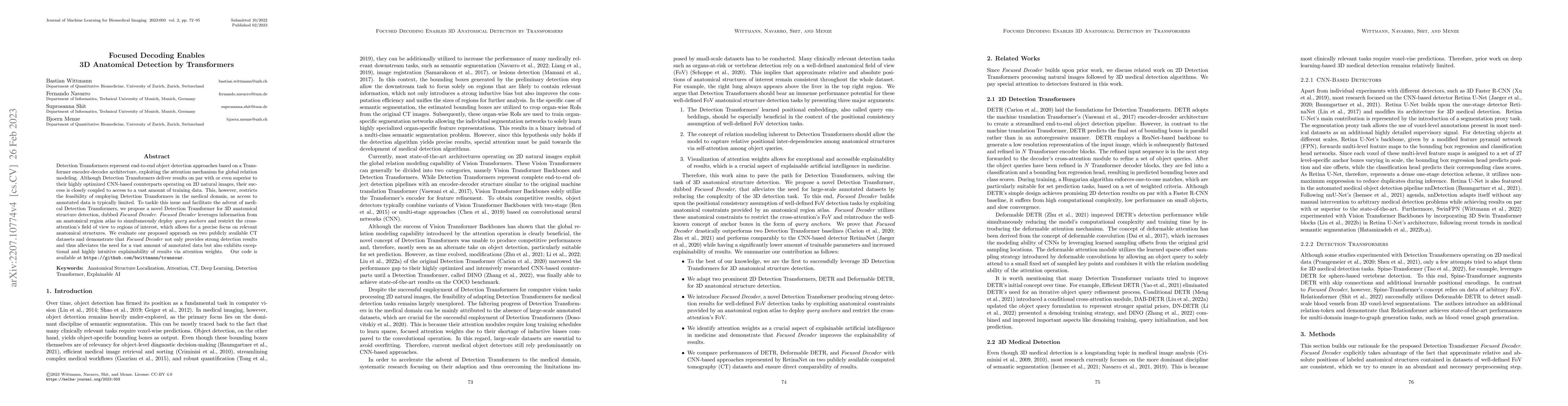
Detection Transformers represent end-to-end object detection approaches based on a Transformer encoder-decoder architecture, exploiting the attention mechanism for global relation modeling. Although...
Magnetic resonance imaging (MRI) is a central modality for stroke imaging. It is used upon patient admission to make treatment decisions such as selecting patients for intravenous thrombolysis or en...
Increasing evidence shows that flaws in machine learning (ML) algorithm validation are an underestimated global problem. Particularly in automatic biomedical image analysis, chosen performance metri...
Human ratings are abstract representations of segmentation quality. To approximate human quality ratings on scarce expert data, we train surrogate quality estimation models. We evaluate on a complex...
Deep convolutional neural networks (CNN) have proven to be remarkably effective in semantic segmentation tasks. Most popular loss functions were introduced targeting improved volumetric scores, such...
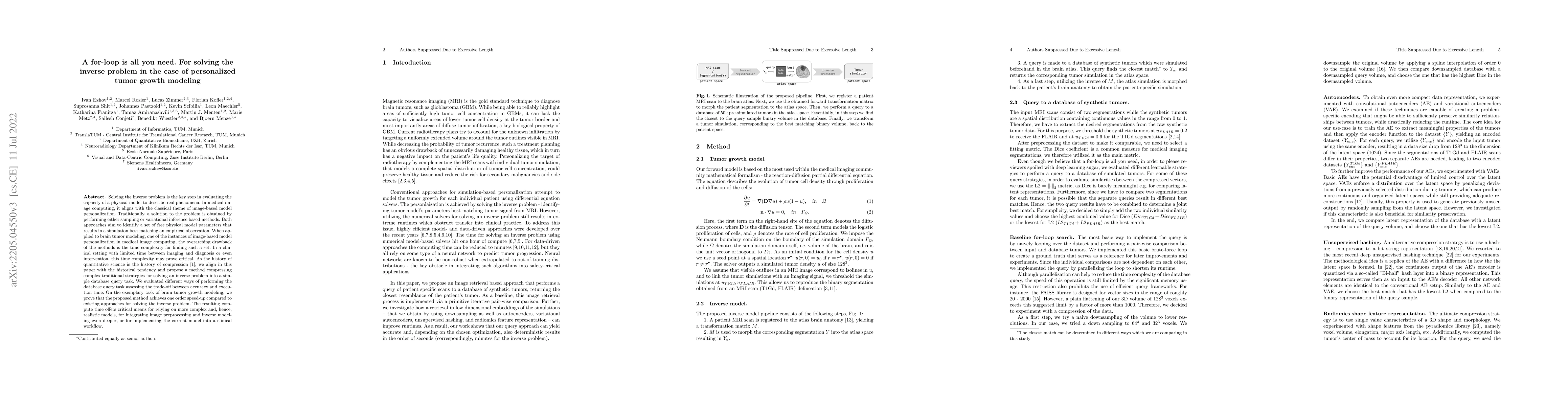
Solving the inverse problem is the key step in evaluating the capacity of a physical model to describe real phenomena. In medical image computing, it aligns with the classical theme of image-based m...
Although machine learning (ML) has shown promise in numerous domains, there are concerns about generalizability to out-of-sample data. This is currently addressed by centrally sharing ample, and imp...
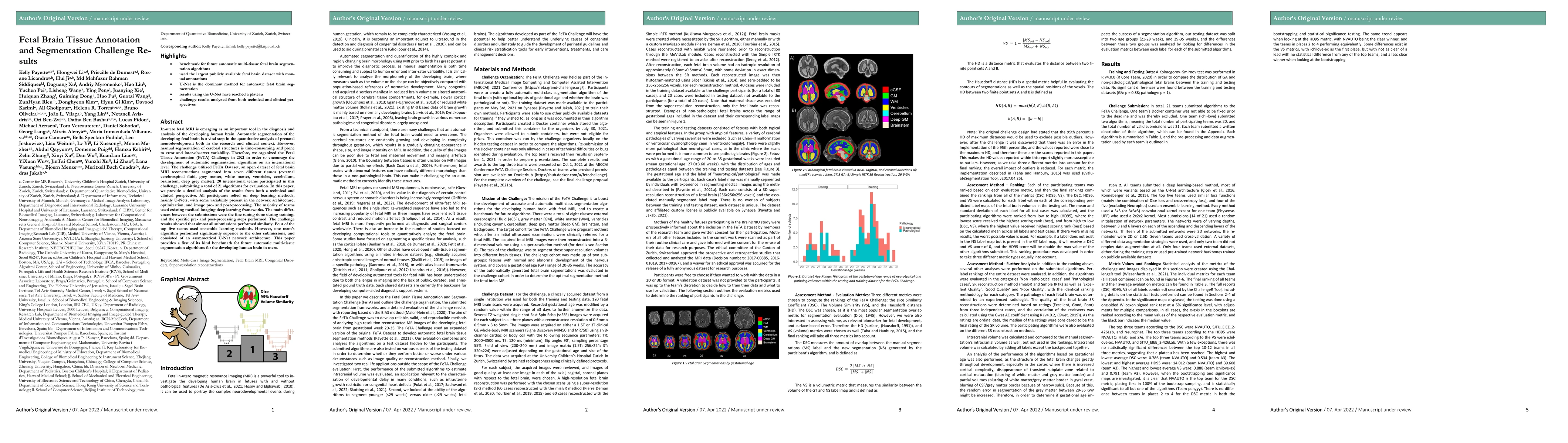
In-utero fetal MRI is emerging as an important tool in the diagnosis and analysis of the developing human brain. Automatic segmentation of the developing fetal brain is a vital step in the quantitat...
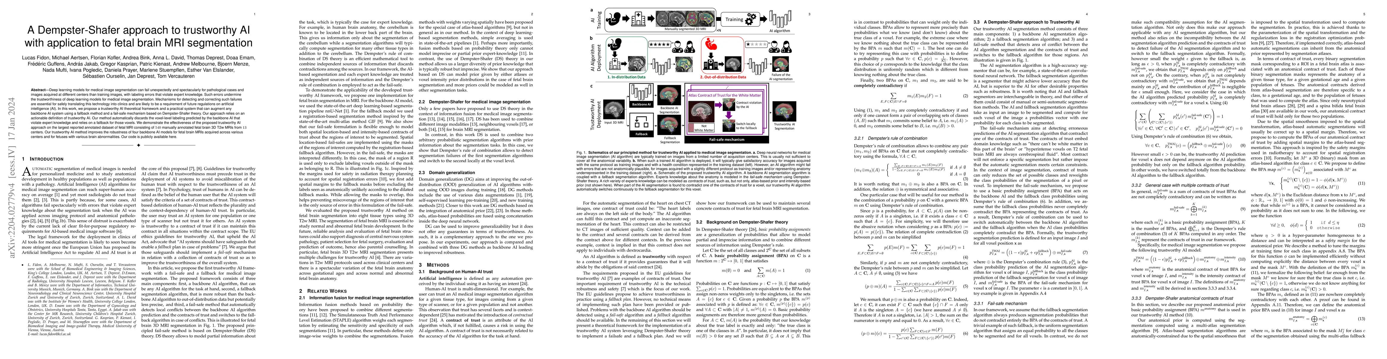
Deep learning models for medical image segmentation can fail unexpectedly and spectacularly for pathological cases and images acquired at different centers than training images, with labeling errors...
In this work, we present a method for synthetic CT (sCT) generation from zero-echo-time (ZTE) MRI aimed at structural and quantitative accuracies of the image, with a particular focus on the accurat...
A comprehensive representation of an image requires understanding objects and their mutual relationship, especially in image-to-graph generation, e.g., road network extraction, blood-vessel network ...
Deep learning (DL) models have provided state-of-the-art performance in various medical imaging benchmarking challenges, including the Brain Tumor Segmentation (BraTS) challenges. However, the task ...
Registration of longitudinal brain MRI scans containing pathologies is challenging due to dramatic changes in tissue appearance. Although there has been progress in developing general-purpose medica...
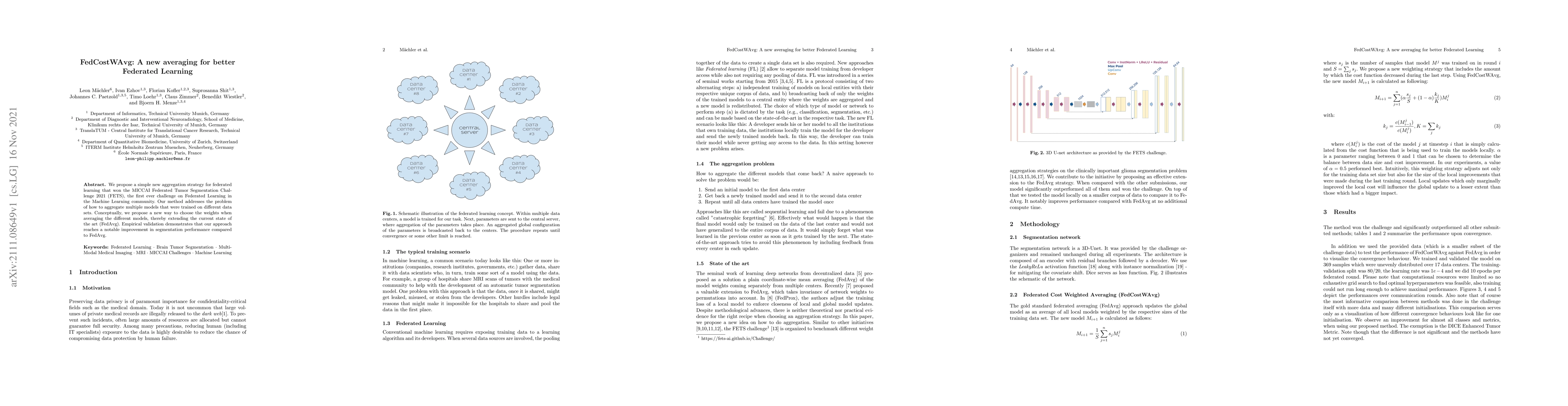
We propose a simple new aggregation strategy for federated learning that won the MICCAI Federated Tumor Segmentation Challenge 2021 (FETS), the first ever challenge on Federated Learning in the Mach...
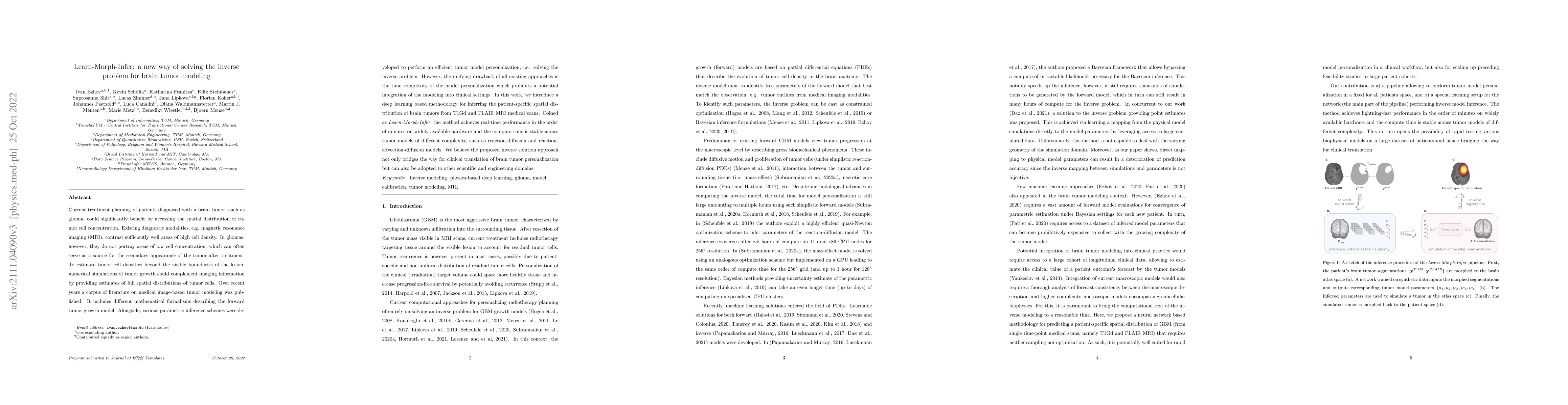
Current treatment planning of patients diagnosed with a brain tumor, such as glioma, could significantly benefit by accessing the spatial distribution of tumor cell concentration. Existing diagnosti...
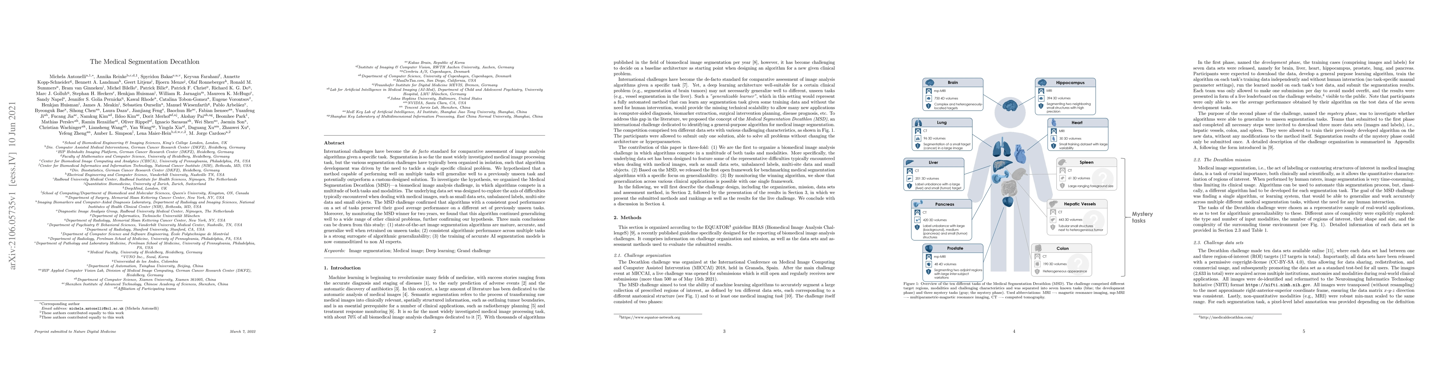
International challenges have become the de facto standard for comparative assessment of image analysis algorithms given a specific task. Segmentation is so far the most widely investigated medical ...
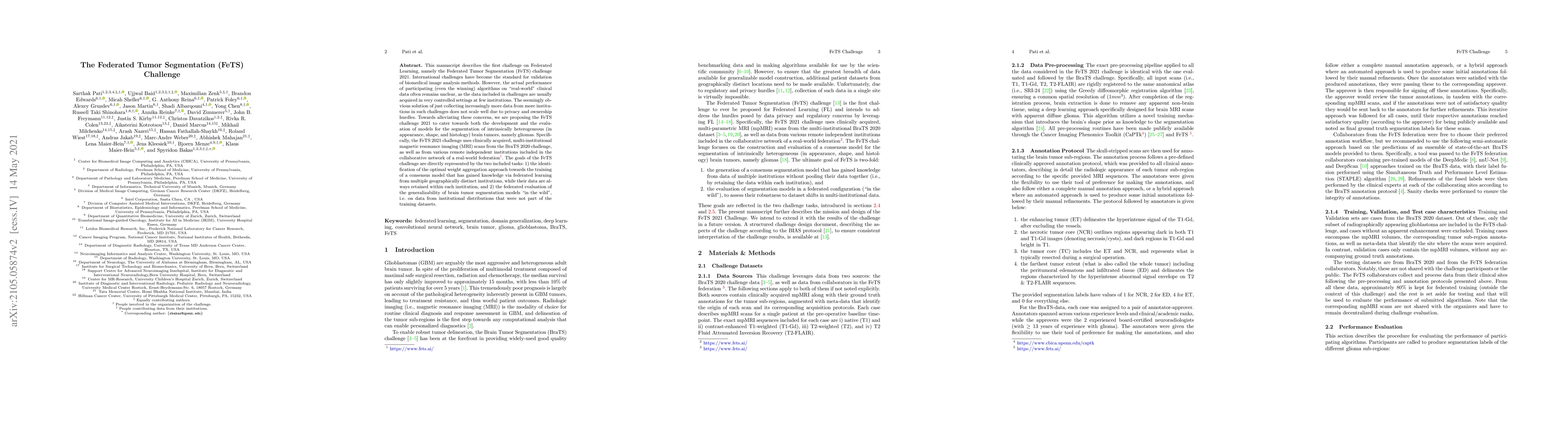
This manuscript describes the first challenge on Federated Learning, namely the Federated Tumor Segmentation (FeTS) challenge 2021. International challenges have become the standard for validation o...
While the importance of automatic image analysis is continuously increasing, recent meta-research revealed major flaws with respect to algorithm validation. Performance metrics are particularly key ...
Purpose: To develop and evaluate MyoMapNet, a rapid myocardial T1 mapping approach that uses neural networks (NN) to estimate voxel-wise myocardial T1 and extracellular (ECV) from T1-weighted images...
Perfusion imaging is the current gold standard for acute ischemic stroke analysis. It allows quantification of the salvageable and non-salvageable tissue regions (penumbra and core areas respectivel...
With the advent of deep learning algorithms, fully automated radiological image analysis is within reach. In spine imaging, several atlas- and shape-based as well as deep learning segmentation algor...
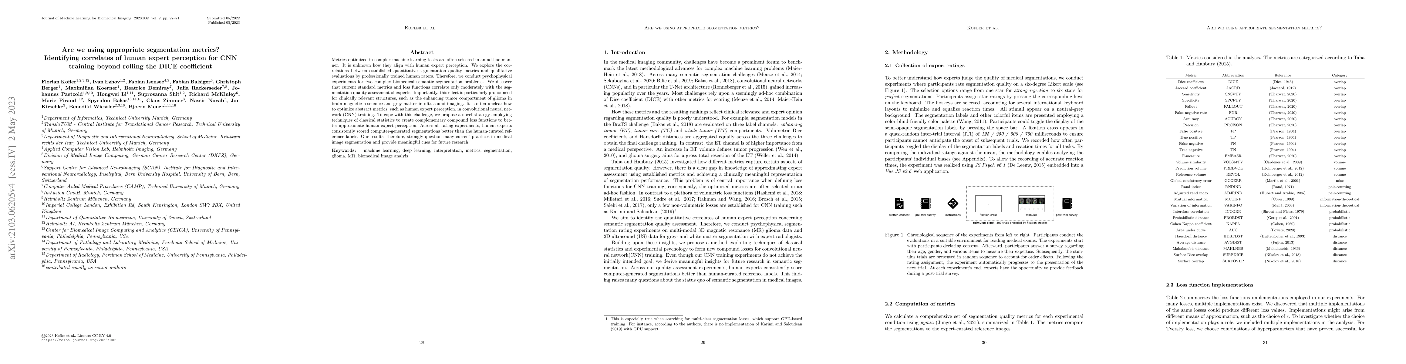
Metrics optimized in complex machine learning tasks are often selected in an ad-hoc manner. It is unknown how they align with human expert perception. We explore the correlations between established...
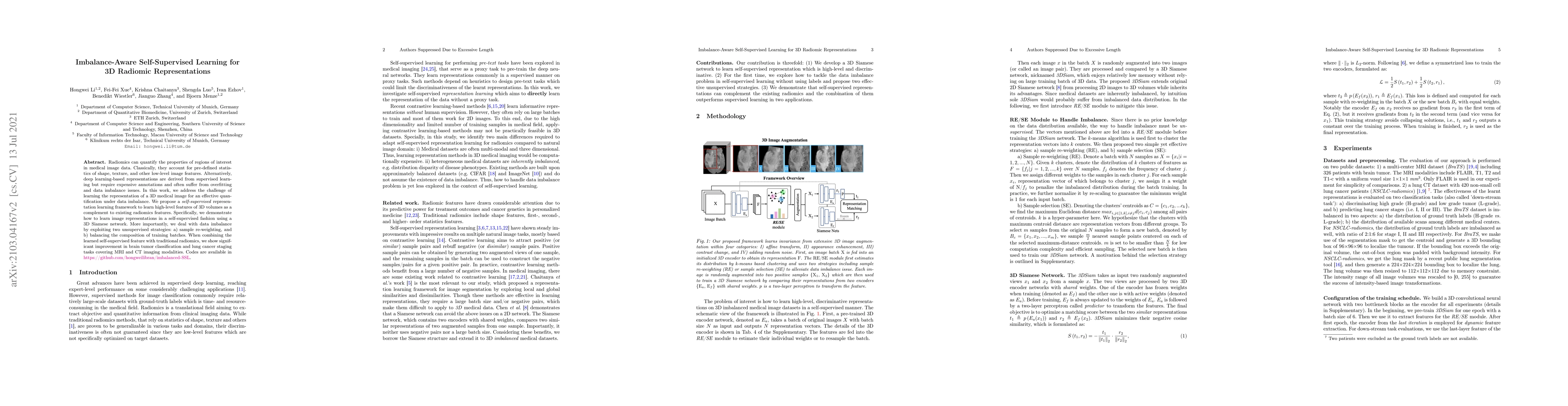
Radiomic representations can quantify properties of regions of interest in medical image data. Classically, they account for pre-defined statistics of shape, texture, and other low-level image featu...
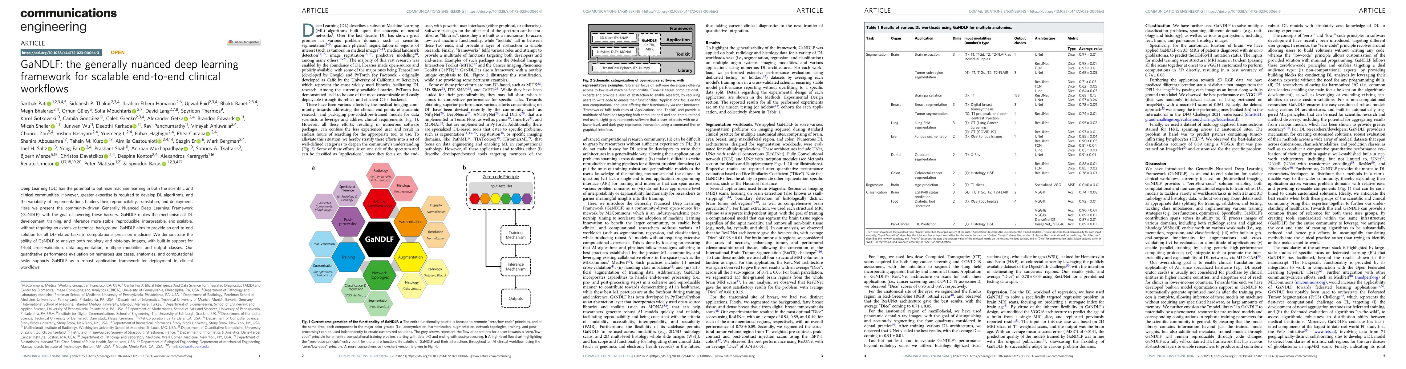
Deep Learning (DL) has the potential to optimize machine learning in both the scientific and clinical communities. However, greater expertise is required to develop DL algorithms, and the variabilit...
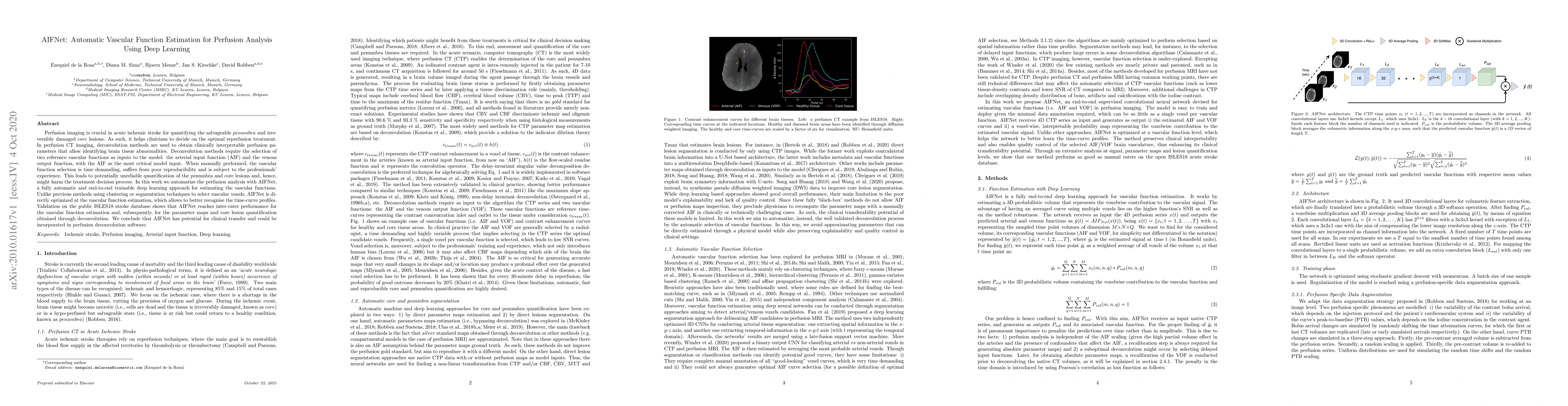
Perfusion imaging is crucial in acute ischemic stroke for quantifying the salvageable penumbra and irreversibly damaged core lesions. As such, it helps clinicians to decide on the optimal reperfusio...
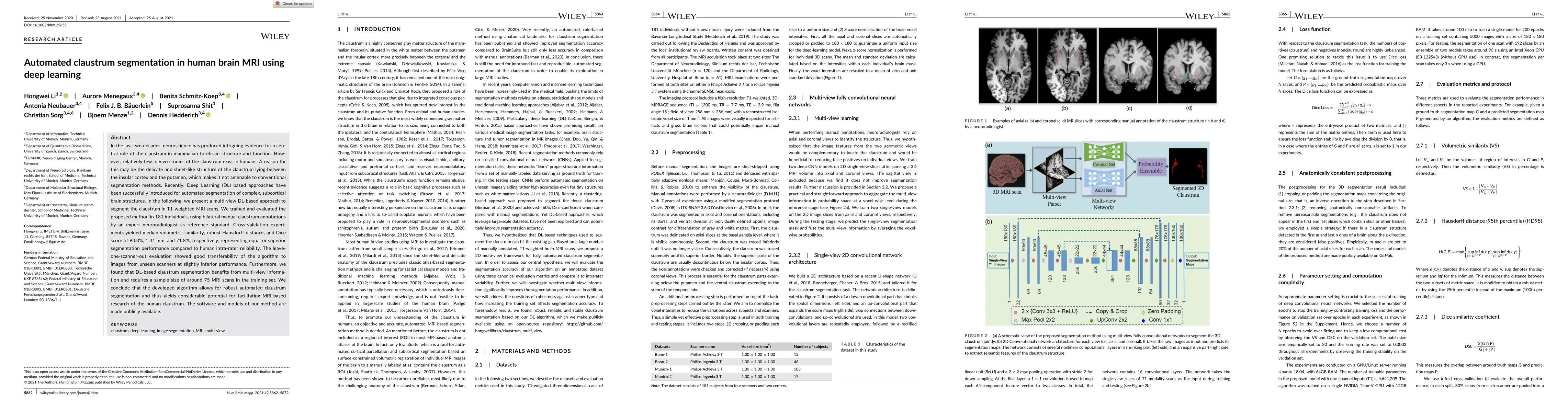
In the last two decades, neuroscience has produced intriguing evidence for a central role of the claustrum in mammalian forebrain structure and function. However, relatively few in vivo studies of t...
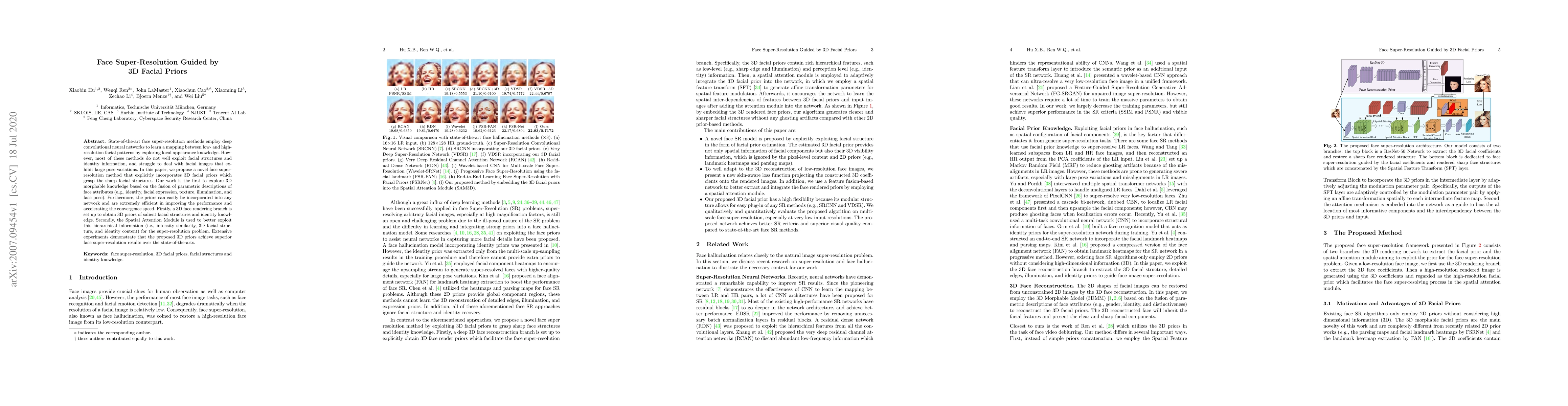
State-of-the-art face super-resolution methods employ deep convolutional neural networks to learn a mapping between low- and high- resolution facial patterns by exploring local appearance knowledge....
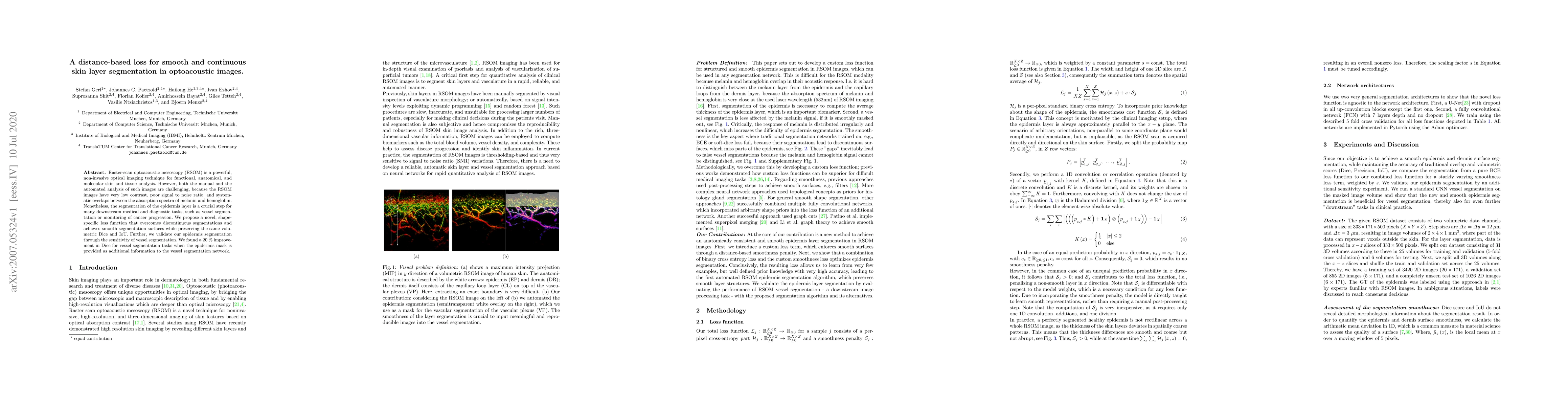
Raster-scan optoacoustic mesoscopy (RSOM) is a powerful, non-invasive optical imaging technique for functional, anatomical, and molecular skin and tissue analysis. However, both the manual and the a...
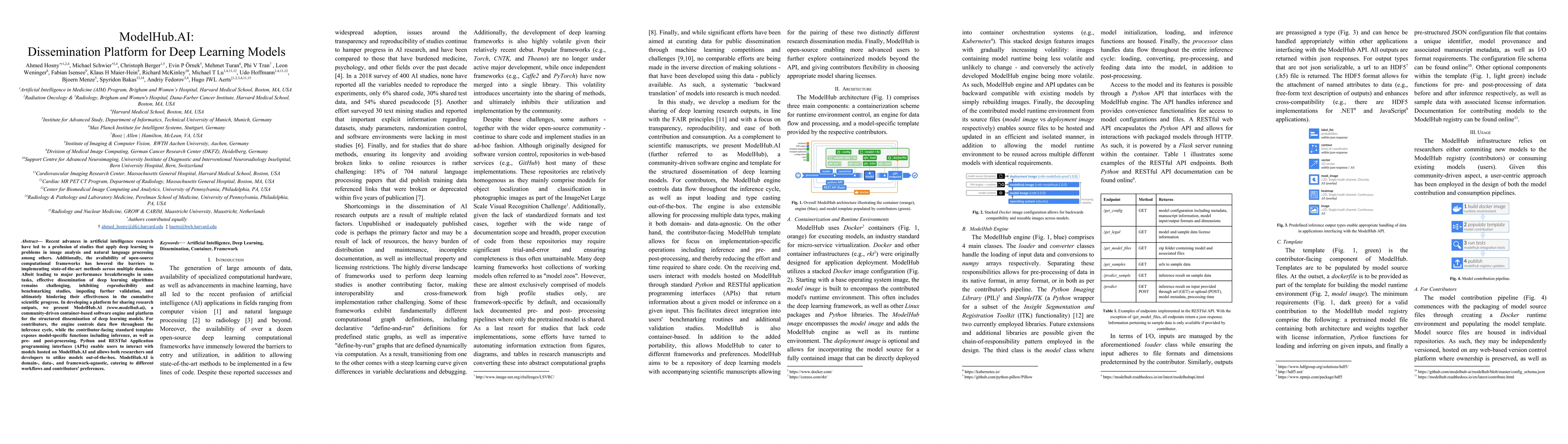
Recent advances in artificial intelligence research have led to a profusion of studies that apply deep learning to problems in image analysis and natural language processing among others. Additional...
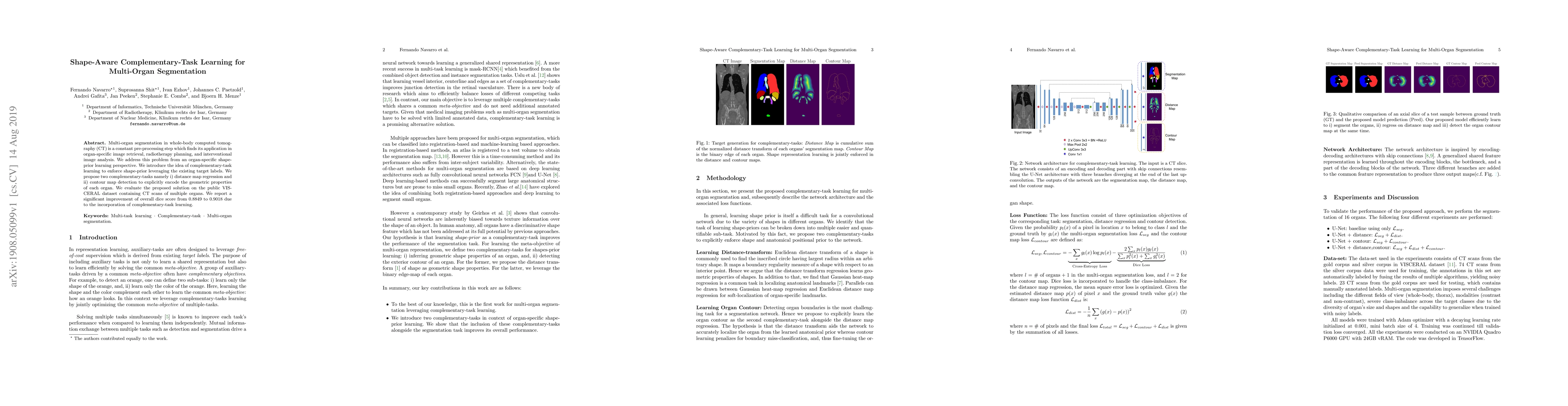
Multi-organ segmentation in whole-body computed tomography (CT) is a constant pre-processing step which finds its application in organ-specific image retrieval, radiotherapy planning, and interventi...
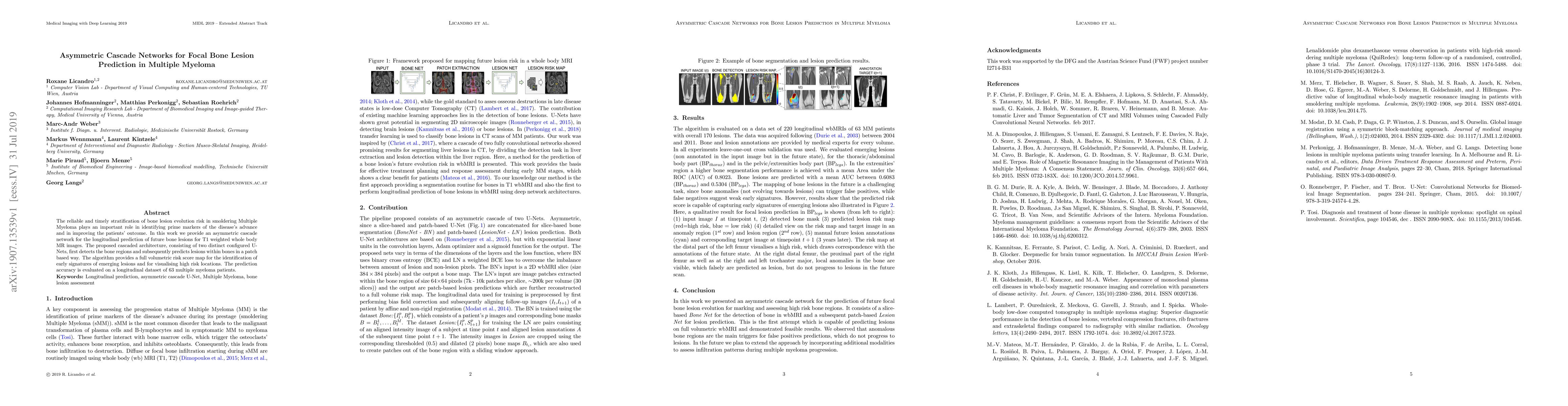
The reliable and timely stratification of bone lesion evolution risk in smoldering Multiple Myeloma plays an important role in identifying prime markers of the disease's advance and in improving the...
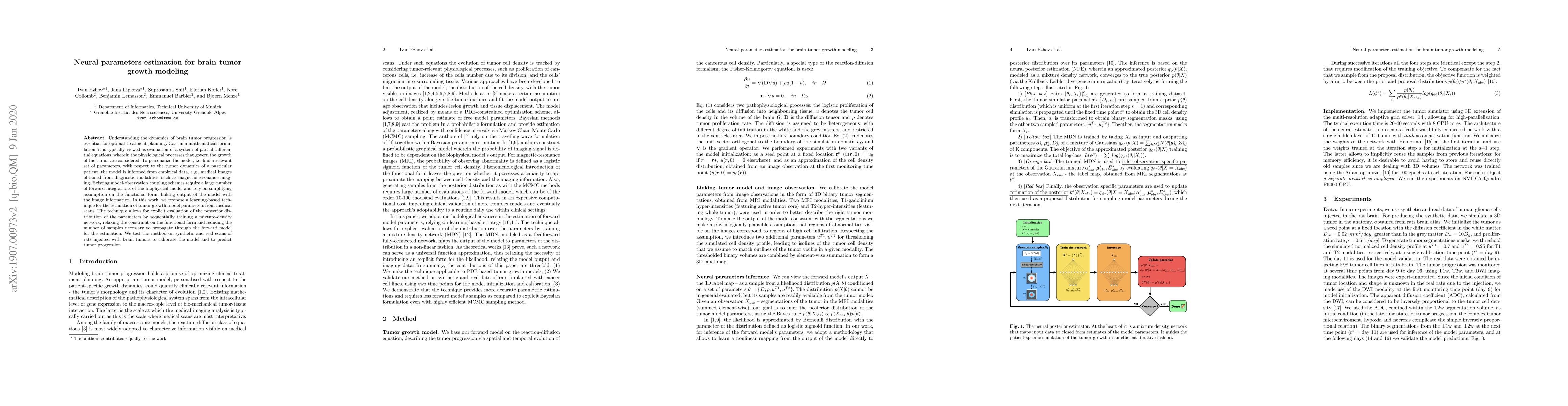
Understanding the dynamics of brain tumor progression is essential for optimal treatment planning. Cast in a mathematical formulation, it is typically viewed as evaluation of a system of partial dif...
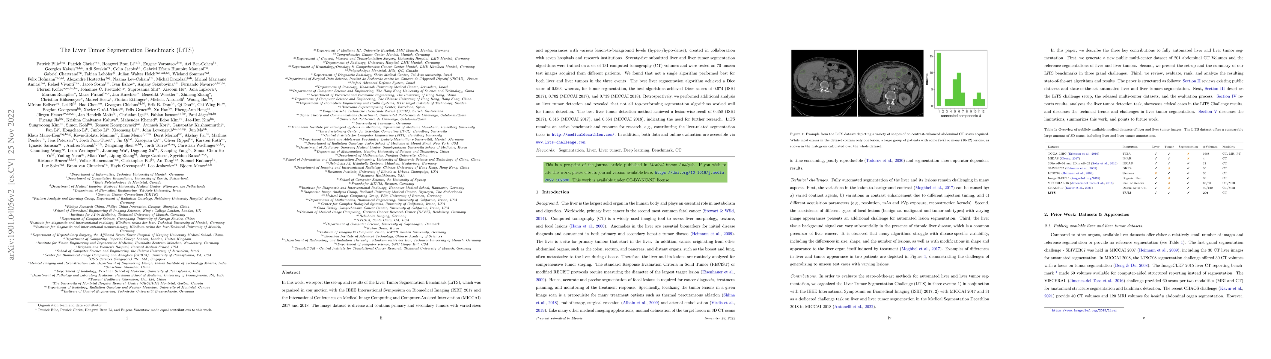
In this work, we report the set-up and results of the Liver Tumor Segmentation Benchmark (LiTS), which was organized in conjunction with the IEEE International Symposium on Biomedical Imaging (ISBI)...
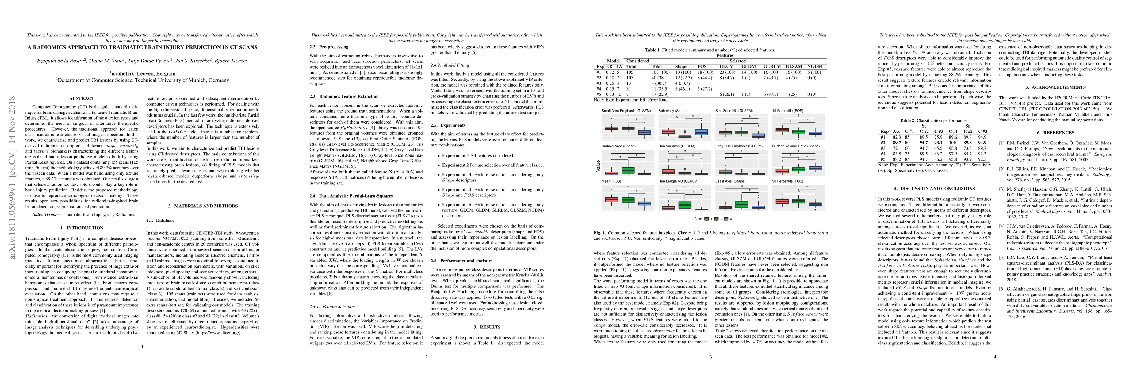
Computer Tomography (CT) is the gold standard technique for brain damage evaluation after acute Traumatic Brain Injury (TBI). It allows identification of most lesion types and determines the need of...
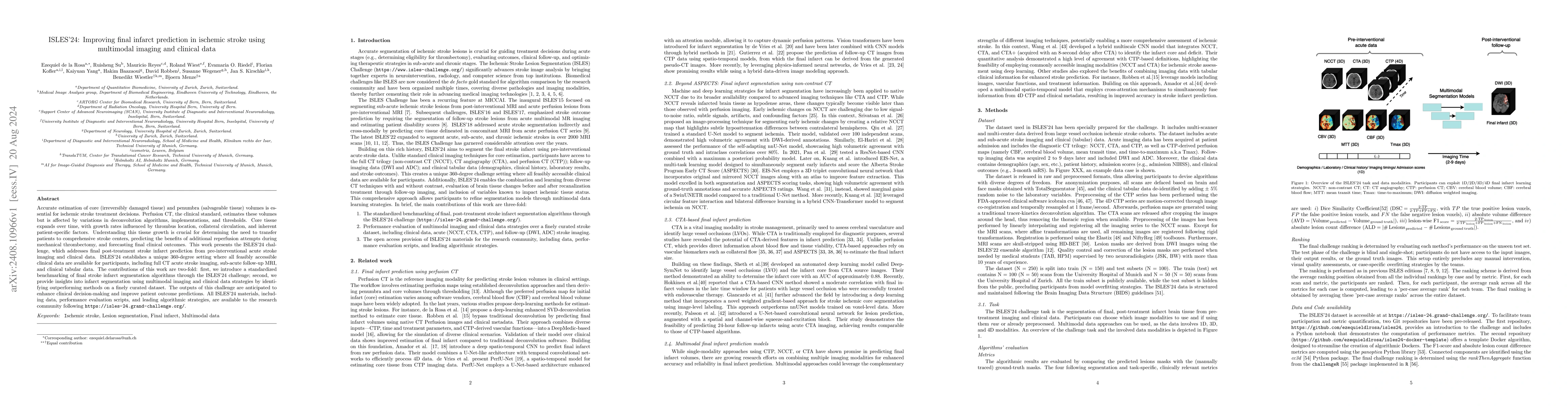
Accurate estimation of core (irreversibly damaged tissue) and penumbra (salvageable tissue) volumes is essential for ischemic stroke treatment decisions. Perfusion CT, the clinical standard, estimates...
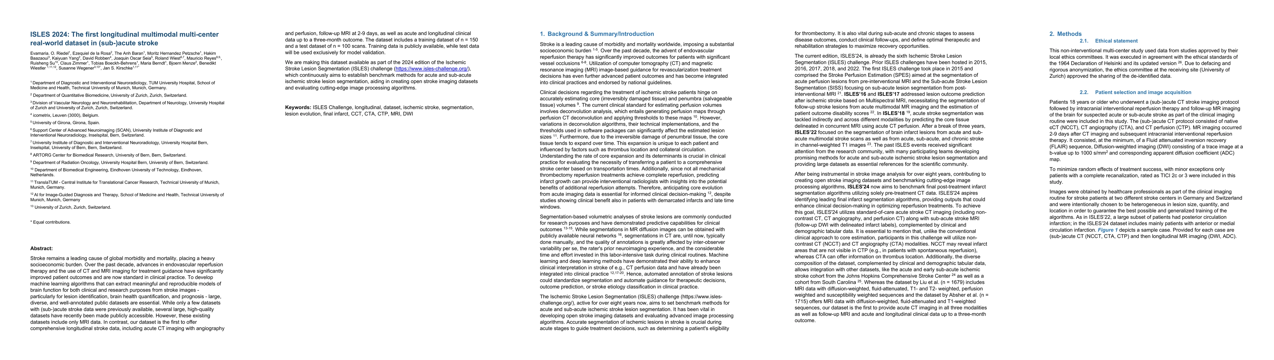
Stroke remains a leading cause of global morbidity and mortality, placing a heavy socioeconomic burden. Over the past decade, advances in endovascular reperfusion therapy and the use of CT and MRI ima...
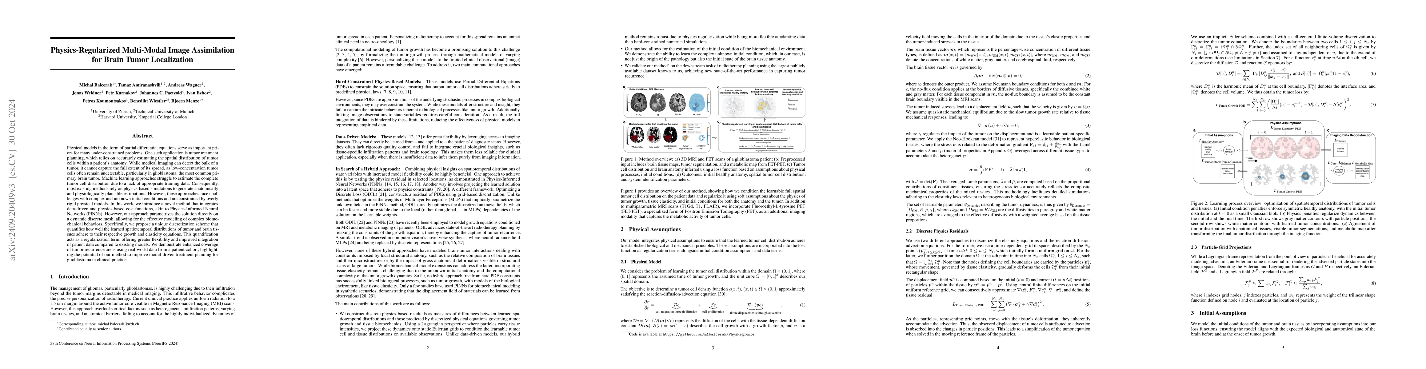
Physical models in the form of partial differential equations represent an important prior for many under-constrained problems. One example is tumor treatment planning, which heavily depends on accura...
Early identification of stroke is crucial for intervention, requiring reliable models. We proposed an efficient retinal image representation together with clinical information to capture a comprehensi...
Accurate prediction of cardiovascular diseases remains imperative for early diagnosis and intervention, necessitating robust and precise predictive models. Recently, there has been a growing interest ...
Predicting stroke risk is a complex challenge that can be enhanced by integrating diverse clinically available data modalities. This study introduces a self-supervised multimodal framework that combin...
Segmenting 3D blood vessels is a critical yet challenging task in medical image analysis. This is due to significant imaging modality-specific variations in artifacts, vascular patterns and scales, si...
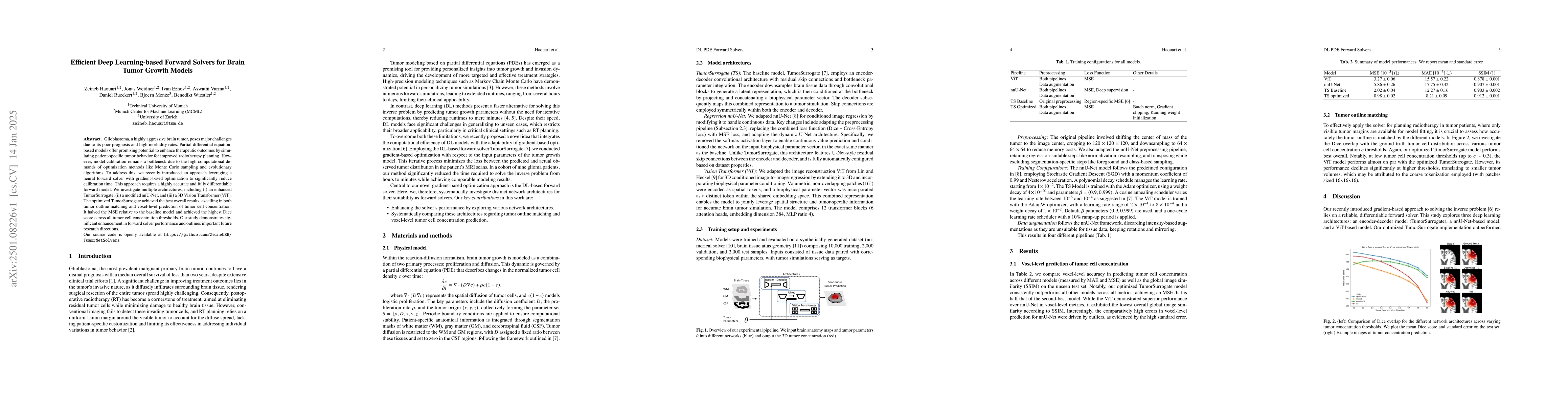
Glioblastoma, a highly aggressive brain tumor, poses major challenges due to its poor prognosis and high morbidity rates. Partial differential equation-based models offer promising potential to enhanc...
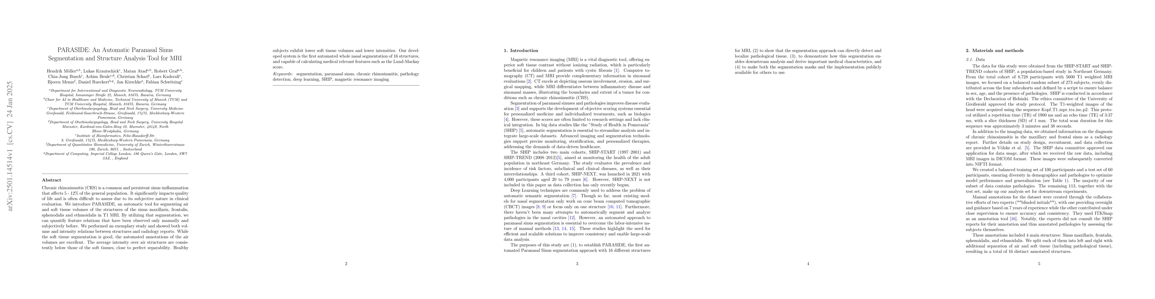
Chronic rhinosinusitis (CRS) is a common and persistent sinus imflammation that affects 5 - 12\% of the general population. It significantly impacts quality of life and is often difficult to assess du...
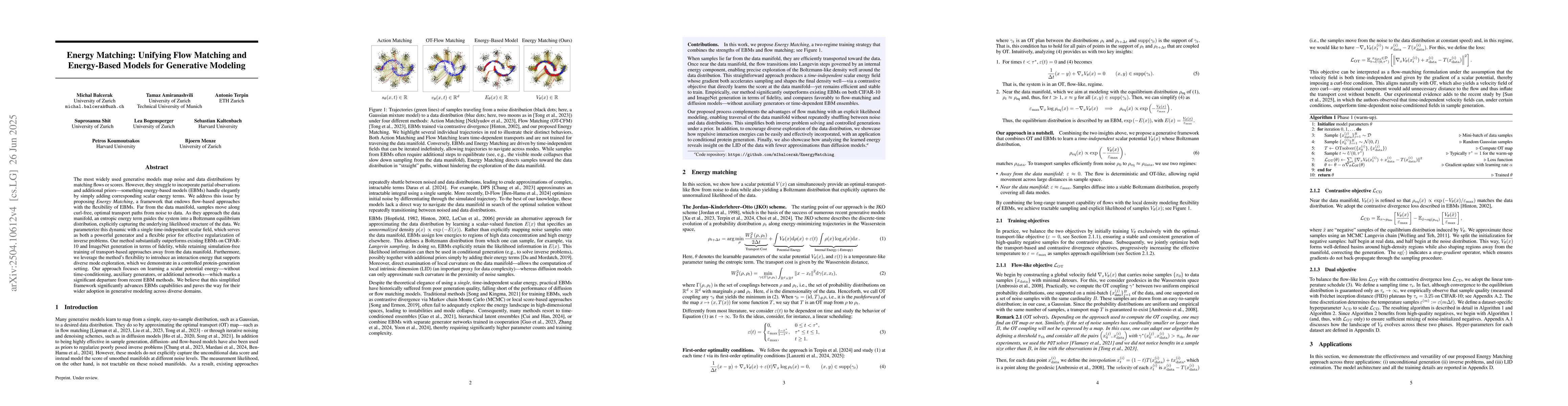
Generative models often map noise to data by matching flows or scores, but these approaches become cumbersome for incorporating partial observations or additional priors. Inspired by recent advances i...
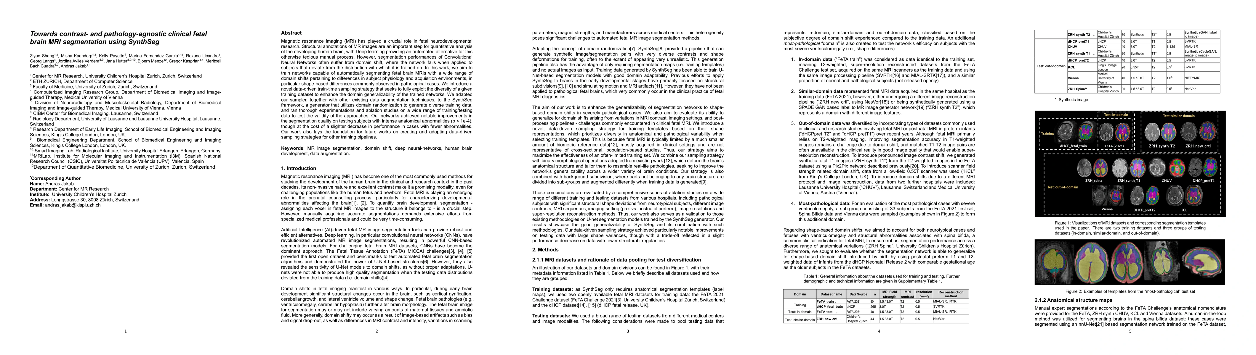
Magnetic resonance imaging (MRI) has played a crucial role in fetal neurodevelopmental research. Structural annotations of MR images are an important step for quantitative analysis of the developing h...
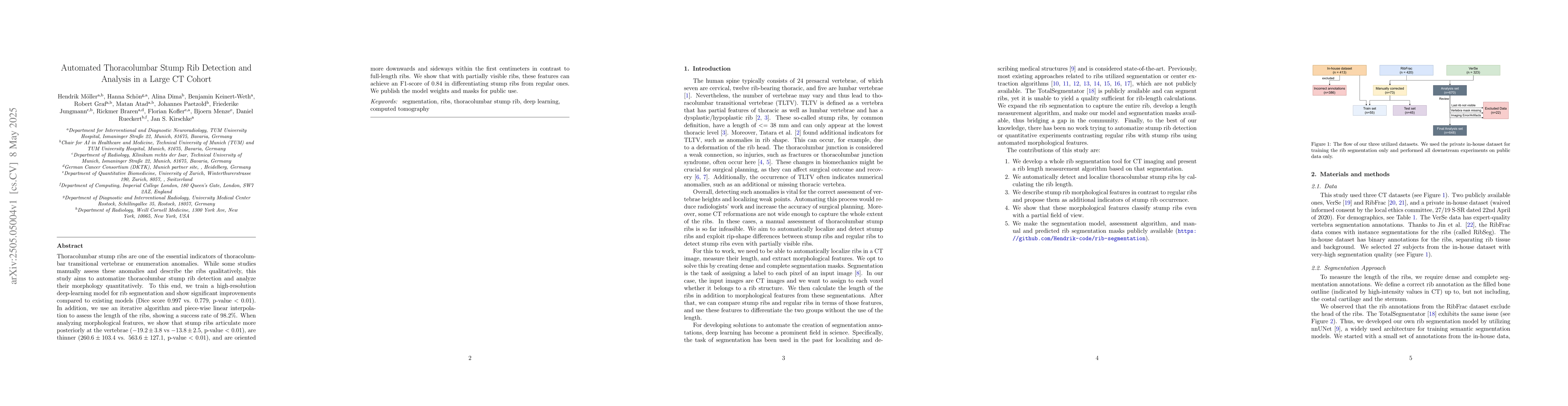
Thoracolumbar stump ribs are one of the essential indicators of thoracolumbar transitional vertebrae or enumeration anomalies. While some studies manually assess these anomalies and describe the ribs ...
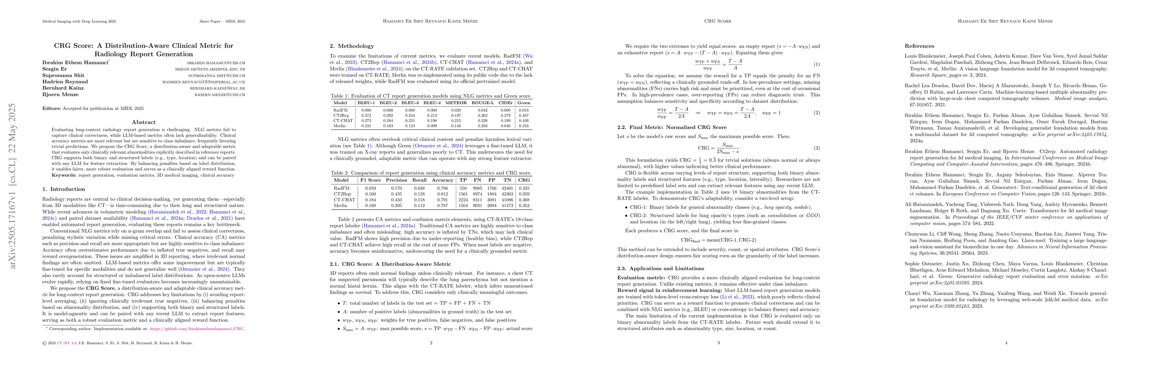
Evaluating long-context radiology report generation is challenging. NLG metrics fail to capture clinical correctness, while LLM-based metrics often lack generalizability. Clinical accuracy metrics are...
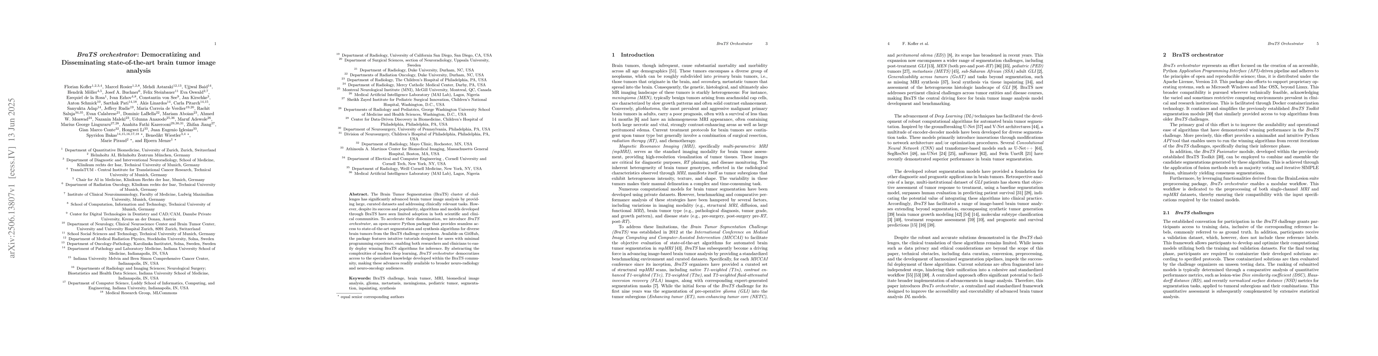
The Brain Tumor Segmentation (BraTS) cluster of challenges has significantly advanced brain tumor image analysis by providing large, curated datasets and addressing clinically relevant tasks. However,...
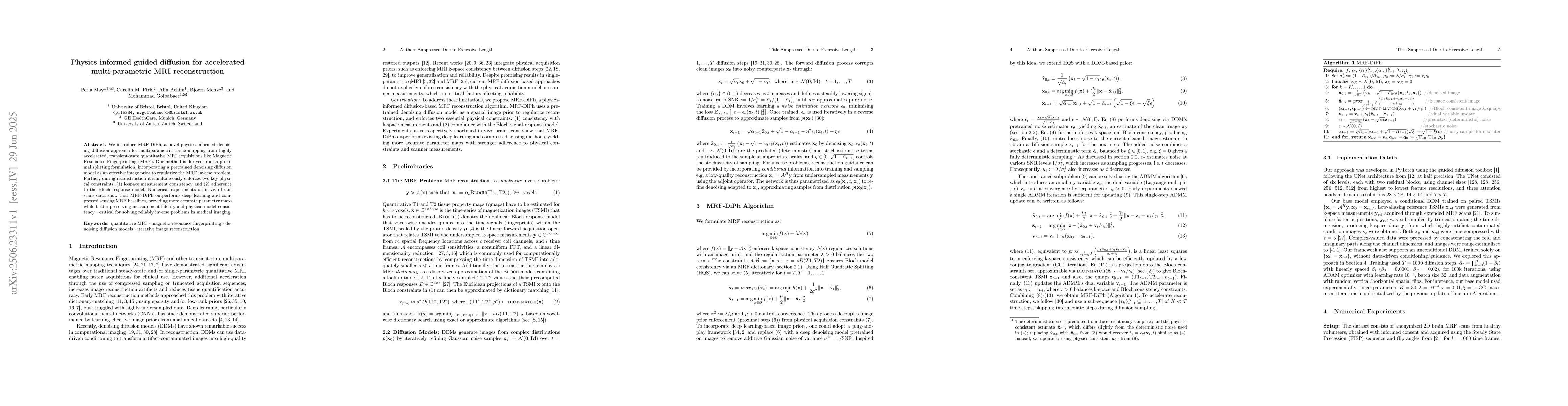
We introduce MRF-DiPh, a novel physics informed denoising diffusion approach for multiparametric tissue mapping from highly accelerated, transient-state quantitative MRI acquisitions like Magnetic Res...

Vessels are complex structures in the body that have been studied extensively in multiple representations. While voxelization is the most common of them, meshes and parametric models are critical in v...
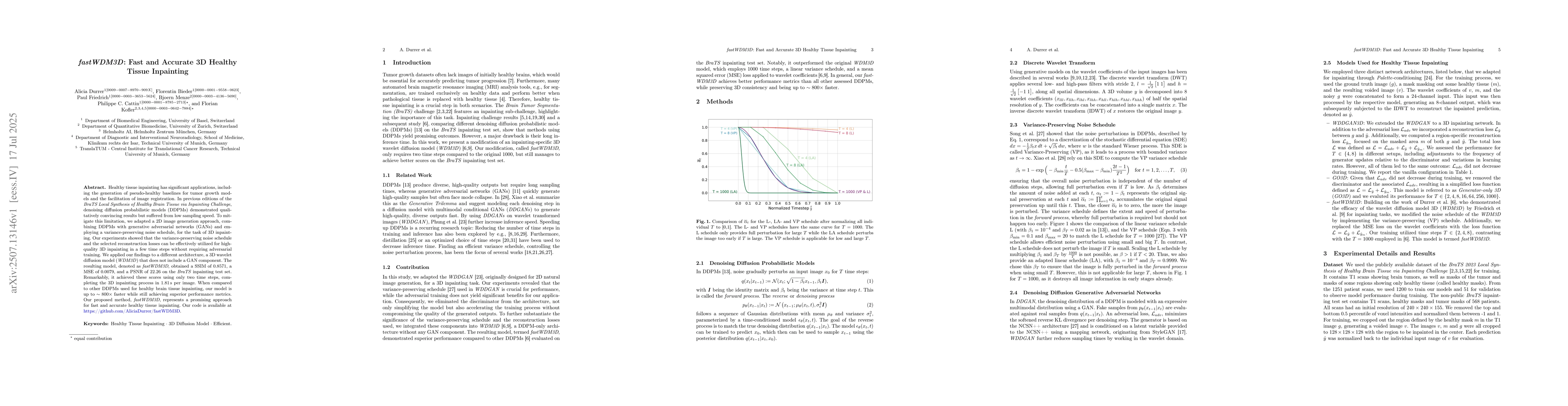
Healthy tissue inpainting has significant applications, including the generation of pseudo-healthy baselines for tumor growth models and the facilitation of image registration. In previous editions of...
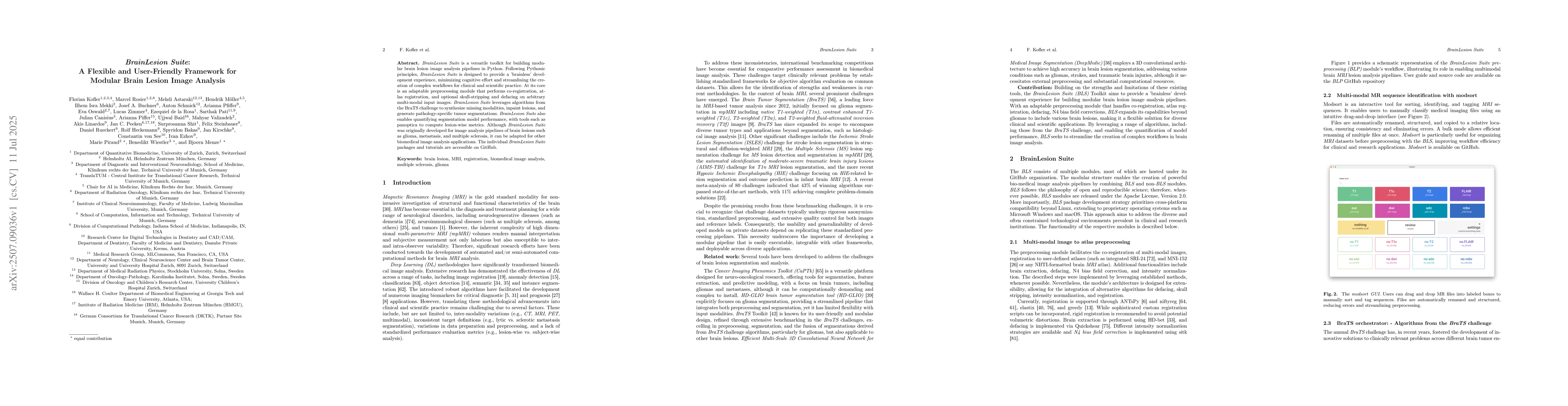
BrainLesion Suite is a versatile toolkit for building modular brain lesion image analysis pipelines in Python. Following Pythonic principles, BrainLesion Suite is designed to provide a 'brainless' dev...
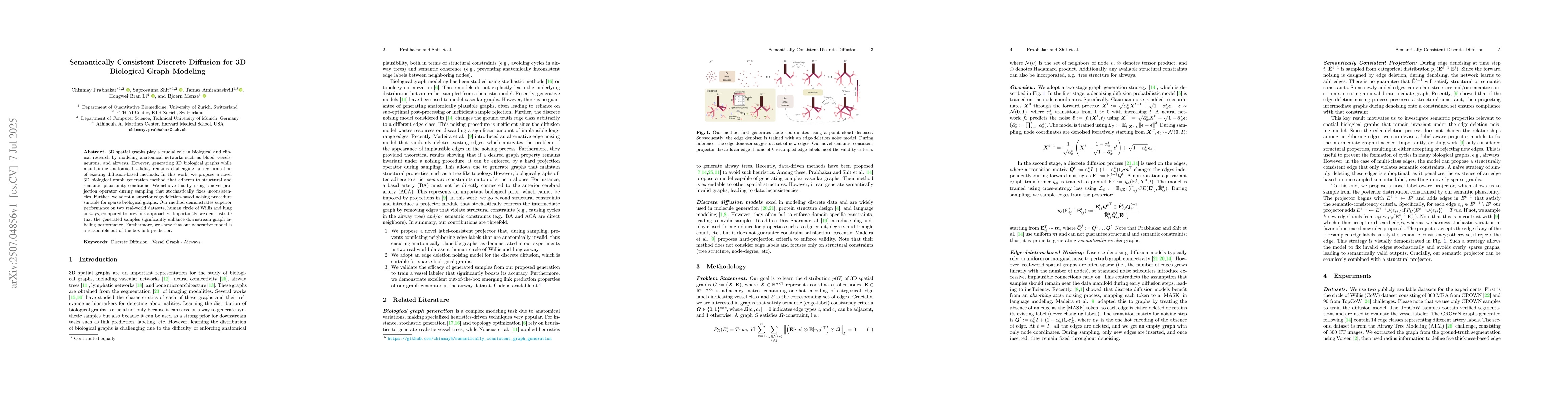
3D spatial graphs play a crucial role in biological and clinical research by modeling anatomical networks such as blood vessels,neurons, and airways. However, generating 3D biological graphs while mai...
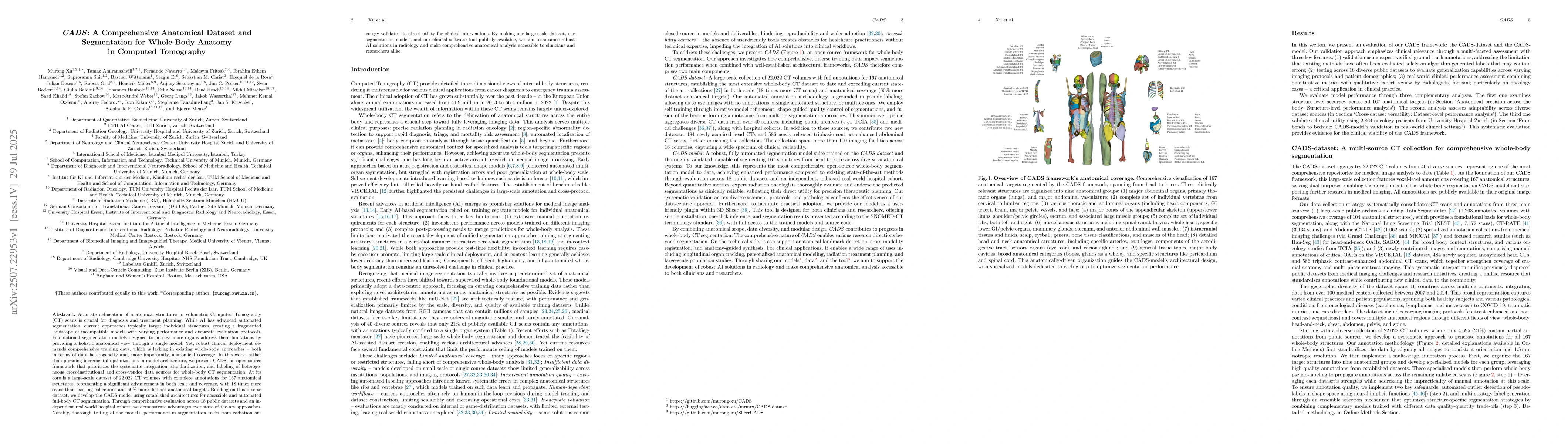
Accurate delineation of anatomical structures in volumetric CT scans is crucial for diagnosis and treatment planning. While AI has advanced automated segmentation, current approaches typically target ...
We present MInDI-3D (Medical Inversion by Direct Iteration in 3D), the first 3D conditional diffusion-based model for real-world sparse-view Cone Beam Computed Tomography (CBCT) artefact removal, aimi...
Early tumor detection save lives. Each year, more than 300 million computed tomography (CT) scans are performed worldwide, offering a vast opportunity for effective cancer screening. However, detectin...
The Circle of Willis (CoW) is a critical network of arteries in the brain, often implicated in cerebrovascular pathologies. Voxel-level segmentation is an important first step toward an automated CoW ...
Most current medical vision language models struggle to jointly generate diagnostic text and pixel-level segmentation masks in response to complex visual questions. This represents a major limitation ...
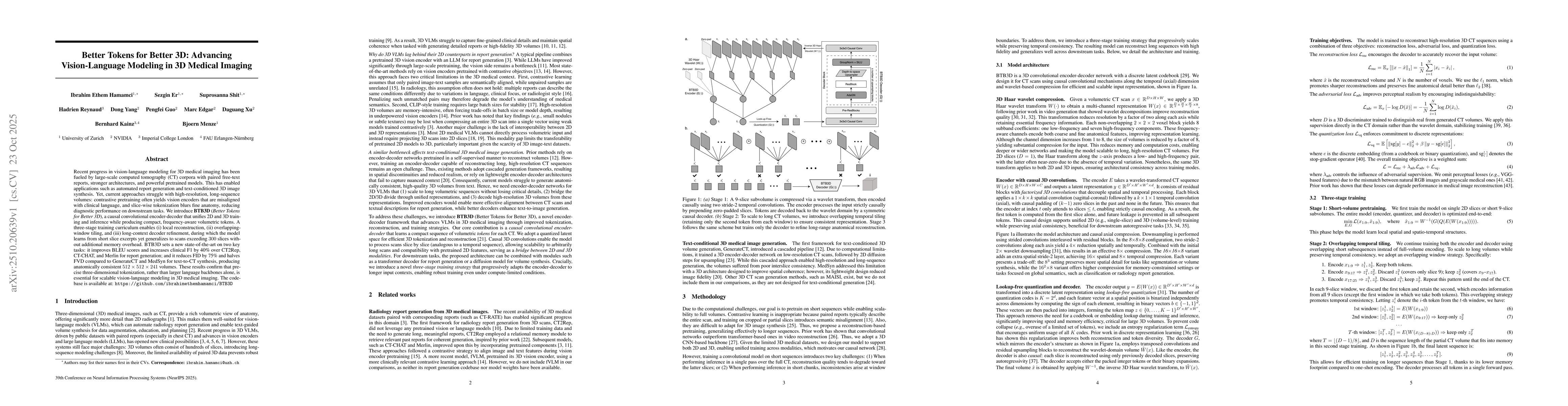
Recent progress in vision-language modeling for 3D medical imaging has been fueled by large-scale computed tomography (CT) corpora with paired free-text reports, stronger architectures, and powerful p...
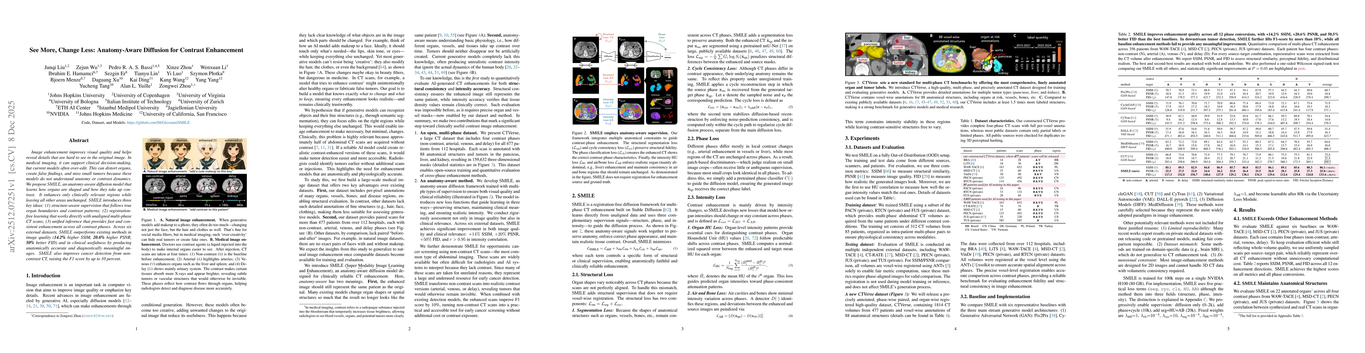
Image enhancement improves visual quality and helps reveal details that are hard to see in the original image. In medical imaging, it can support clinical decision-making, but current models often ove...
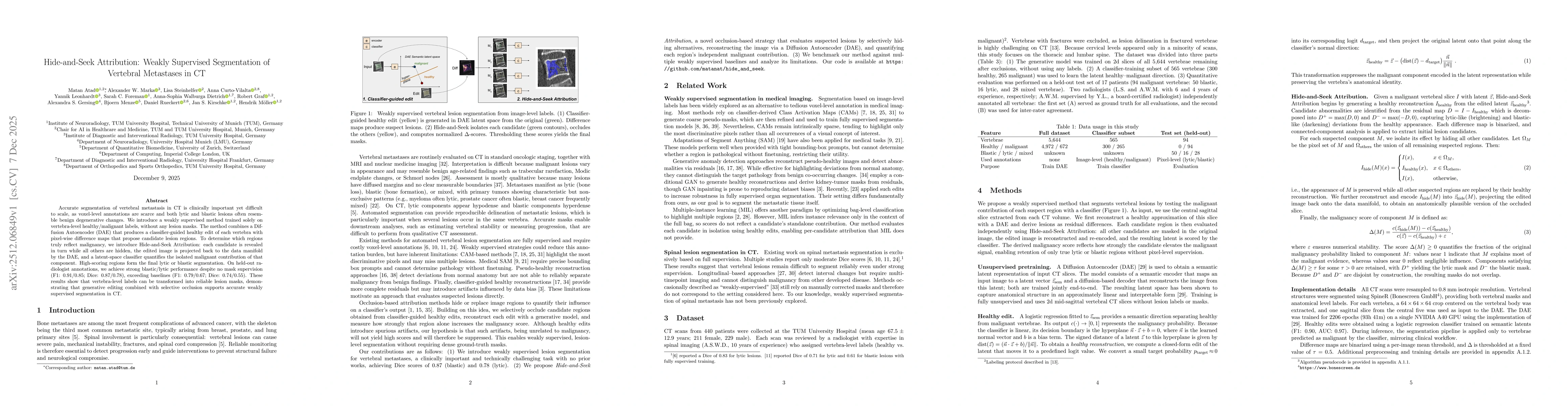
Accurate segmentation of vertebral metastasis in CT is clinically important yet difficult to scale, as voxel-level annotations are scarce and both lytic and blastic lesions often resemble benign degen...
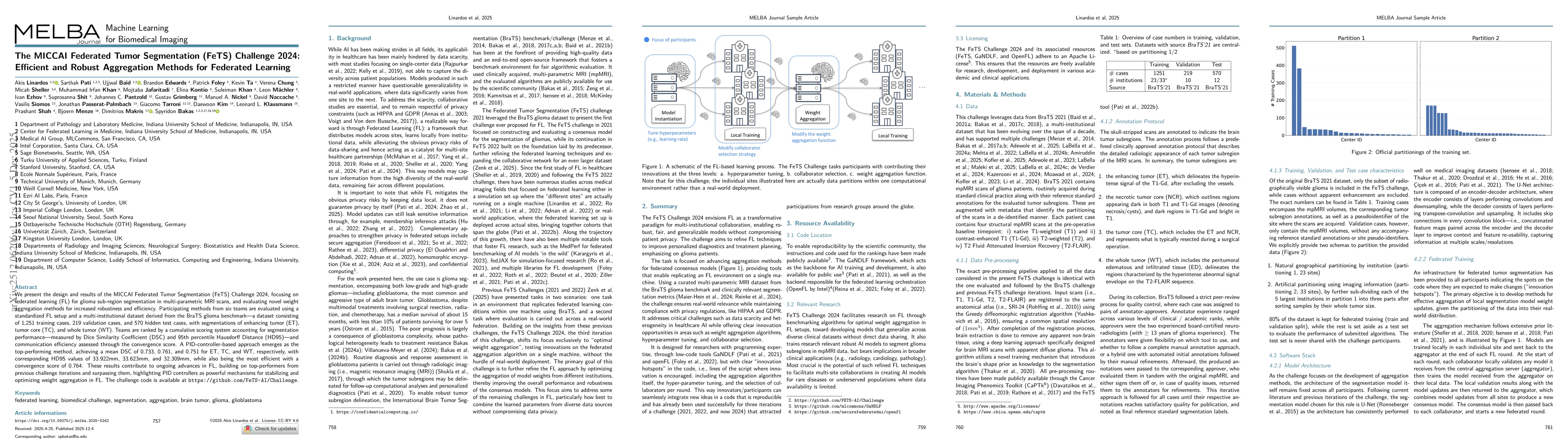
We present the design and results of the MICCAI Federated Tumor Segmentation (FeTS) Challenge 2024, which focuses on federated learning (FL) for glioma sub-region segmentation in multi-parametric MRI ...
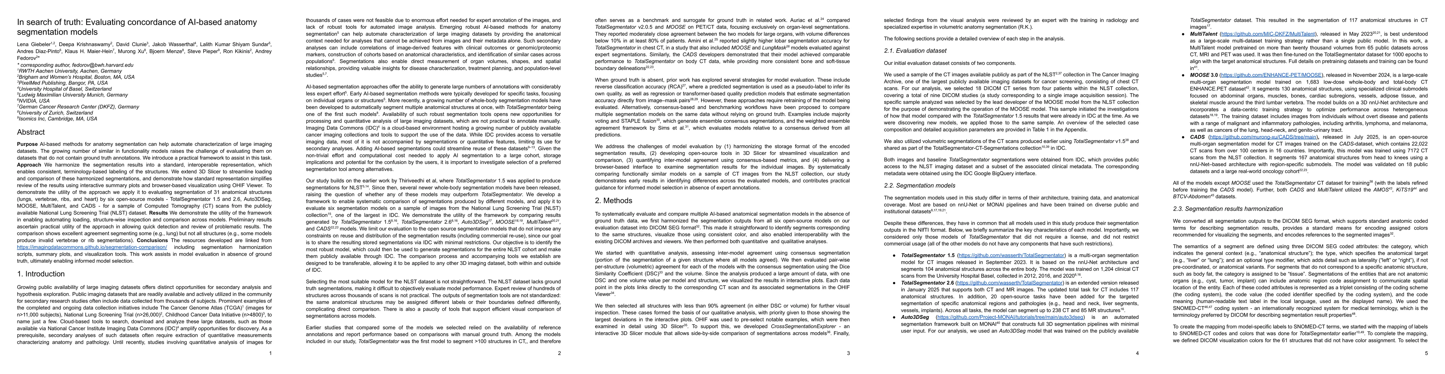
Purpose AI-based methods for anatomy segmentation can help automate characterization of large imaging datasets. The growing number of similar in functionality models raises the challenge of evaluating...
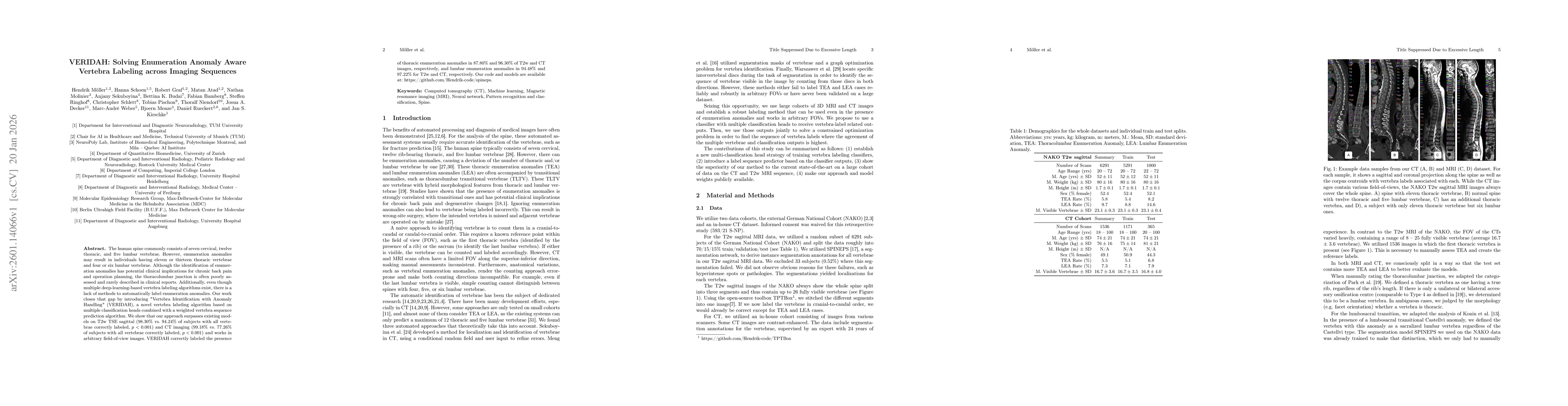
The human spine commonly consists of seven cervical, twelve thoracic, and five lumbar vertebrae. However, enumeration anomalies may result in individuals having eleven or thirteen thoracic vertebrae a...
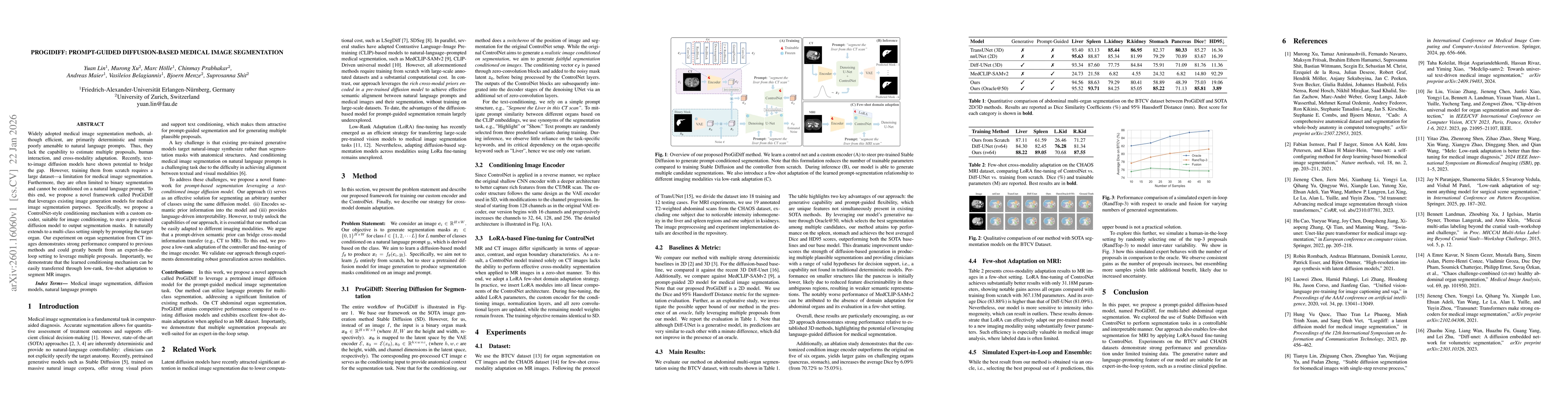
Widely adopted medical image segmentation methods, although efficient, are primarily deterministic and remain poorly amenable to natural language prompts. Thus, they lack the capability to estimate mu...
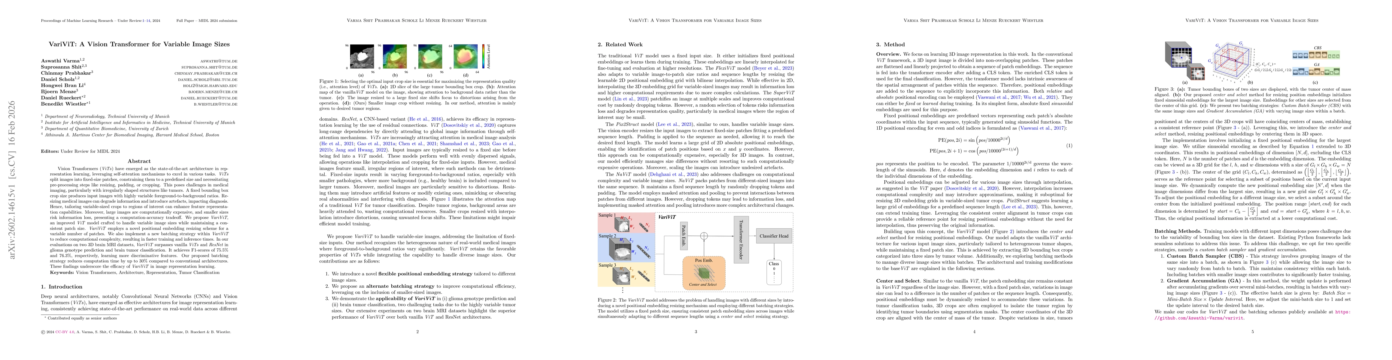
Vision Transformers (ViTs) have emerged as the state-of-the-art architecture in representation learning, leveraging self-attention mechanisms to excel in various tasks. ViTs split images into fixed-si...
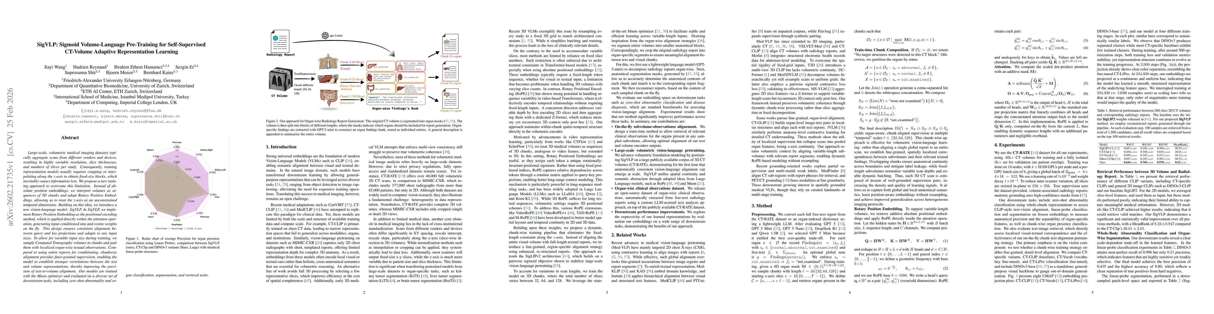
Large-scale, volumetric medical imaging datasets typically aggregate scans from different vendors and devices, resulting in highly variable resolution, slice thicknesses, and numbers of slices per stu...
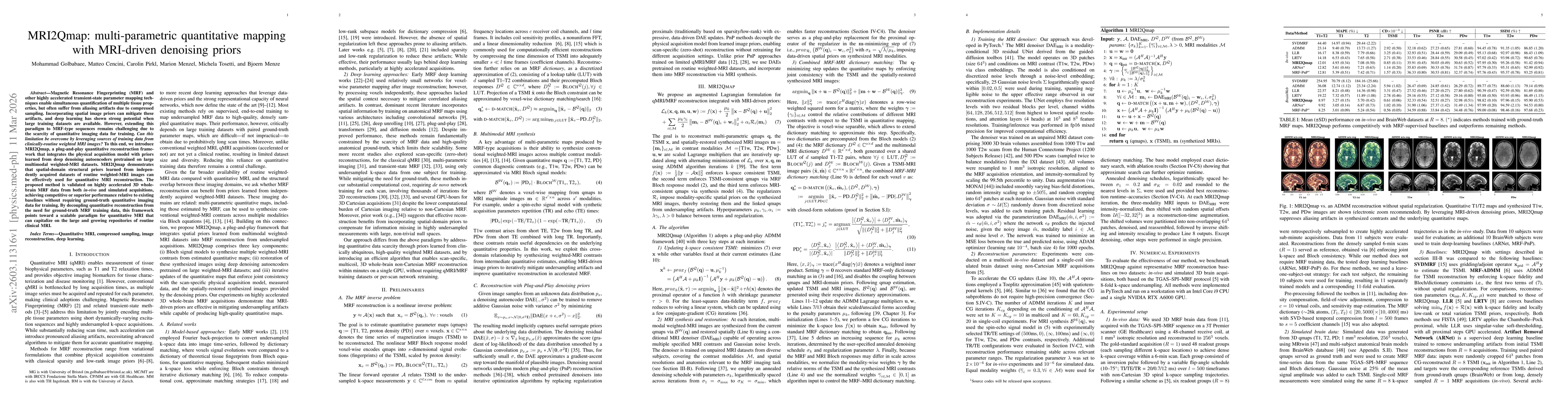
Magnetic Resonance Fingerprinting (MRF) and other highly accelerated transient-state parameter mapping techniques enable simultaneous quantification of multiple tissue properties, but often suffer fro...
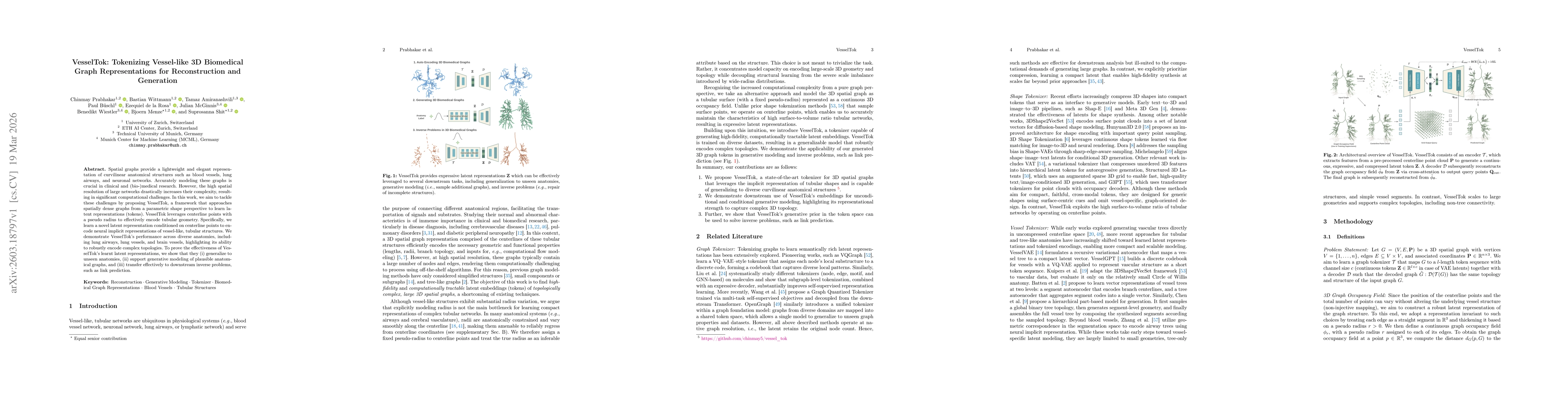
Spatial graphs provide a lightweight and elegant representation of curvilinear anatomical structures such as blood vessels, lung airways, and neuronal networks. Accurately modeling these graphs is cru...
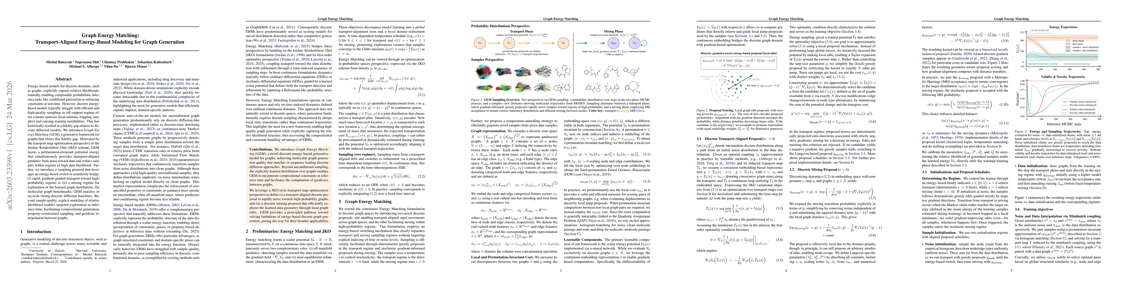
Energy-based models for discrete domains, such as graphs, explicitly capture relative likelihoods, naturally enabling composable probabilistic inference tasks like conditional generation or enforcing ...
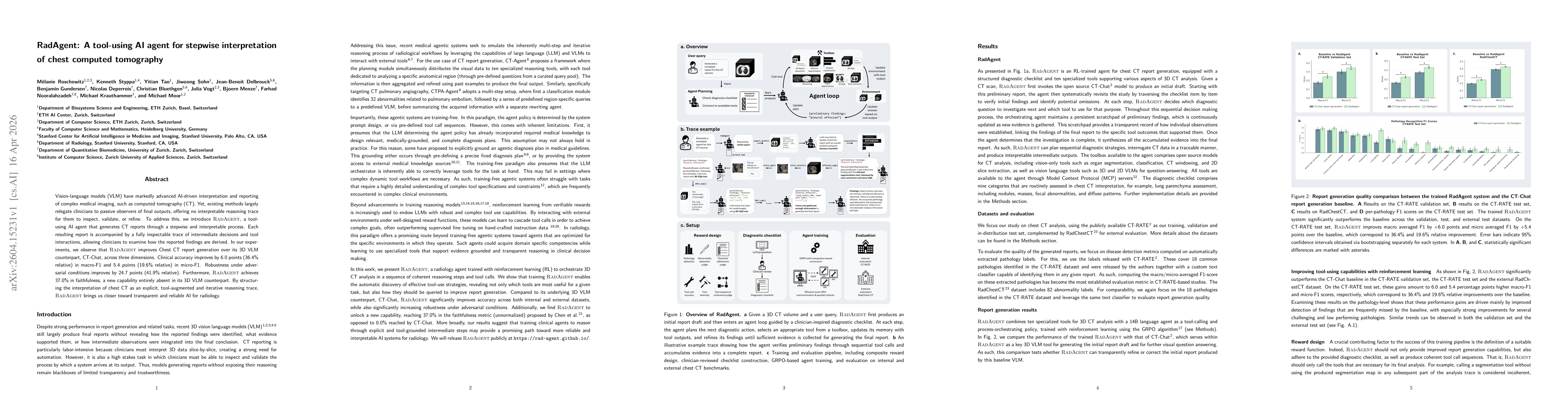
Vision-language models (VLM) have markedly advanced AI-driven interpretation and reporting of complex medical imaging, such as computed tomography (CT). Yet, existing methods largely relegate clinicia...
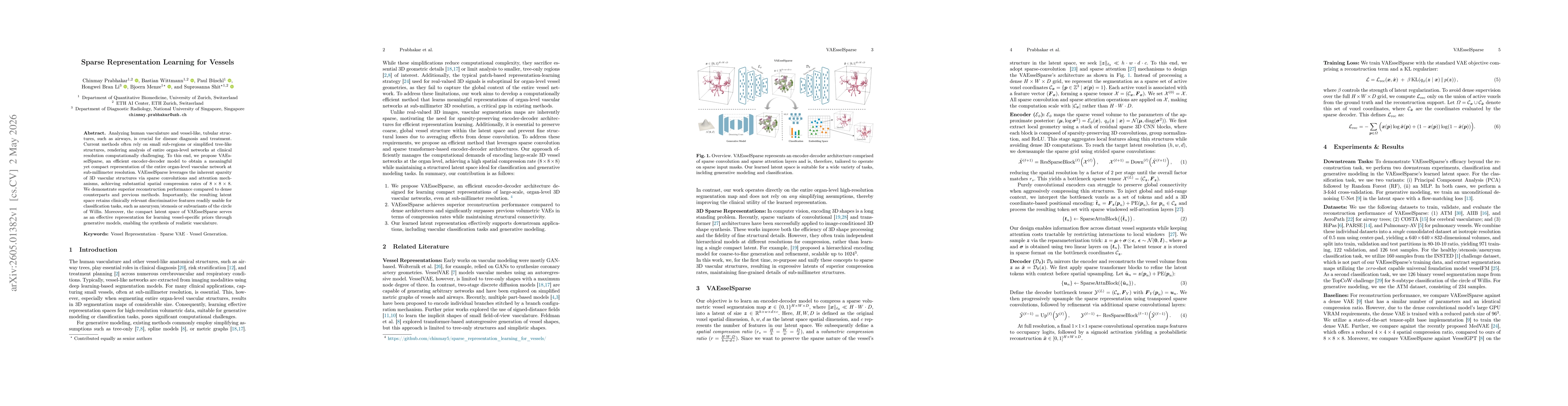
Analyzing human vasculature and vessel-like, tubular structures, such as airways, is crucial for disease diagnosis and treatment. Current methods often rely on small sub-regions or simplified tree-lik...
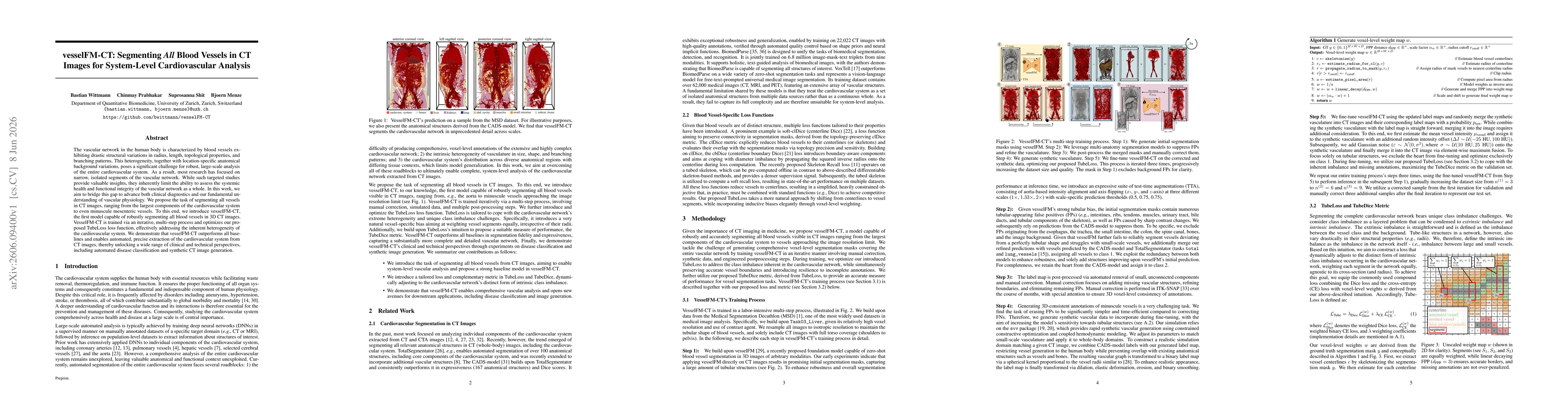
The vascular network in the human body is characterized by blood vessels exhibiting drastic structural variations in radius, length, topological properties, and branching patterns. This heterogeneity,...
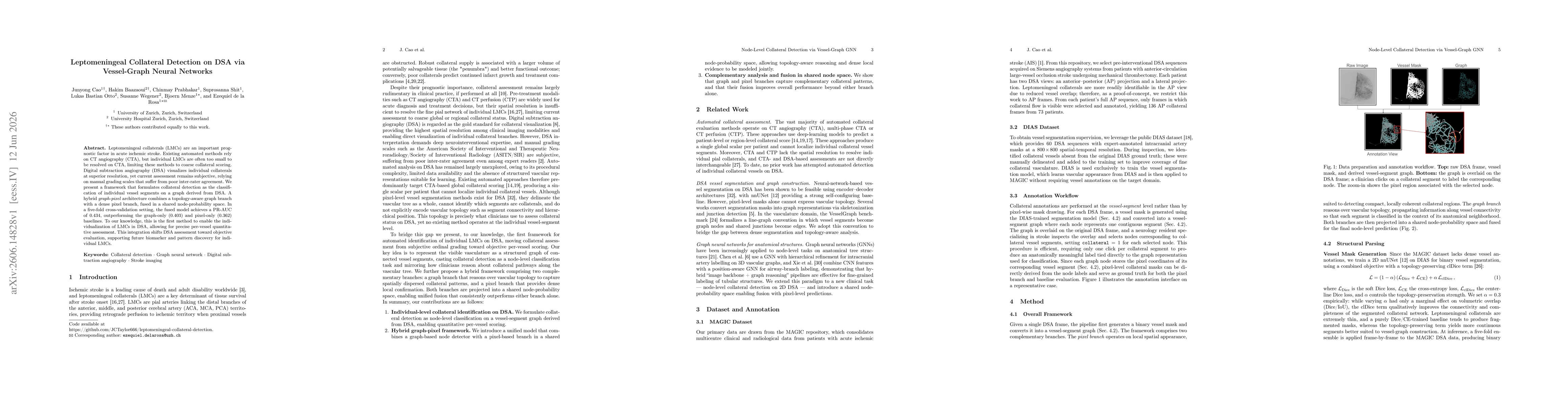
Leptomeningeal collaterals (LMCs) are an important prognostic factor in acute ischemic stroke. Existing automated methods rely on CT angiography (CTA), but individual LMCs are often too small to be re...
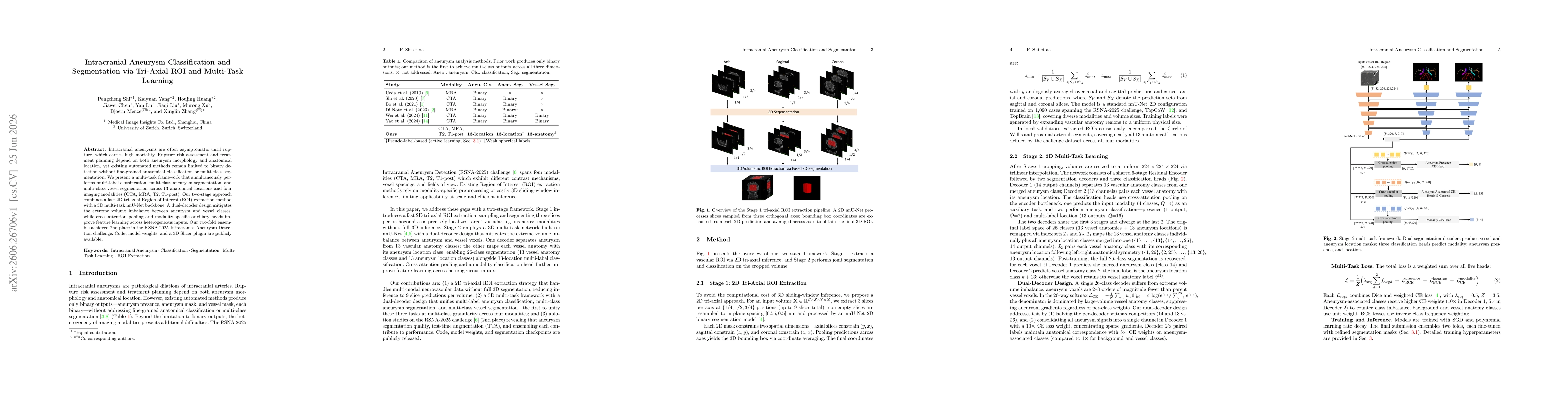
Intracranial aneurysms are often asymptomatic until rupture, which carries high mortality. Rupture risk assessment and treatment planning depend on both aneurysm morphology and anatomical location, ye...
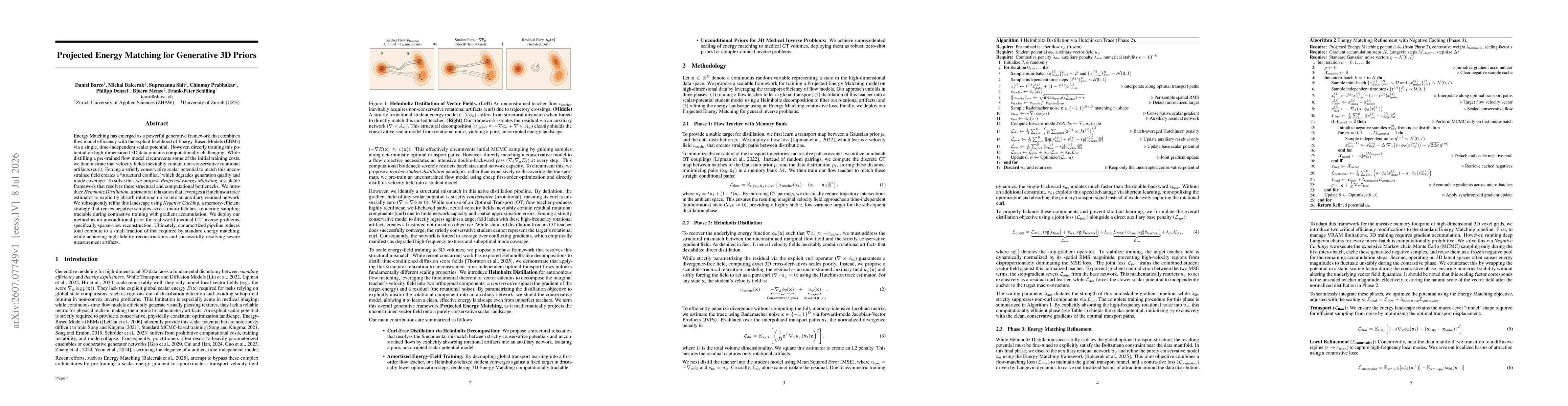
Energy Matching has emerged as a powerful generative framework that combines flow model efficiency with the explicit likelihood of Energy-Based Models (EBMs) via a single, time-independent scalar pote...