Academic Profile
Statistics
Similar Authors
Papers on arXiv
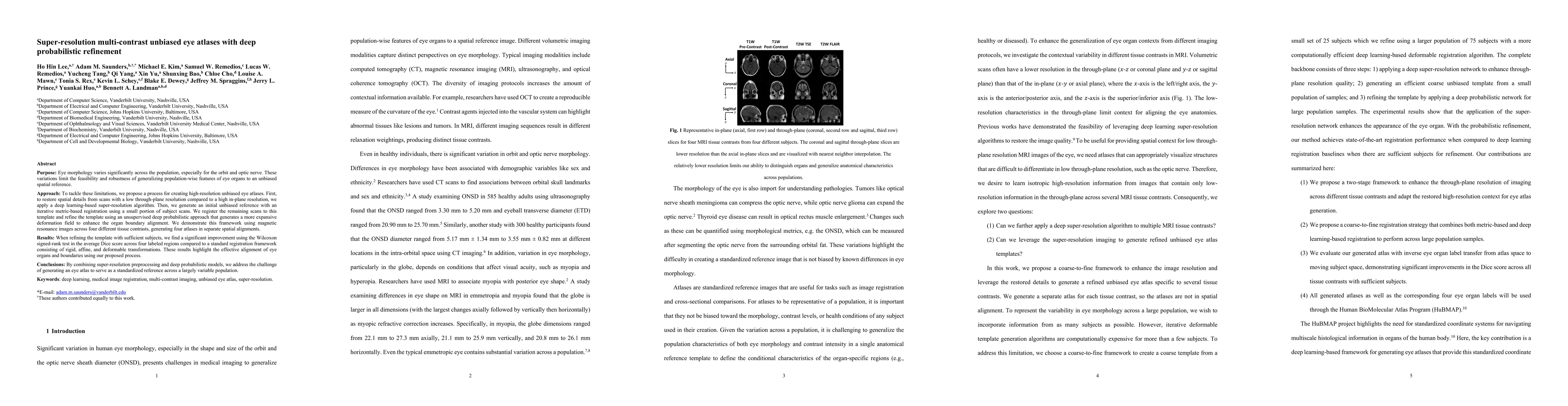
Purpose: Eye morphology varies significantly across the population, especially for the orbit and optic nerve. These variations limit the feasibility and robustness of generalizing population-wise fe...
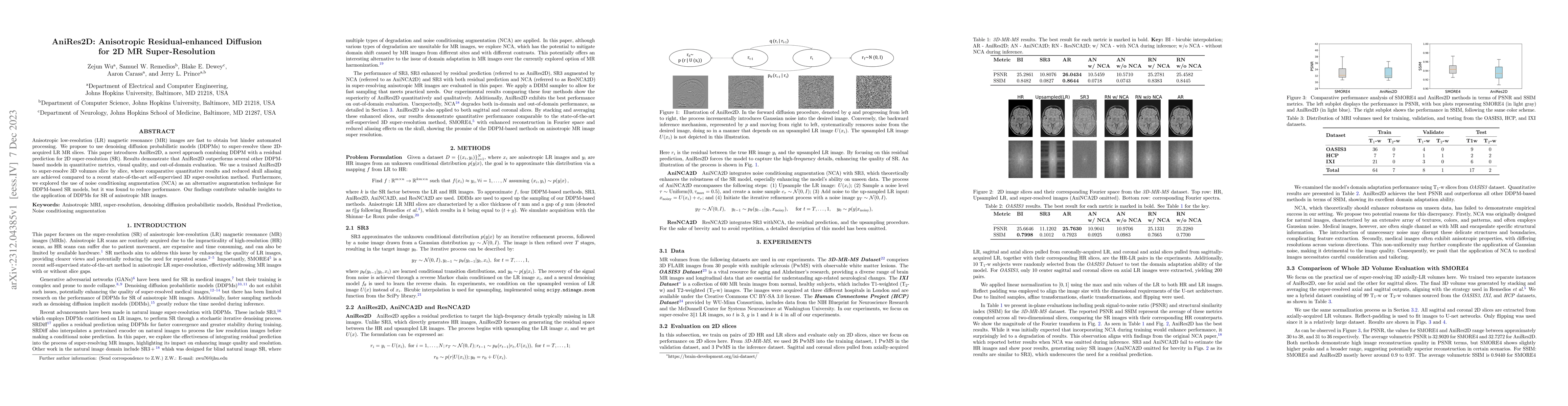
Anisotropic low-resolution (LR) magnetic resonance (MR) images are fast to obtain but hinder automated processing. We propose to use denoising diffusion probabilistic models (DDPMs) to super-resolve...
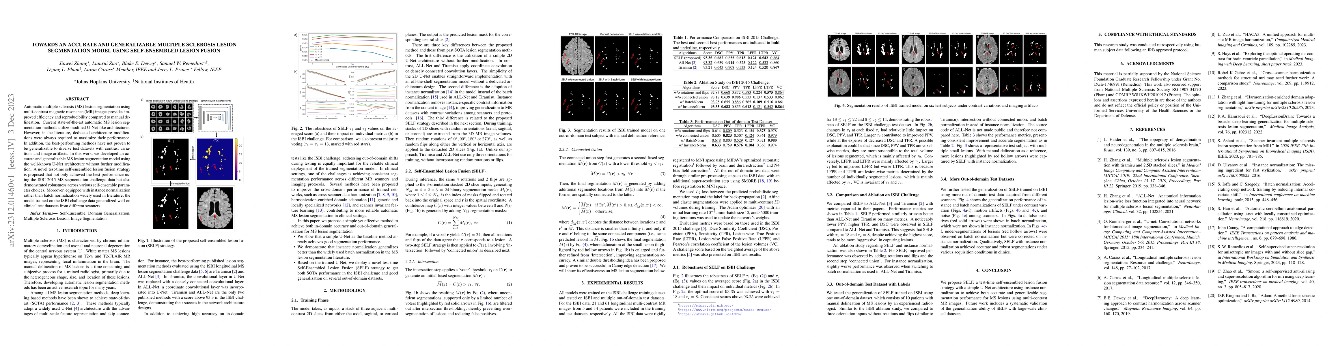
Automatic multiple sclerosis (MS) lesion segmentation using multi-contrast magnetic resonance (MR) images provides improved efficiency and reproducibility compared to manual delineation. Current sta...
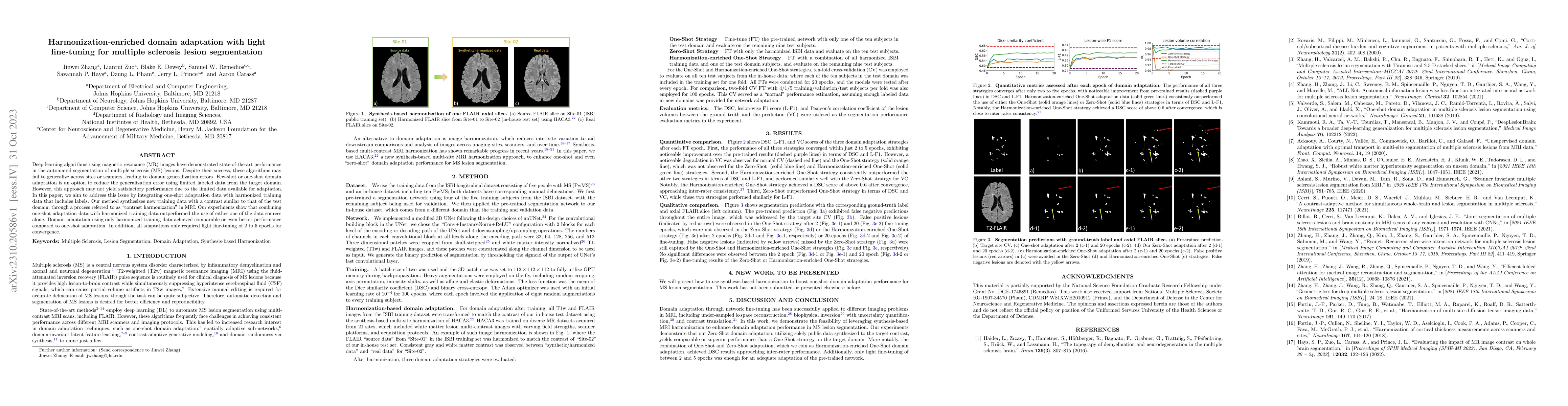
Deep learning algorithms utilizing magnetic resonance (MR) images have demonstrated cutting-edge proficiency in autonomously segmenting multiple sclerosis (MS) lesions. Despite their achievements, t...
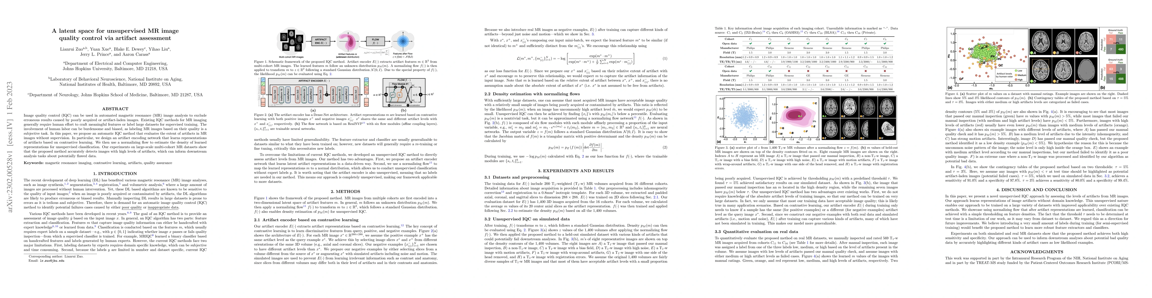
Image quality control (IQC) can be used in automated magnetic resonance (MR) image analysis to exclude erroneous results caused by poorly acquired or artifact-laden images. Existing IQC methods for ...
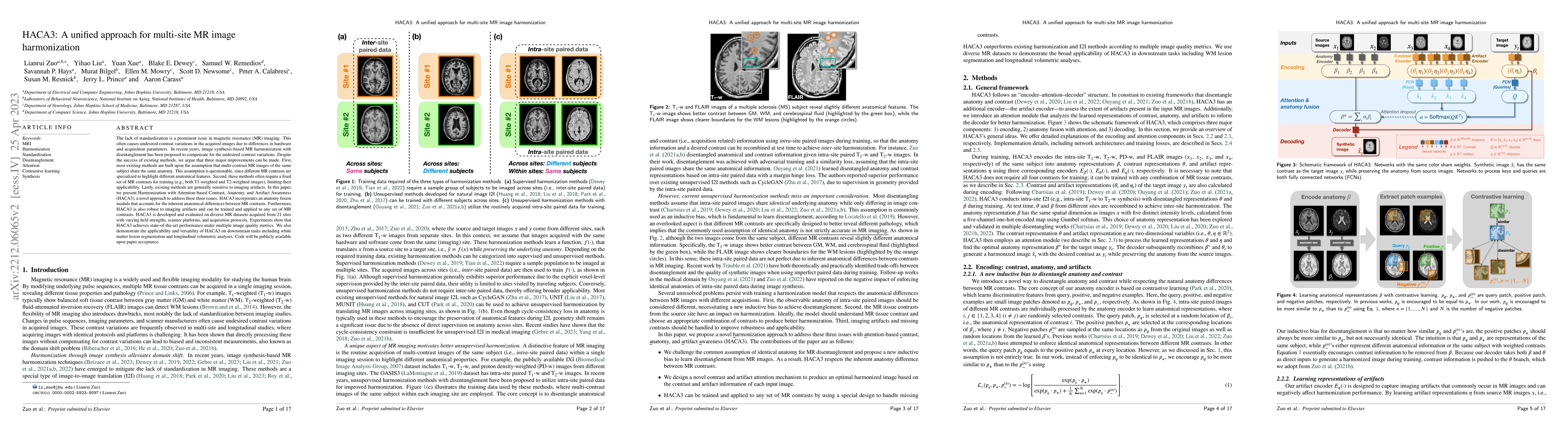
The lack of standardization is a prominent issue in magnetic resonance (MR) imaging. This often causes undesired contrast variations in the acquired images due to differences in hardware and acquisi...
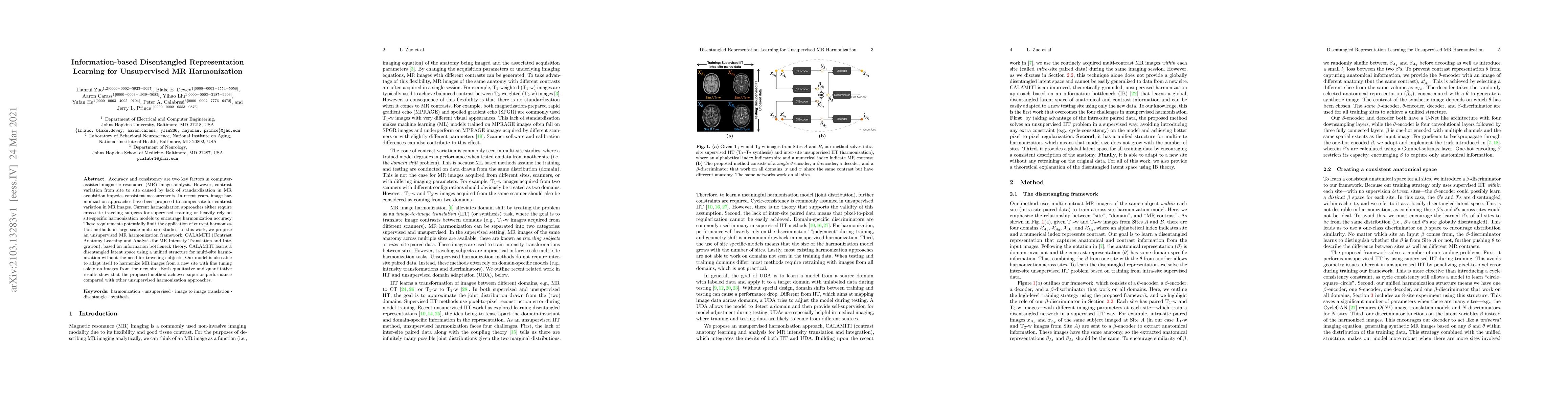
Accuracy and consistency are two key factors in computer-assisted magnetic resonance (MR) image analysis. However, contrast variation from site to site caused by lack of standardization in MR acquis...
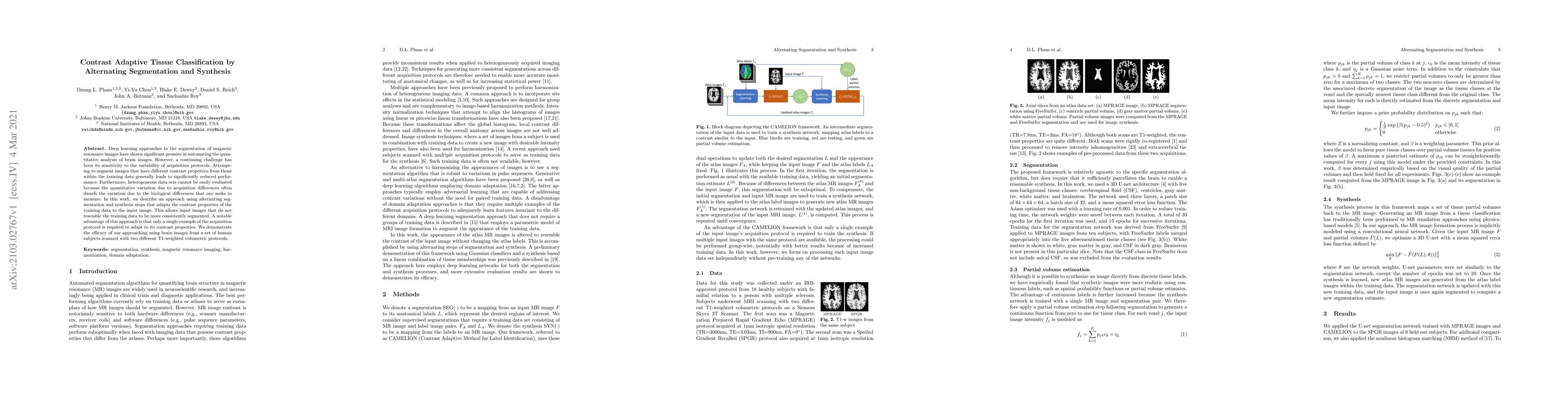
Deep learning approaches to the segmentation of magnetic resonance images have shown significant promise in automating the quantitative analysis of brain images. However, a continuing challenge has ...
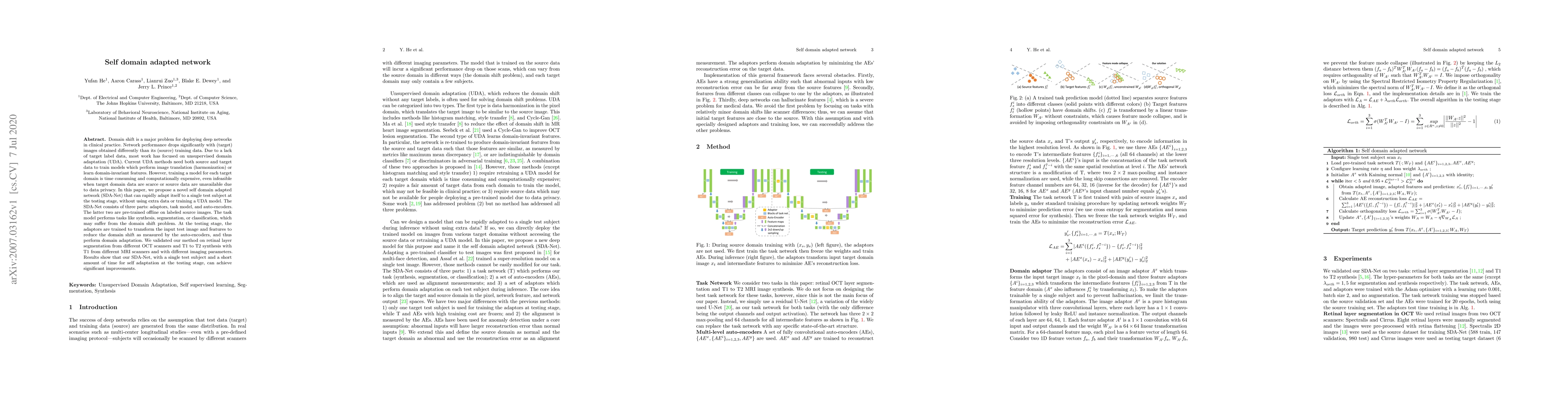
Domain shift is a major problem for deploying deep networks in clinical practice. Network performance drops significantly with (target) images obtained differently than its (source) training data. D...
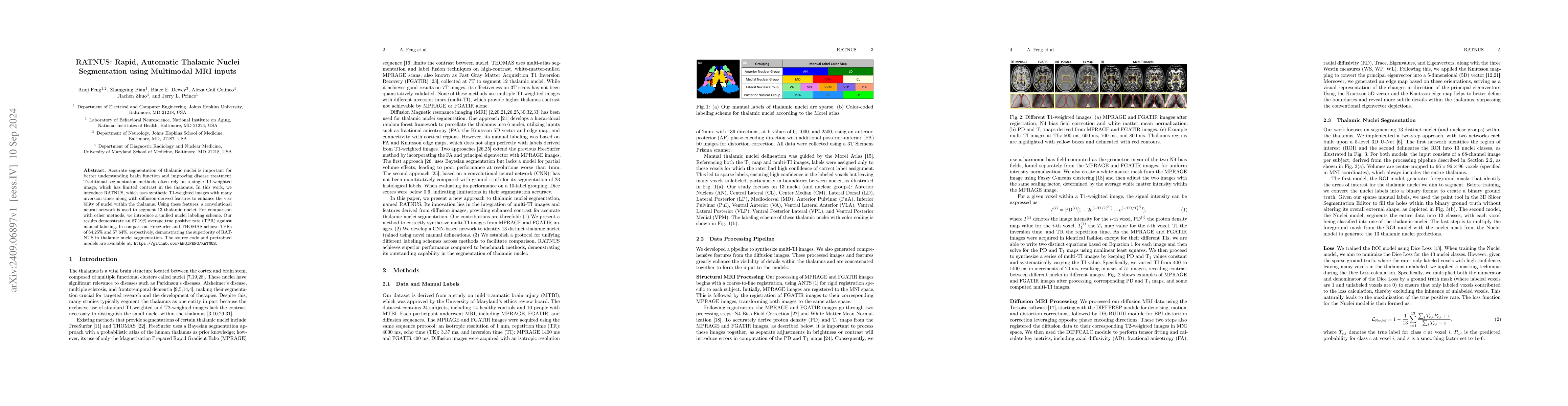
Accurate segmentation of thalamic nuclei is important for better understanding brain function and improving disease treatment. Traditional segmentation methods often rely on a single T1-weighted image...
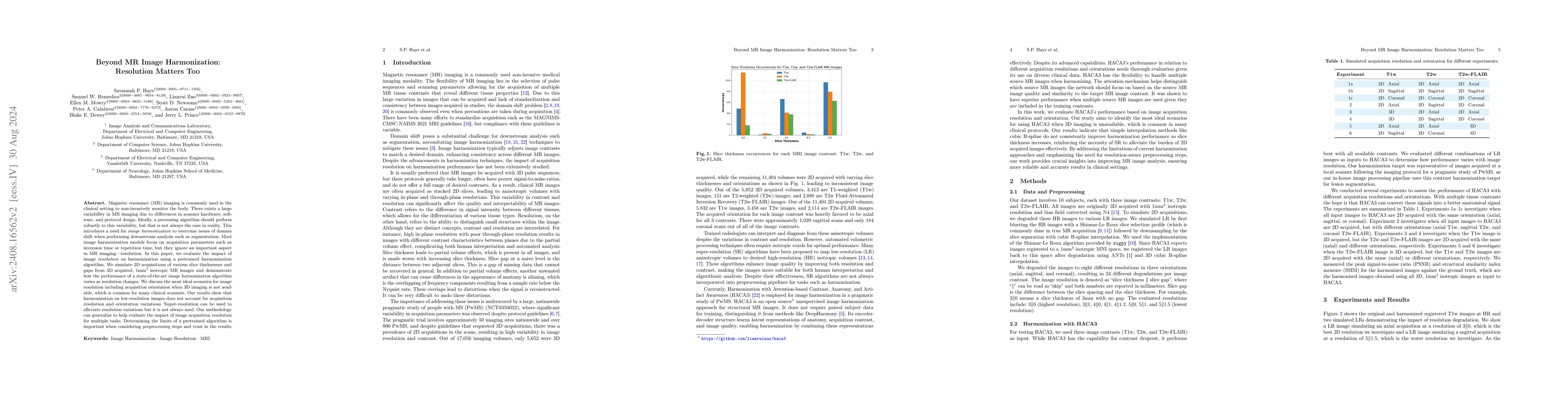
Magnetic resonance (MR) imaging is commonly used in the clinical setting to non-invasively monitor the body. There exists a large variability in MR imaging due to differences in scanner hardware, soft...
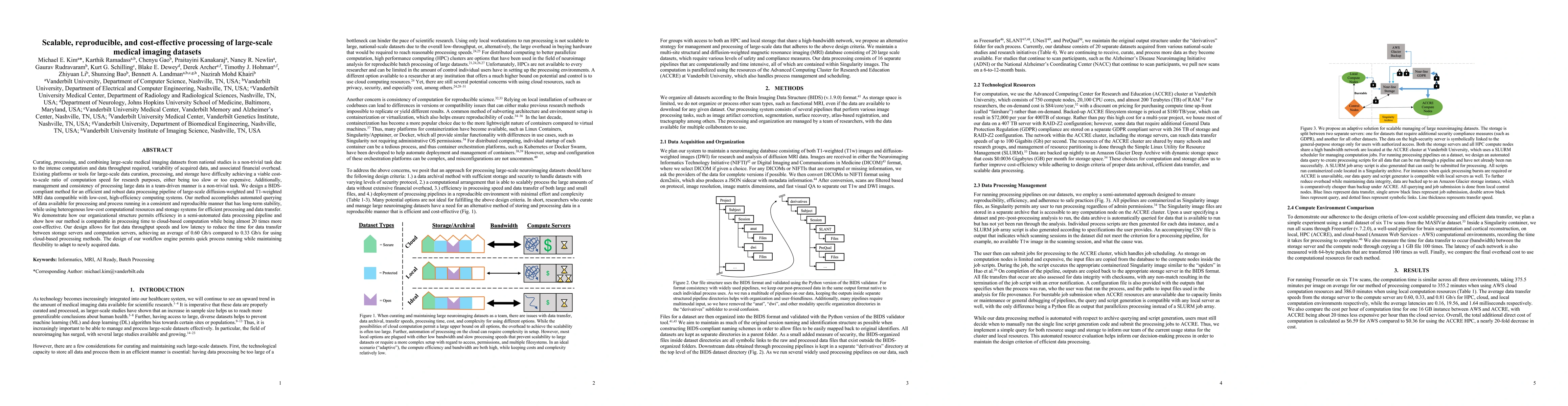
Curating, processing, and combining large-scale medical imaging datasets from national studies is a non-trivial task due to the intense computation and data throughput required, variability of acquire...
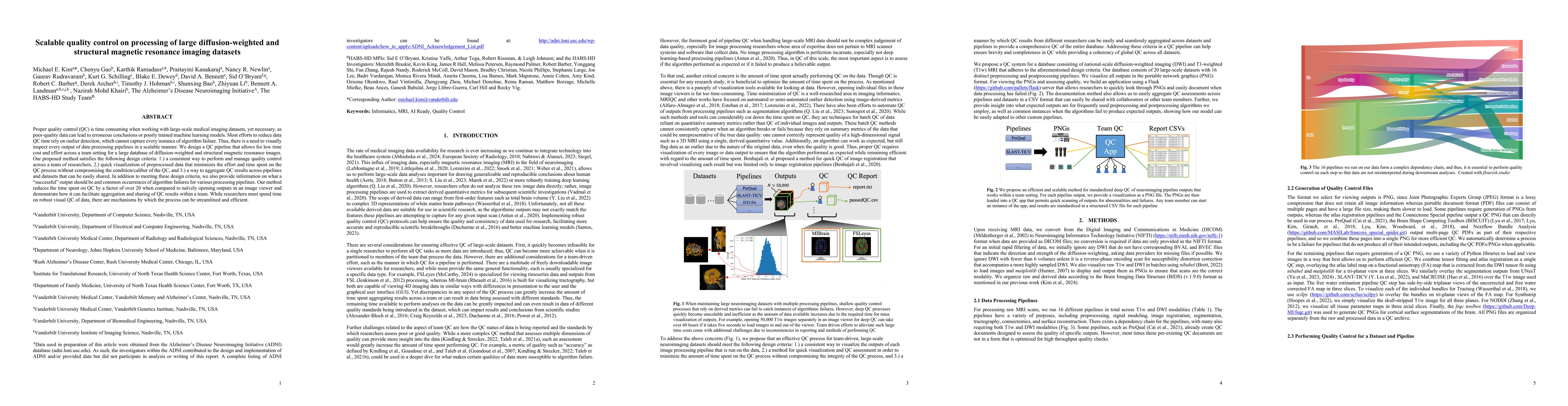
Proper quality control (QC) is time consuming when working with large-scale medical imaging datasets, yet necessary, as poor-quality data can lead to erroneous conclusions or poorly trained machine le...
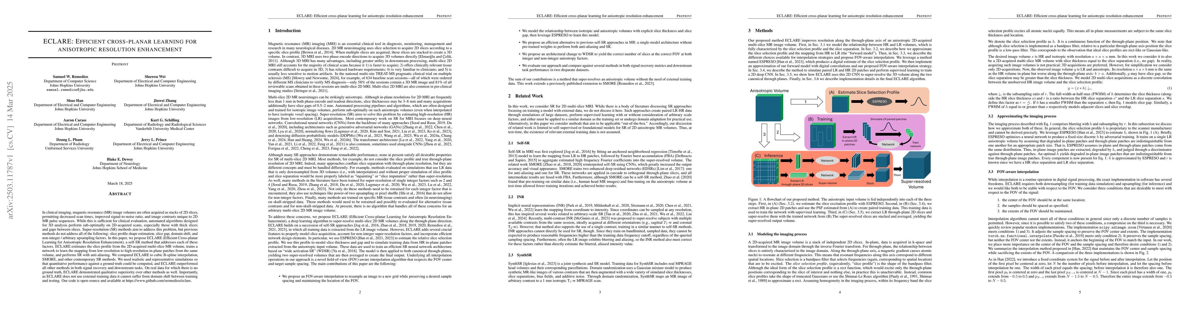
In clinical imaging, magnetic resonance (MR) image volumes are often acquired as stacks of 2D slices, permitting decreased scan times, improved signal-to-noise ratio, and image contrasts unique to 2D ...
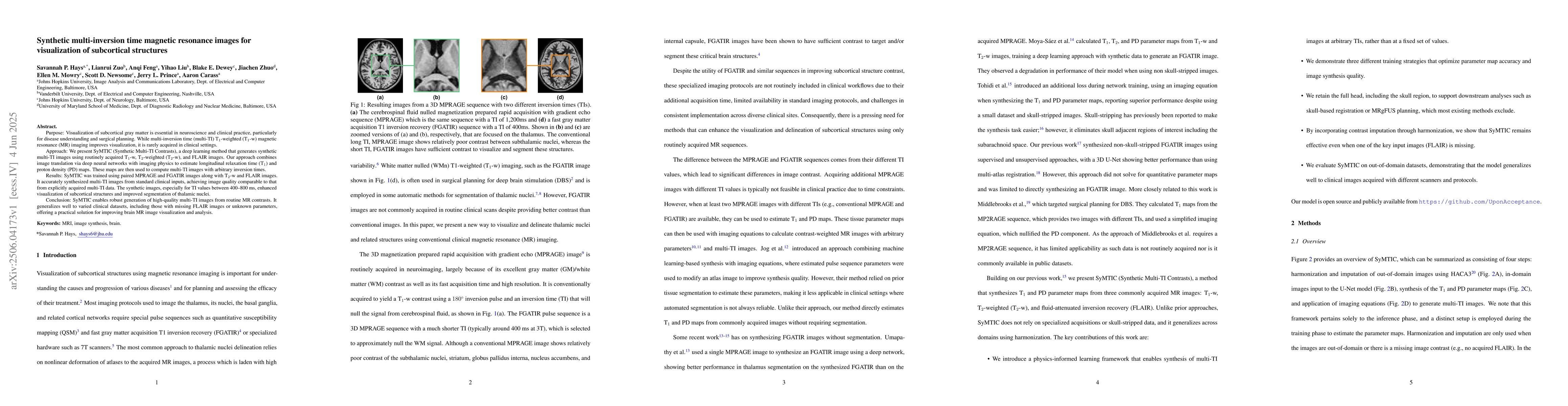
Purpose: Visualization of subcortical gray matter is essential in neuroscience and clinical practice, particularly for disease understanding and surgical planning.While multi-inversion time (multi-TI)...
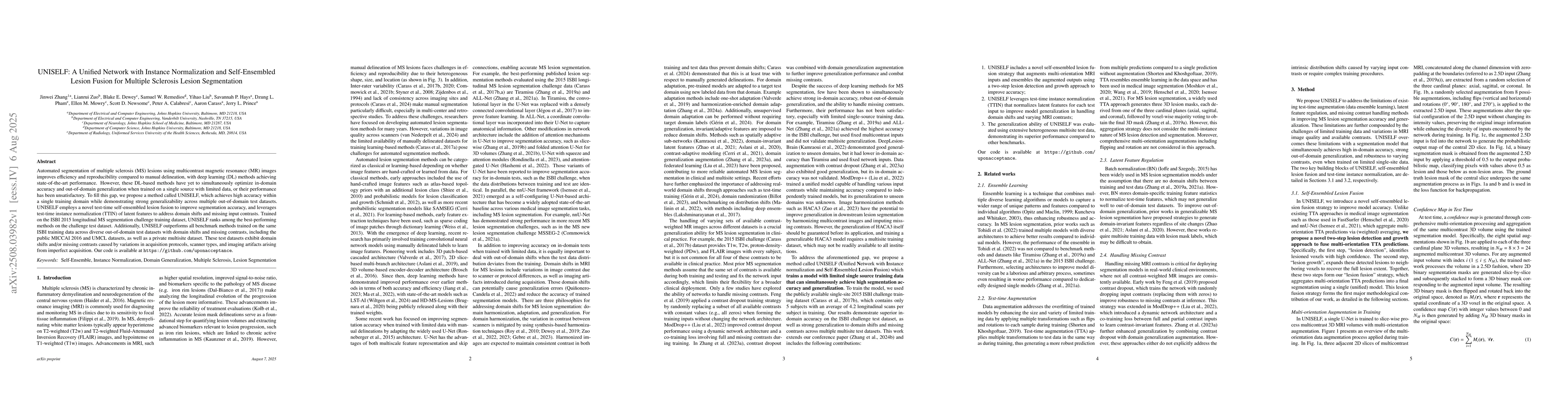
Automated segmentation of multiple sclerosis (MS) lesions using multicontrast magnetic resonance (MR) images improves efficiency and reproducibility compared to manual delineation, with deep learning ...
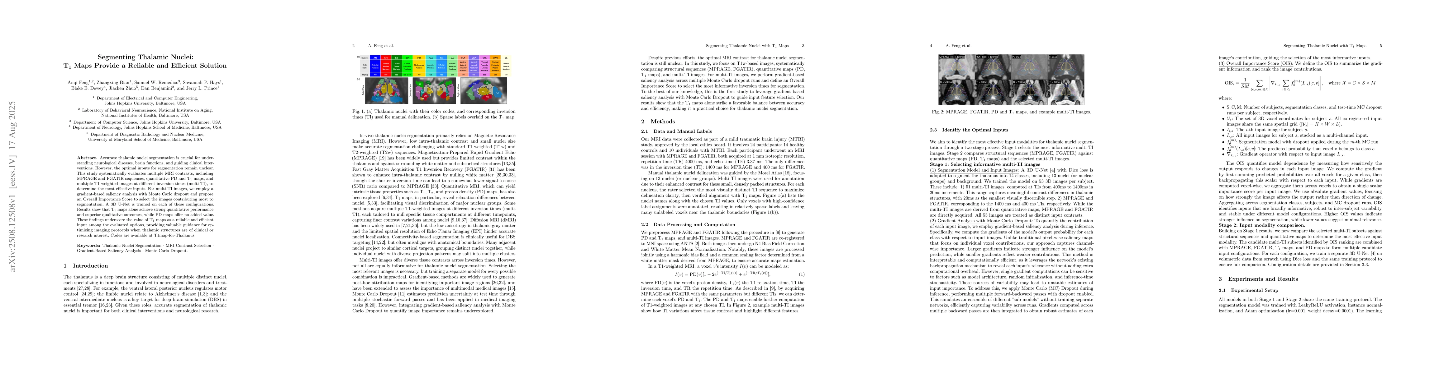
Accurate thalamic nuclei segmentation is crucial for understanding neurological diseases, brain functions, and guiding clinical interventions. However, the optimal inputs for segmentation remain uncle...
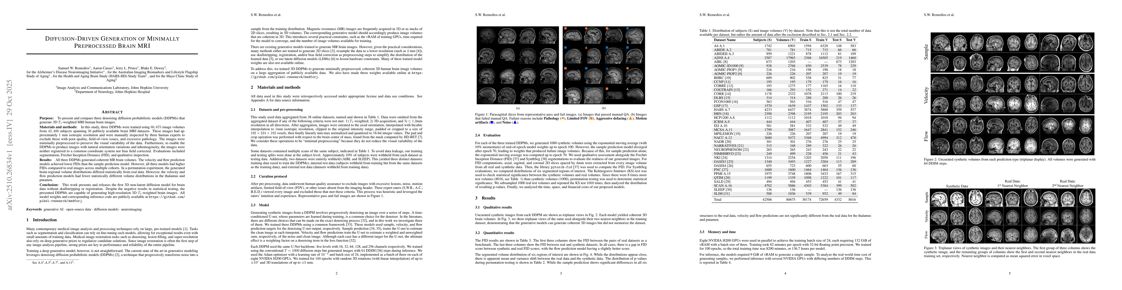
The purpose of this study is to present and compare three denoising diffusion probabilistic models (DDPMs) that generate 3D $T_1$-weighted MRI human brain images. Three DDPMs were trained using 80,675...
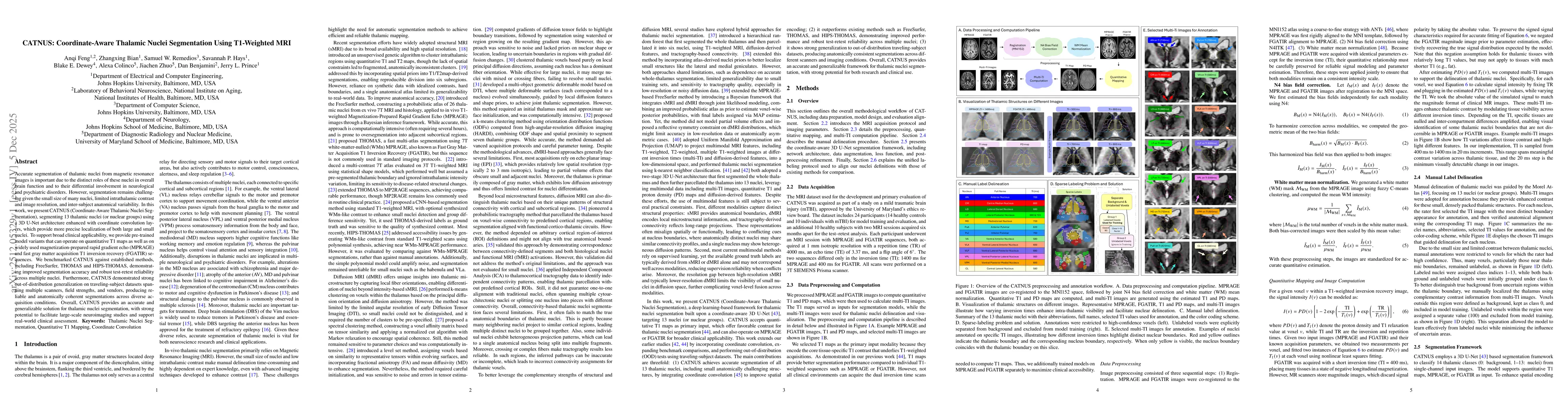
Accurate segmentation of thalamic nuclei from magnetic resonance images is important due to the distinct roles of these nuclei in overall brain function and to their differential involvement in neurol...
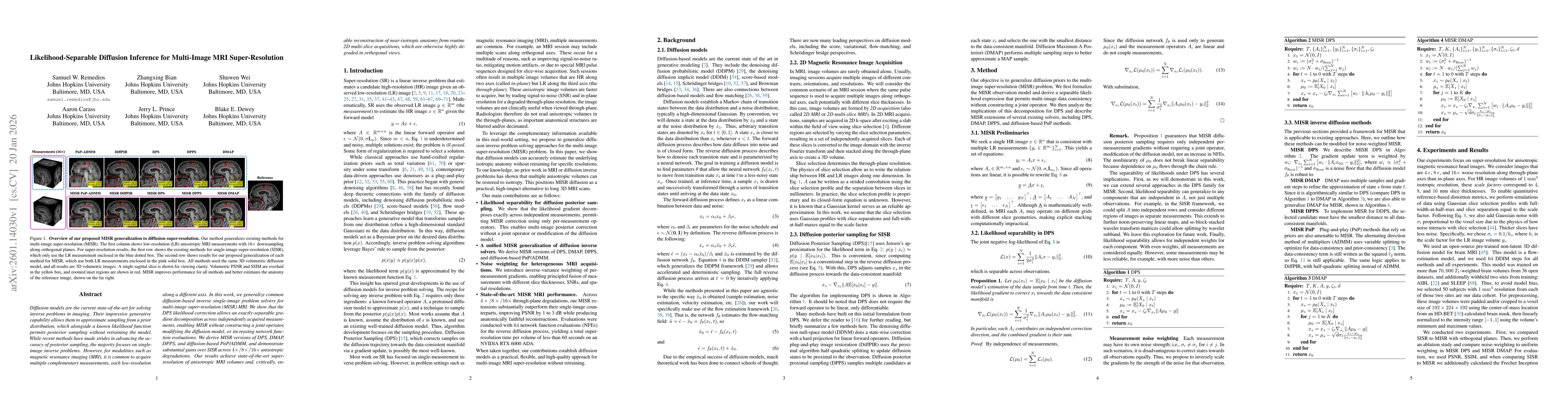
Diffusion models are the current state-of-the-art for solving inverse problems in imaging. Their impressive generative capability allows them to approximate sampling from a prior distribution, which a...
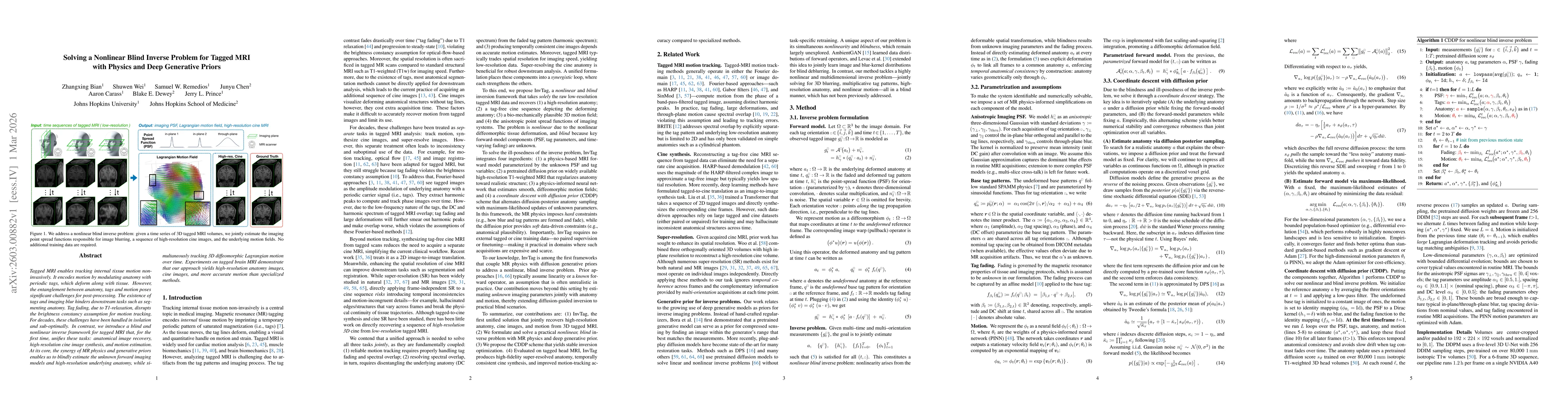
Tagged MRI enables tracking internal tissue motion non-invasively. It encodes motion by modulating anatomy with periodic tags, which deform along with tissue. However, the entanglement between anatomy...
Reliable harmonization of heterogeneous magnetic resonance~(MR) image datasets, especially those acquired in pragmatic clinical trials, is critical to advance multi-center neuroimaging studies and tra...
Purpose: Diffusion MRI (dMRI) provides a diverse set of quantitative measures and derived datatypes to assess white matter microstructure and macrostructure. Coupled with the increasing size of imagin...