Academic Profile
Statistics
Similar Authors
Papers on arXiv
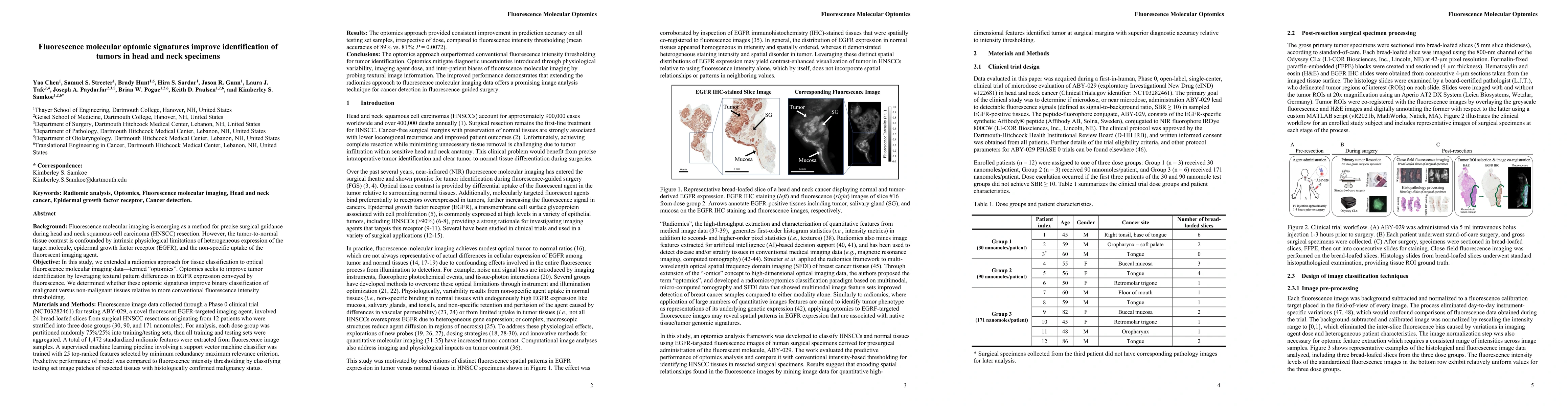
In this study, a radiomics approach was extended to optical fluorescence molecular imaging data for tissue classification, termed 'optomics'. Fluorescence molecular imaging is emerging for precise s...
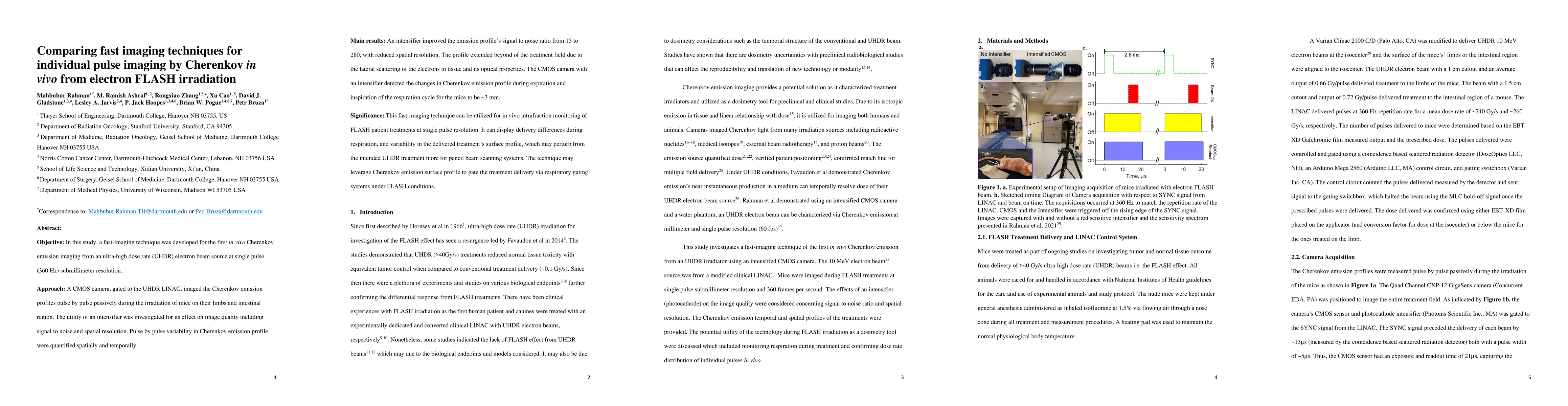
Objective: In this study, a fast imaging technique was developed for the first in vivo Cherenkov emission imaging from an ultra-high dose rate (UHDR) electron beam source at single pulse (360 Hz) su...
Purpose: A diode EDGE Detector with a newly designed electrometer has been characterized for use in an UHDR electron beam and demonstrated appropriateness for UHDR FLASH radiotherapy dosimetry. Me...
Background: Use of a linear accelerator in ultra-high dose rate (UHDR) mode can provide a conduit for wider access to UHDR FLASH effects, sparing normal tissue, but care needs to be taken in the use...
Ultra-high dose rate electron sources require dose rate independent dosimeters and a calibrated dose control system for accurate delivery. In this study, we developed a single-pulse dose monitoring ...
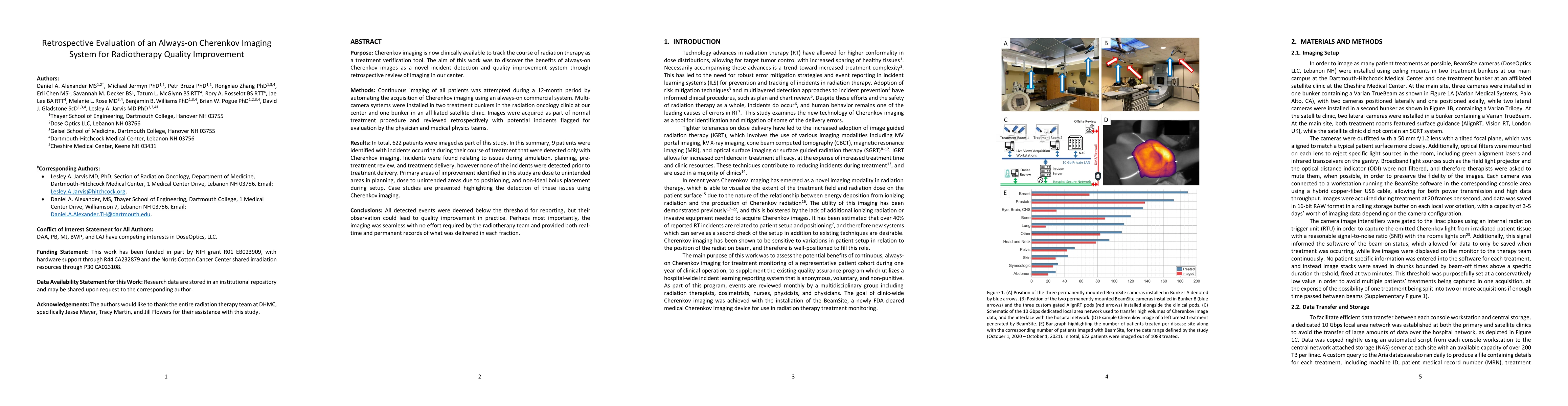
Purpose: Cherenkov imaging is now clinically available to track the course of radiation therapy as a treatment verification tool. The aim of this work was to discover the benefits of always-on Chere...
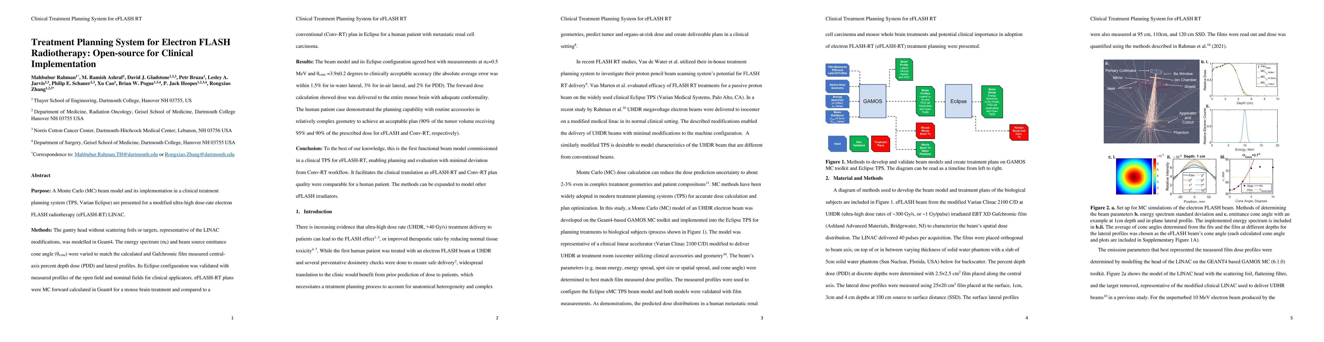
Purpose: A Monte Carlo (MC) beam model and its implementation in a clinical treatment planning system (TPS, Varian Eclipse) are presented for a modified ultra-high dose-rate electron FLASH radiother...
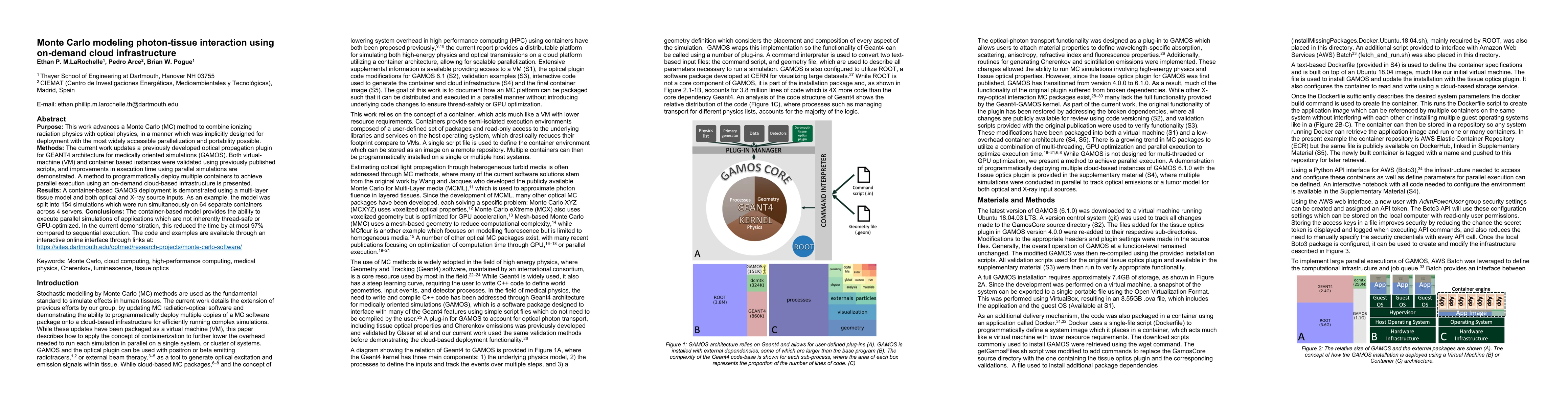
Purpose: This work advances a Monte Carlo (MC) method to combine ionizing radiation physics with optical physics, in a manner which was implicitly designed for deployment with the most widely access...
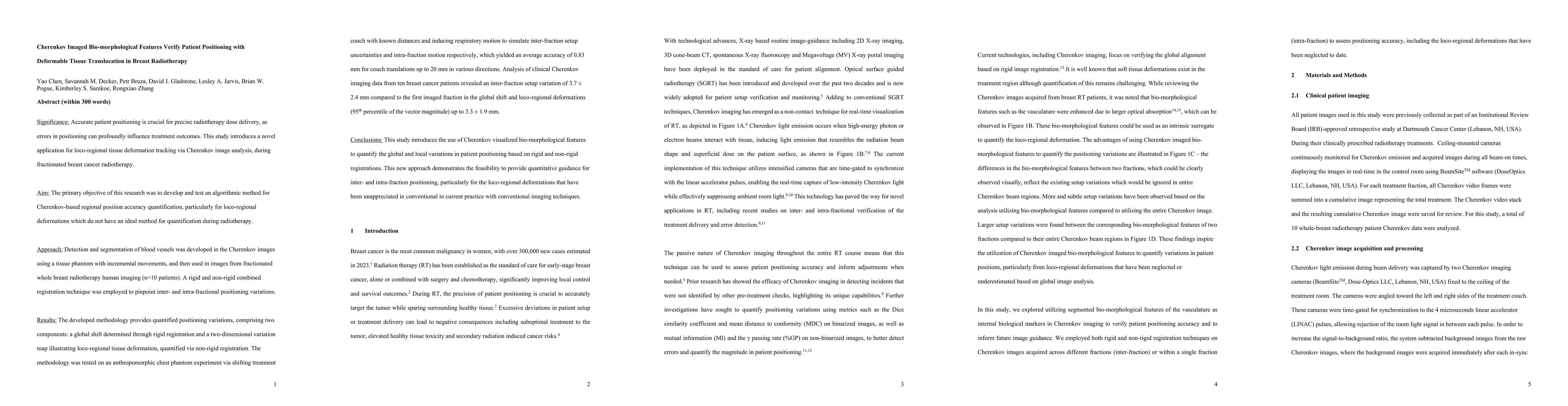
Accurate patient positioning is critical for precise radiotherapy dose delivery, as positioning errors can significantly affect treatment outcomes. This study introduces a novel method for tracking lo...
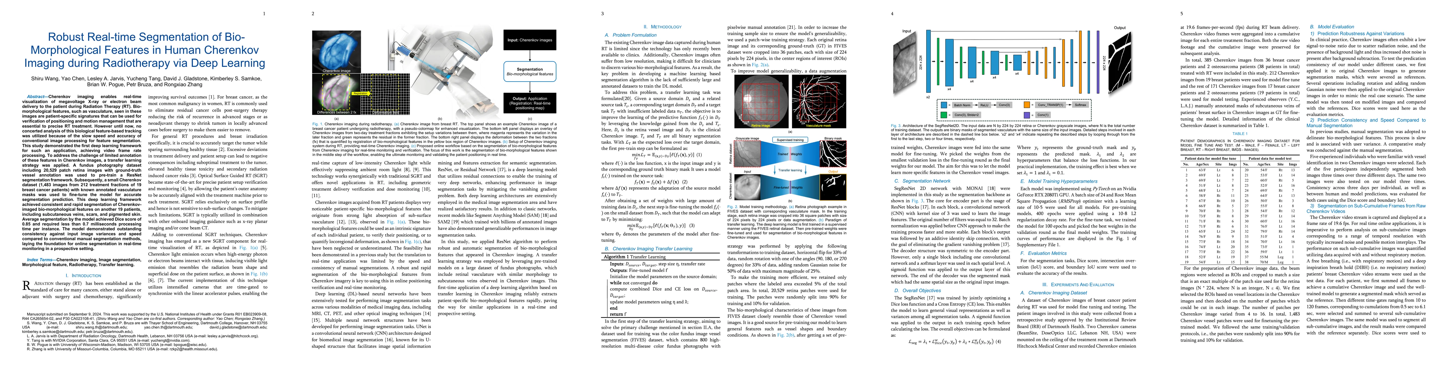
Cherenkov imaging enables real-time visualization of megavoltage X-ray or electron beam delivery to the patient during Radiation Therapy (RT). Bio-morphological features, such as vasculature, seen in ...
Background: Ultra-high dose rate proton therapy shows promise in tissue sparing by enhancing therapeutic ratio through the FLASH effect. In radiotherapy, accurate in vivo dosimetry is crucial for qual...
Medical Physics education is delivered through accredited programs with admissions and funding for students determined by individual institutions providing the educational experiences. Public data fro...
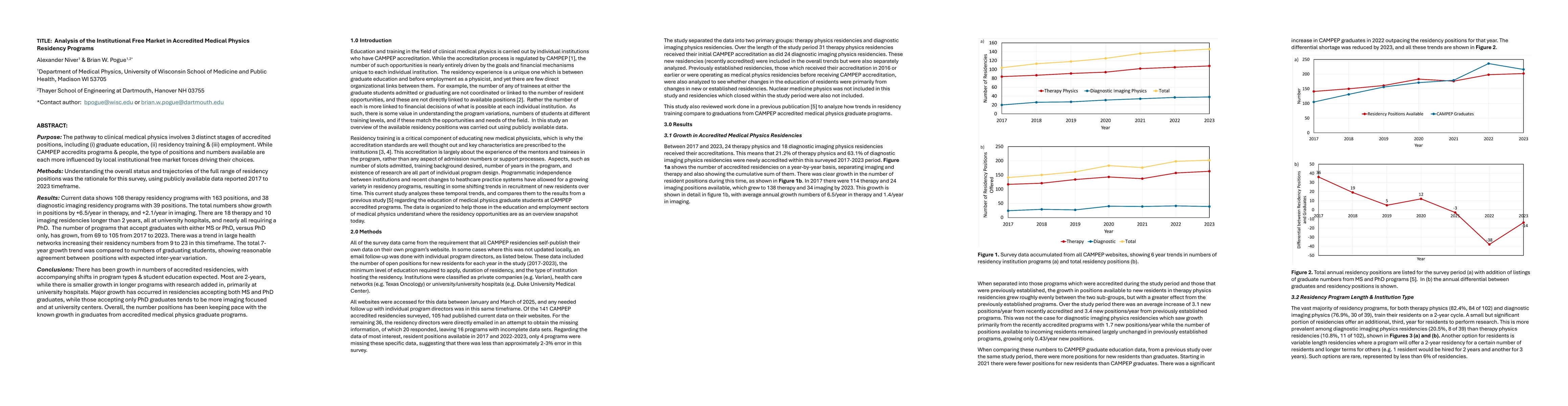
Purpose: The pathway to clinical medical physics involves 3 distinct stages of accredited positions, including (i) graduate education, (ii) residency training & (iii) employment. While CAMPEP accredit...
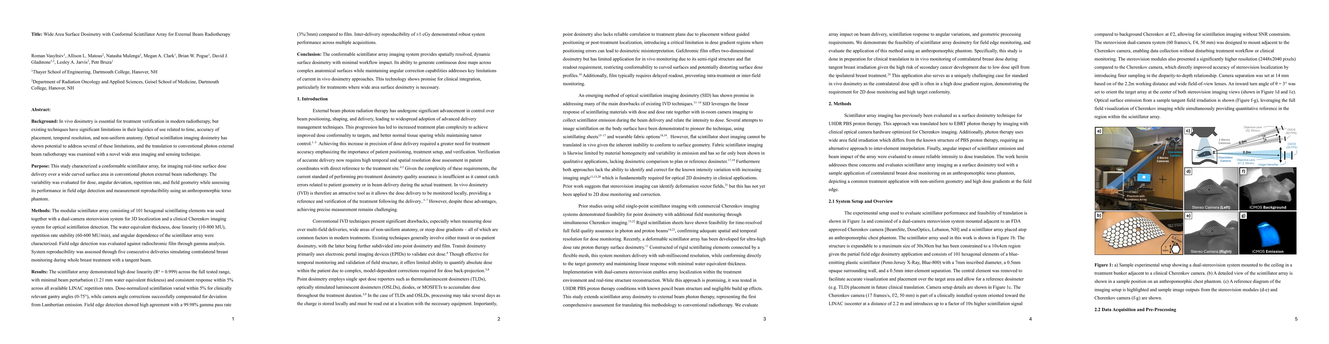
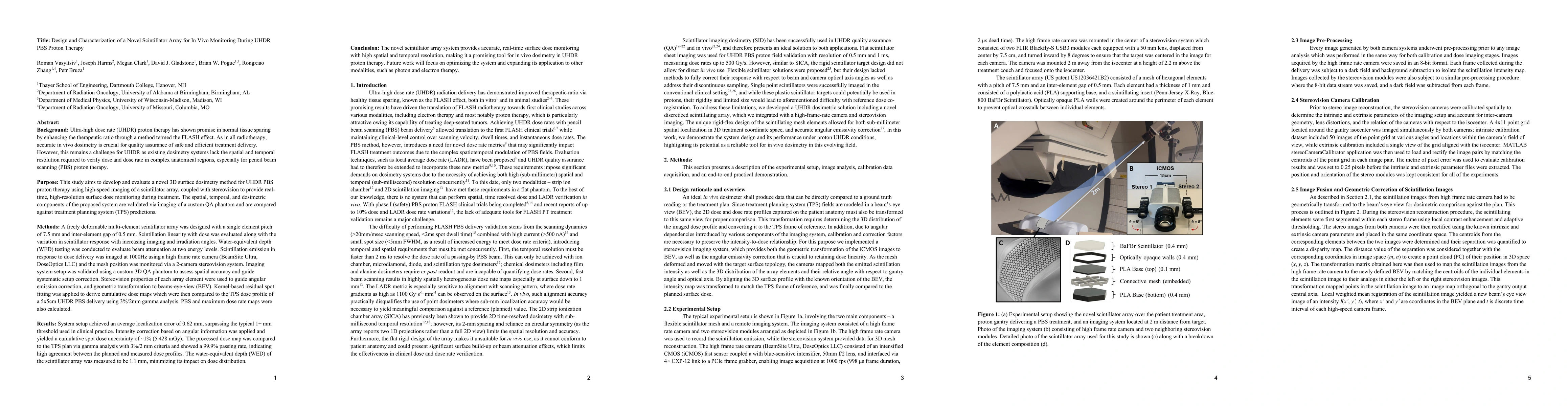
Background: In vivo dosimetry is essential for treatment verification in modern radiotherapy, but existing techniques are limited by spatiotemporal resolution and performance on non-uniform anatomy. S...
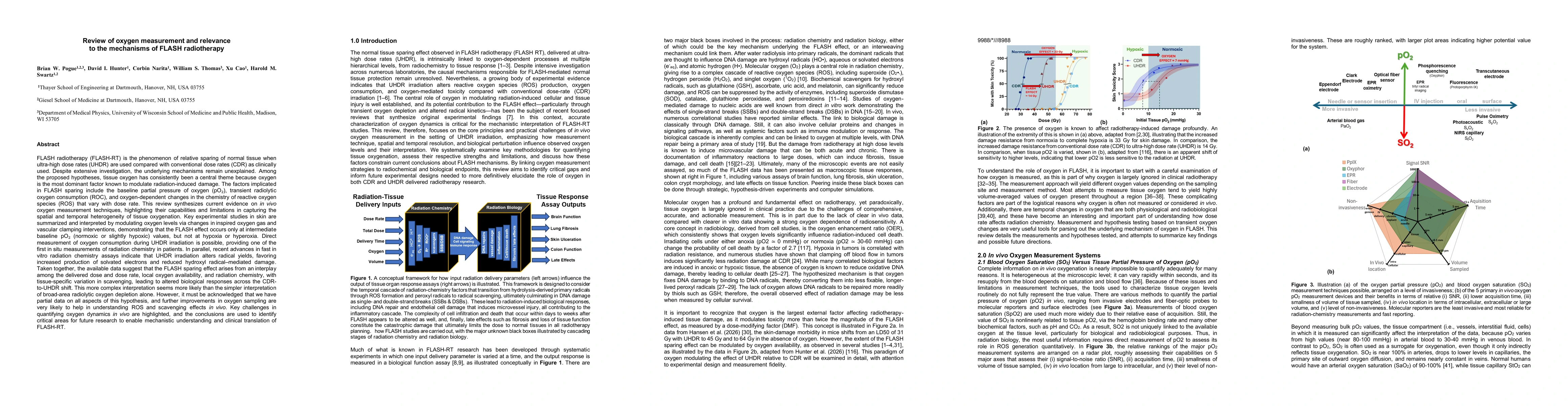
FLASH radiotherapy (FLASH-RT) is the phenomenon of relative sparing of normal tissue when ultra-high dose rates (UHDR) are used compared with conventional dose rates (CDR) as clinically used. Despite ...