Academic Profile
Statistics
Similar Authors
Papers on arXiv
Together with the molecular knowledge of genes and proteins, biological images promise to significantly enhance the scientific understanding of complex cellular systems and to advance predictive and...
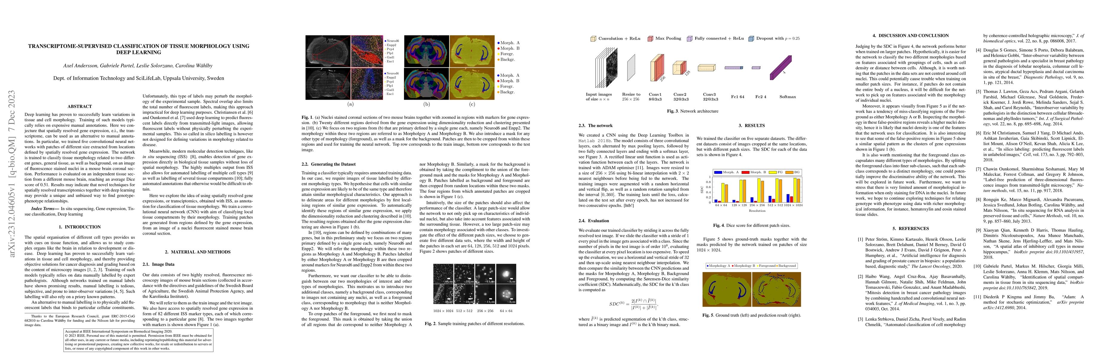
Deep learning has proven to successfully learn variations in tissue and cell morphology. Training of such models typically relies on expensive manual annotations. Here we conjecture that spatially r...
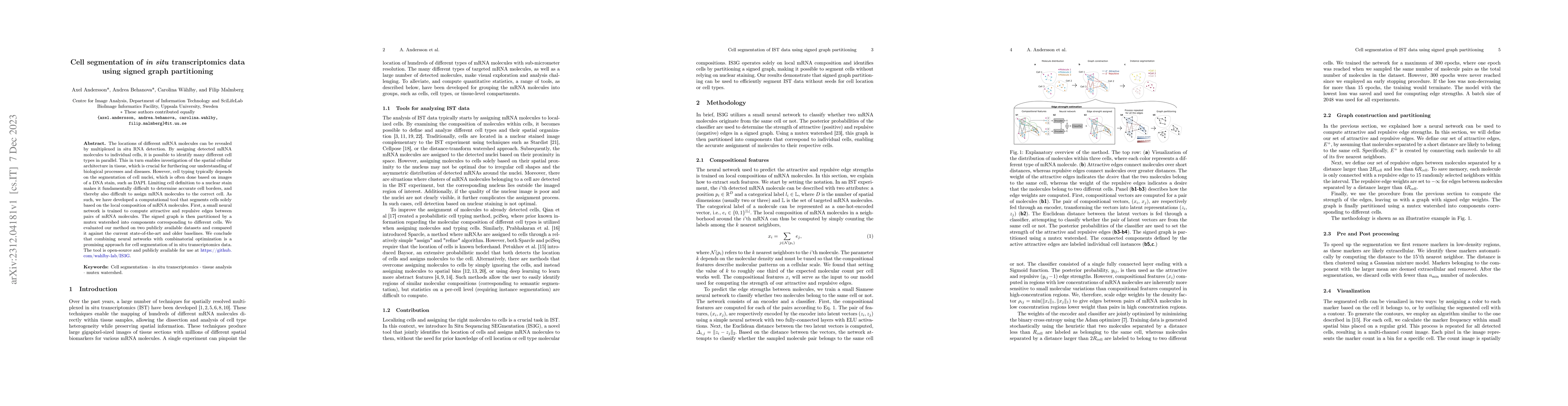
The locations of different mRNA molecules can be revealed by multiplexed in situ RNA detection. By assigning detected mRNA molecules to individual cells, it is possible to identify many different ce...
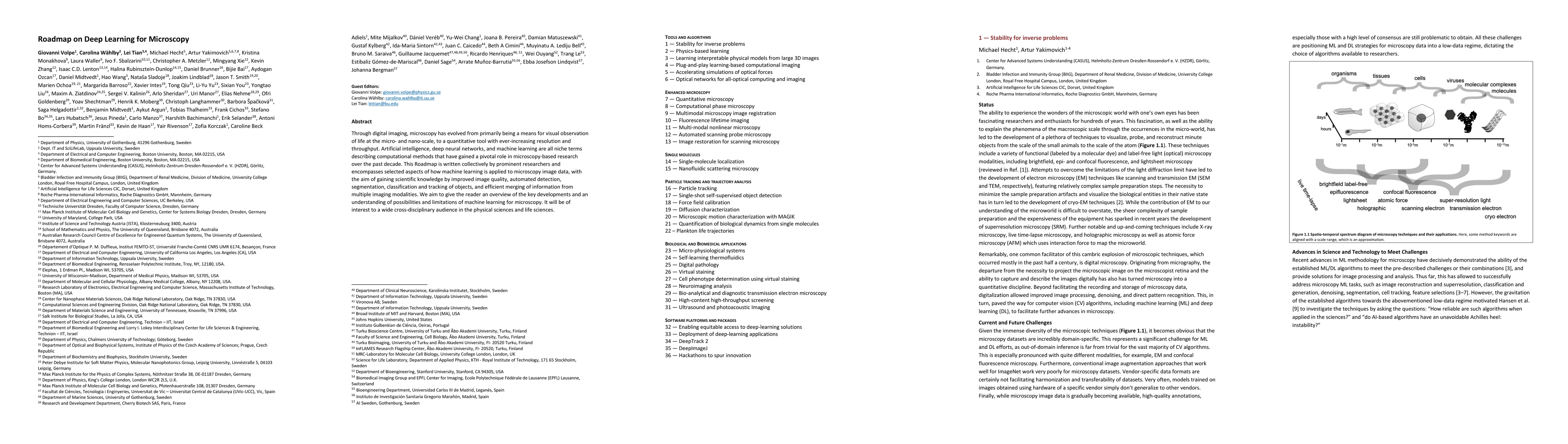
Through digital imaging, microscopy has evolved from primarily being a means for visual observation of life at the micro- and nano-scale, to a quantitative tool with ever-increasing resolution and t...
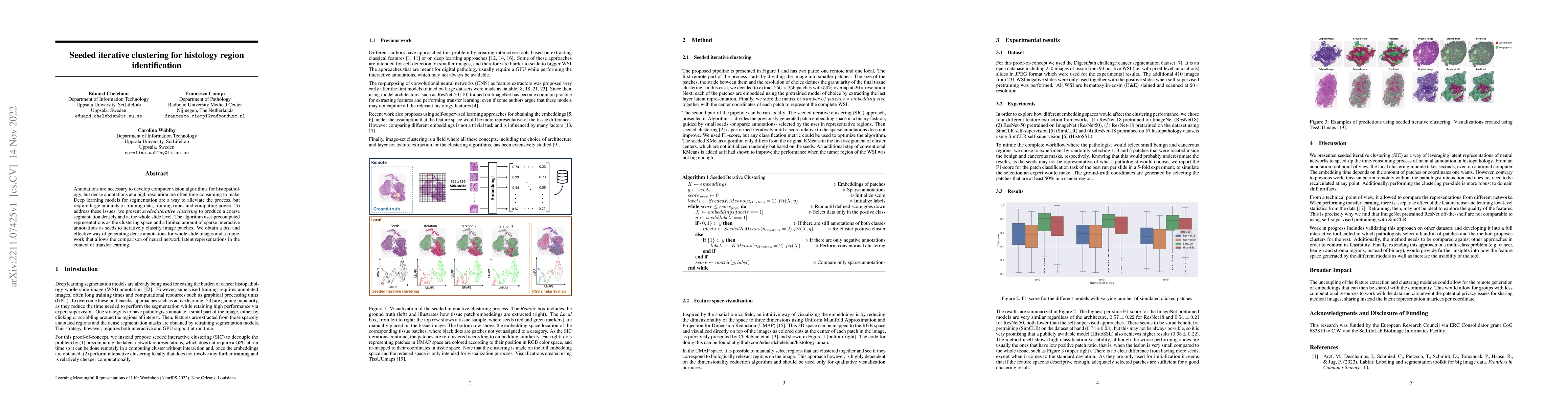
Annotations are necessary to develop computer vision algorithms for histopathology, but dense annotations at a high resolution are often time-consuming to make. Deep learning models for segmentation...
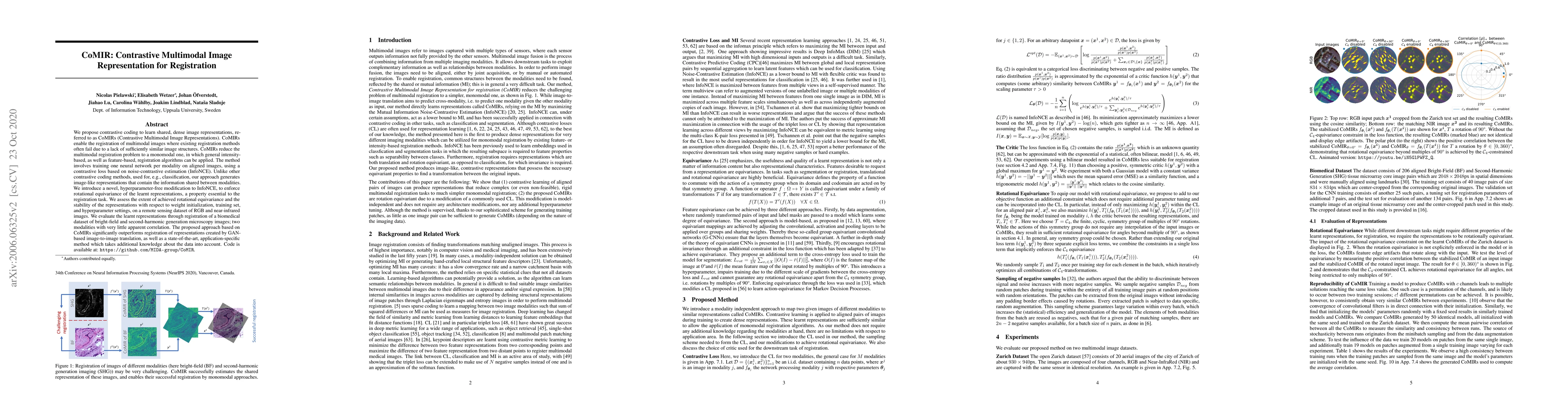
We propose contrastive coding to learn shared, dense image representations, referred to as CoMIRs (Contrastive Multimodal Image Representations). CoMIRs enable the registration of multimodal images ...
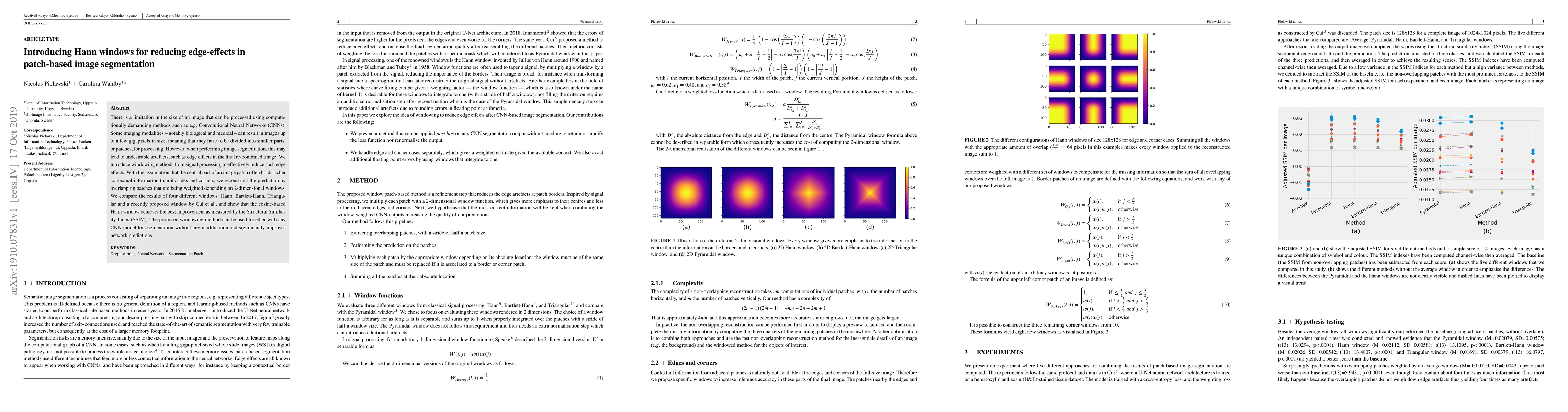
There is a limitation in the size of an image that can be processed using computationally demanding methods such as e.g. Convolutional Neural Networks (CNNs). Some imaging modalities - notably biolo...
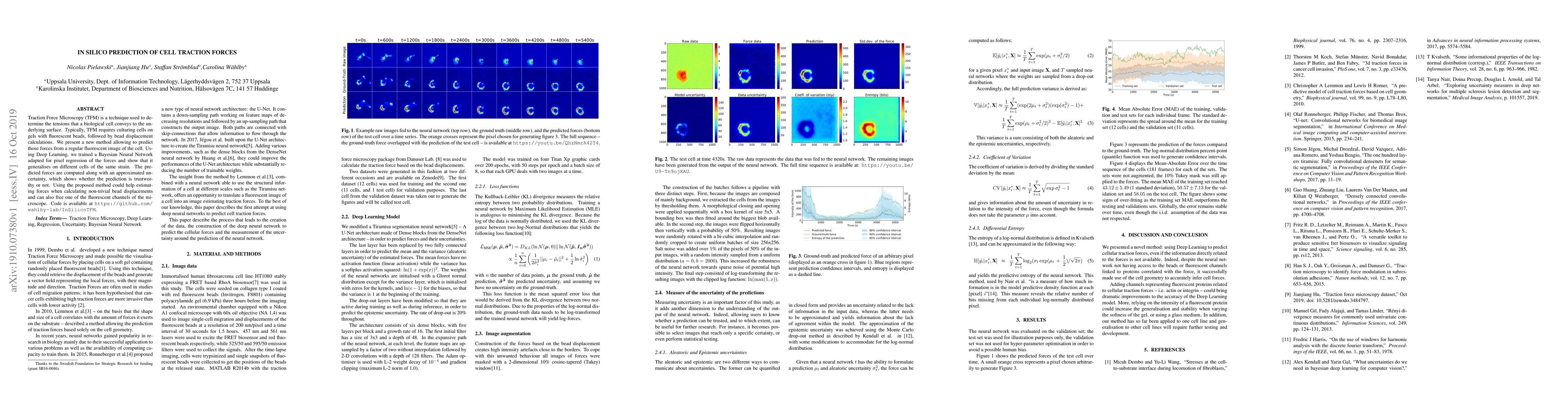
Traction Force Microscopy (TFM) is a technique used to determine the tensions that a biological cell conveys to the underlying surface. Typically, TFM requires culturing cells on gels with fluoresce...
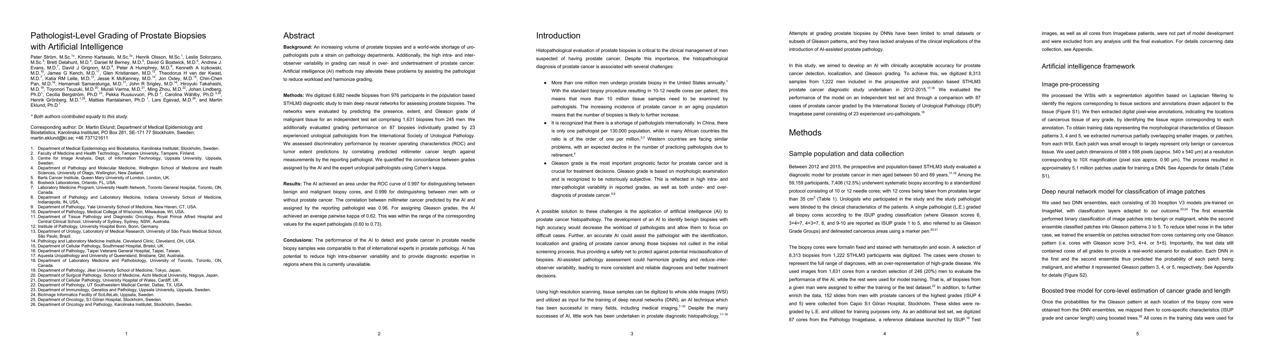
Background: An increasing volume of prostate biopsies and a world-wide shortage of uro-pathologists puts a strain on pathology departments. Additionally, the high intra- and inter-observer variabili...
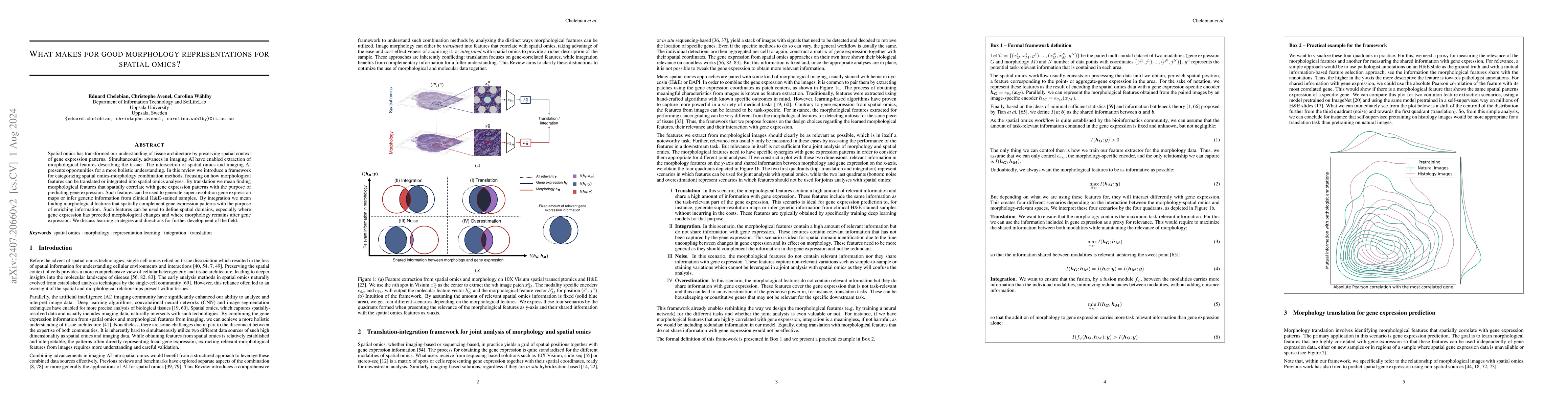
Spatial omics has transformed our understanding of tissue architecture by preserving spatial context of gene expression patterns. Simultaneously, advances in imaging AI have enabled extraction of morp...
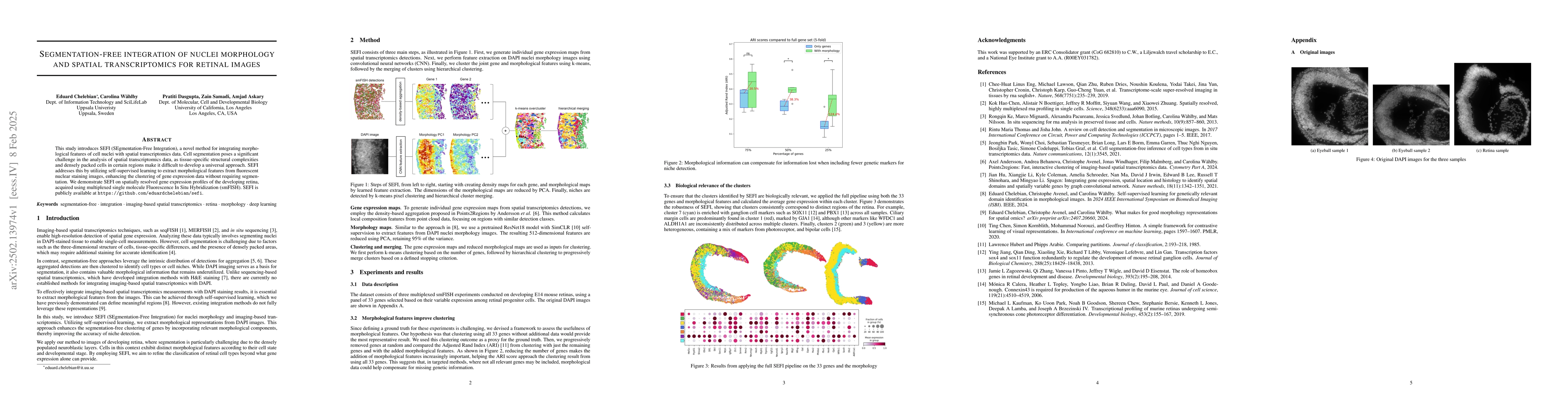
This study introduces SEFI (SEgmentation-Free Integration), a novel method for integrating morphological features of cell nuclei with spatial transcriptomics data. Cell segmentation poses a significan...