Academic Profile
Statistics
Similar Authors
Papers on arXiv
Whole slide imaging is routinely adopted for carcinoma diagnosis and prognosis. Abundant experience is required for pathologists to achieve accurate and reliable diagnostic results of whole slide imag...
Vision Language Models (VLMs) like CLIP have attracted substantial attention in pathology, serving as backbones for applications such as zero-shot image classification and Whole Slide Image (WSI) anal...
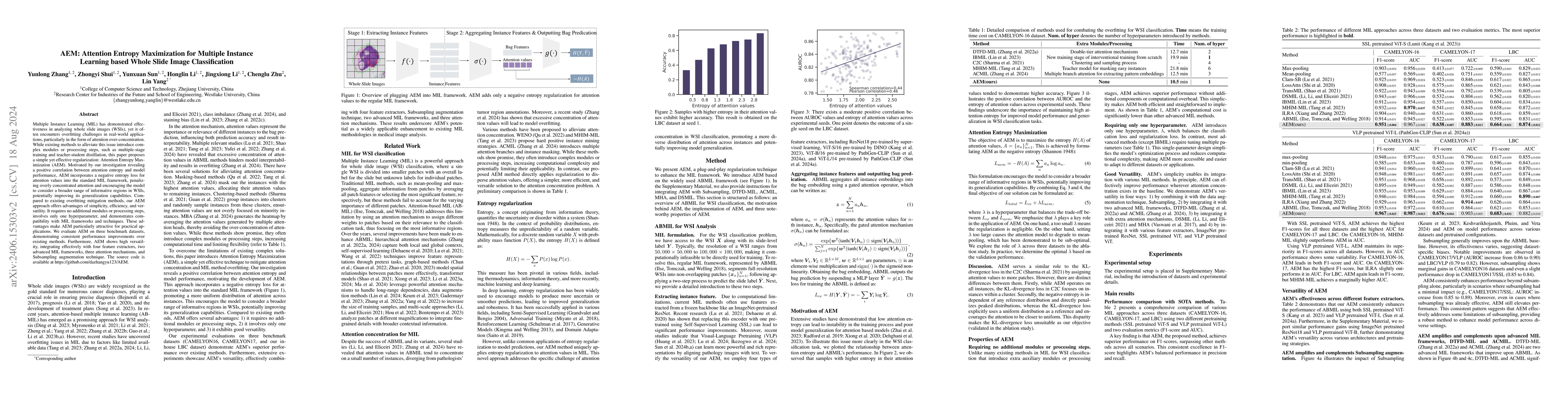
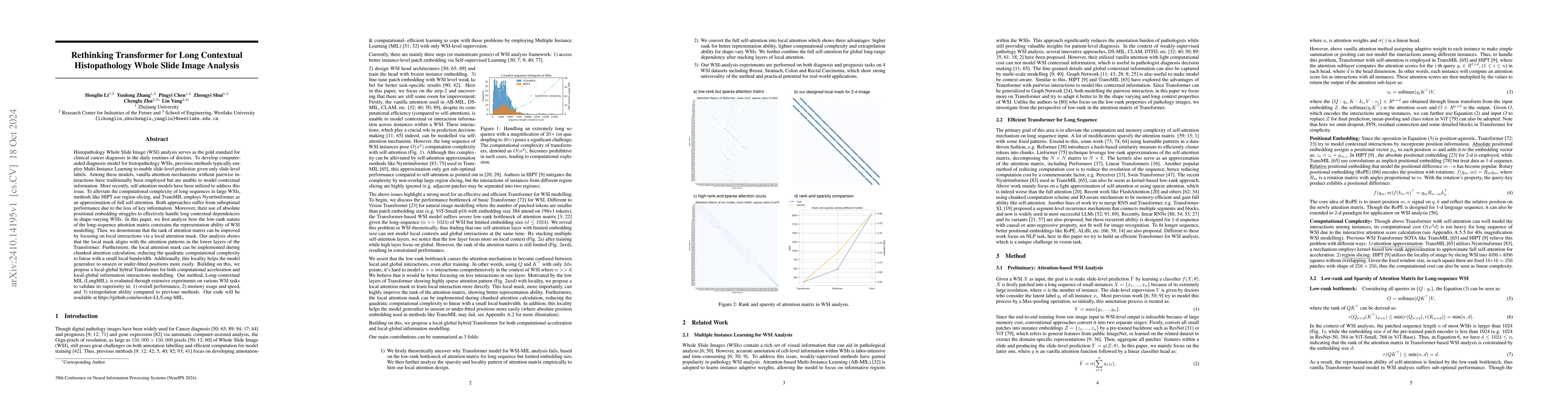
Multiple Instance Learning (MIL) has demonstrated effectiveness in analyzing whole slide images (WSIs), yet it often encounters overfitting challenges in real-world applications. This paper reveals th...
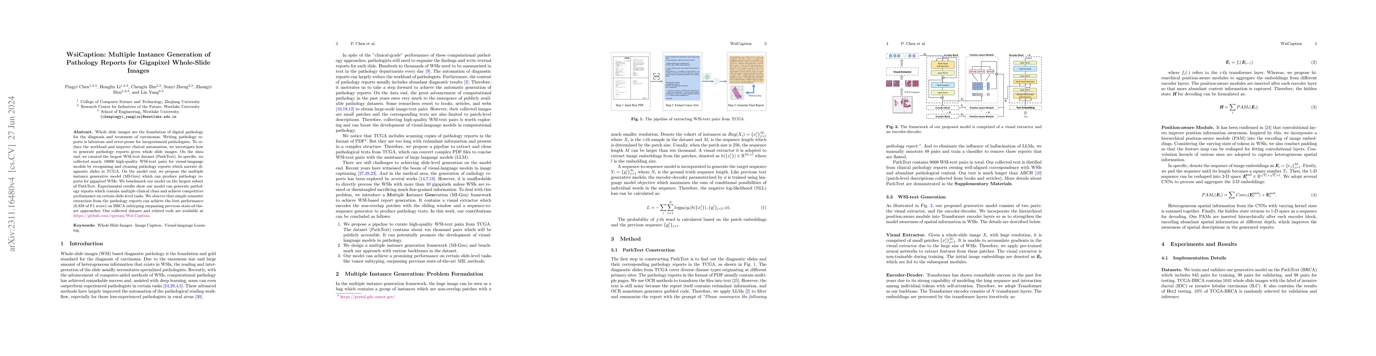
Whole slide images are the foundation of digital pathology for the diagnosis and treatment of carcinomas. Writing pathology reports is laborious and error-prone for inexperienced pathologists. To redu...
The emergence of large multimodal models has unlocked remarkable potential in AI, particularly in pathology. However, the lack of specialized, high-quality benchmark impeded their development and pr...
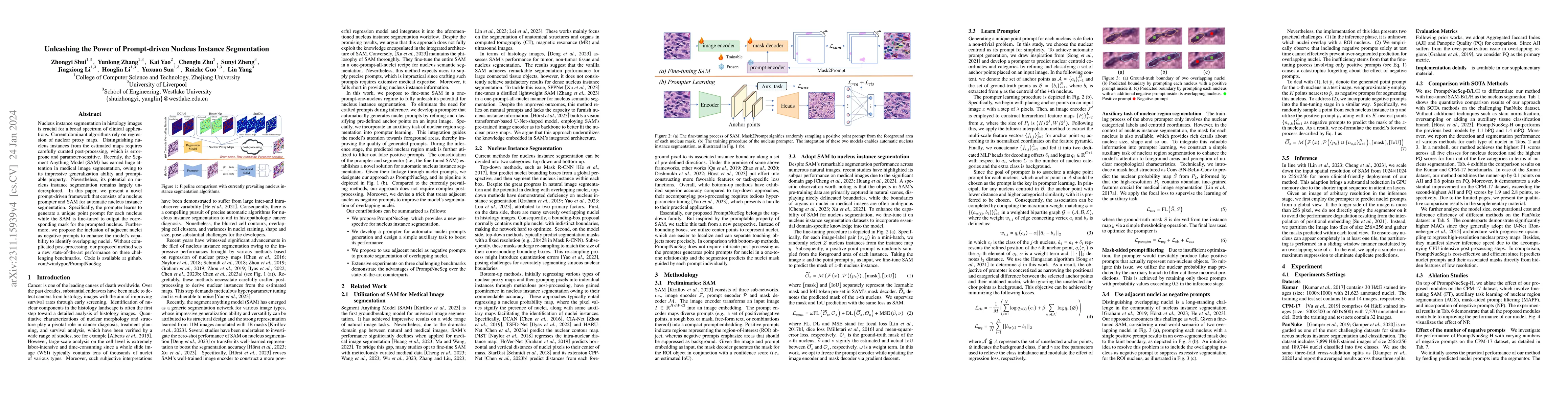
Nucleus instance segmentation in histology images is crucial for a broad spectrum of clinical applications. Current dominant algorithms rely on regression of nuclear proxy maps. Distinguishing nucle...
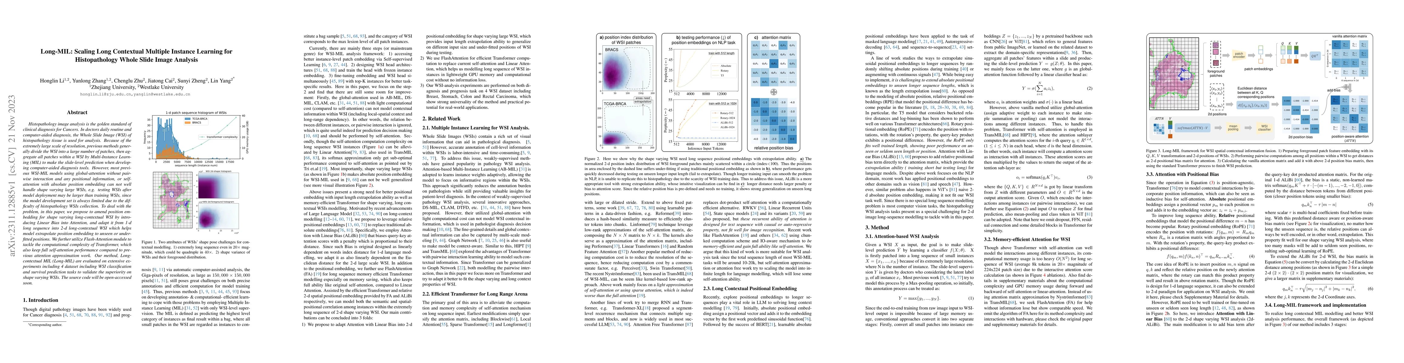
Histopathology image analysis is the golden standard of clinical diagnosis for Cancers. In doctors daily routine and computer-aided diagnosis, the Whole Slide Image (WSI) of histopathology tissue is...
Although deep learning-based segmentation models have achieved impressive performance on public benchmarks, generalizing well to unseen environments remains a major challenge. To improve the model's...
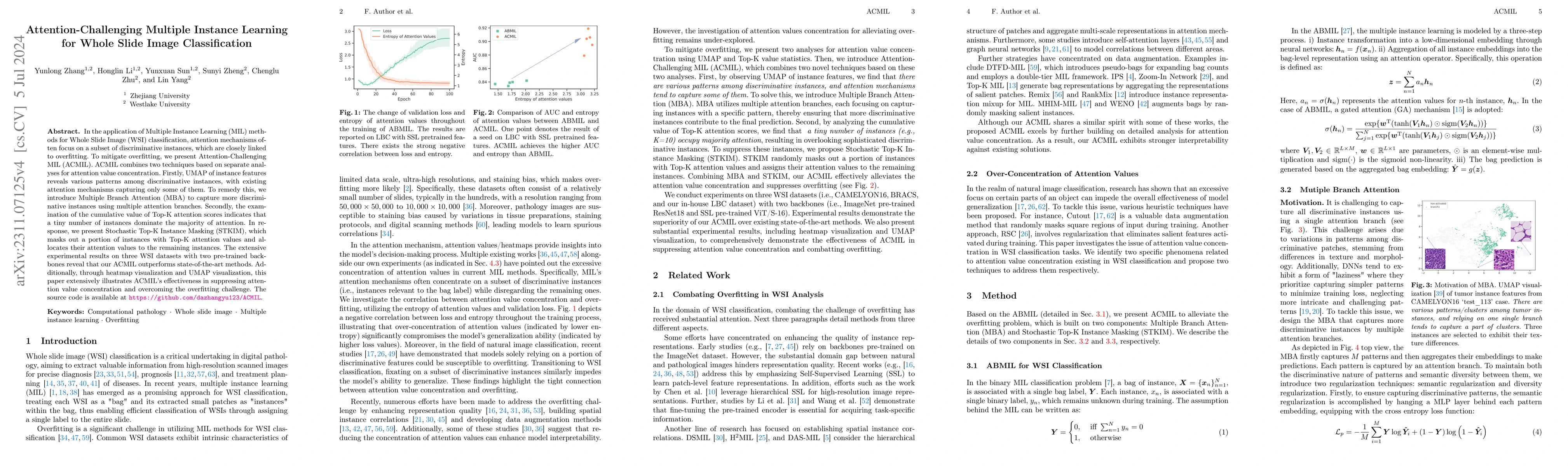
In the application of Multiple Instance Learning (MIL) methods for Whole Slide Image (WSI) classification, attention mechanisms often focus on a subset of discriminative instances, which are closely...
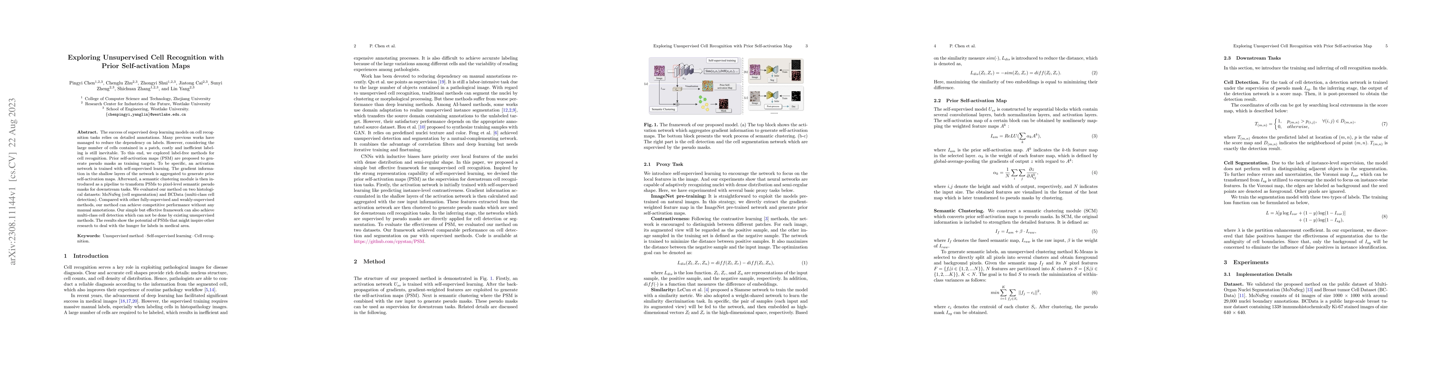
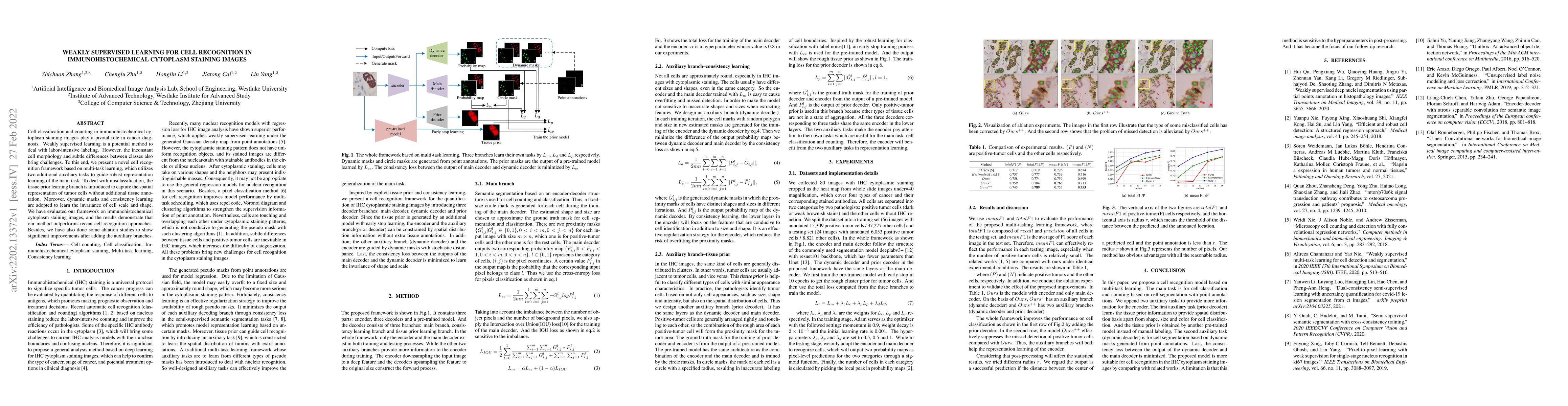
The success of supervised deep learning models on cell recognition tasks relies on detailed annotations. Many previous works have managed to reduce the dependency on labels. However, considering the...
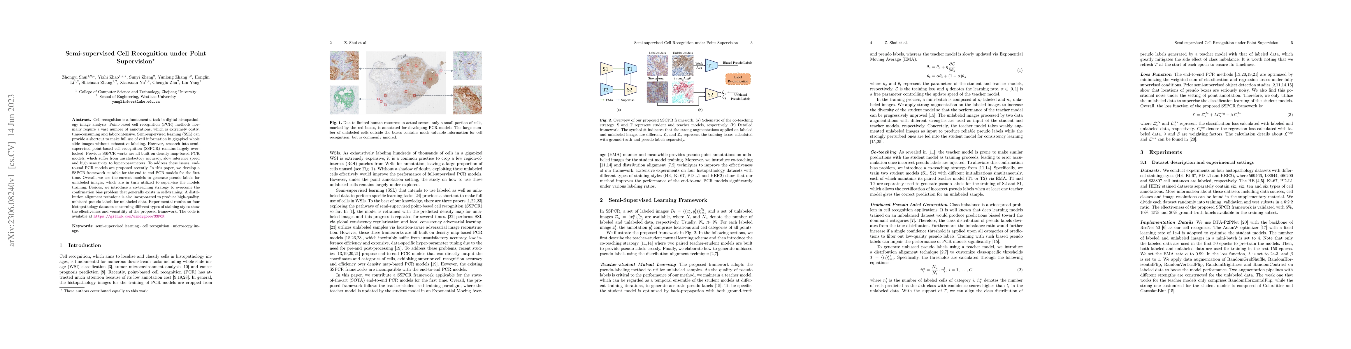
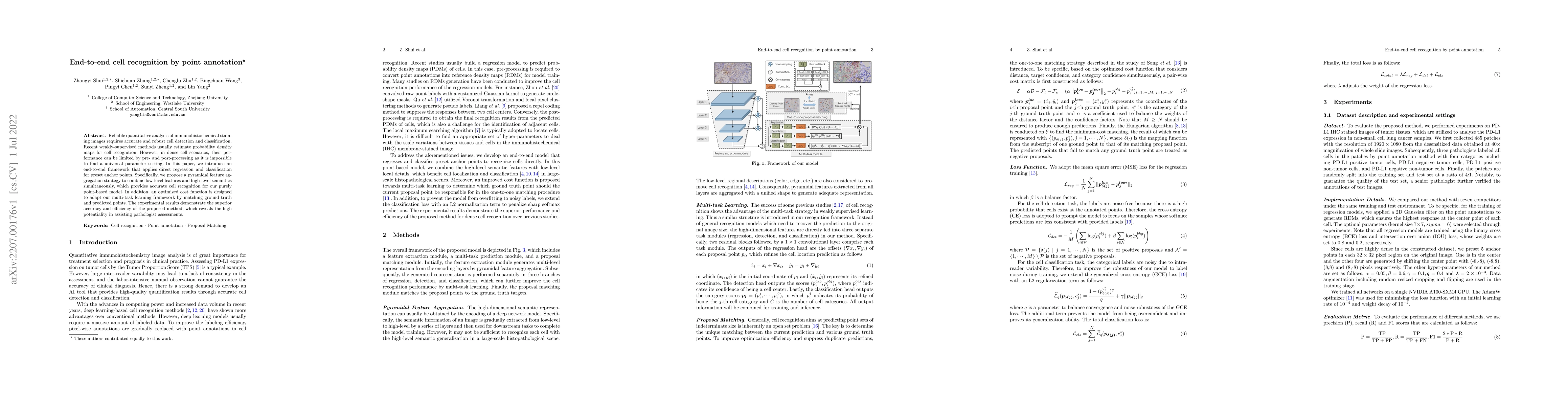
Cell recognition is a fundamental task in digital histopathology image analysis. Point-based cell recognition (PCR) methods normally require a vast number of annotations, which is extremely costly, ...
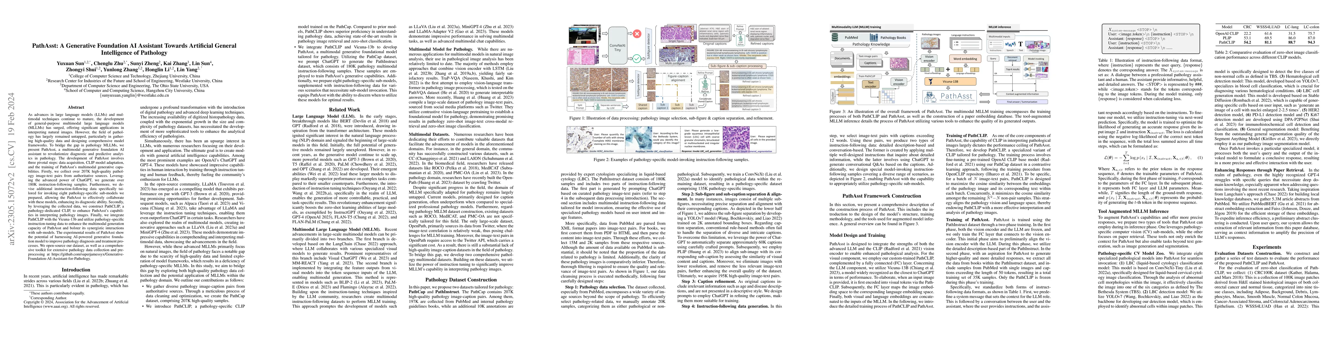
As advances in large language models (LLMs) and multimodal techniques continue to mature, the development of general-purpose multimodal large language models (MLLMs) has surged, offering significant...
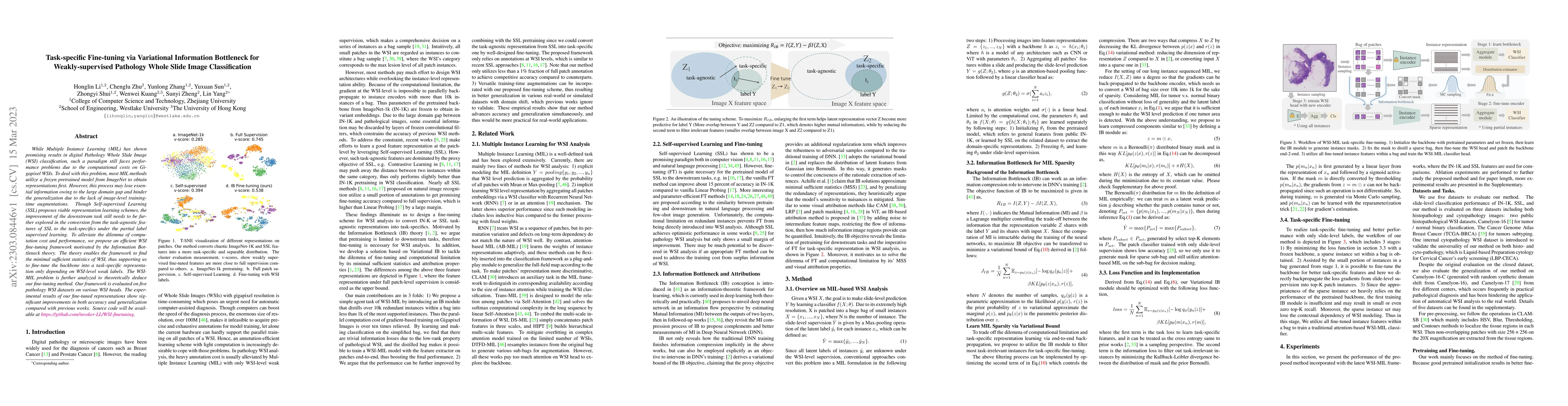
While Multiple Instance Learning (MIL) has shown promising results in digital Pathology Whole Slide Image (WSI) classification, such a paradigm still faces performance and generalization problems du...
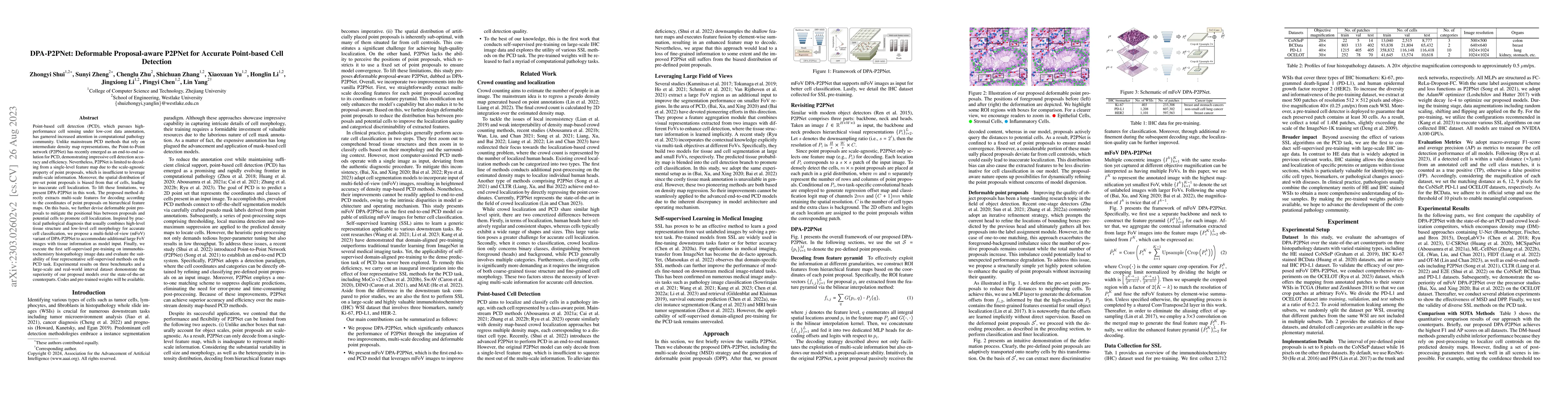
Point-based cell detection (PCD), which pursues high-performance cell sensing under low-cost data annotation, has garnered increased attention in computational pathology community. Unlike mainstream...
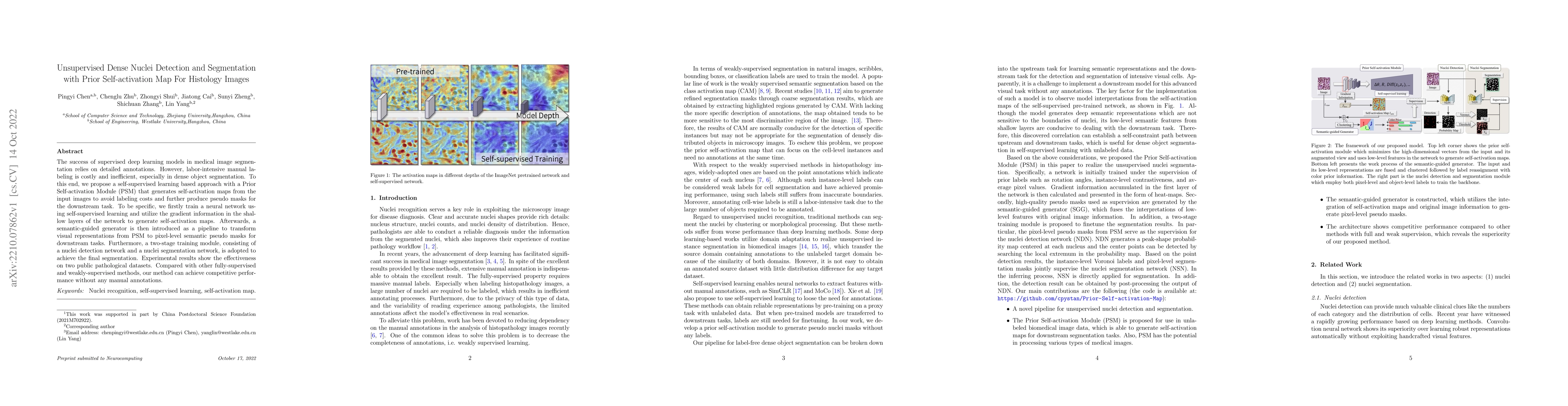
The success of supervised deep learning models in medical image segmentation relies on detailed annotations. However, labor-intensive manual labeling is costly and inefficient, especially in dense o...
Reliable quantitative analysis of immunohistochemical staining images requires accurate and robust cell detection and classification. Recent weakly-supervised methods usually estimate probability de...
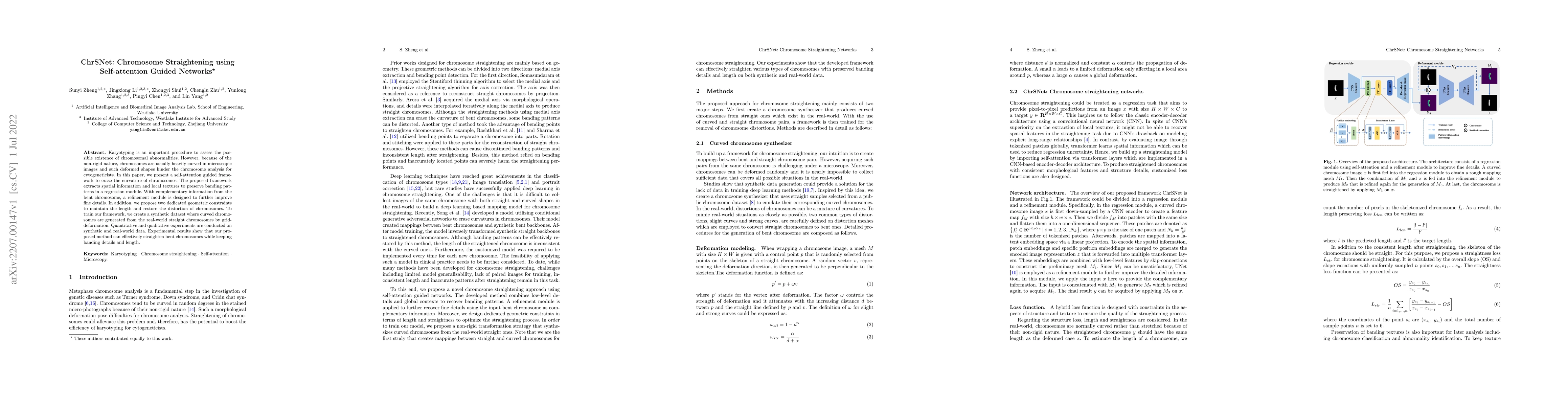
Karyotyping is an important procedure to assess the possible existence of chromosomal abnormalities. However, because of the non-rigid nature, chromosomes are usually heavily curved in microscopic i...
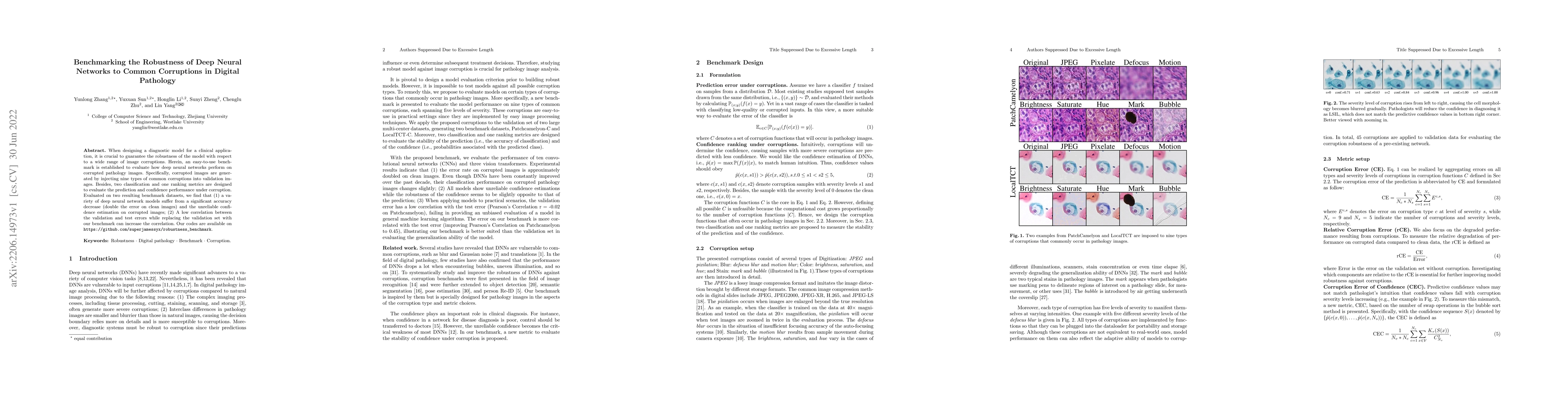
When designing a diagnostic model for a clinical application, it is crucial to guarantee the robustness of the model with respect to a wide range of image corruptions. Herein, an easy-to-use benchma...
Cell classification and counting in immunohistochemical cytoplasm staining images play a pivotal role in cancer diagnosis. Weakly supervised learning is a potential method to deal with labor-intensi...
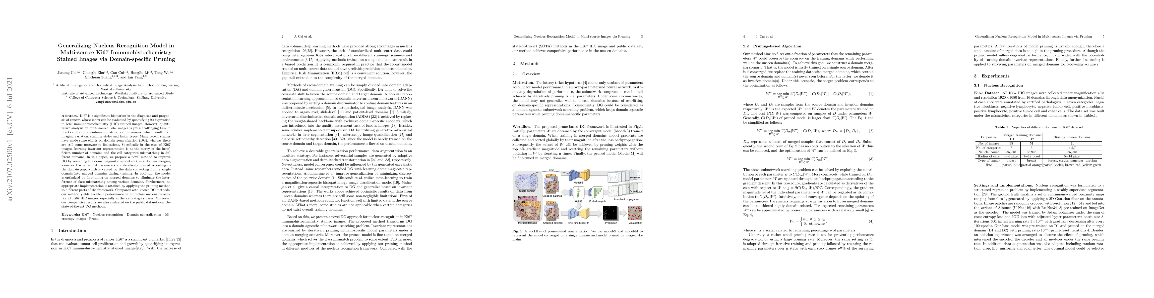
Ki67 is a significant biomarker in the diagnosis and prognosis of cancer, whose index can be evaluated by quantifying its expression in Ki67 immunohistochemistry (IHC) stained images. However, quant...
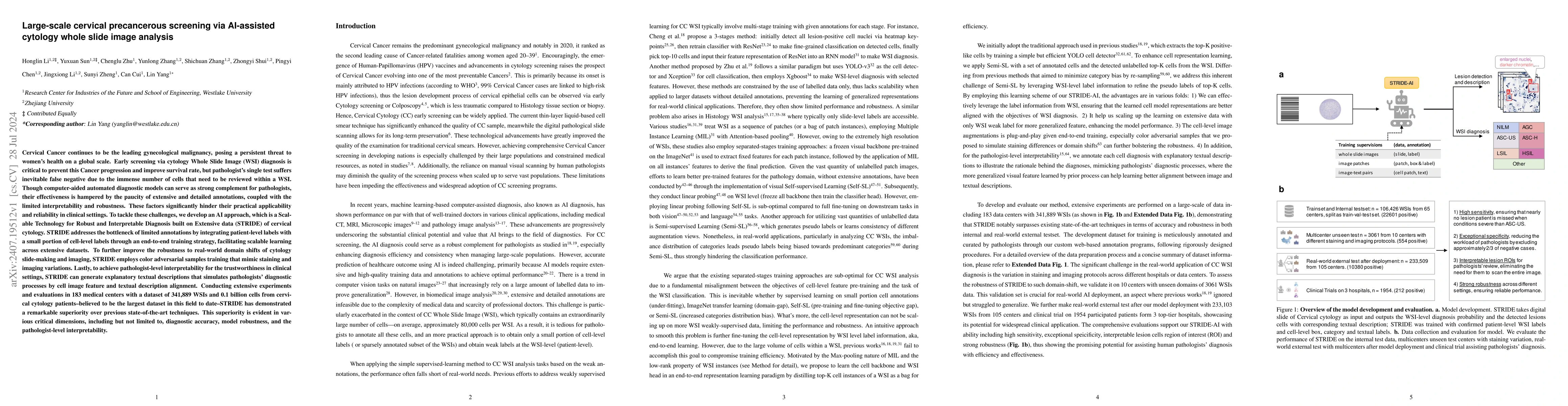
Cervical Cancer continues to be the leading gynecological malignancy, posing a persistent threat to women's health on a global scale. Early screening via cytology Whole Slide Image (WSI) diagnosis is ...
Histopathology Whole Slide Image (WSI) analysis serves as the gold standard for clinical cancer diagnosis in the daily routines of doctors. To develop computer-aided diagnosis model for WSIs, previous...
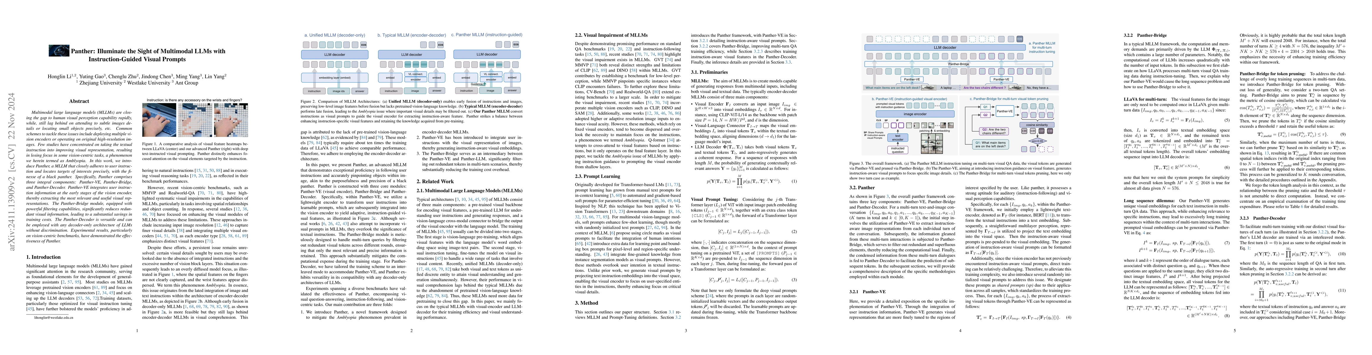
Multimodal large language models (MLLMs) are closing the gap to human visual perception capability rapidly, while, still lag behind on attending to subtle images details or locating small objects prec...
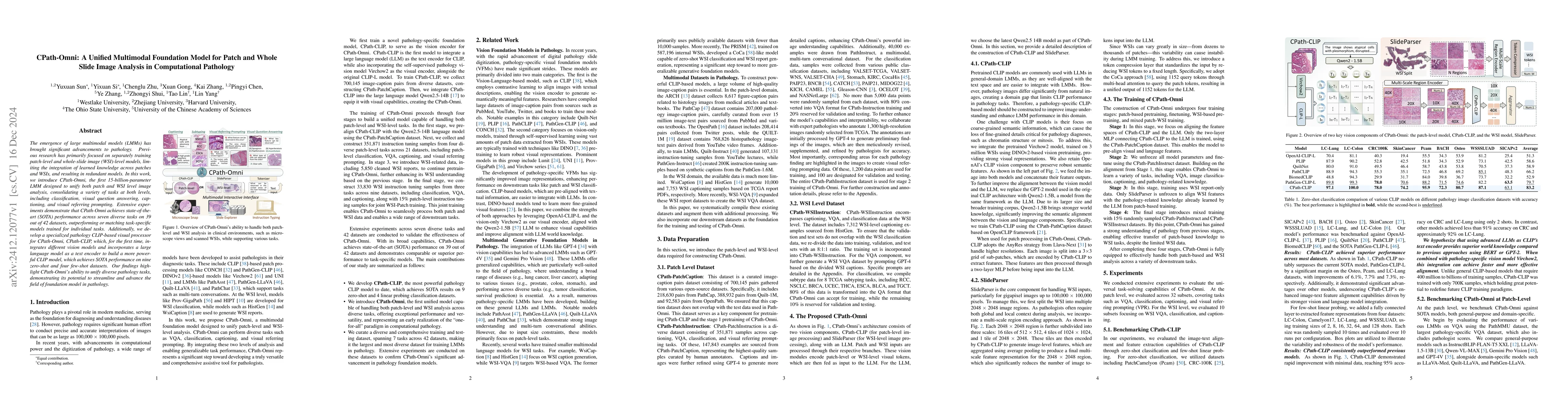
The emergence of large multimodal models (LMMs) has brought significant advancements to pathology. Previous research has primarily focused on separately training patch-level and whole-slide image (WSI...
Nucleus detection in histopathology whole slide images (WSIs) is crucial for a broad spectrum of clinical applications. Current approaches for nucleus detection in gigapixel WSIs utilize a sliding win...
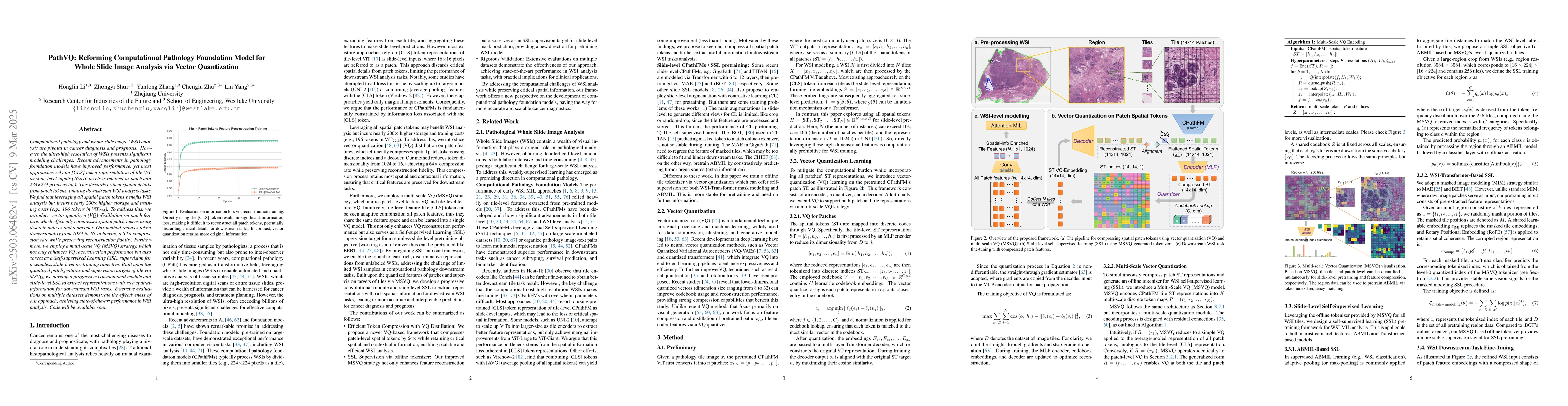
Computational pathology and whole-slide image (WSI) analysis are pivotal in cancer diagnosis and prognosis. However, the ultra-high resolution of WSIs presents significant modeling challenges. Recent ...
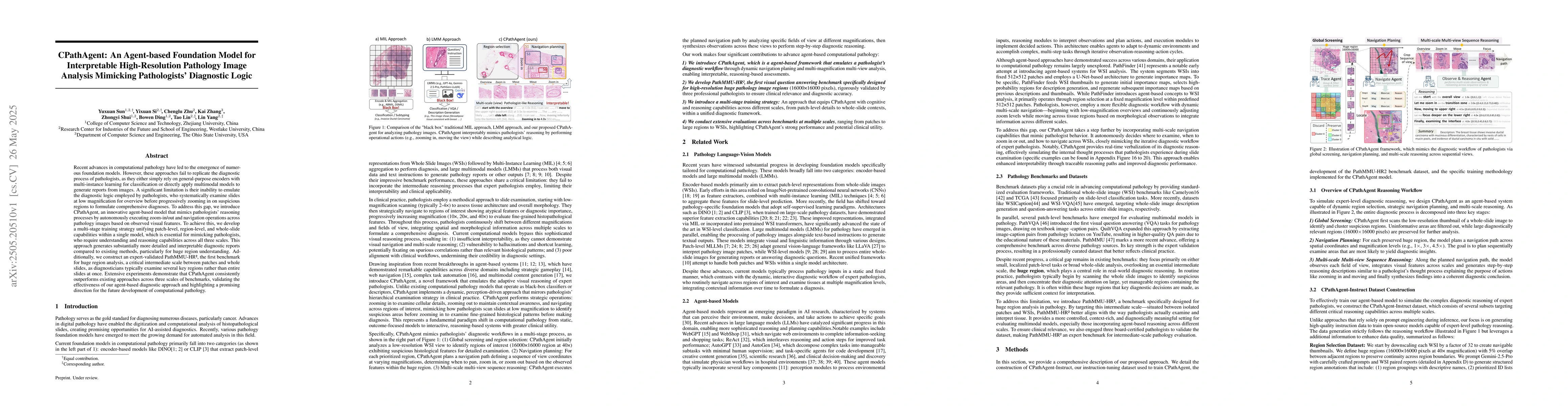
Recent advances in computational pathology have led to the emergence of numerous foundation models. However, these approaches fail to replicate the diagnostic process of pathologists, as they either s...
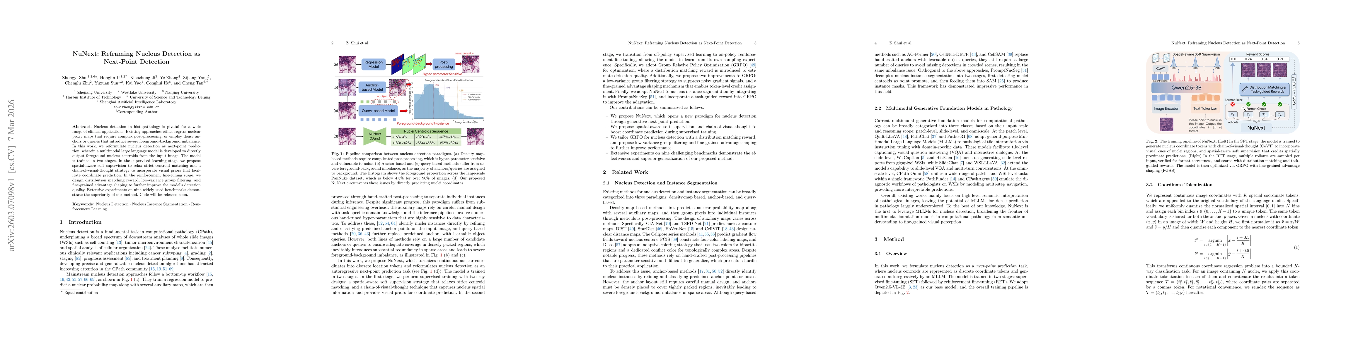
Nucleus detection in histopathology is pivotal for a wide range of clinical applications. Existing approaches either regress nuclear proxy maps that require complex post-processing, or employ dense an...