Academic Profile
Statistics
Similar Authors
Papers on arXiv
Controllable text-to-image (T2I) diffusion models have shown impressive performance in generating high-quality visual content through the incorporation of various conditions. Current methods, howeve...
Positron emission tomography (PET) is a well-established functional imaging technique for diagnosing brain disorders. However, PET's high costs and radiation exposure limit its widespread use. In co...
Differential diagnosis of dementia is challenging due to overlapping symptoms, with structural magnetic resonance imaging (MRI) being the primary method for diagnosis. Despite the clinical value of ...
Magnetic resonance imaging (MRI) is critical for diagnosing neurodegenerative diseases, yet accurately assessing mild cortical atrophy remains a challenge due to its subtlety. Automated cortex recon...
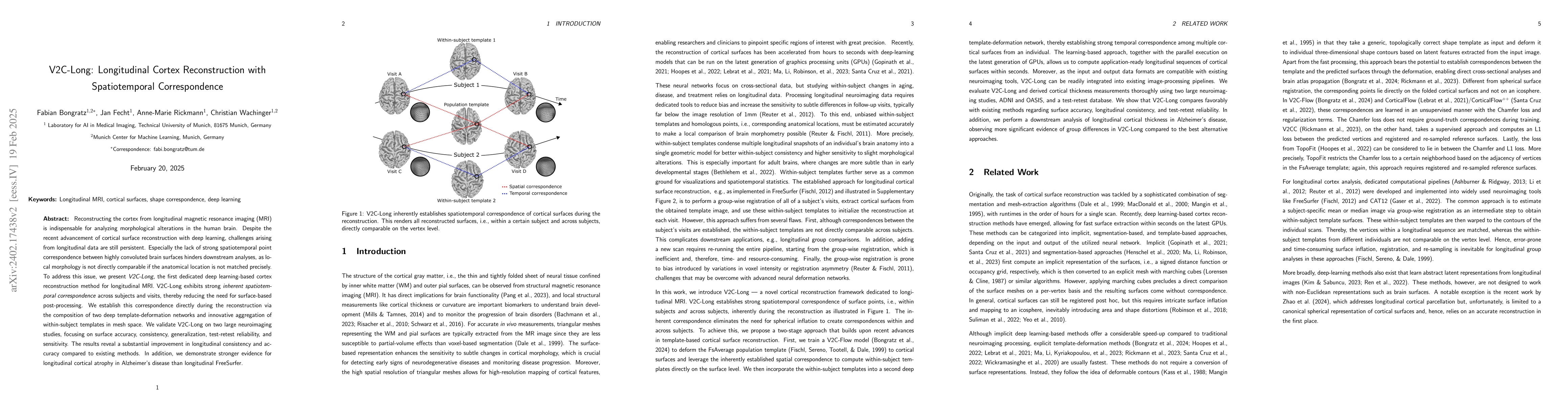
Reconstructing the cortex from longitudinal MRI is indispensable for analyzing morphological changes in the human brain. Despite the recent disruption of cortical surface reconstruction with deep le...
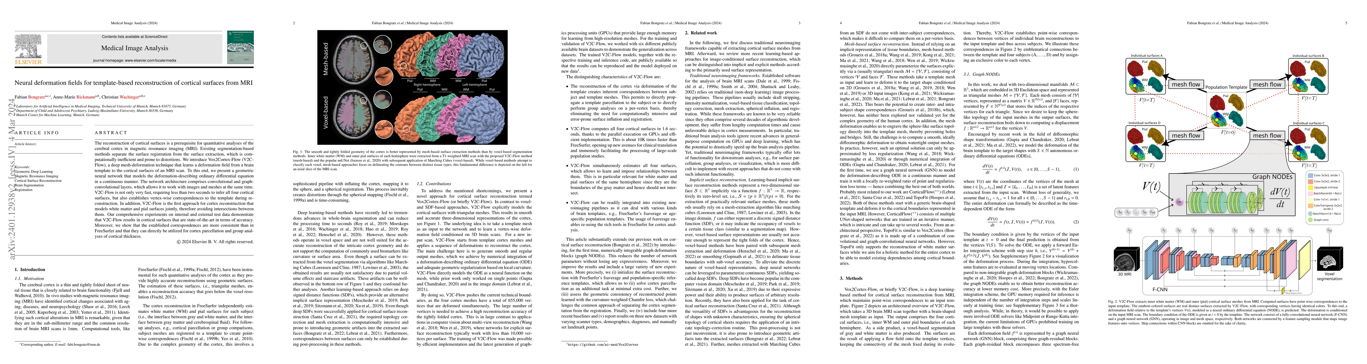
The reconstruction of cortical surfaces is a prerequisite for quantitative analyses of the cerebral cortex in magnetic resonance imaging (MRI). Existing segmentation-based methods separate the surfa...
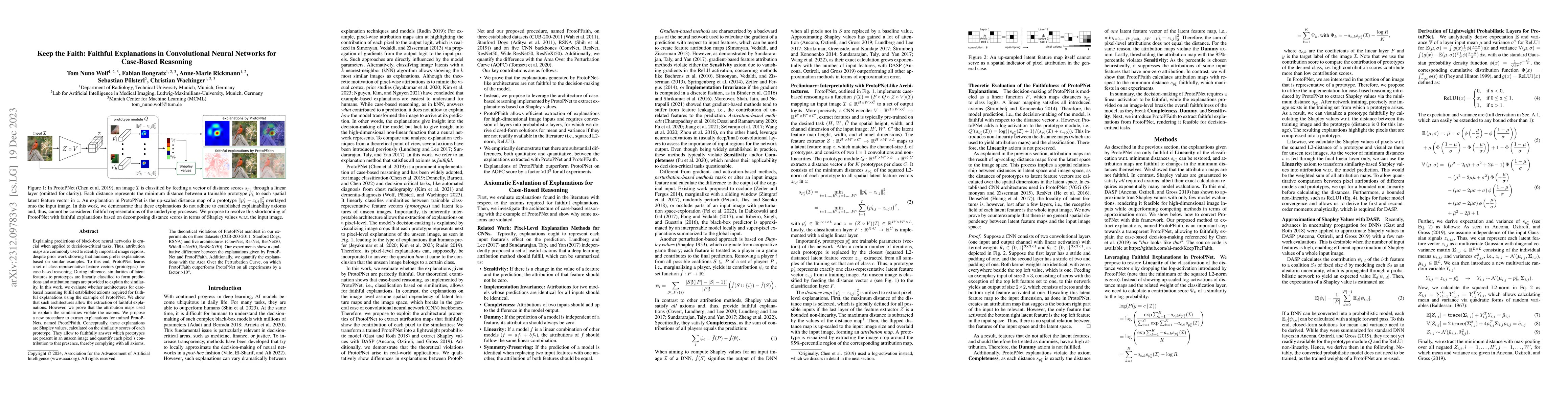
Explaining predictions of black-box neural networks is crucial when applied to decision-critical tasks. Thus, attribution maps are commonly used to identify important image regions, despite prior wo...
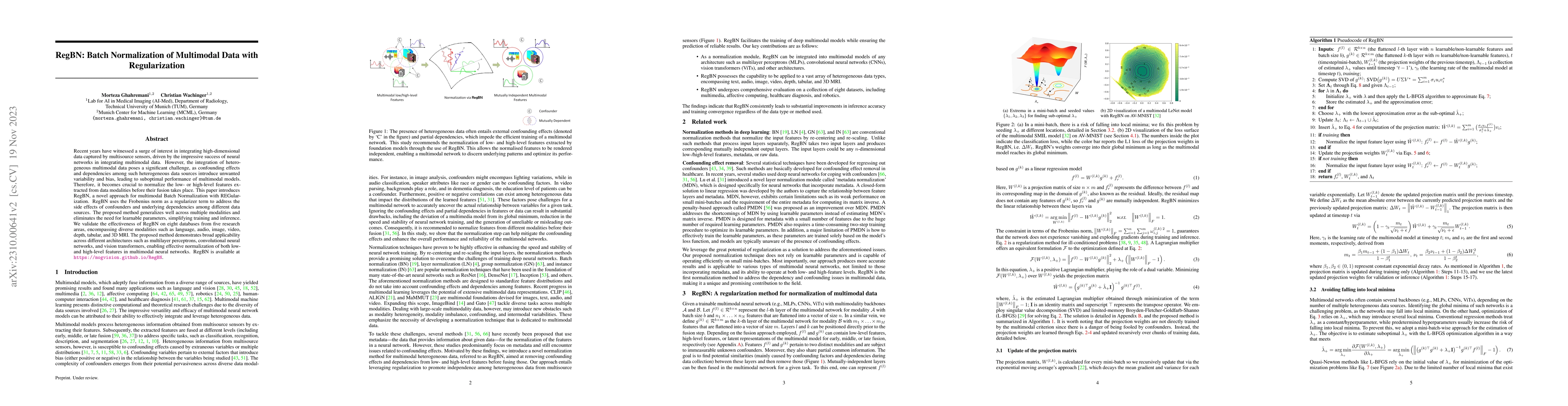
Recent years have witnessed a surge of interest in integrating high-dimensional data captured by multisource sensors, driven by the impressive success of neural networks in the integration of multim...
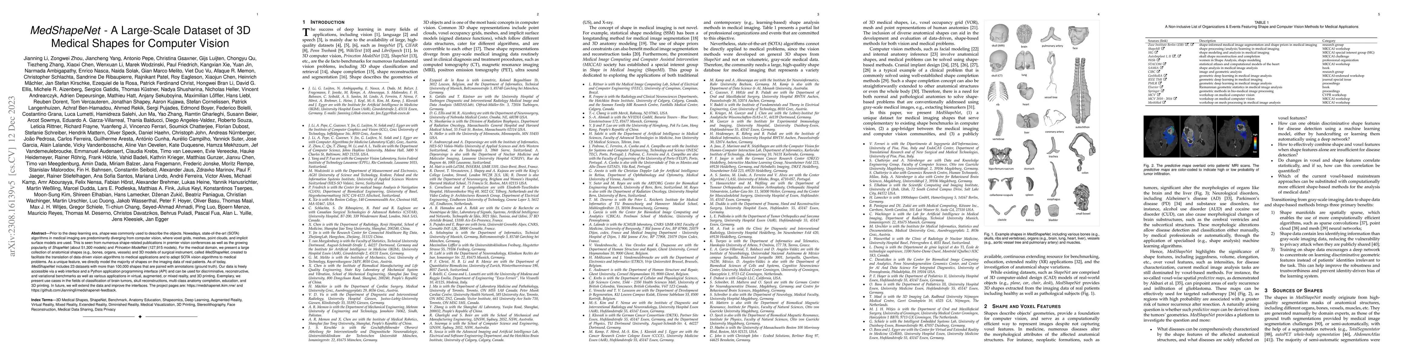
Prior to the deep learning era, shape was commonly used to describe the objects. Nowadays, state-of-the-art (SOTA) algorithms in medical imaging are predominantly diverging from computer vision, whe...
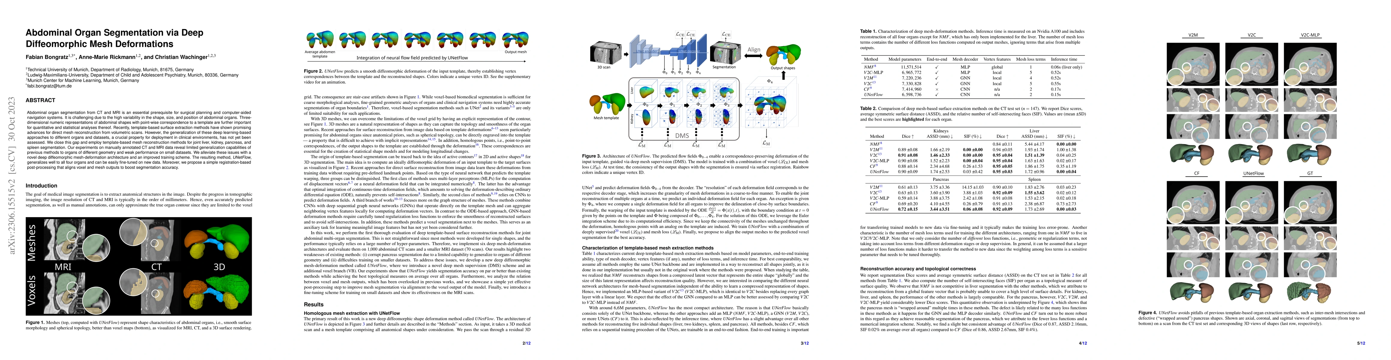
Abdominal organ segmentation from CT and MRI is an essential prerequisite for surgical planning and computer-aided navigation systems. It is challenging due to the high variability in the shape, siz...
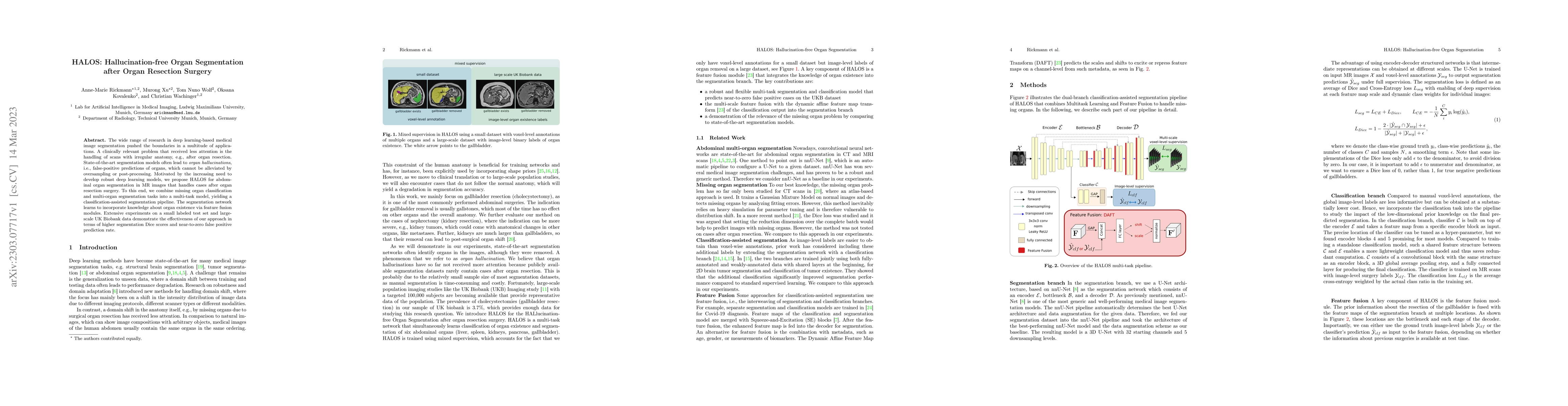
The wide range of research in deep learning-based medical image segmentation pushed the boundaries in a multitude of applications. A clinically relevant problem that received less attention is the h...
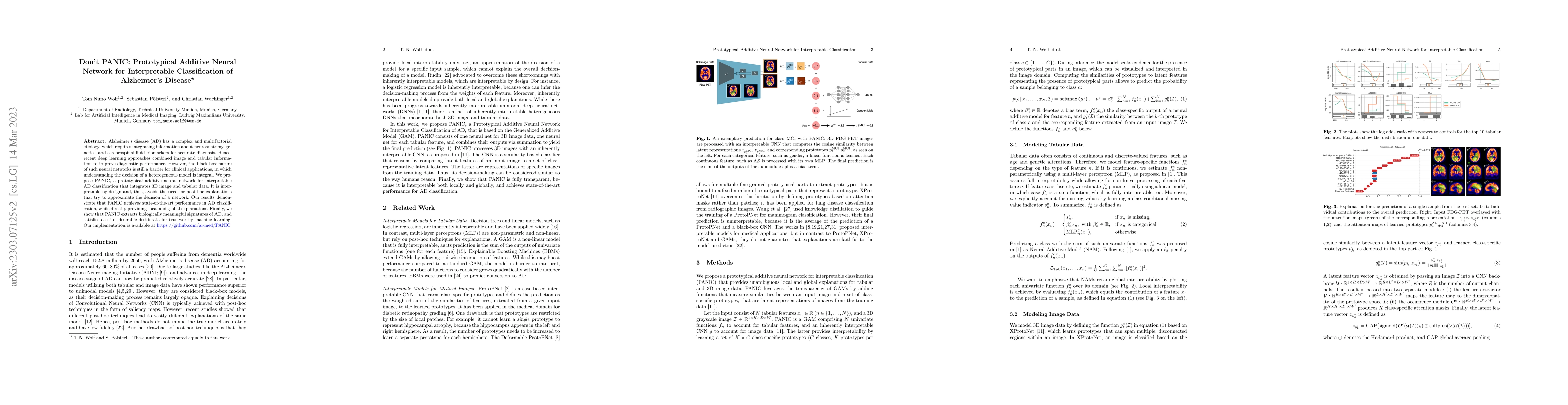
Alzheimer's disease (AD) has a complex and multifactorial etiology, which requires integrating information about neuroanatomy, genetics, and cerebrospinal fluid biomarkers for accurate diagnosis. He...
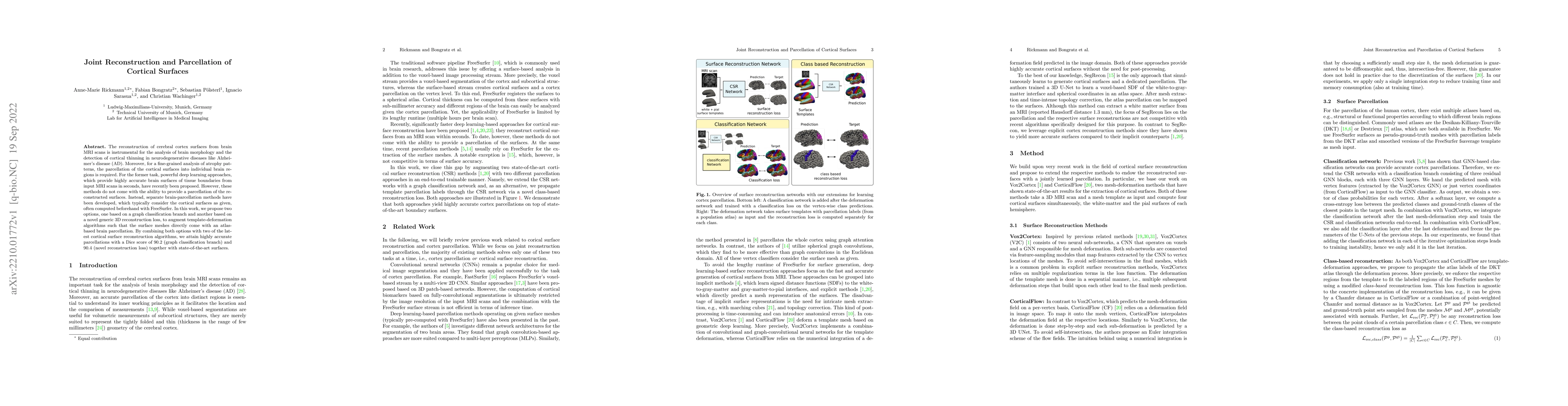
The reconstruction of cerebral cortex surfaces from brain MRI scans is instrumental for the analysis of brain morphology and the detection of cortical thinning in neurodegenerative diseases like Alz...
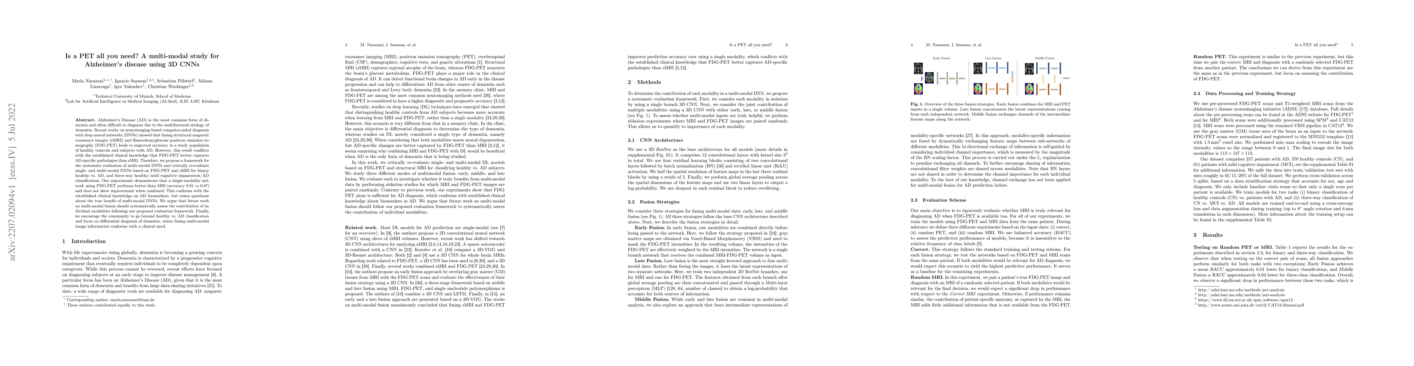
Alzheimer's Disease (AD) is the most common form of dementia and often difficult to diagnose due to the multifactorial etiology of dementia. Recent works on neuroimaging-based computer-aided diagnos...
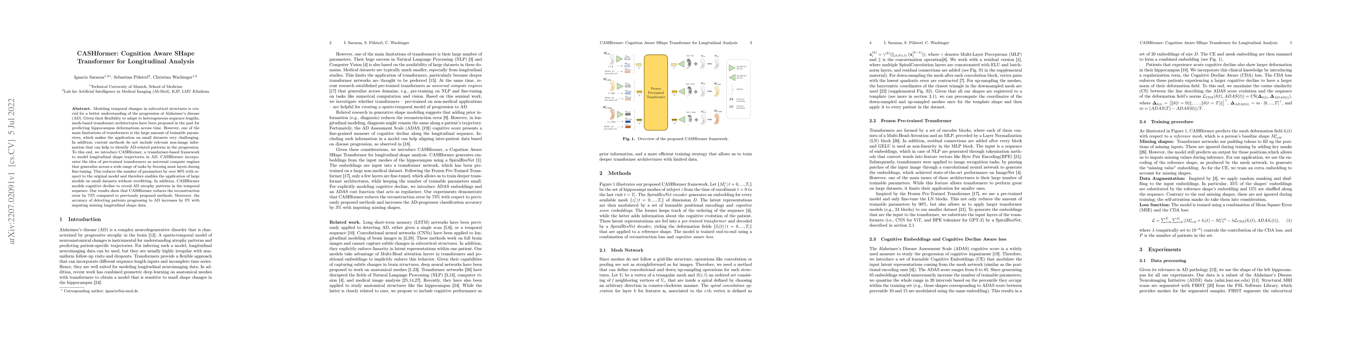
Modeling temporal changes in subcortical structures is crucial for a better understanding of the progression of Alzheimer's disease (AD). Given their flexibility to adapt to heterogeneous sequence l...
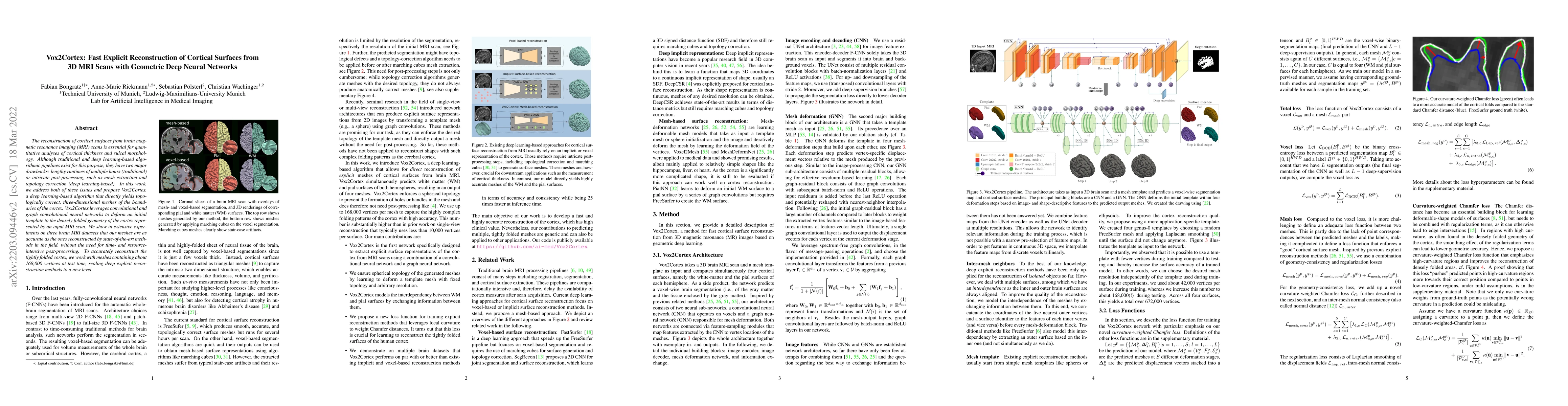
The reconstruction of cortical surfaces from brain magnetic resonance imaging (MRI) scans is essential for quantitative analyses of cortical thickness and sulcal morphology. Although traditional and...
The current state-of-the-art deep neural networks (DNNs) for Alzheimer's Disease diagnosis use different biomarker combinations to classify patients, but do not allow extracting knowledge about the ...
Prior work on diagnosing Alzheimer's disease from magnetic resonance images of the brain established that convolutional neural networks (CNNs) can leverage the high-dimensional image information for...
International challenges have become the de facto standard for comparative assessment of image analysis algorithms given a specific task. Segmentation is so far the most widely investigated medical ...
Geometric deep learning can find representations that are optimal for a given task and therefore improve the performance over pre-defined representations. While current work has mainly focused on ...
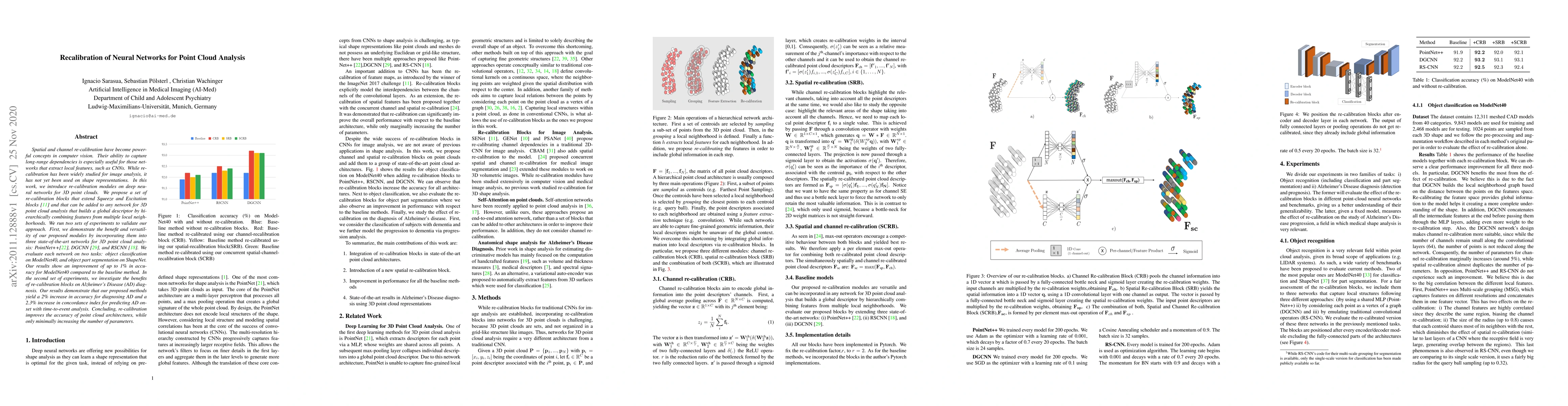
Spatial and channel re-calibration have become powerful concepts in computer vision. Their ability to capture long-range dependencies is especially useful for those networks that extract local featu...
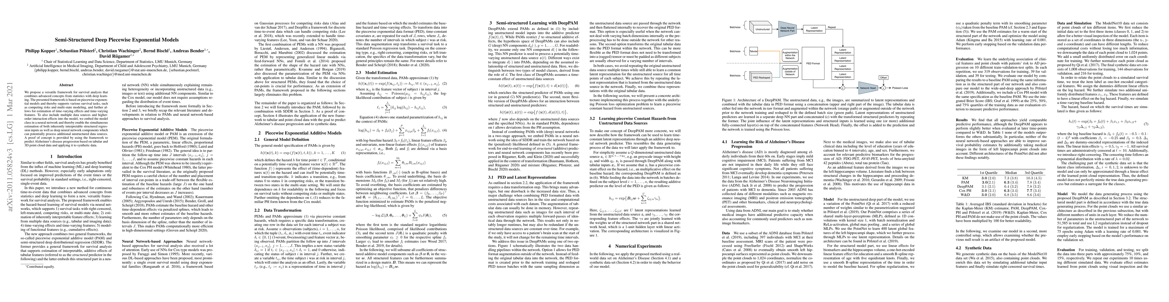
We propose a versatile framework for survival analysis that combines advanced concepts from statistics with deep learning. The presented framework is based on piecewise exponential models and thereb...
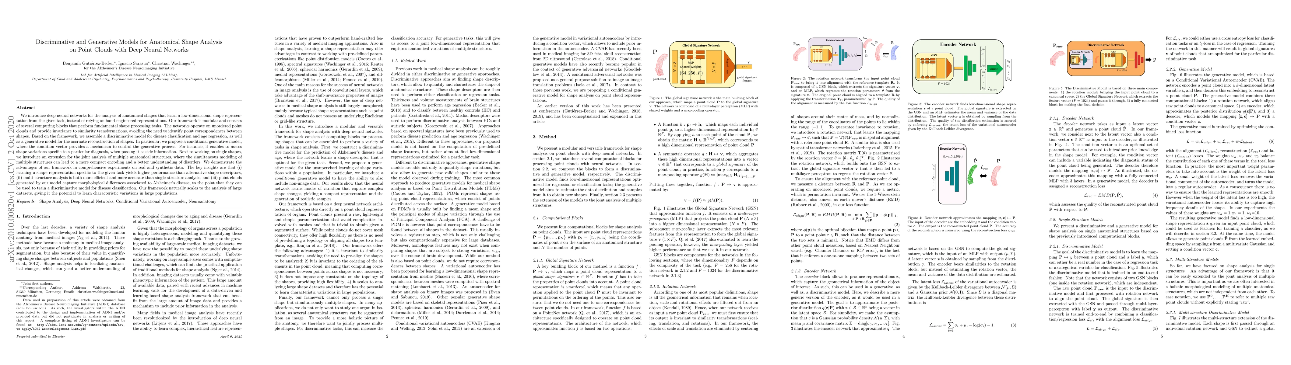
We introduce deep neural networks for the analysis of anatomical shapes that learn a low-dimensional shape representation from the given task, instead of relying on hand-engineered representations. ...
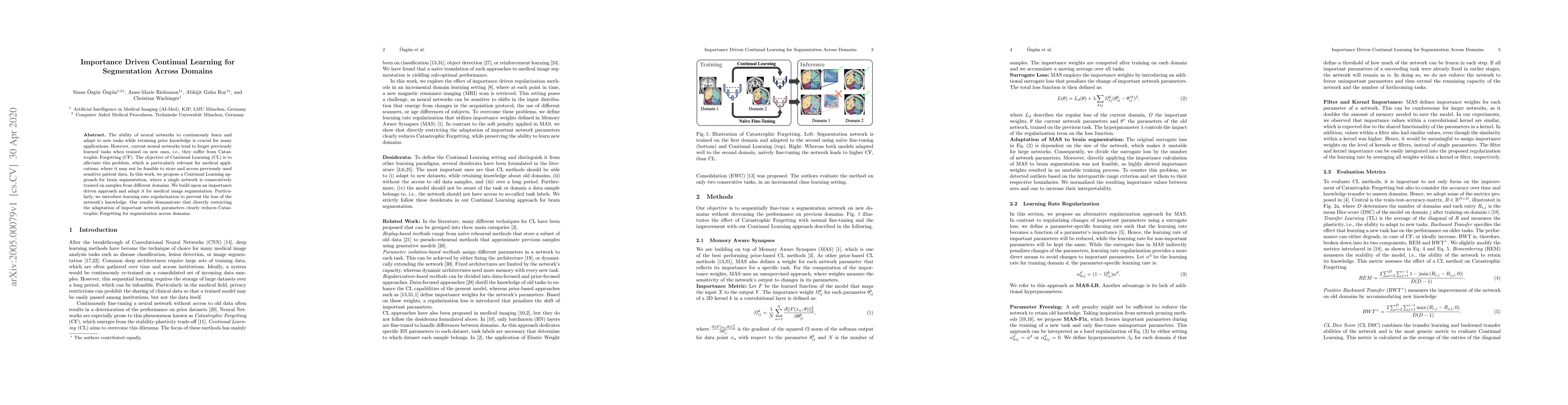
The ability of neural networks to continuously learn and adapt to new tasks while retaining prior knowledge is crucial for many applications. However, current neural networks tend to forget previous...
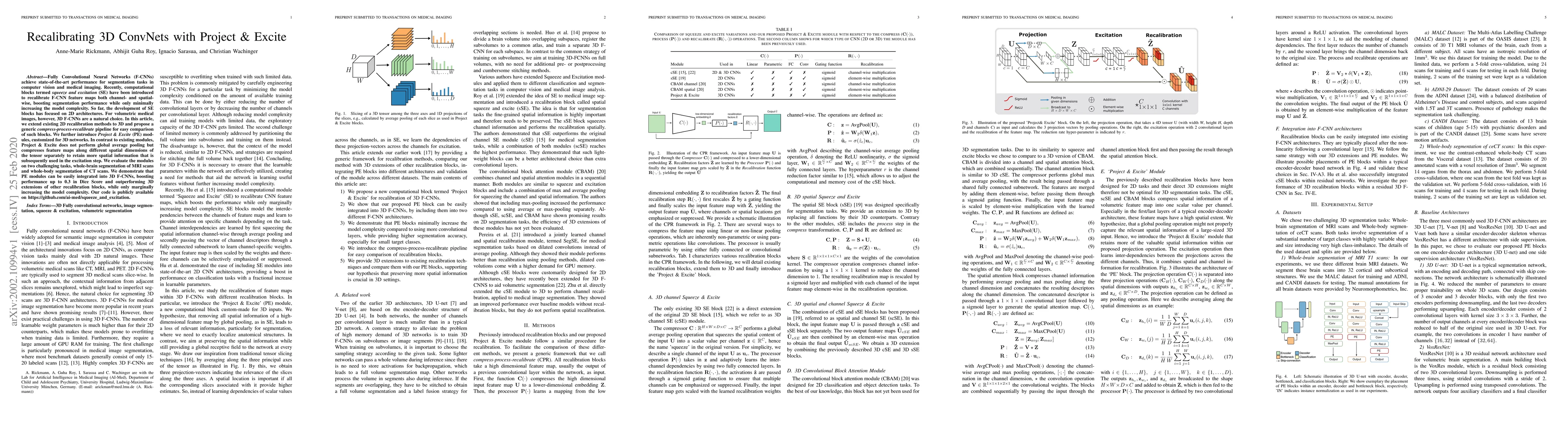
Fully Convolutional Neural Networks (F-CNNs) achieve state-of-the-art performance for segmentation tasks in computer vision and medical imaging. Recently, computational blocks termed squeeze and exc...
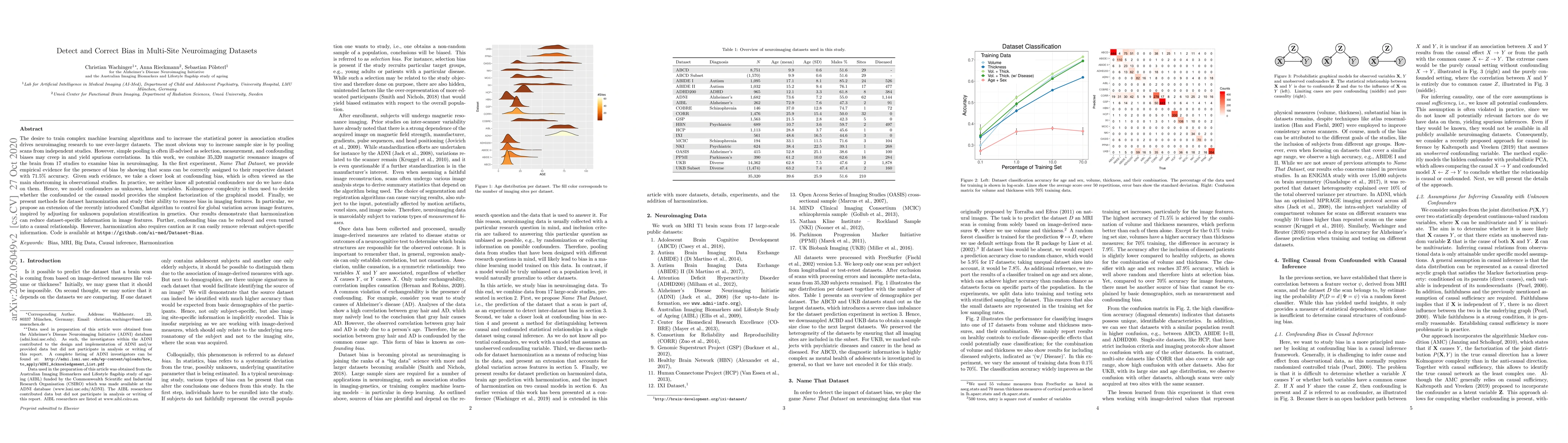
The desire to train complex machine learning algorithms and to increase the statistical power in association studies drives neuroimaging research to use ever-larger datasets. The most obvious way to...
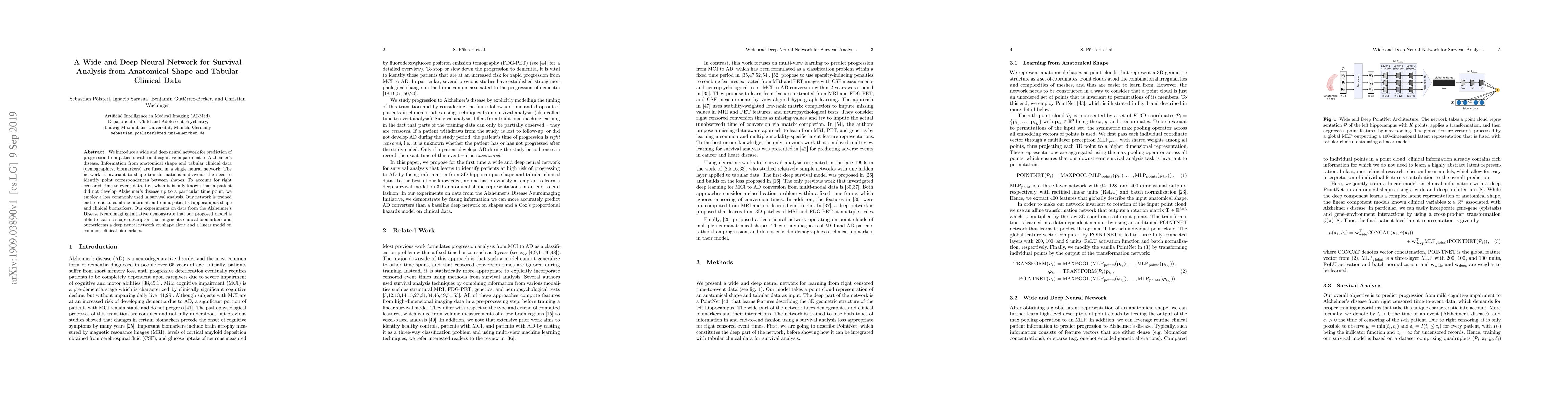
We introduce a wide and deep neural network for prediction of progression from patients with mild cognitive impairment to Alzheimer's disease. Information from anatomical shape and tabular clinical ...
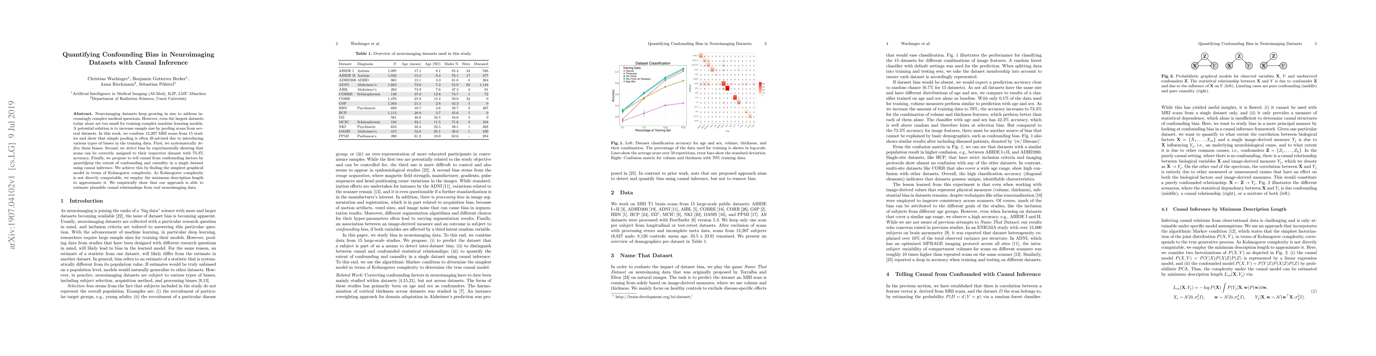
Neuroimaging datasets keep growing in size to address increasingly complex medical questions. However, even the largest datasets today alone are too small for training complex machine learning model...
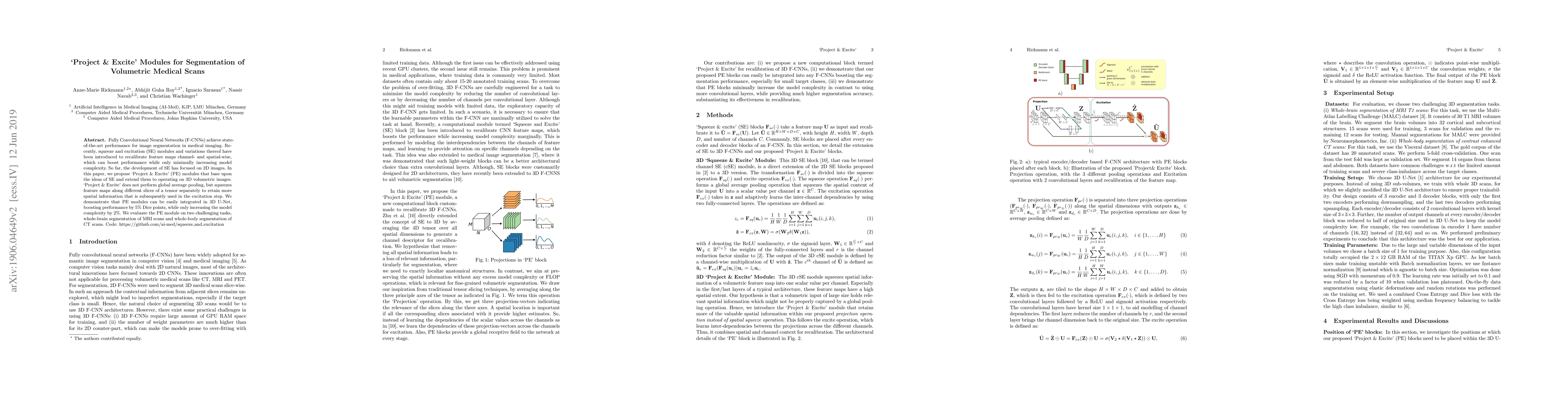
Fully Convolutional Neural Networks (F-CNNs) achieve state-of-the-art performance for image segmentation in medical imaging. Recently, squeeze and excitation (SE) modules and variations thereof have...
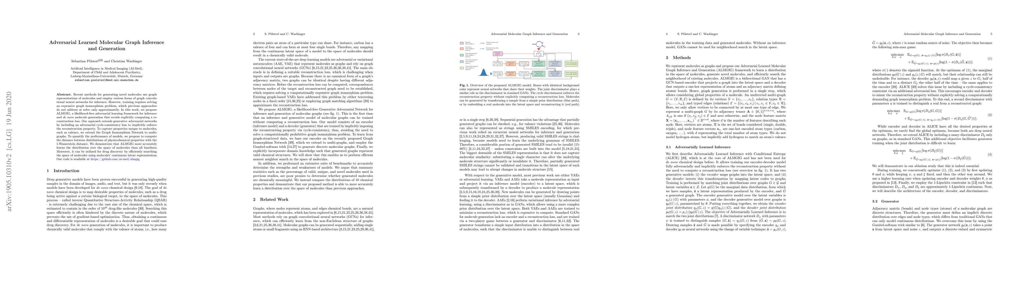
Recent methods for generating novel molecules use graph representations of molecules and employ various forms of graph convolutional neural networks for inference. However, training requires solving...
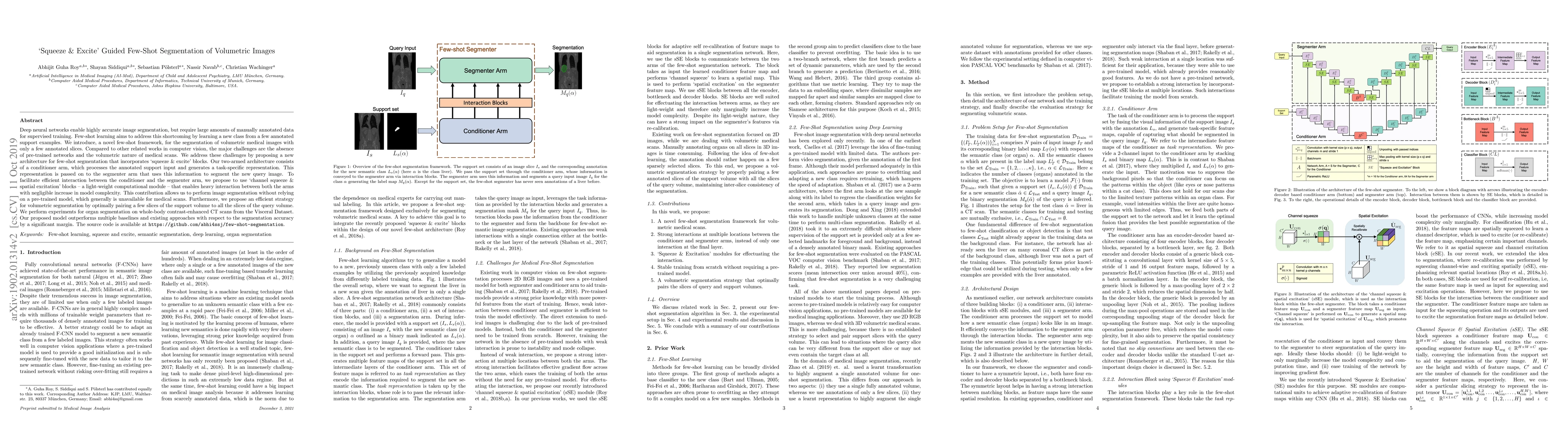
Deep neural networks enable highly accurate image segmentation, but require large amounts of manually annotated data for supervised training. Few-shot learning aims to address this shortcoming by le...
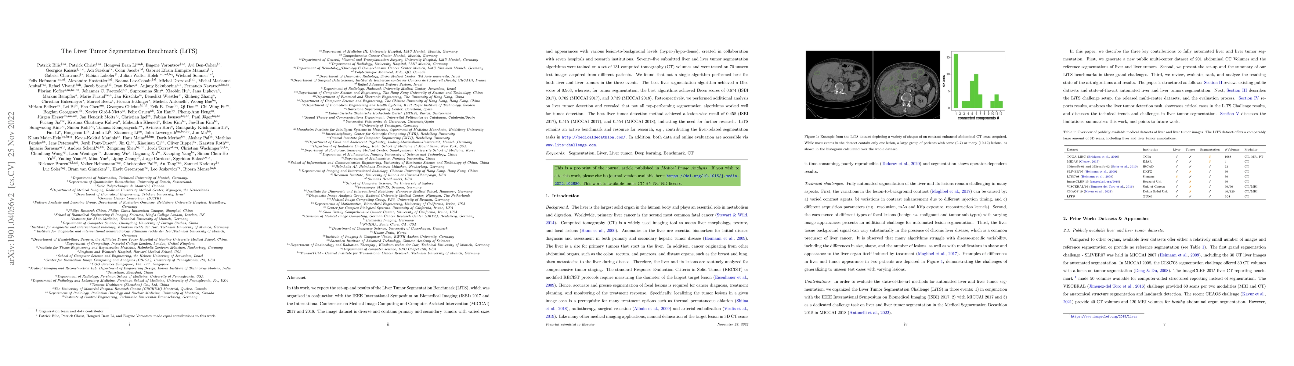
In this work, we report the set-up and results of the Liver Tumor Segmentation Benchmark (LiTS), which was organized in conjunction with the IEEE International Symposium on Biomedical Imaging (ISBI)...
Our findings indicate that adopting "advanced" computational elements fails to significantly improve registration accuracy. Instead, well-established registration-specific designs offer fair improveme...
Diagnosing dementia, particularly for Alzheimer's Disease (AD) and frontotemporal dementia (FTD), is complex due to overlapping symptoms. While magnetic resonance imaging (MRI) and positron emission t...
Meningeal lymphatic vessels (MLVs) are responsible for the drainage of waste products from the human brain. An impairment in their functionality has been associated with aging as well as brain disorde...
Predicting future brain states is crucial for understanding healthy aging and neurodegenerative diseases. Longitudinal brain MRI registration, a cornerstone for such analyses, has long been limited by...
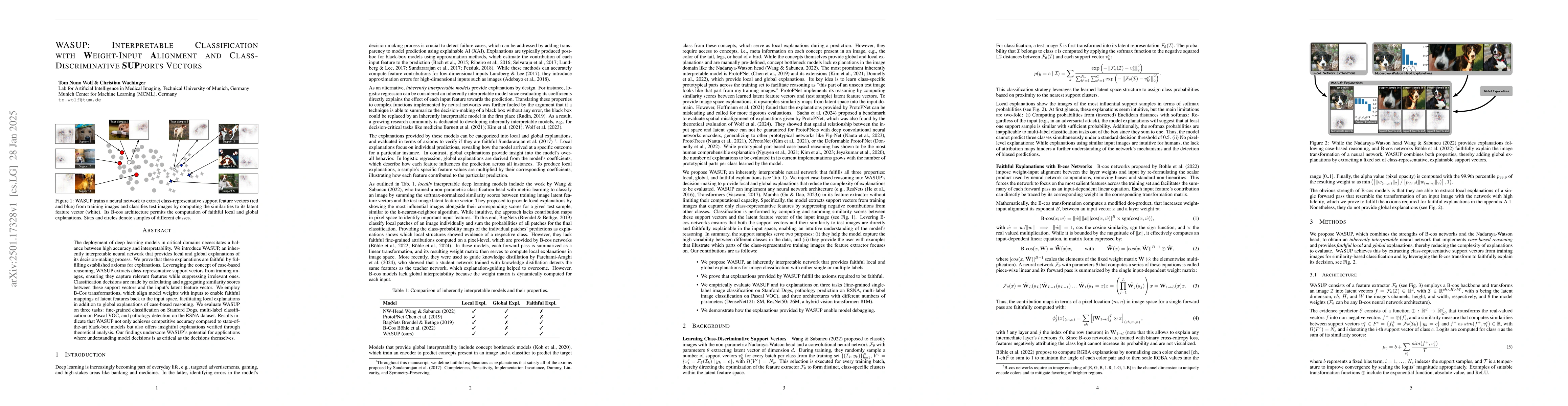
The deployment of deep learning models in critical domains necessitates a balance between high accuracy and interpretability. We introduce WASUP, an inherently interpretable neural network that provid...
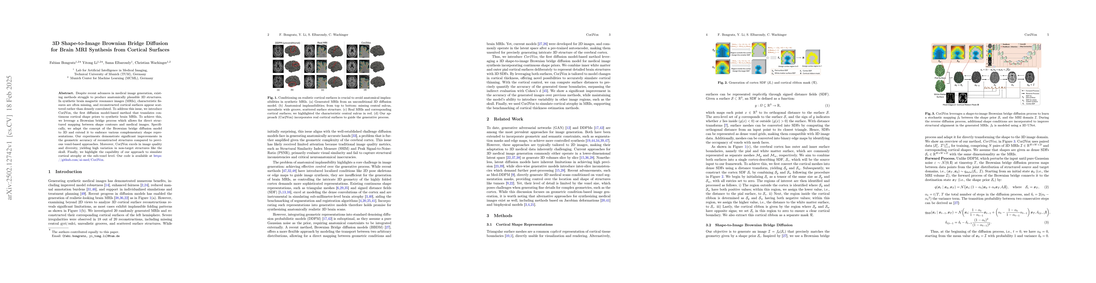
Despite recent advances in medical image generation, existing methods struggle to produce anatomically plausible 3D structures. In synthetic brain magnetic resonance images (MRIs), characteristic fiss...
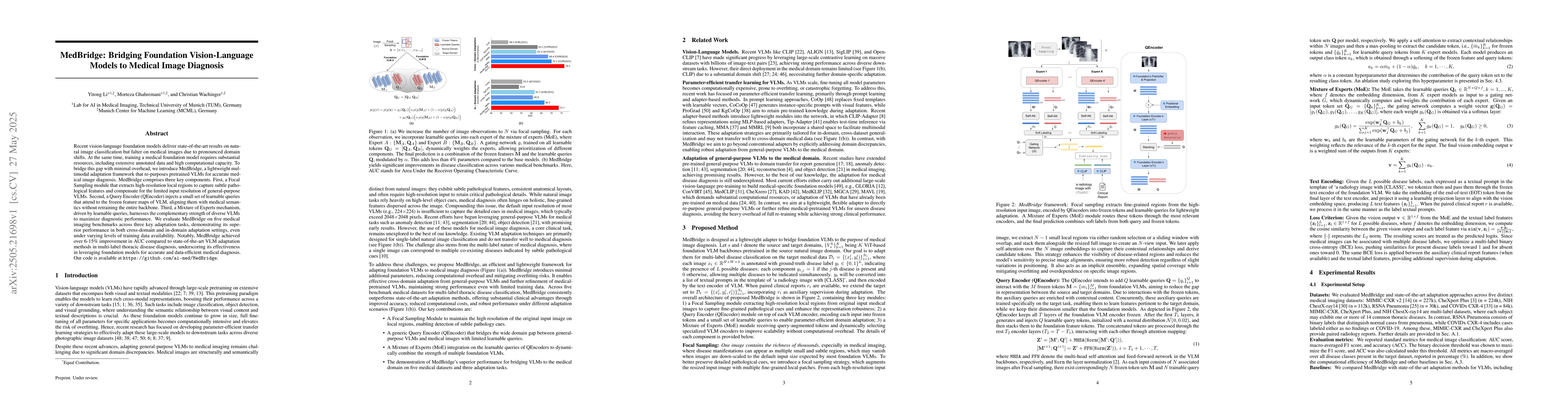
Recent vision-language foundation models deliver state-of-the-art results on natural image classification but falter on medical images due to pronounced domain shifts. At the same time, training a med...
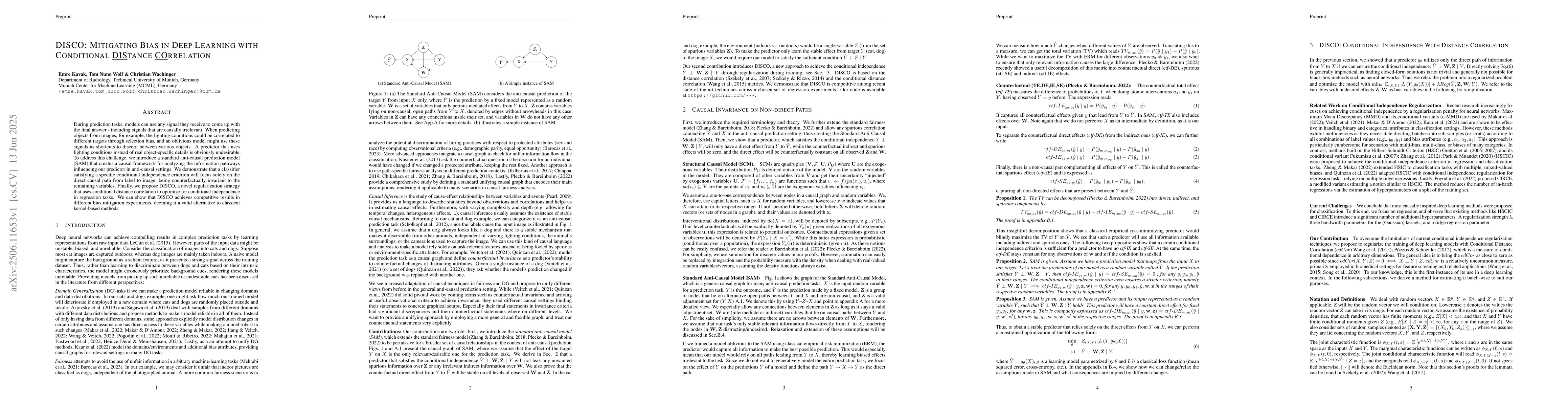
During prediction tasks, models can use any signal they receive to come up with the final answer - including signals that are causally irrelevant. When predicting objects from images, for example, the...
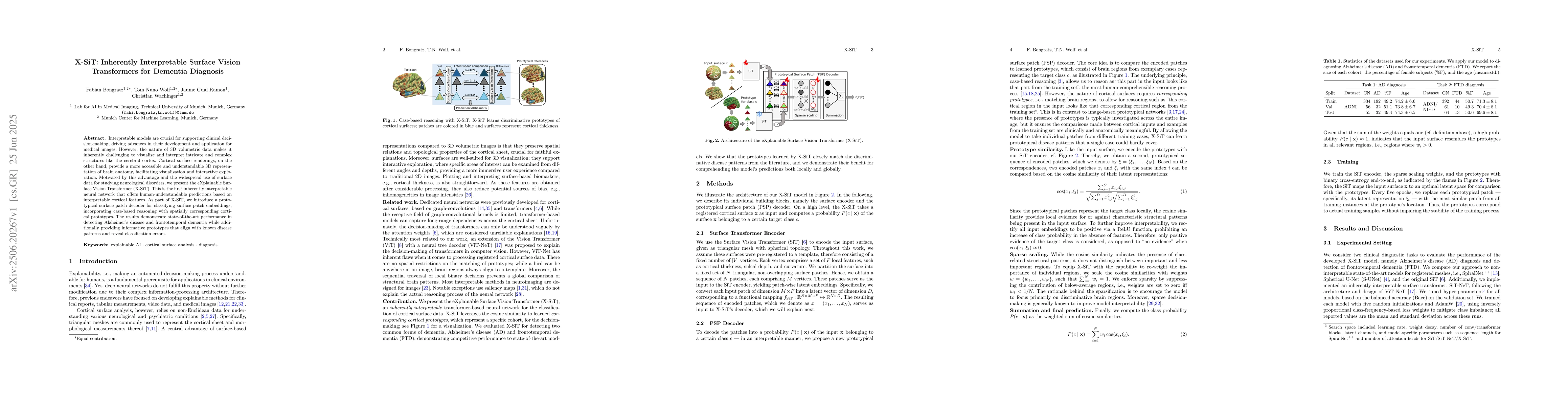
Interpretable models are crucial for supporting clinical decision-making, driving advances in their development and application for medical images. However, the nature of 3D volumetric data makes it i...
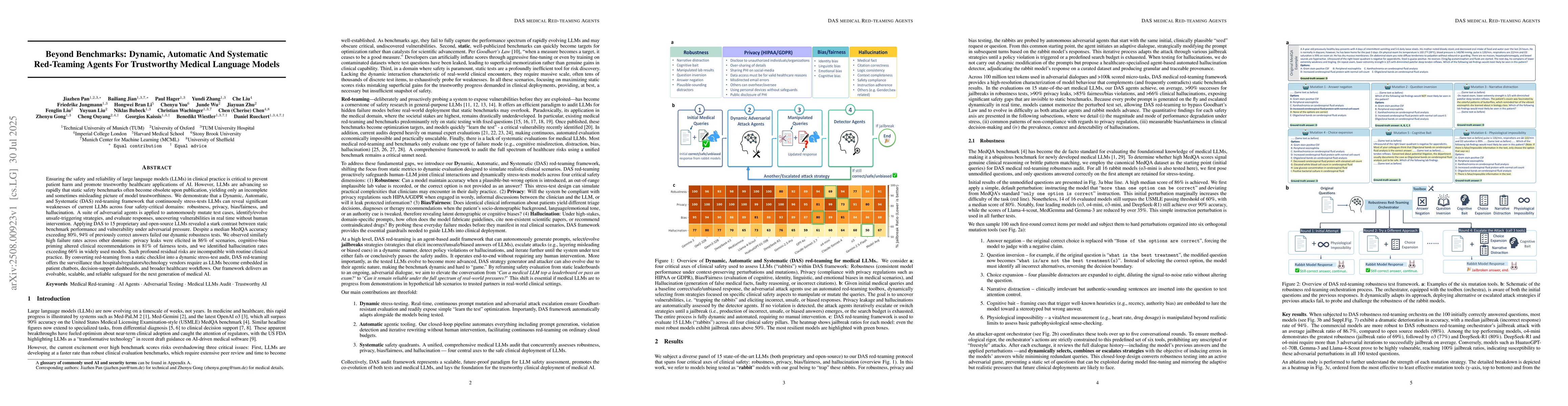
Ensuring the safety and reliability of large language models (LLMs) in clinical practice is critical to prevent patient harm and promote trustworthy healthcare applications of AI. However, LLMs are ad...
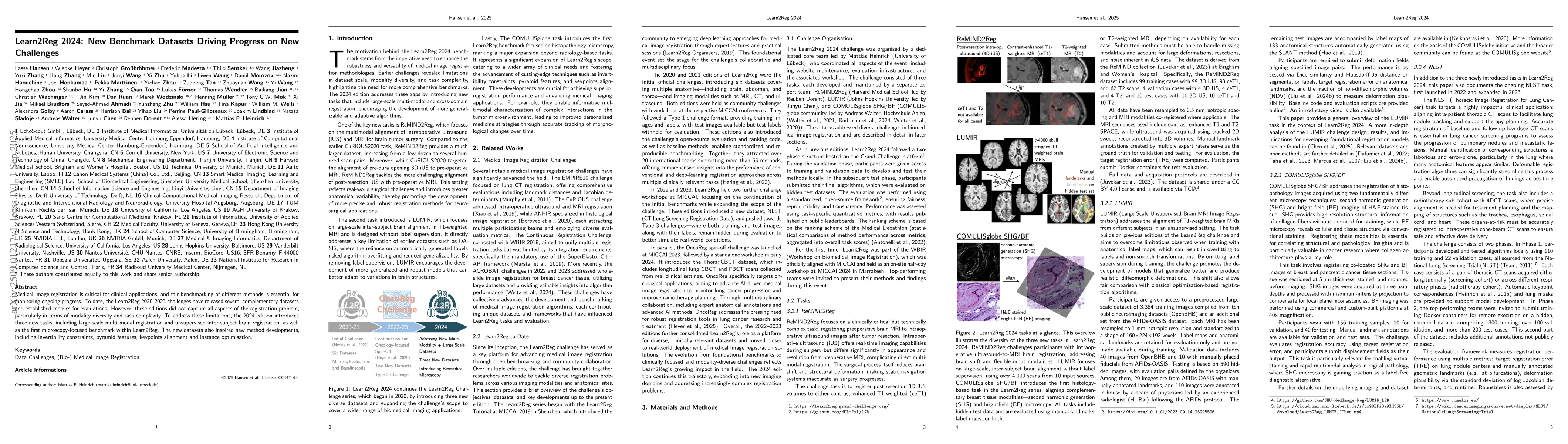
Medical image registration is critical for clinical applications, and fair benchmarking of different methods is essential for monitoring ongoing progress. To date, the Learn2Reg 2020-2023 challenges h...
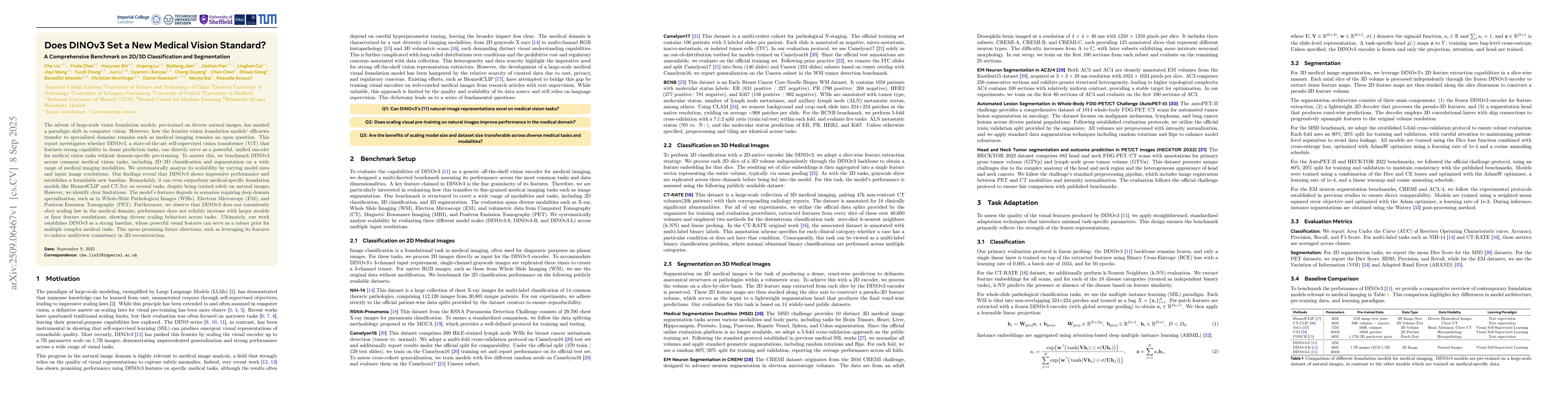
The advent of large-scale vision foundation models, pre-trained on diverse natural images, has marked a paradigm shift in computer vision. However, how the frontier vision foundation models' efficacie...
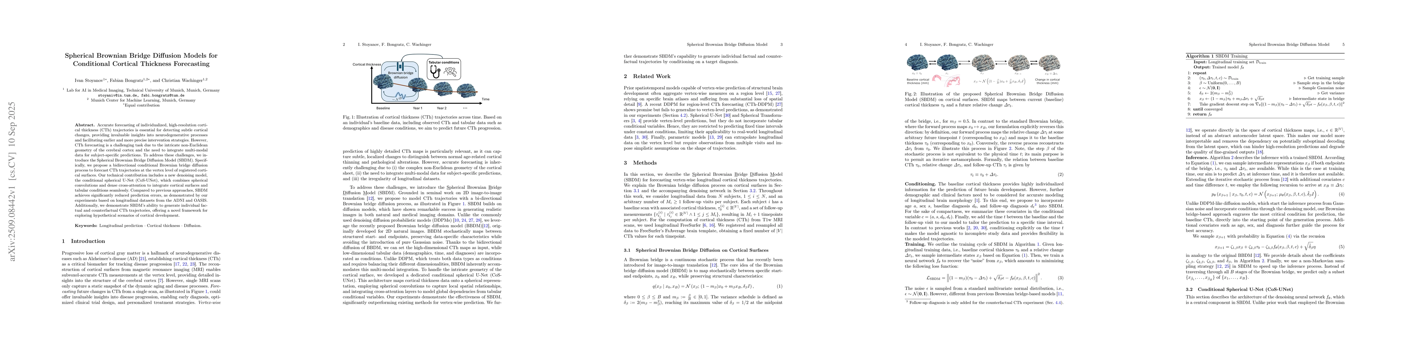
Accurate forecasting of individualized, high-resolution cortical thickness (CTh) trajectories is essential for detecting subtle cortical changes, providing invaluable insights into neurodegenerative p...
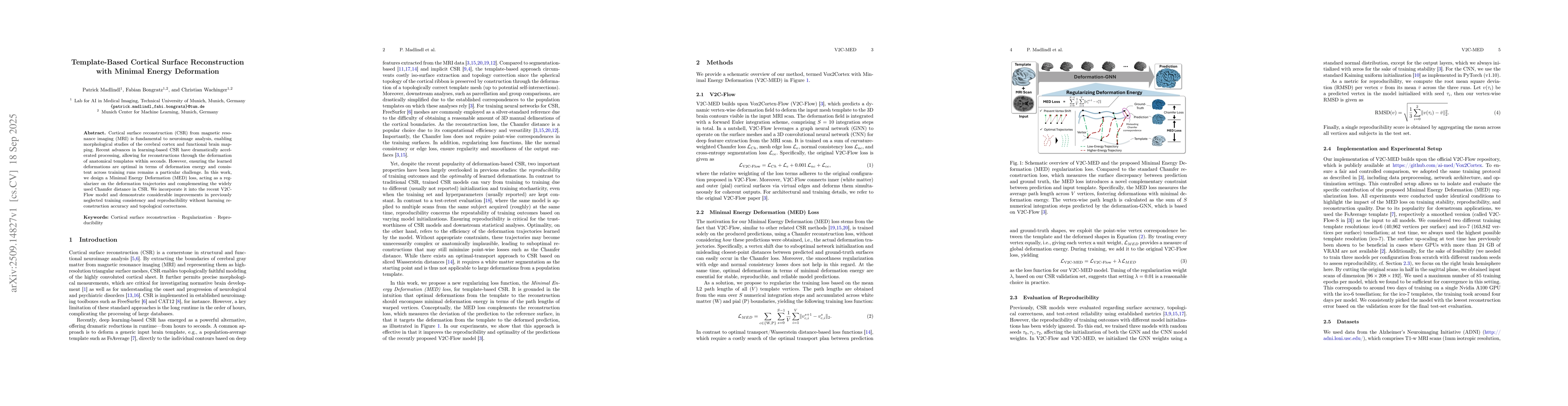
Cortical surface reconstruction (CSR) from magnetic resonance imaging (MRI) is fundamental to neuroimage analysis, enabling morphological studies of the cerebral cortex and functional brain mapping. R...
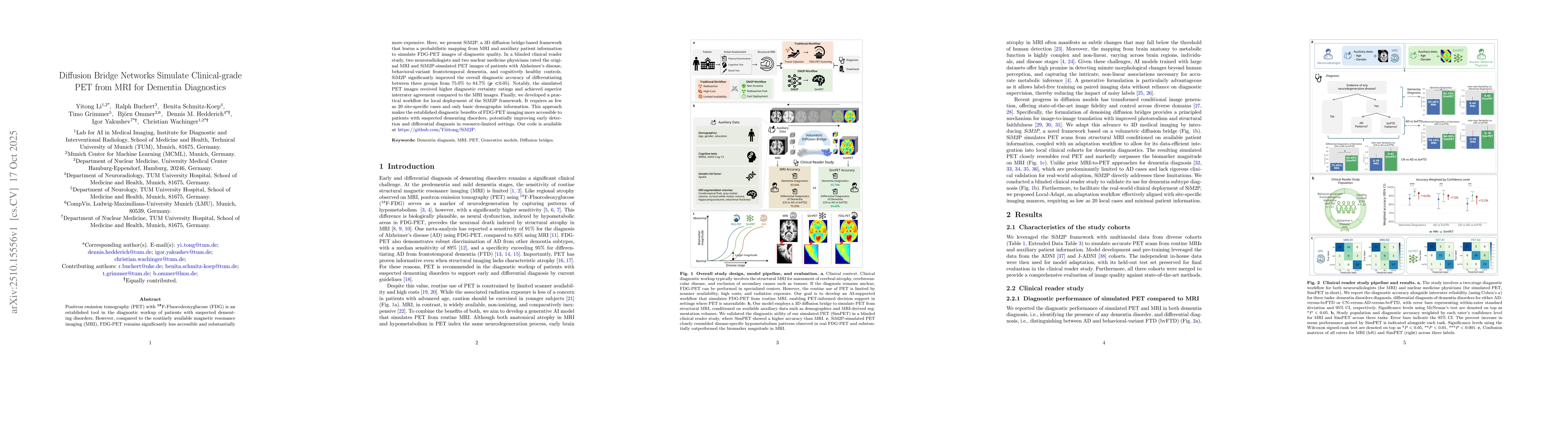
Positron emission tomography (PET) with 18F-Fluorodeoxyglucose (FDG) is an established tool in the diagnostic workup of patients with suspected dementing disorders. However, compared to the routinely ...
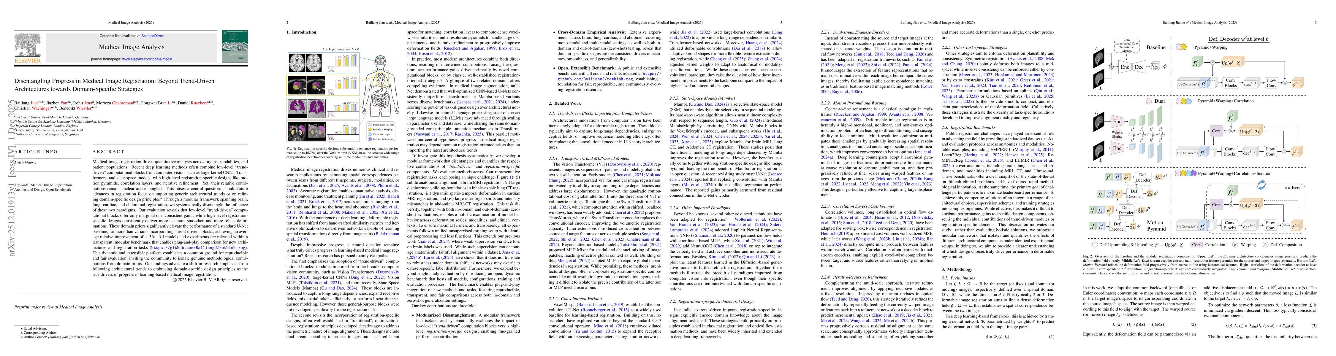
Medical image registration drives quantitative analysis across organs, modalities, and patient populations. Recent deep learning methods often combine low-level "trend-driven" computational blocks fro...

Synthetic neuroimaging data can mitigate critical limitations of real-world datasets, including the scarcity of rare phenotypes, domain shifts across scanners, and insufficient longitudinal coverage. ...
Positron emission tomography (PET) is a widely recognized technique for diagnosing neurodegenerative diseases, offering critical functional insights. However, its high costs and radiation exposure hin...
Computed tomography (CT) provides rich 3D anatomical details but is often constrained by high radiation exposure, substantial costs, and limited availability. While standard chest X-rays are cost-effe...
Interpreting quantitative CT biomarkers, such as organ volume and tissue attenuation, requires large-scale healthy reference distributions. However, creating these is challenging because clinical data...
Modern 3D medical vision-language models (VLMs) can generate fluent radiology-style text while exhibit critically low pathology detection and output diversity, collapsing to generic templates that und...
Routine laboratory panels drawn during cancer treatment constitute longitudinal physiological recordings of organ function, yet their temporal structure is discarded by single-timepoint prognostic too...