Academic Profile
Statistics
Similar Authors
Papers on arXiv
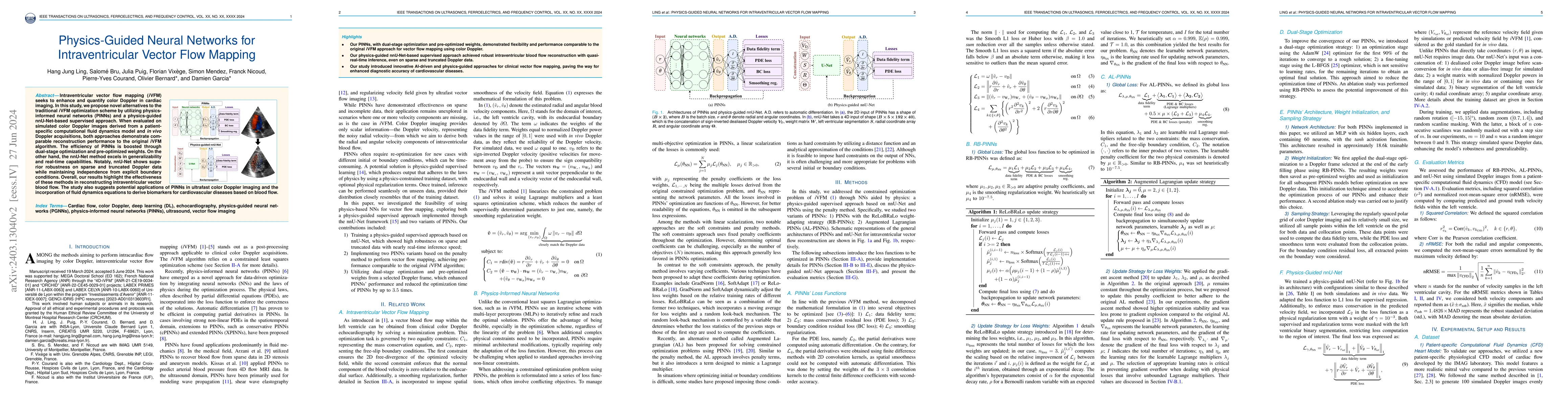
Intraventricular vector flow mapping (iVFM) seeks to enhance and quantify color Doppler in cardiac imaging. In this study, we propose novel alternatives to the traditional iVFM optimization scheme by ...
Color Doppler echocardiography enables visualization of blood flow within the heart. However, the limited frame rate impedes the quantitative assessment of blood velocity throughout the cardiac cycl...
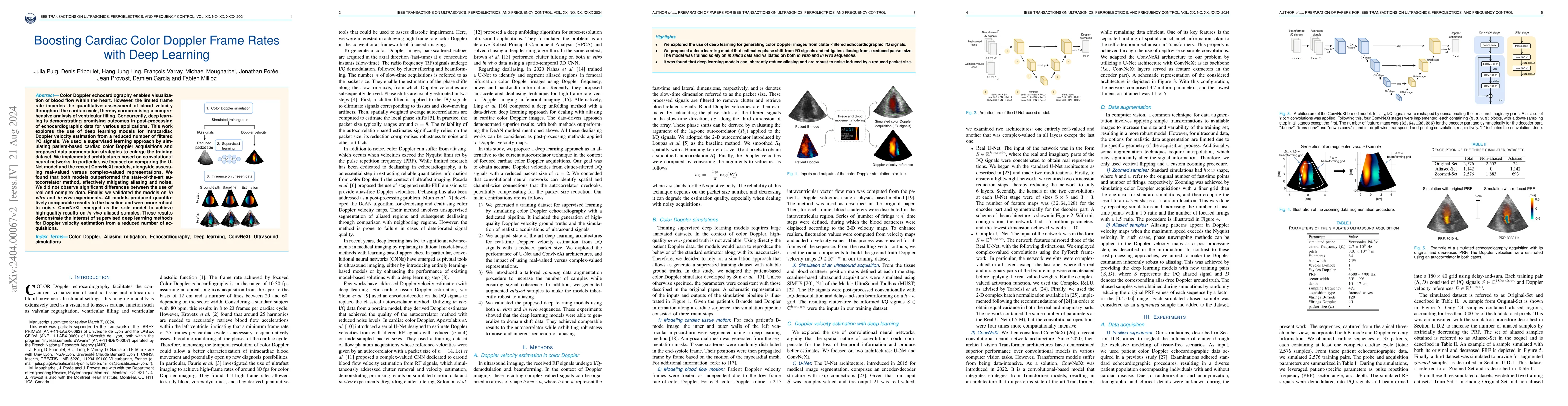
High-quality ultrafast ultrasound imaging is based on coherent compounding from multiple transmissions of plane waves (PW) or diverging waves (DW). However, compounding results in reduced frame rate...
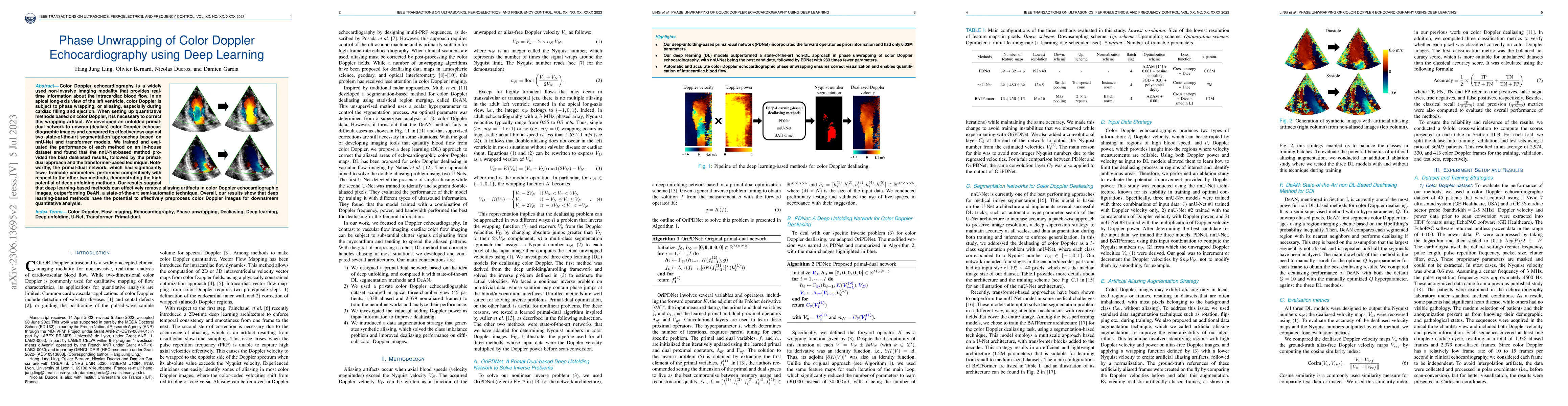
Color Doppler echocardiography is a widely used non-invasive imaging modality that provides real-time information about the intracardiac blood flow. In an apical long-axis view of the left ventricle...
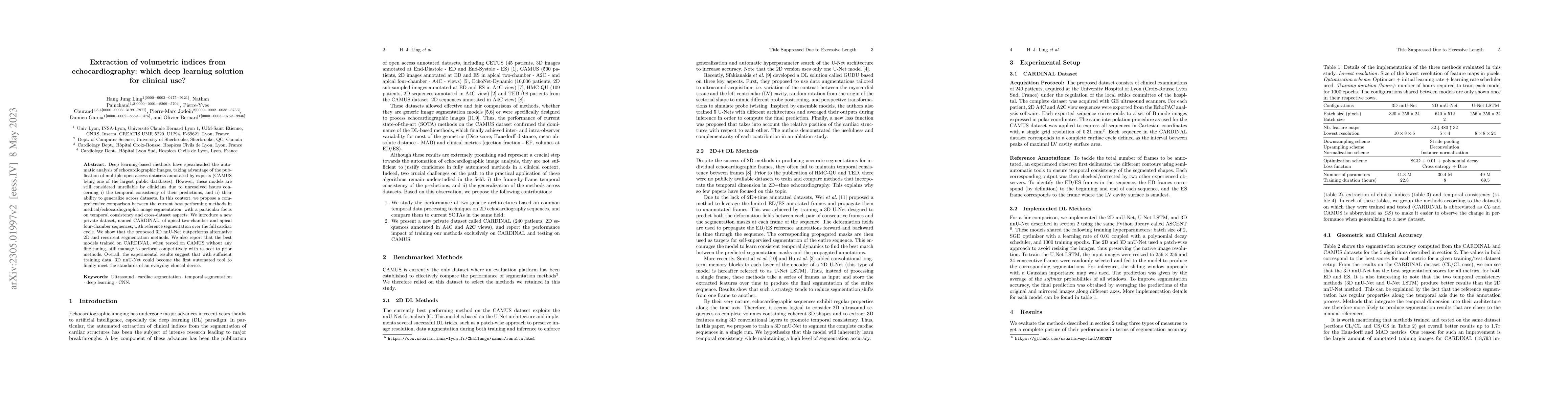
Deep learning-based methods have spearheaded the automatic analysis of echocardiographic images, taking advantage of the publication of multiple open access datasets annotated by experts (CAMUS bein...
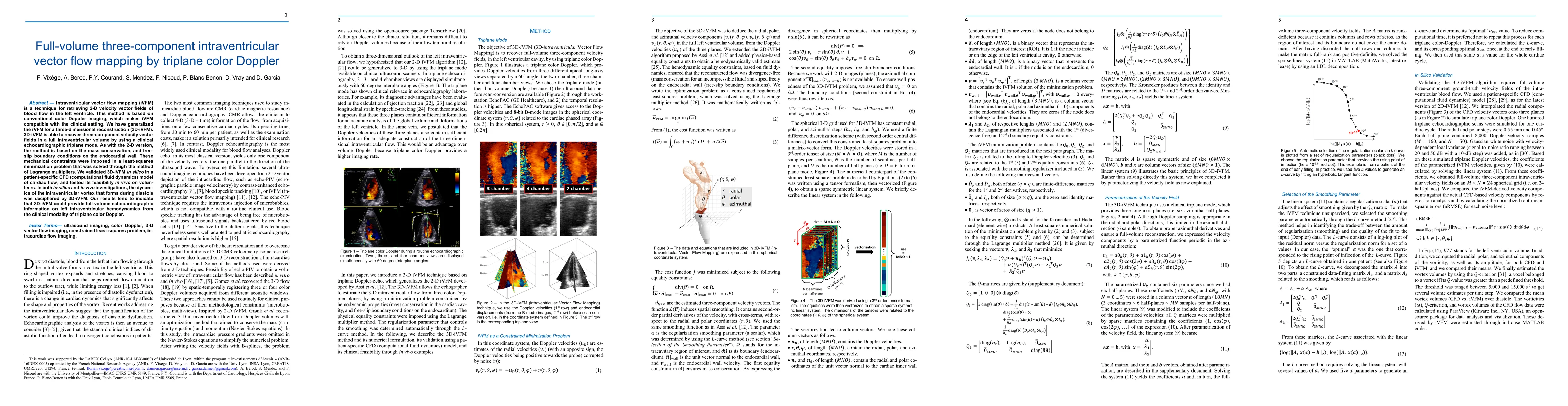
Intraventricular vector flow mapping (iVFM) is a technique for retrieving 2-D velocity vector fields of blood flow in the left ventricle. This method is based on conventional color Doppler imaging, ...
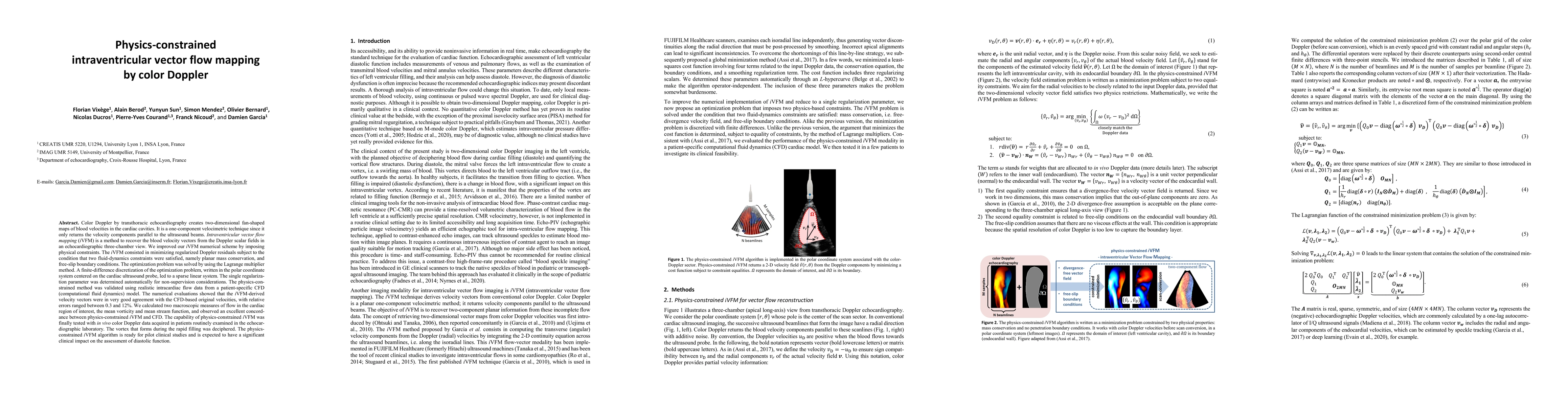
Color Doppler by transthoracic echocardiography creates 2-D fan-shaped maps of blood velocities in the cardiac cavities. It is a one-component velocimetric technique since it only returns the veloci...
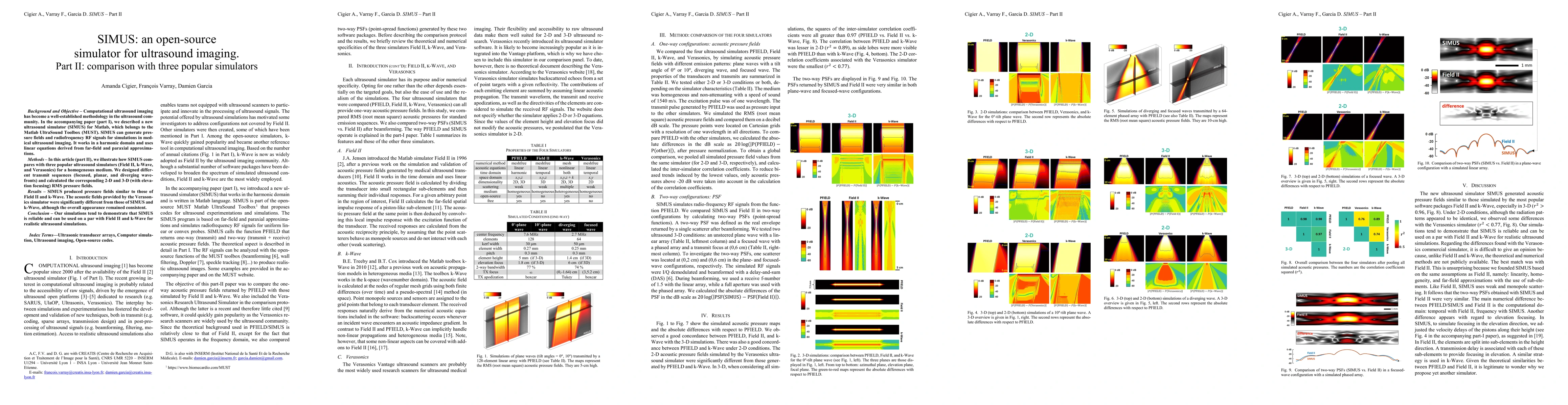
Computational ultrasound imaging has become a well-established methodology in the ultrasound community. In the accompanying paper (part I), we described a new ultrasound simulator (SIMUS) for Matlab...
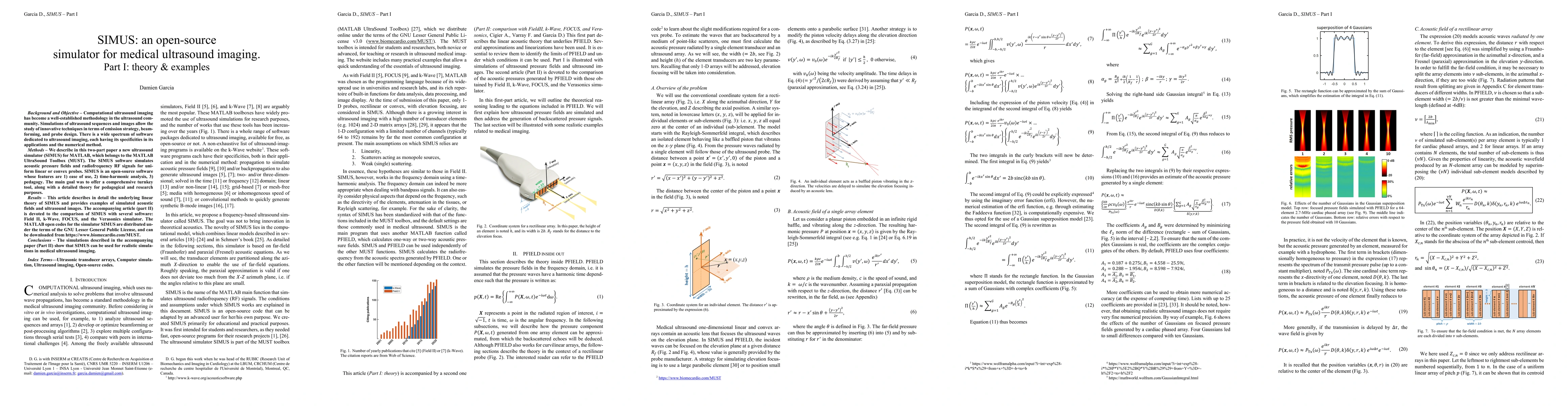
Background and Objective: Computational ultrasound imaging has become a well-established methodology in the ultrasound community. Simulations of ultrasound sequences and images allow the study of in...

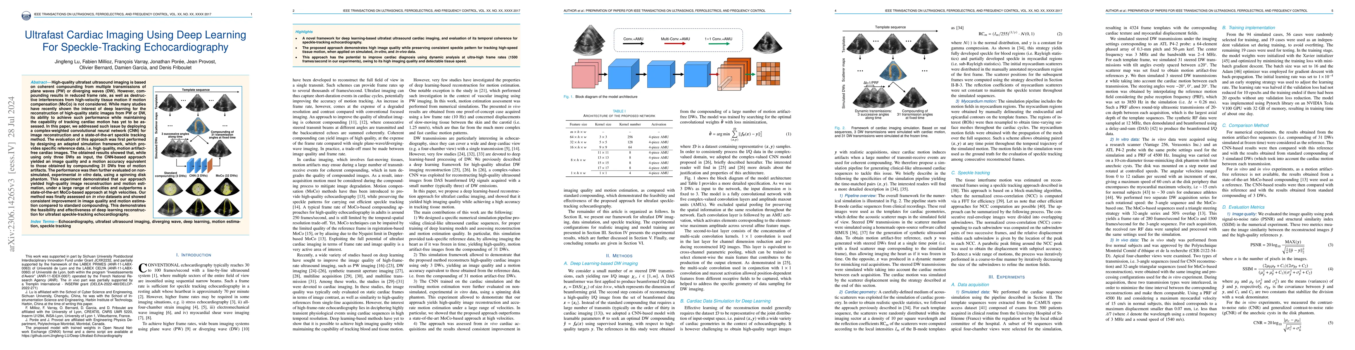
Ultrafast ultrasound imaging remains an active area of interest in the ultrasound community due to its ultra-high frame rates. Recently, a wide variety of studies based on deep learning have sought ...
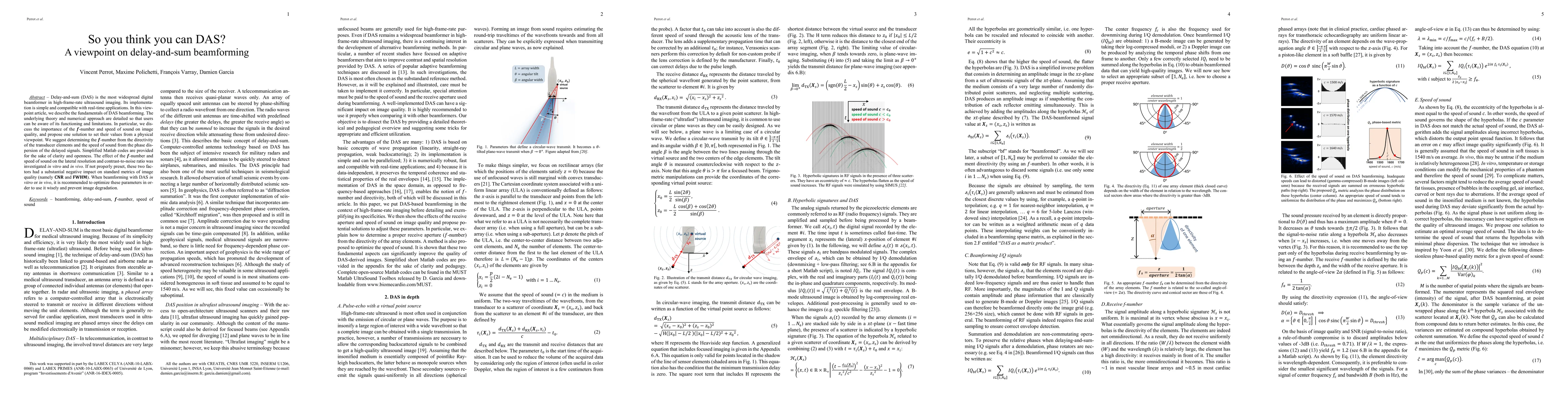
Delay-and-sum (DAS) is the most widespread digital beamformer in high-frame-rate ultrasound imaging. Its implementation is simple and compatible with real-time applications. In this viewpoint articl...
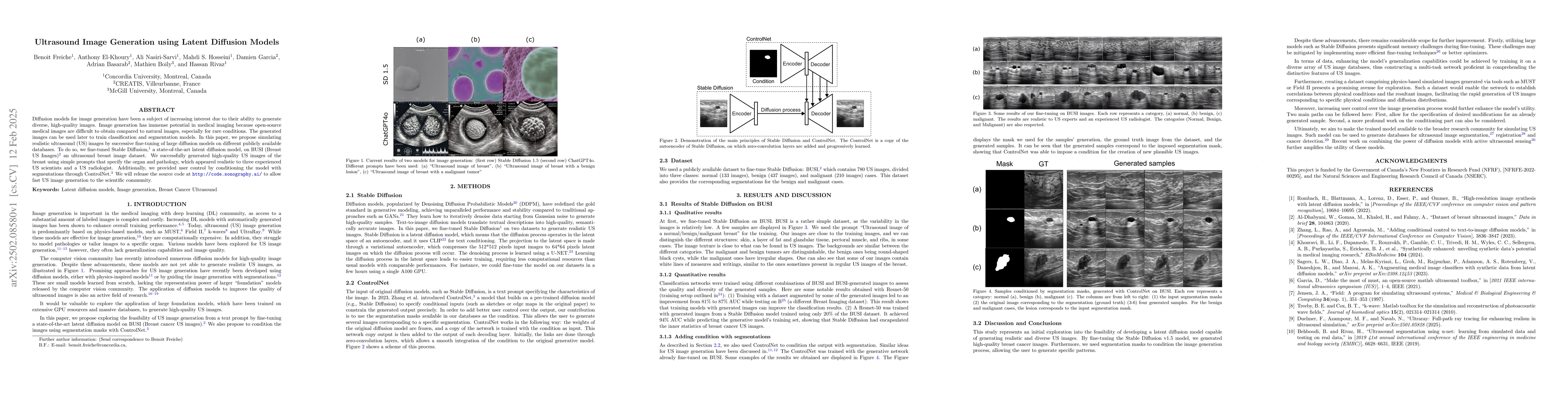
Diffusion models for image generation have been a subject of increasing interest due to their ability to generate diverse, high-quality images. Image generation has immense potential in medical imagin...