Academic Profile
Statistics
Similar Authors
Papers on arXiv
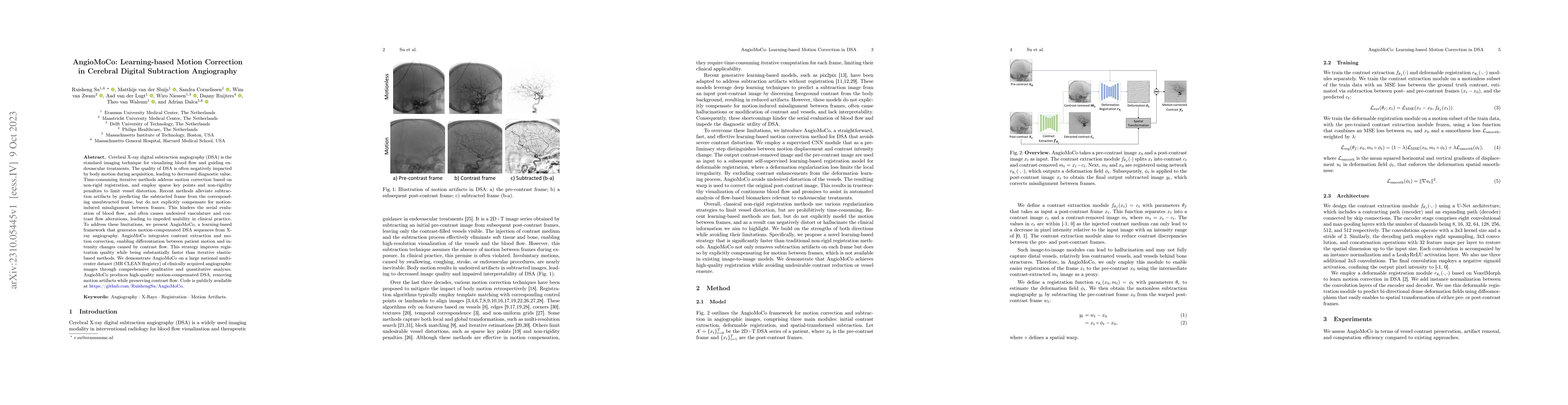
Cerebral X-ray digital subtraction angiography (DSA) is the standard imaging technique for visualizing blood flow and guiding endovascular treatments. The quality of DSA is often negatively impacted...
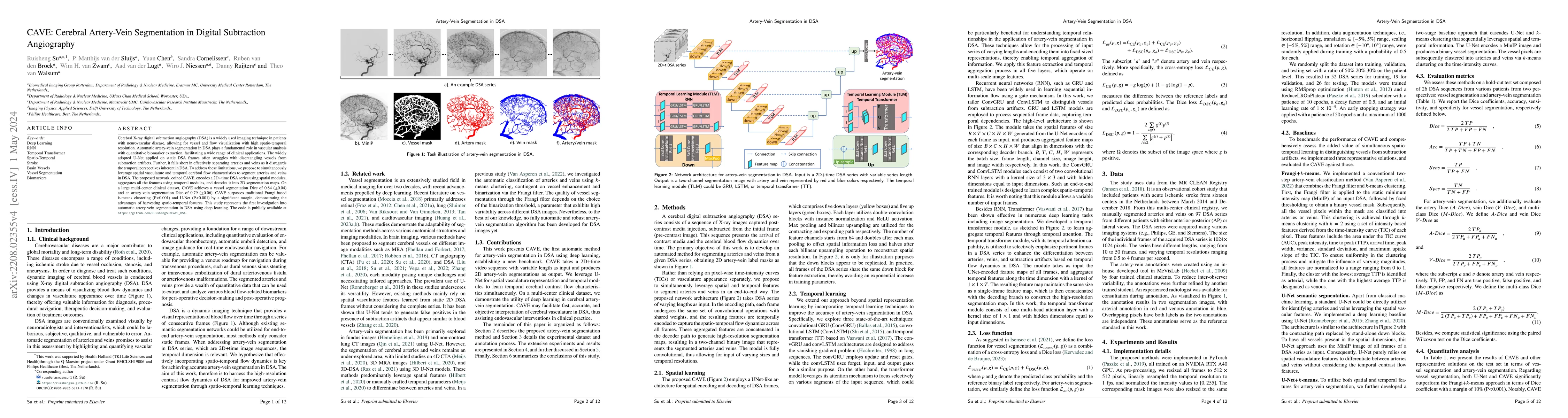
Cerebral X-ray digital subtraction angiography (DSA) is a widely used imaging technique in patients with neurovascular disease, allowing for vessel and flow visualization with high spatio-temporal r...
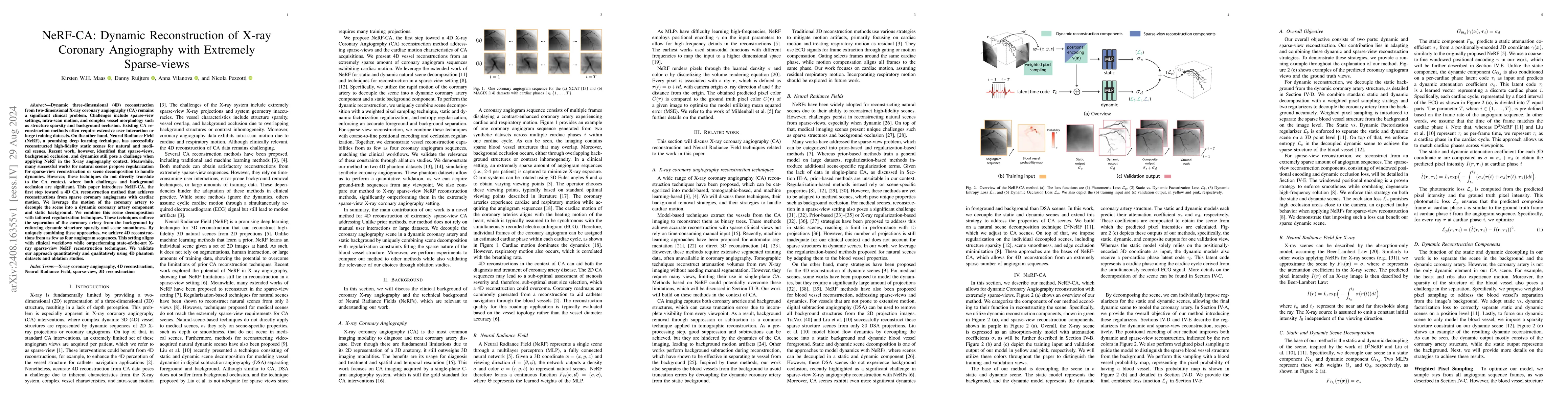
Dynamic three-dimensional (4D) reconstruction from two-dimensional X-ray coronary angiography (CA) remains a significant clinical problem. Challenges include sparse-view settings, intra-scan motion, a...
Functional magnetic resonance imaging (fMRI) has become instrumental in researching brain function. One application of fMRI is investigating potential neural features that distinguish people with auti...
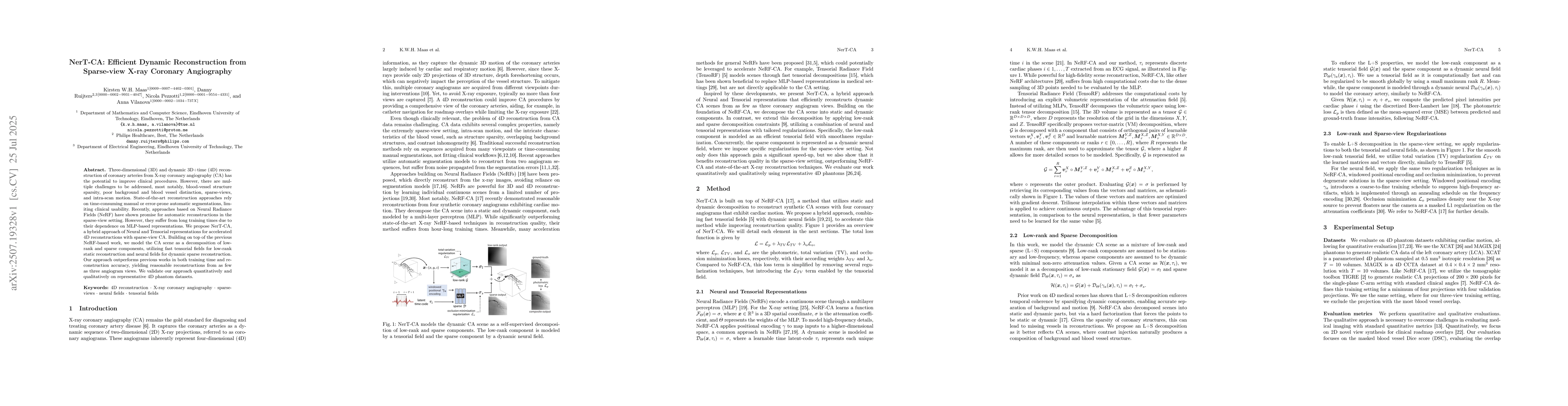
Three-dimensional (3D) and dynamic 3D+time (4D) reconstruction of coronary arteries from X-ray coronary angiography (CA) has the potential to improve clinical procedures. However, there are multiple c...
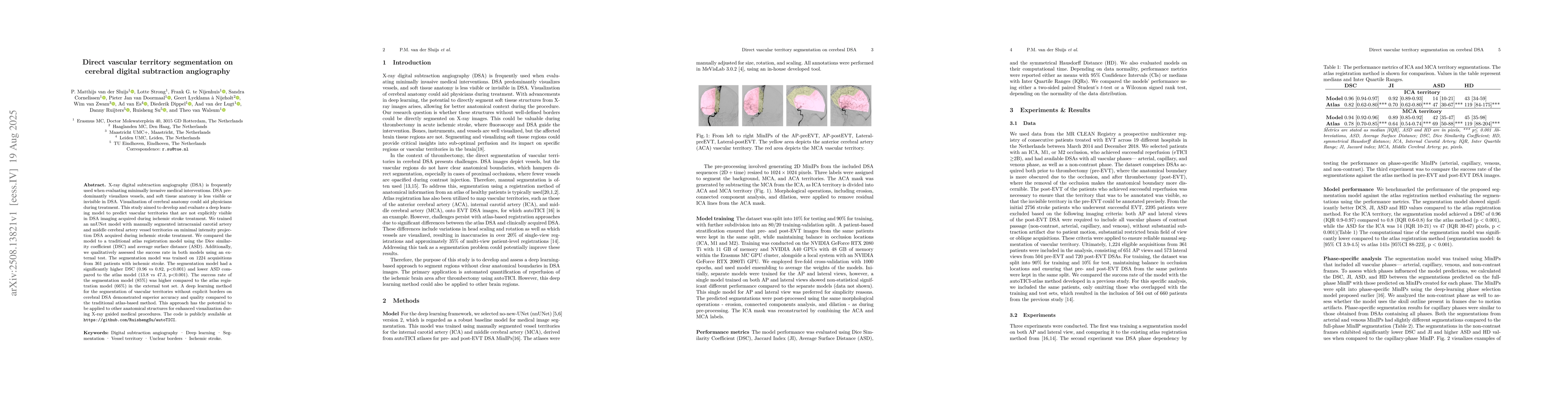
X-ray digital subtraction angiography (DSA) is frequently used when evaluating minimally invasive medical interventions. DSA predominantly visualizes vessels, and soft tissue anatomy is less visible o...
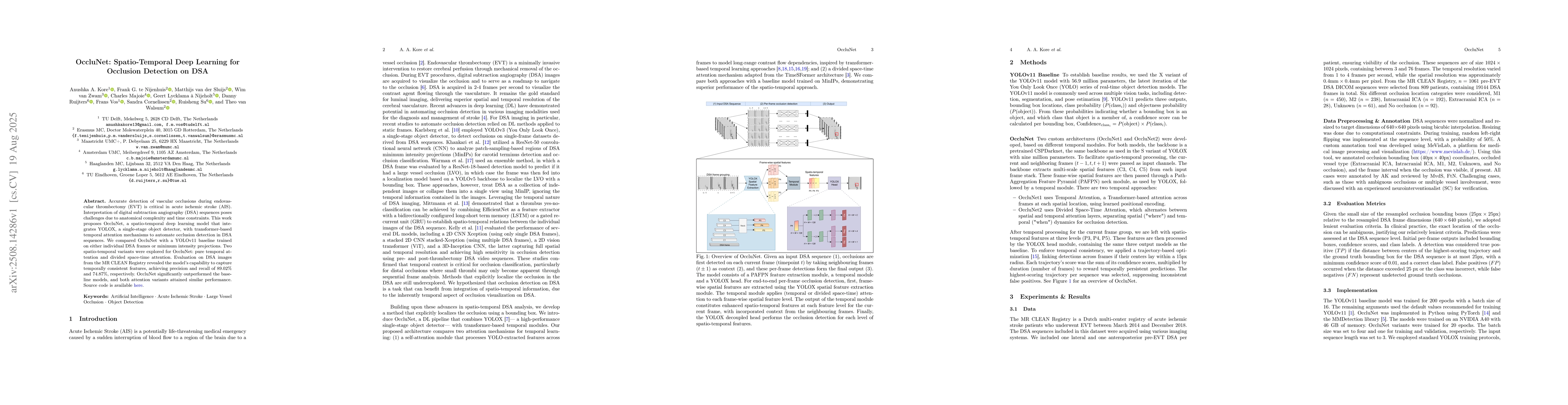
Accurate detection of vascular occlusions during endovascular thrombectomy (EVT) is critical in acute ischemic stroke (AIS). Interpretation of digital subtraction angiography (DSA) sequences poses cha...
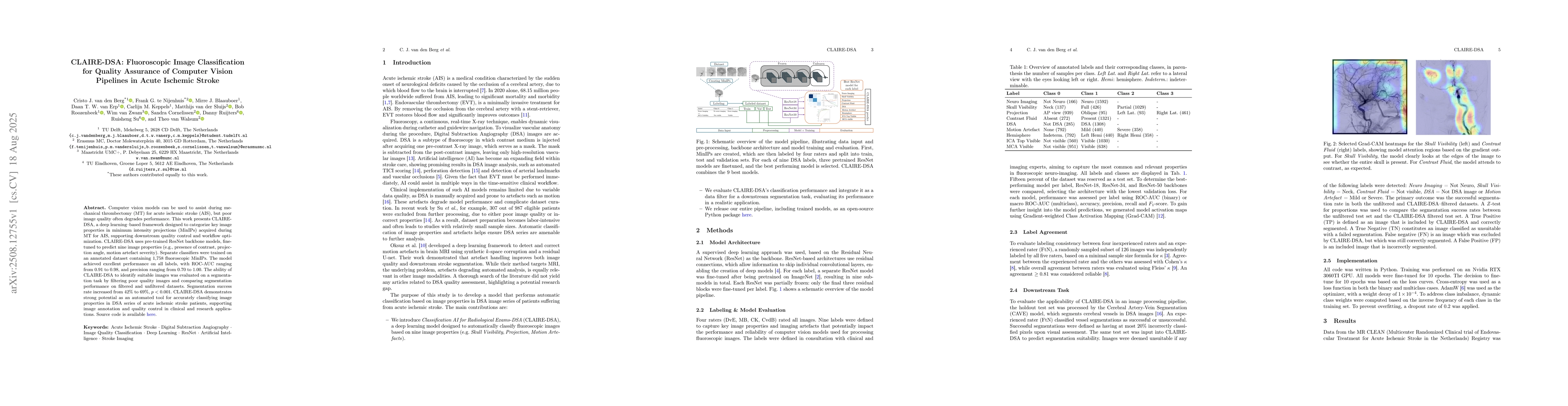
Computer vision models can be used to assist during mechanical thrombectomy (MT) for acute ischemic stroke (AIS), but poor image quality often degrades performance. This work presents CLAIRE-DSA, a de...