Academic Profile
Statistics
Similar Authors
Papers on arXiv
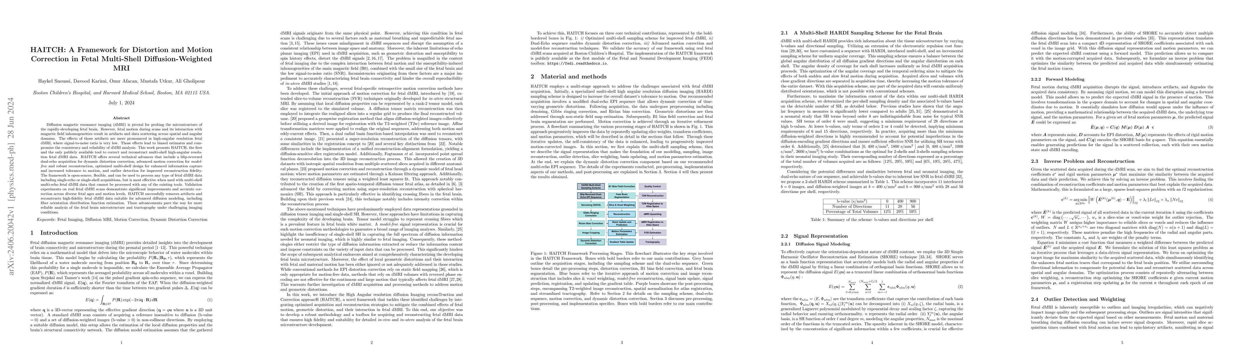
Diffusion magnetic resonance imaging (dMRI) is pivotal for probing the microstructure of the rapidly-developing fetal brain. However, fetal motion during scans and its interaction with magnetic field ...
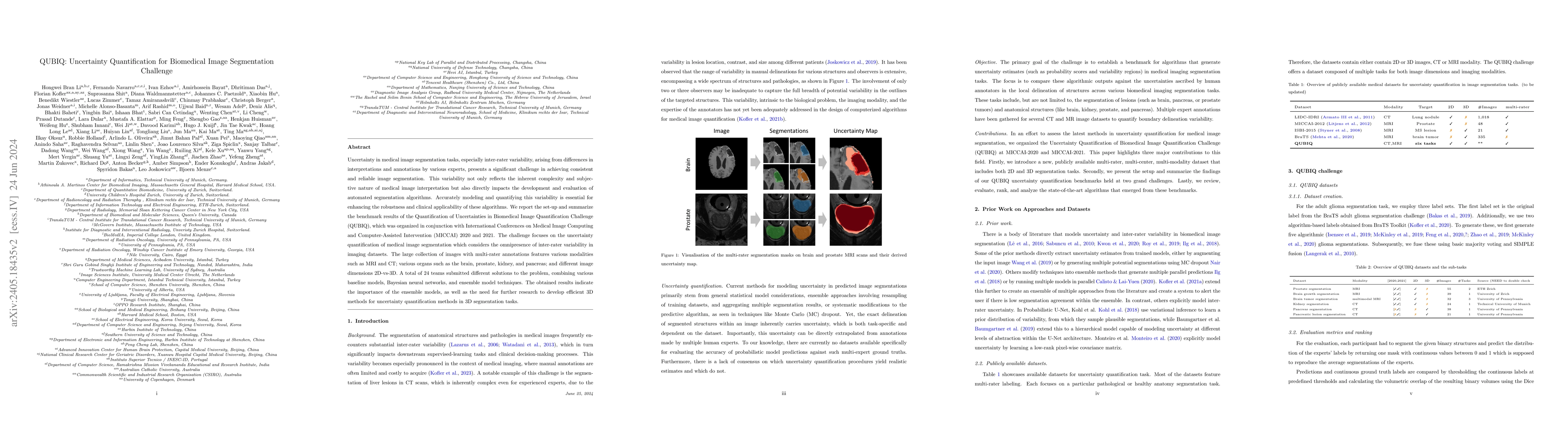
Uncertainty in medical image segmentation tasks, especially inter-rater variability, arising from differences in interpretations and annotations by various experts, presents a significant challenge in...
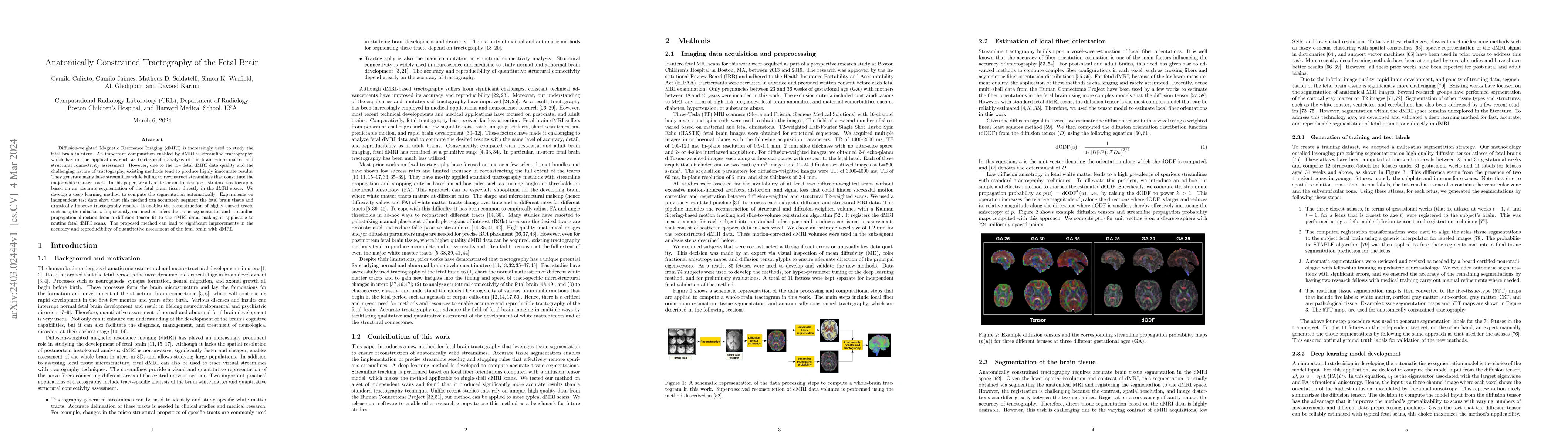
Diffusion-weighted Magnetic Resonance Imaging (dMRI) is increasingly used to study the fetal brain in utero. An important computation enabled by dMRI is streamline tractography, which has unique app...
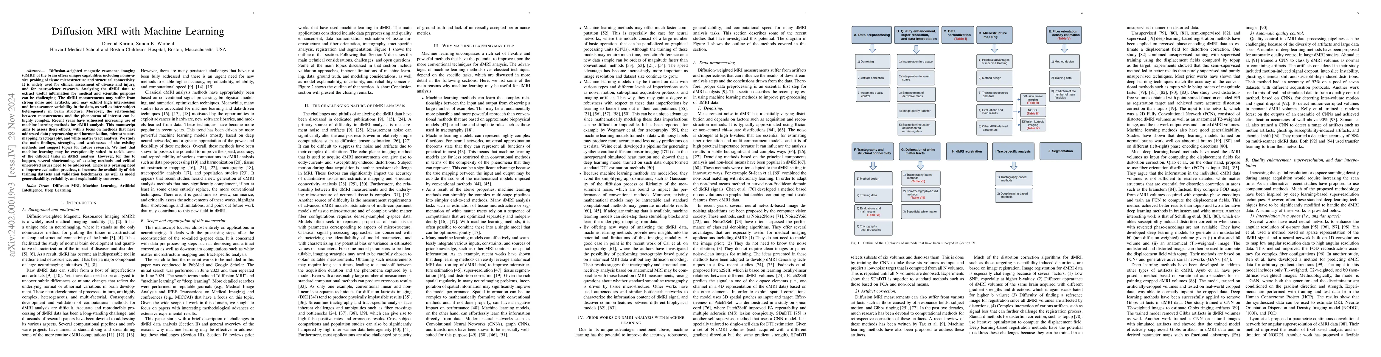
Diffusion-weighted magnetic resonance imaging (dMRI) offers unique capabilities such as noninvasive assessment of brain's micro-structure and structural connectivity. However, analyzing the dMRI dat...
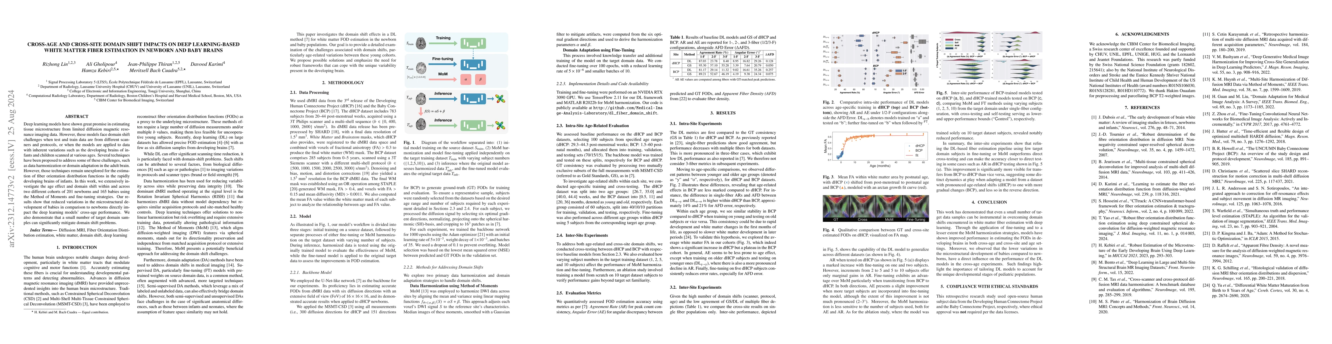
Deep learning models have shown great promise in estimating tissue microstructure from limited diffusion magnetic resonance imaging data. However, these models face domain shift challenges when test...
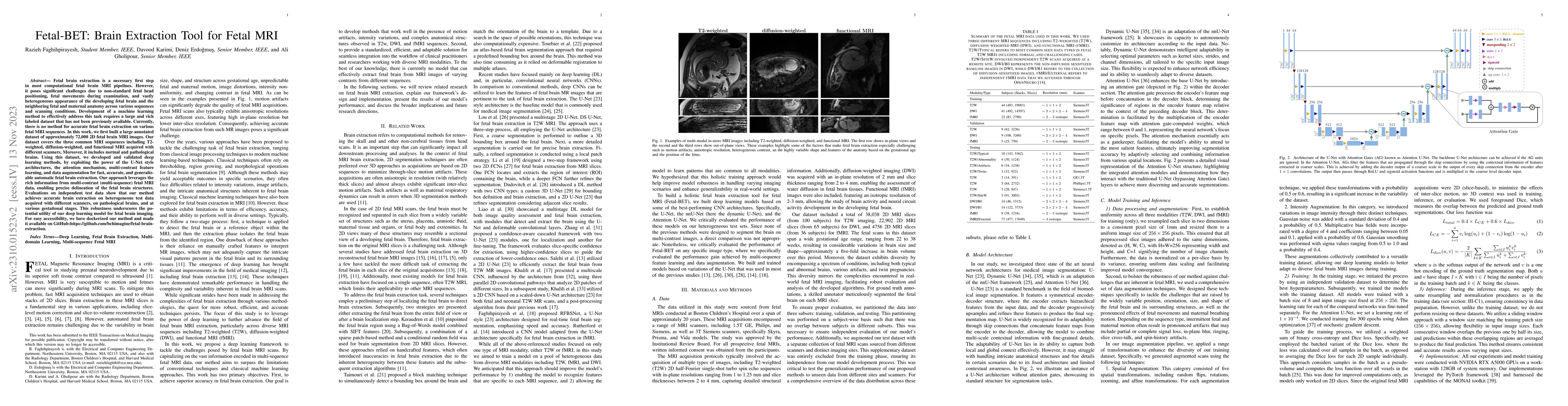
Fetal brain extraction is a necessary first step in most computational fetal brain MRI pipelines. However, it has been a very challenging task due to non-standard fetal head pose, fetal movements du...
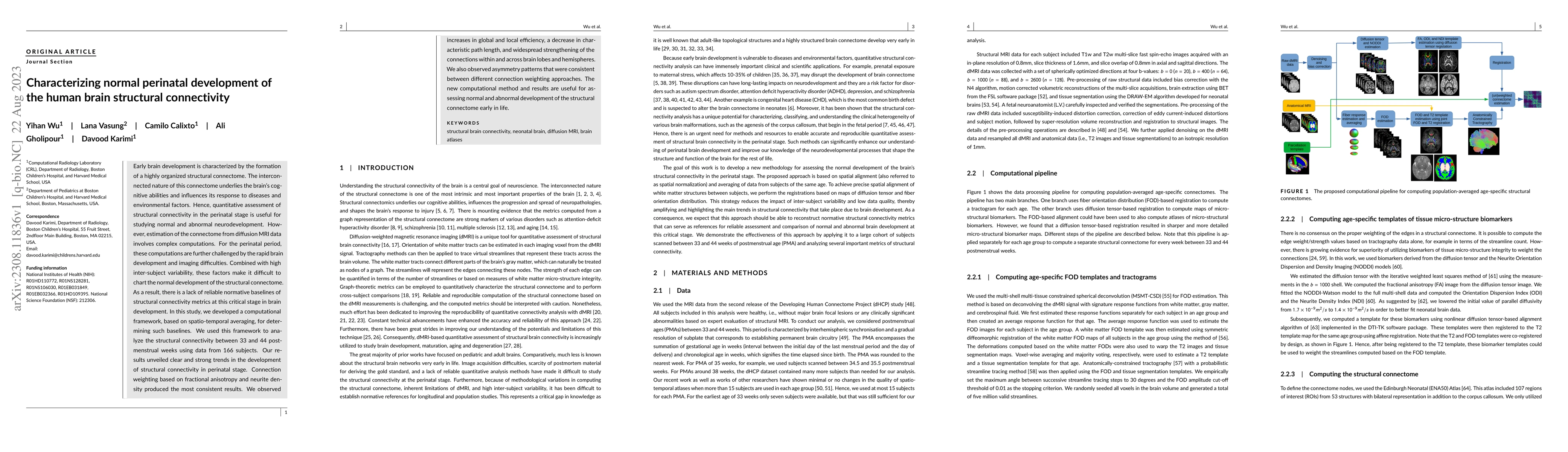
Early brain development is characterized by the formation of a highly organized structural connectome. The interconnected nature of this connectome underlies the brain's cognitive abilities and infl...
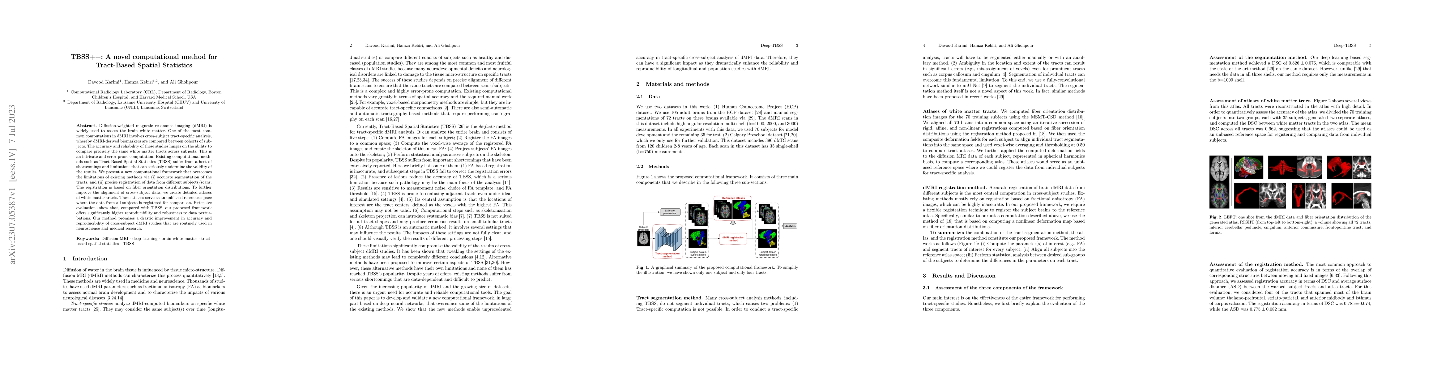
Diffusion-weighted magnetic resonance imaging (dMRI) is widely used to assess the brain white matter. One of the most common computations in dMRI involves cross-subject tract-specific analysis, wher...
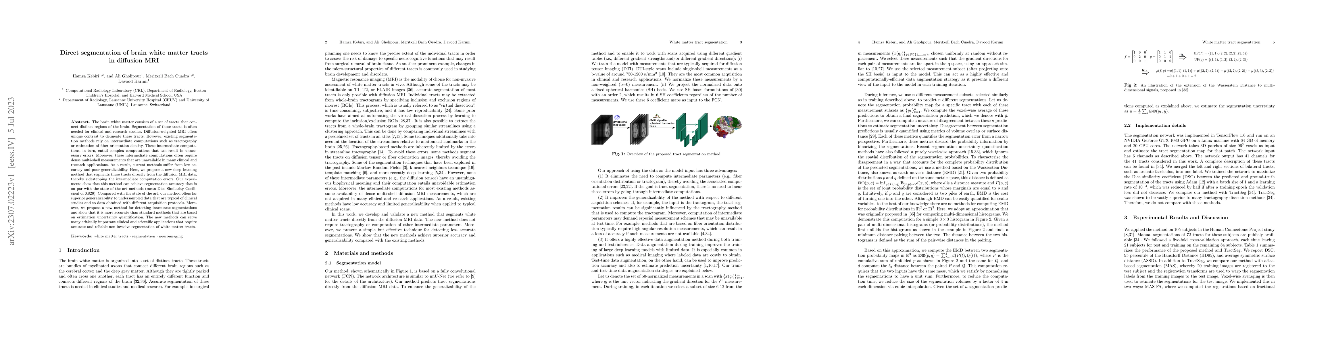
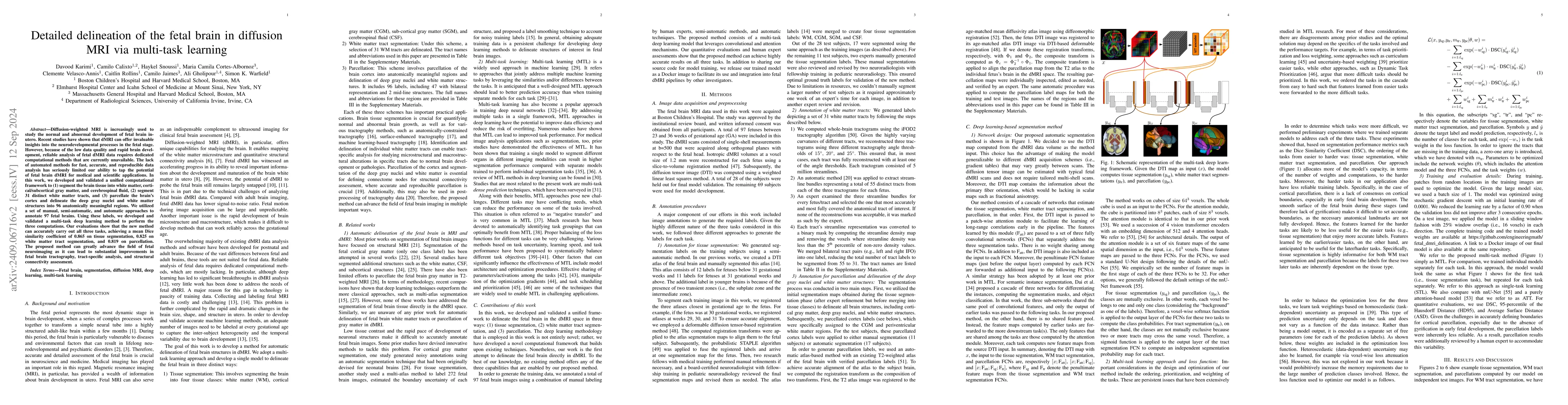
The brain white matter consists of a set of tracts that connect distinct regions of the brain. Segmentation of these tracts is often needed for clinical and research studies. Diffusion-weighted MRI ...
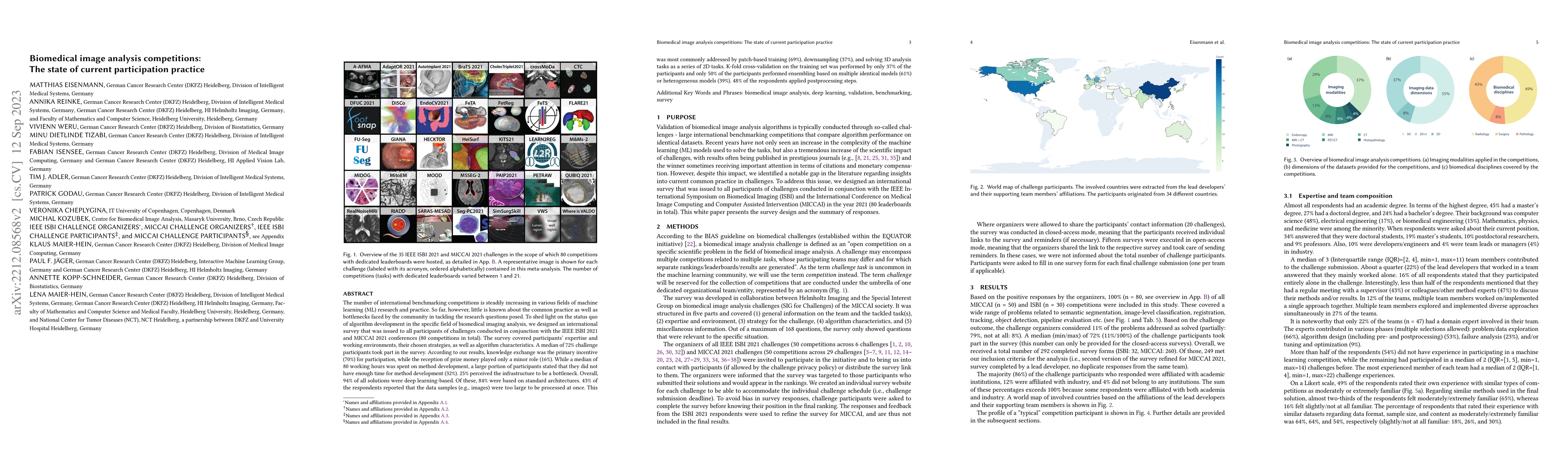
The number of international benchmarking competitions is steadily increasing in various fields of machine learning (ML) research and practice. So far, however, little is known about the common pract...
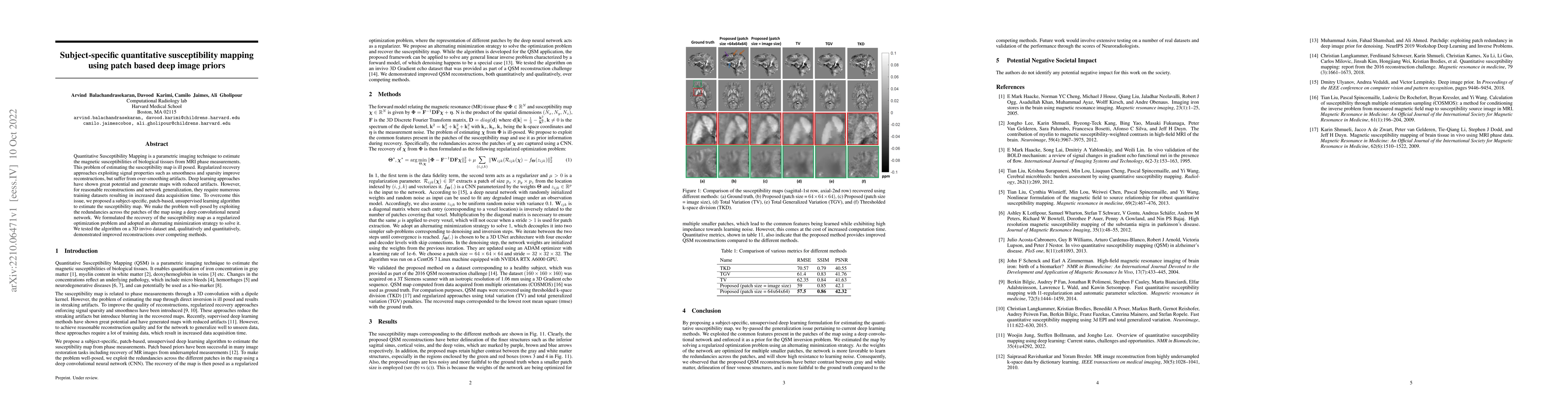
Quantitative Susceptibility Mapping is a parametric imaging technique to estimate the magnetic susceptibilities of biological tissues from MRI phase measurements. This problem of estimating the susc...
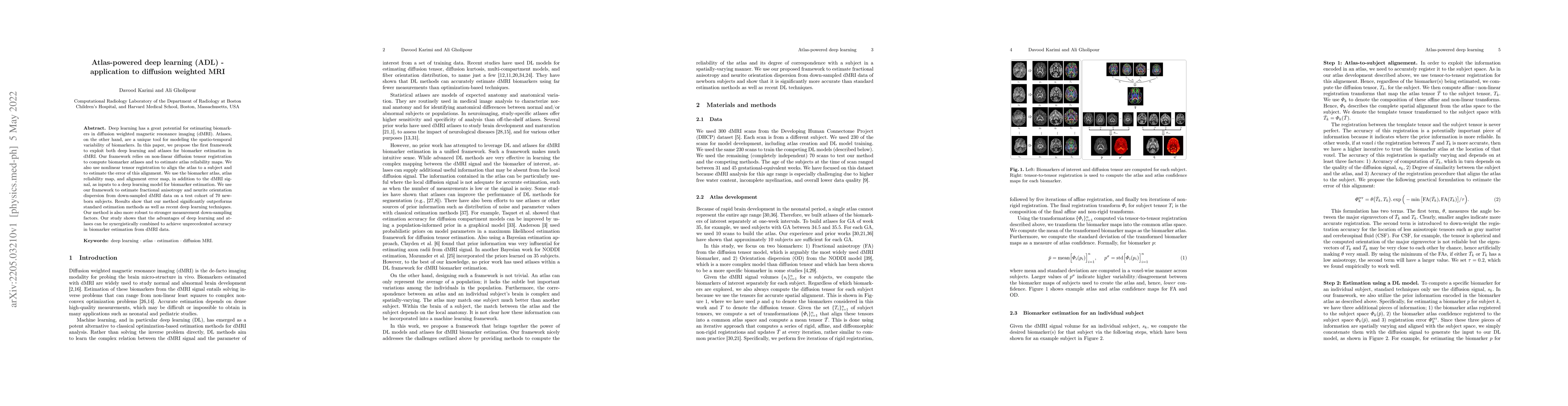
Deep learning has a great potential for estimating biomarkers in diffusion weighted magnetic resonance imaging (dMRI). Atlases, on the other hand, are a unique tool for modeling the spatio-temporal ...
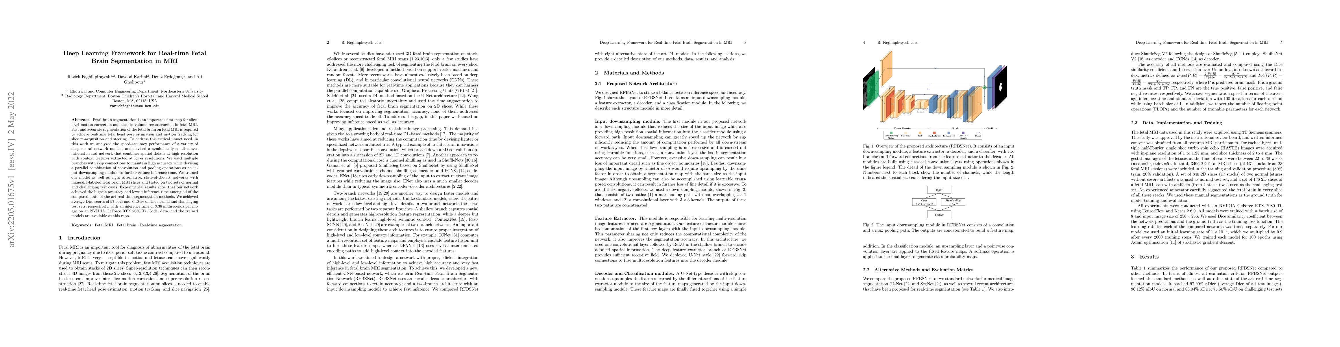
Fetal brain segmentation is an important first step for slice-level motion correction and slice-to-volume reconstruction in fetal MRI. Fast and accurate segmentation of the fetal brain on fetal MRI ...
In-utero fetal MRI is emerging as an important tool in the diagnosis and analysis of the developing human brain. Automatic segmentation of the developing fetal brain is a vital step in the quantitat...
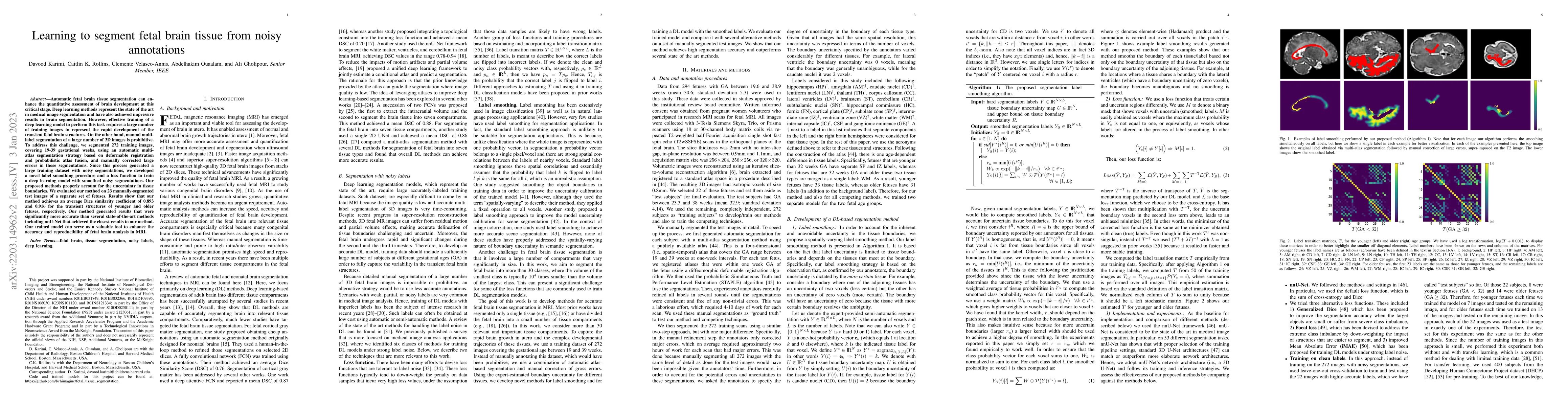
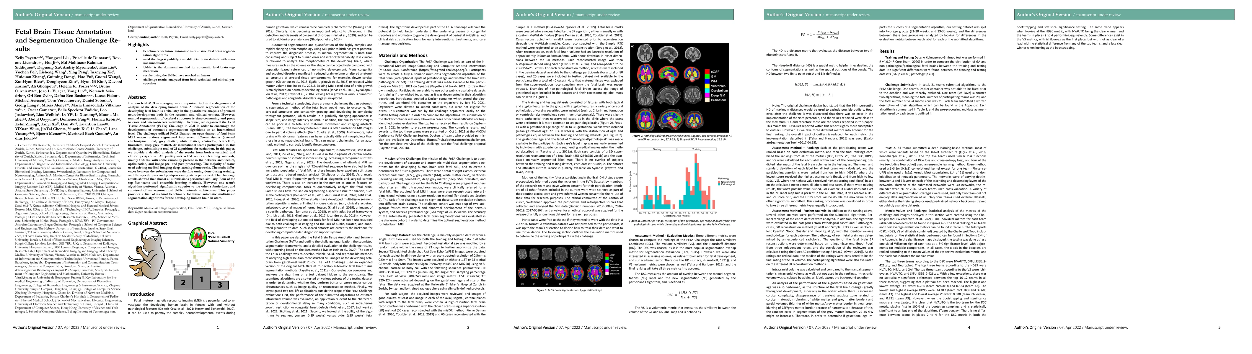
Automatic fetal brain tissue segmentation can enhance the quantitative assessment of brain development at this critical stage. Deep learning methods represent the state of the art in medical image s...
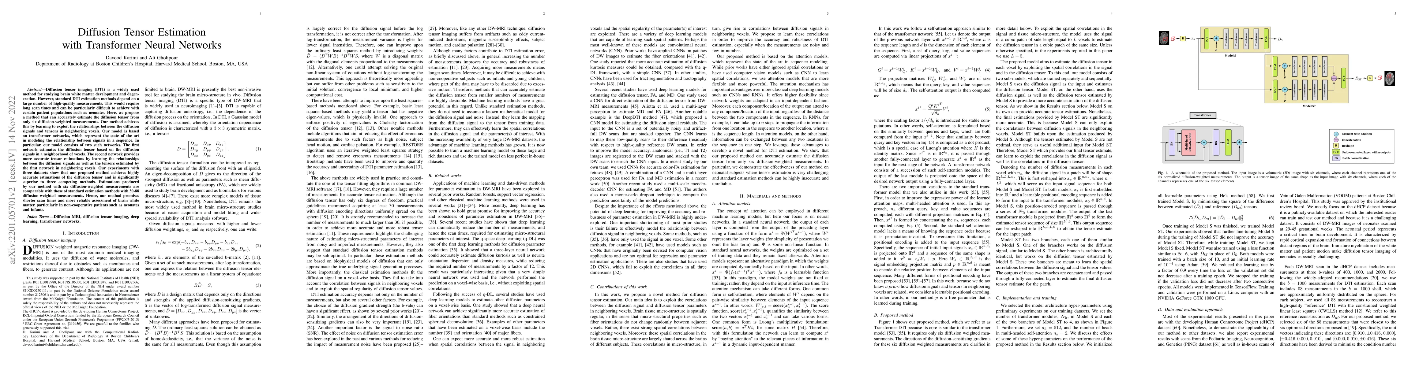
Diffusion tensor imaging (DTI) is a widely used method for studying brain white matter development and degeneration. However, standard DTI estimation methods depend on a large number of high-quality...
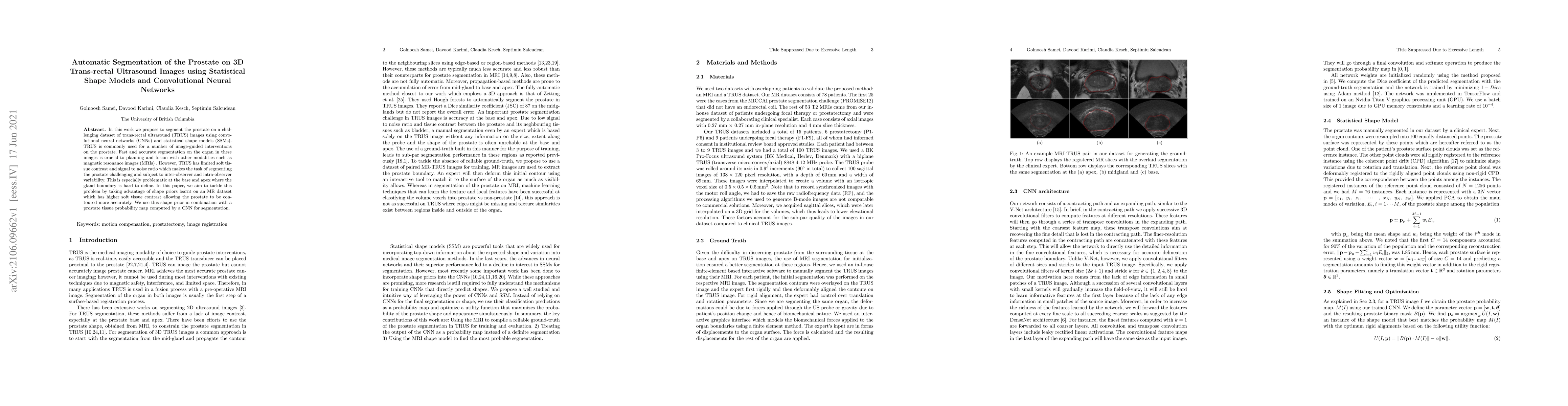
In this work we propose to segment the prostate on a challenging dataset of trans-rectal ultrasound (TRUS) images using convolutional neural networks (CNNs) and statistical shape models (SSMs). TRUS...
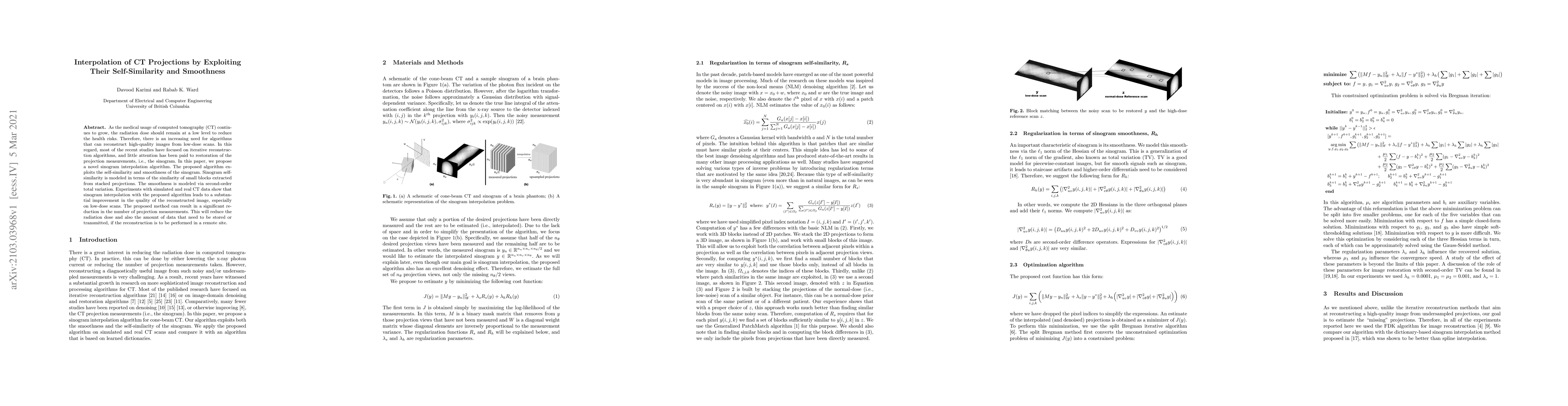
As the medical usage of computed tomography (CT) continues to grow, the radiation dose should remain at a low level to reduce the health risks. Therefore, there is an increasing need for algorithms ...
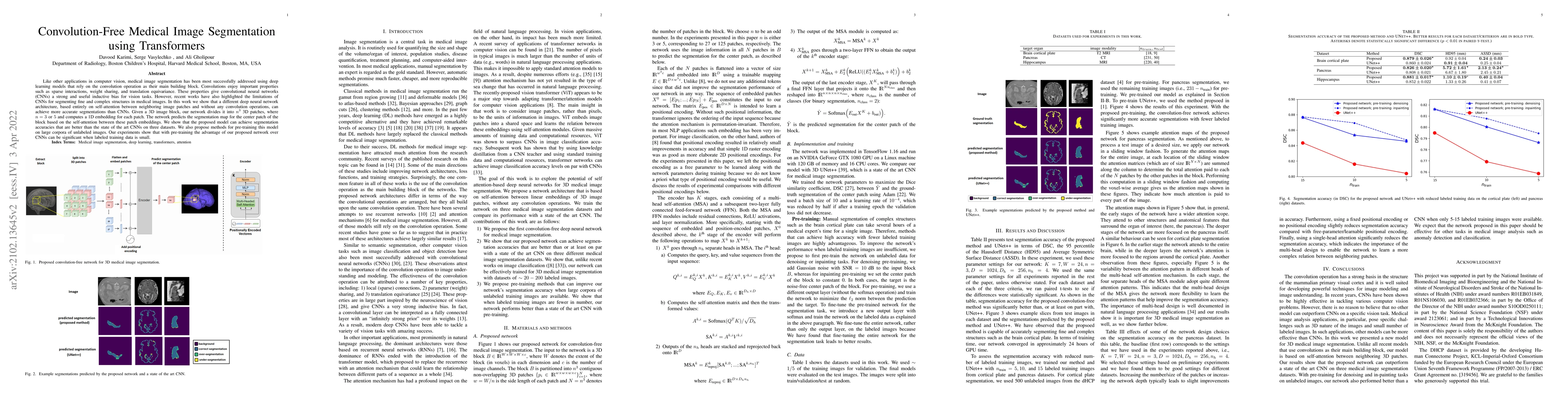
Like other applications in computer vision, medical image segmentation has been most successfully addressed using deep learning models that rely on the convolution operation as their main building b...
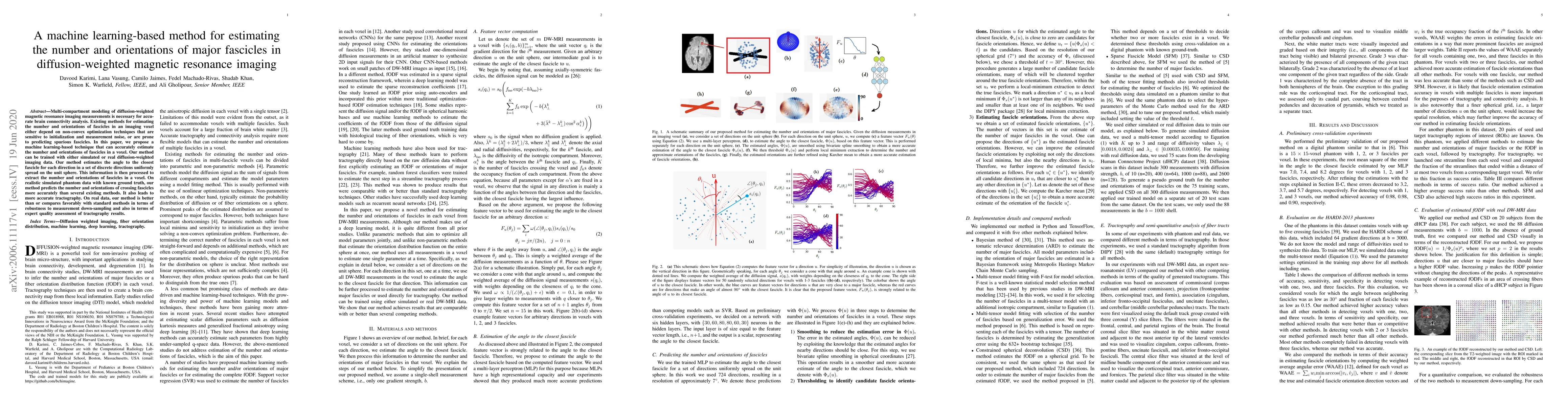
Multi-compartment modeling of diffusion-weighted magnetic resonance imaging measurements is necessary for accurate brain connectivity analysis. Existing methods for estimating the number and orienta...
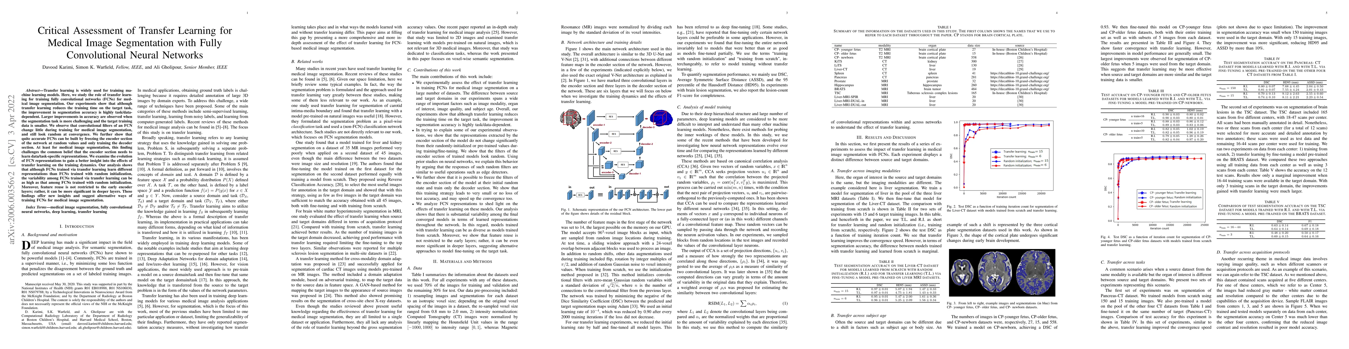
Transfer learning is widely used for training machine learning models. Here, we study the role of transfer learning for training fully convolutional networks (FCNs) for medical image segmentation. O...
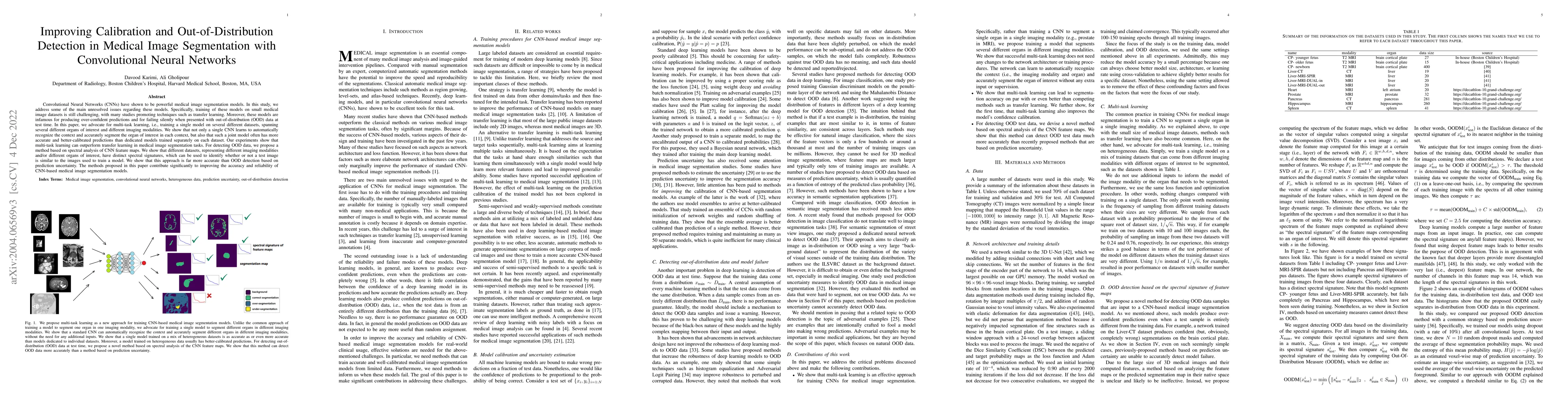
Convolutional Neural Networks (CNNs) have shown to be powerful medical image segmentation models. In this study, we address some of the main unresolved issues regarding these models. Specifically, t...
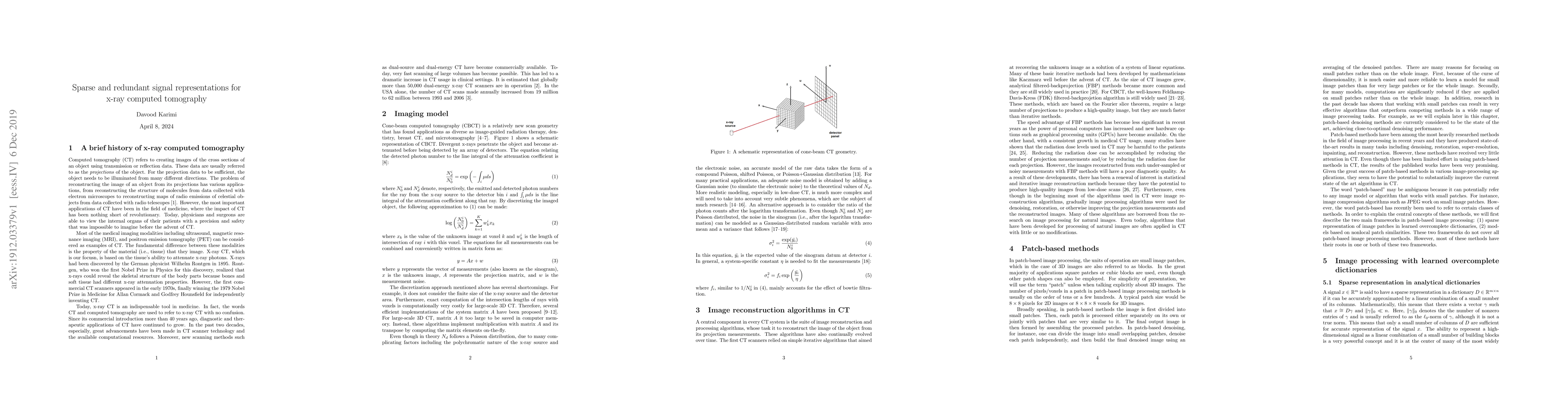
Image models are central to all image processing tasks. The great advancements in digital image processing would not have been made possible without powerful models which, themselves, have evolved o...
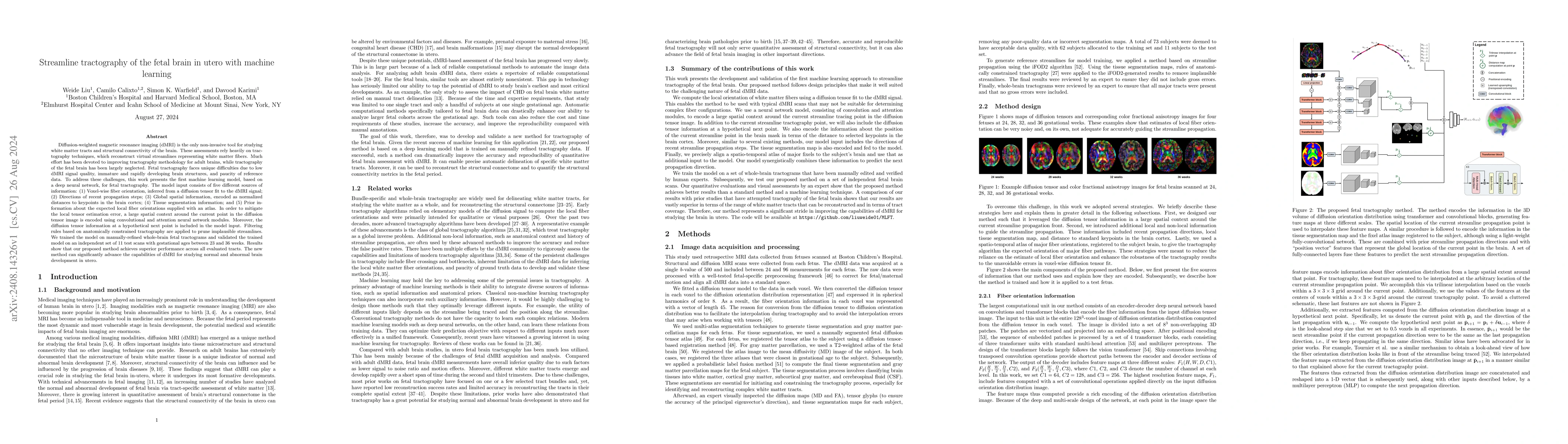
Diffusion-weighted magnetic resonance imaging (dMRI) is the only non-invasive tool for studying white matter tracts and structural connectivity of the brain. These assessments rely heavily on tractogr...
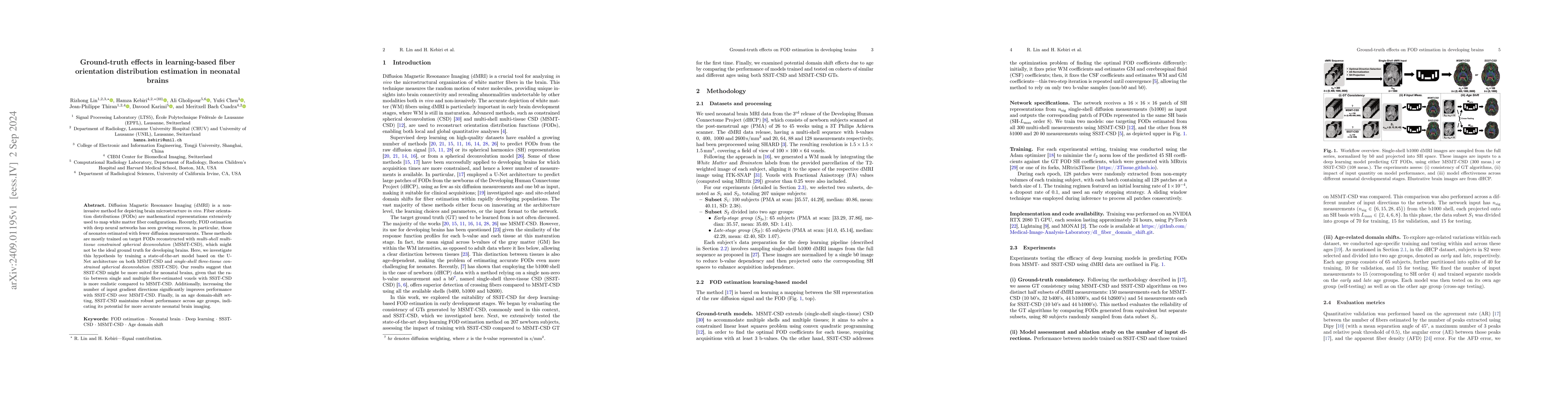
Diffusion Magnetic Resonance Imaging (dMRI) is a non-invasive method for depicting brain microstructure in vivo. Fiber orientation distributions (FODs) are mathematical representations extensively use...
Diffusion-weighted MRI is increasingly used to study the normal and abnormal development of fetal brain in-utero. Recent studies have shown that dMRI can offer invaluable insights into the neurodevelo...
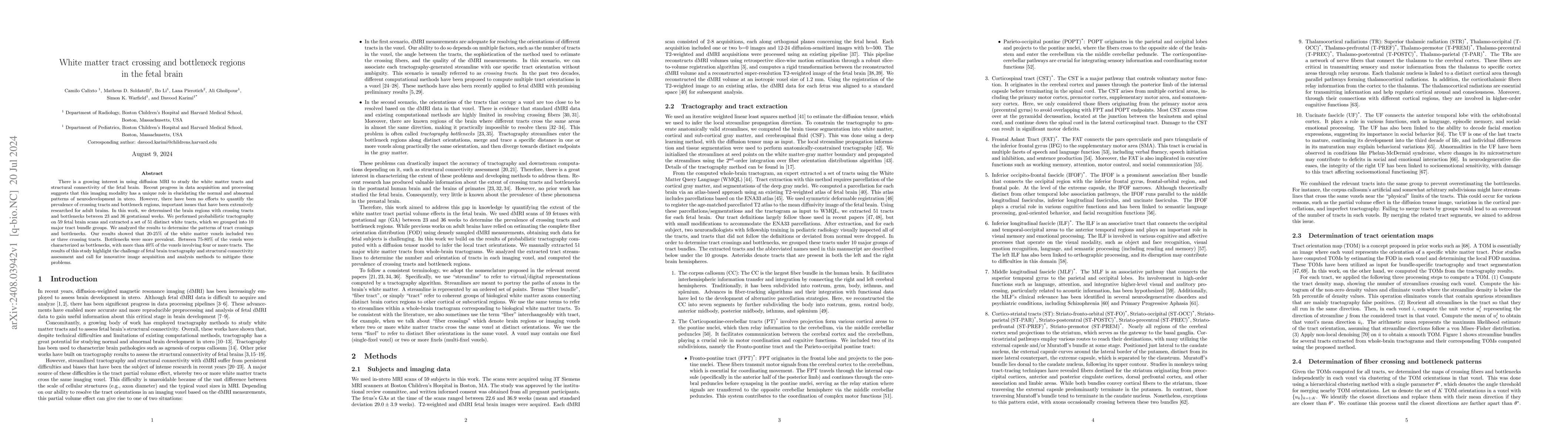
There is a growing interest in using diffusion MRI to study the white matter tracts and structural connectivity of the fetal brain. Recent progress in data acquisition and processing suggests that thi...
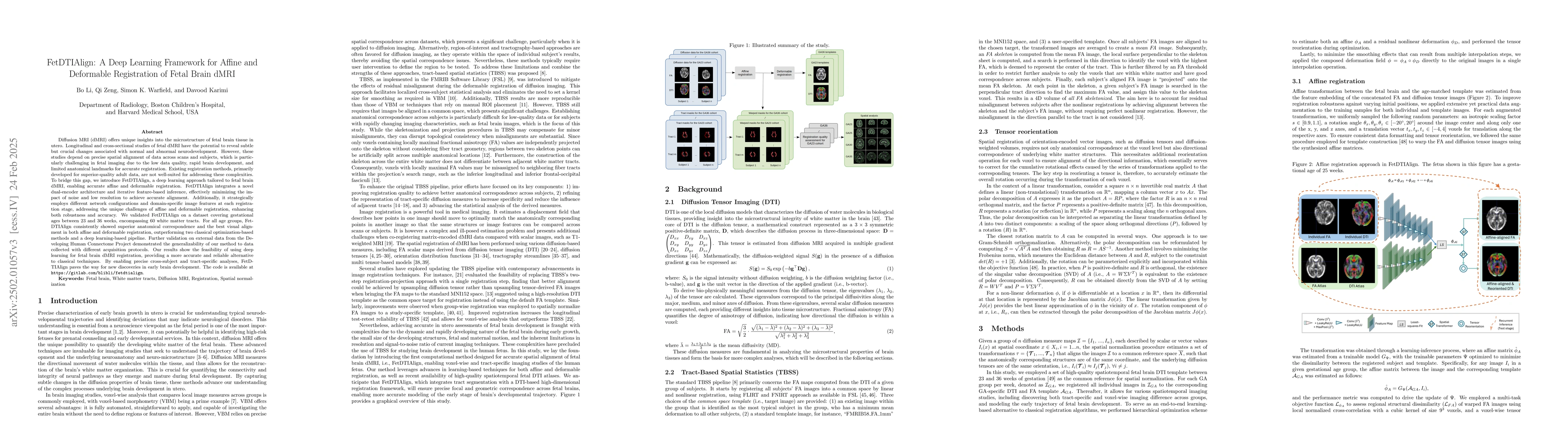
Diffusion MRI (dMRI) provides unique insights into fetal brain microstructure in utero. Longitudinal and cross-sectional fetal dMRI studies can reveal crucial neurodevelopmental changes but require pr...
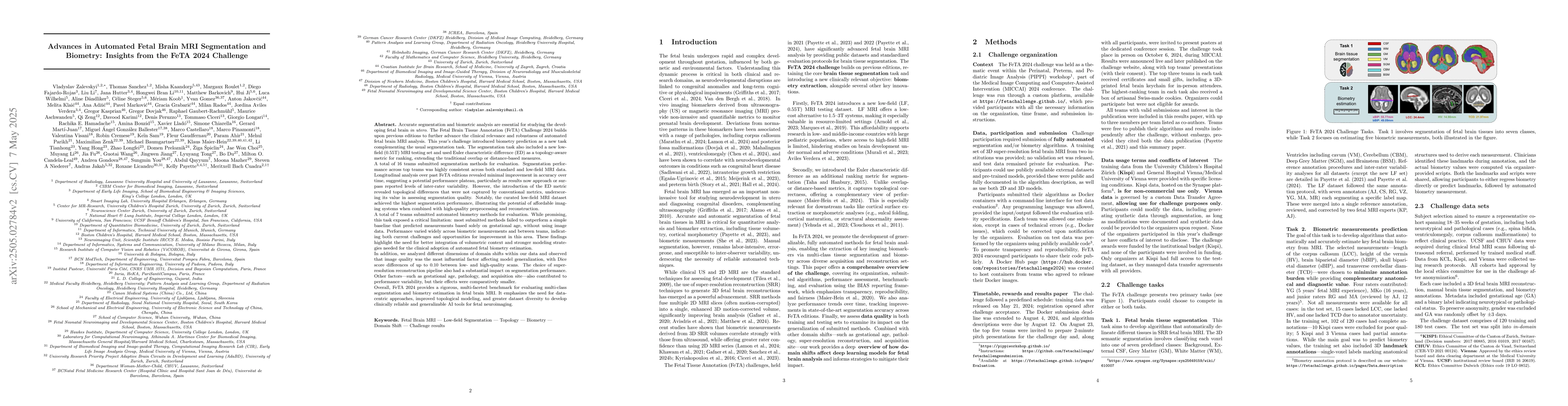
Accurate fetal brain tissue segmentation and biometric analysis are essential for studying brain development in utero. The FeTA Challenge 2024 advanced automated fetal brain MRI analysis by introducin...
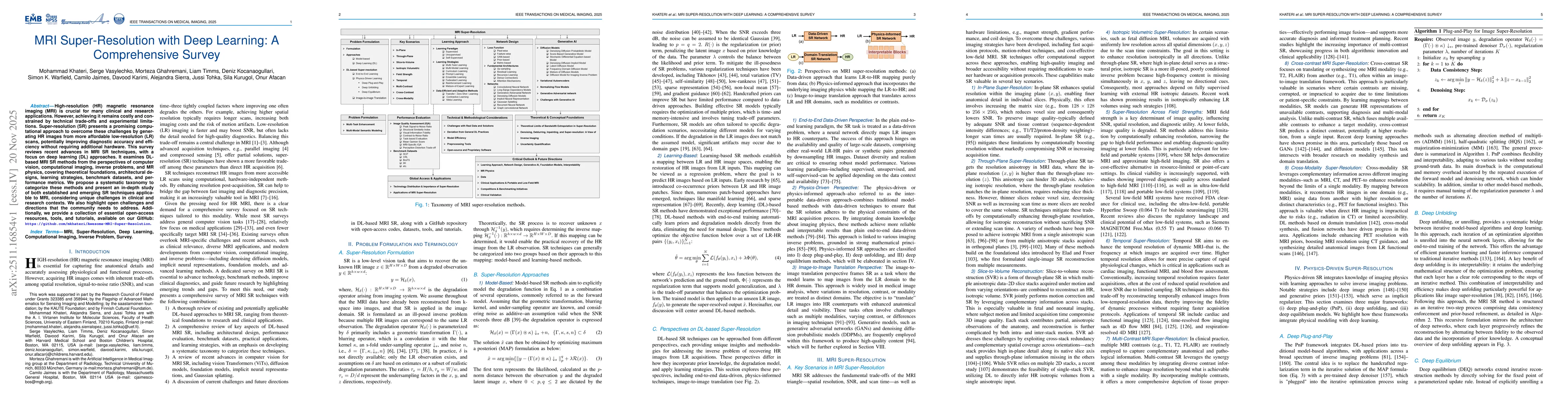
High-resolution (HR) magnetic resonance imaging (MRI) is crucial for many clinical and research applications. However, achieving it remains costly and constrained by technical trade-offs and experimen...
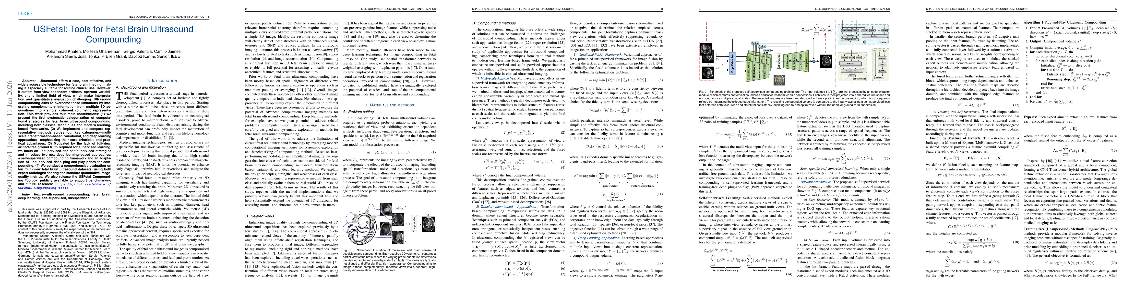
Ultrasound offers a safe, cost-effective, and widely accessible technology for fetal brain imaging, making it especially suitable for routine clinical use. However, it suffers from view-dependent arti...
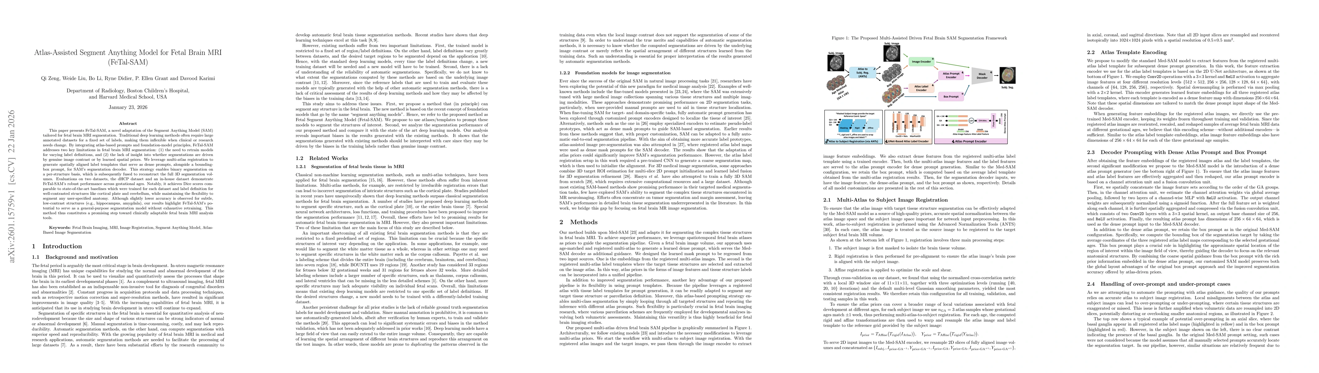
This paper presents FeTal-SAM, a novel adaptation of the Segment Anything Model (SAM) tailored for fetal brain MRI segmentation. Traditional deep learning methods often require large annotated dataset...