Academic Profile
Statistics
Similar Authors
Papers on arXiv
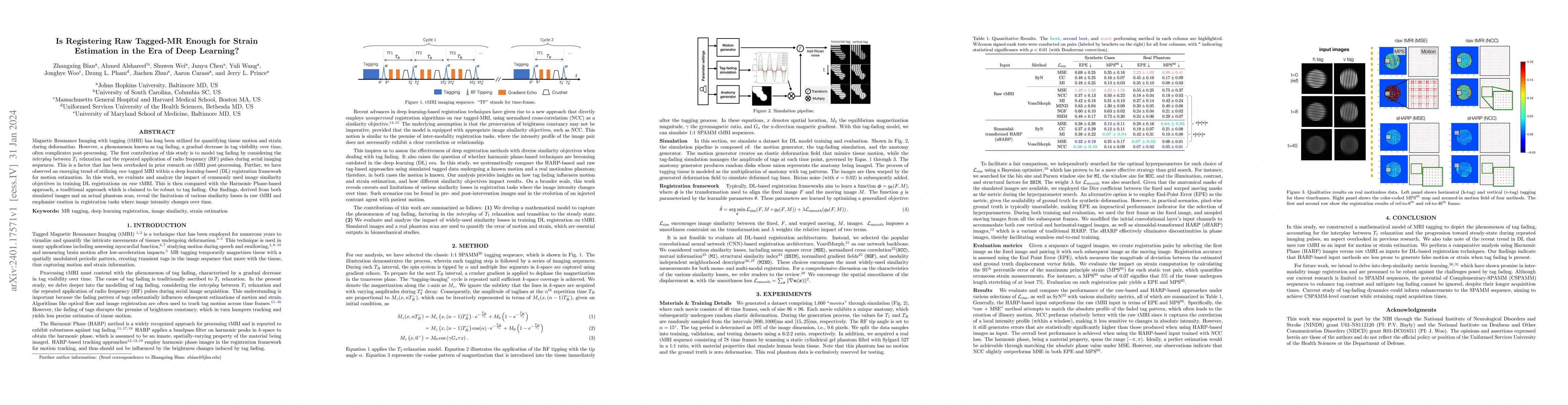
Magnetic Resonance Imaging with tagging (tMRI) has long been utilized for quantifying tissue motion and strain during deformation. However, a phenomenon known as tag fading, a gradual decrease in ta...
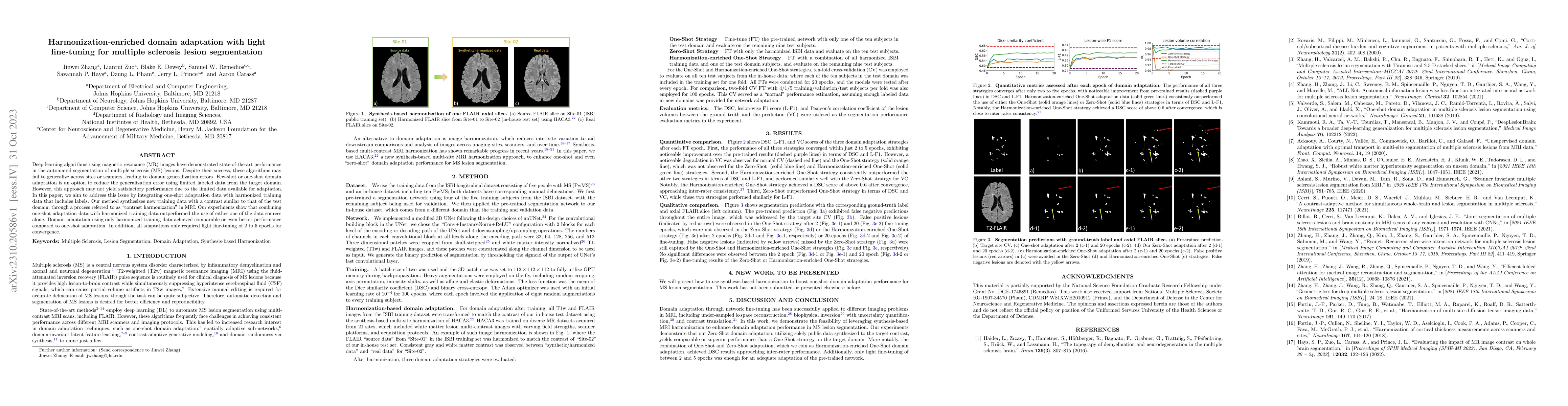
Automatic multiple sclerosis (MS) lesion segmentation using multi-contrast magnetic resonance (MR) images provides improved efficiency and reproducibility compared to manual delineation. Current sta...
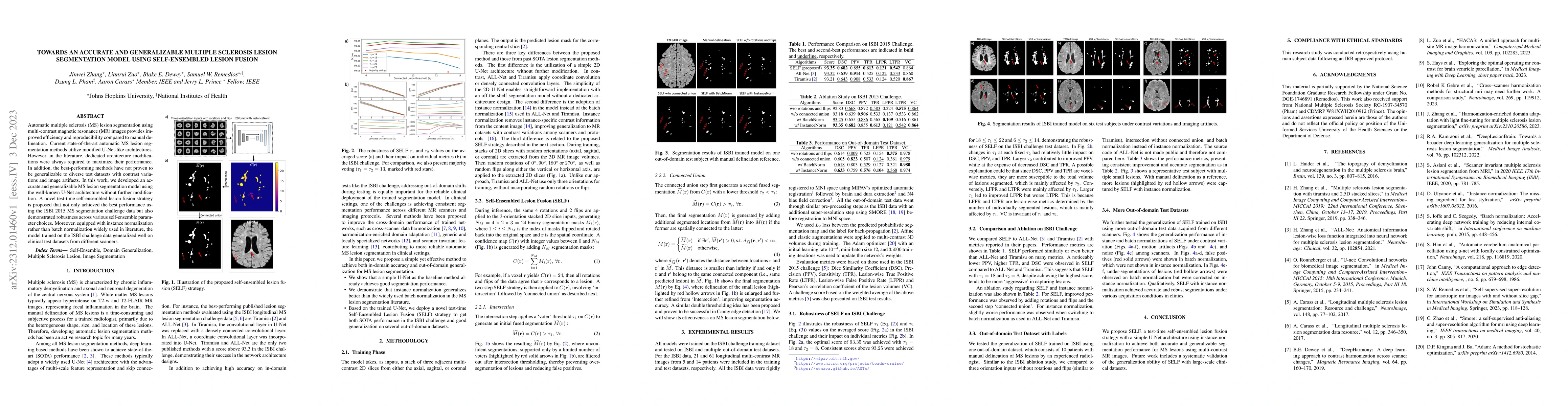
Deep learning algorithms utilizing magnetic resonance (MR) images have demonstrated cutting-edge proficiency in autonomously segmenting multiple sclerosis (MS) lesions. Despite their achievements, t...
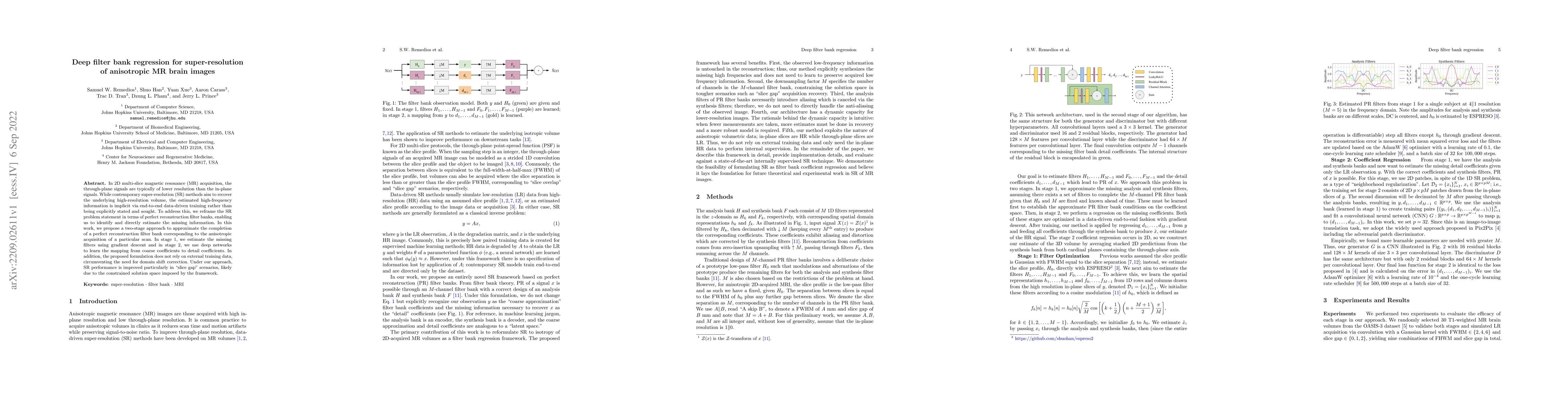
In 2D multi-slice magnetic resonance (MR) acquisition, the through-plane signals are typically of lower resolution than the in-plane signals. While contemporary super-resolution (SR) methods aim to ...
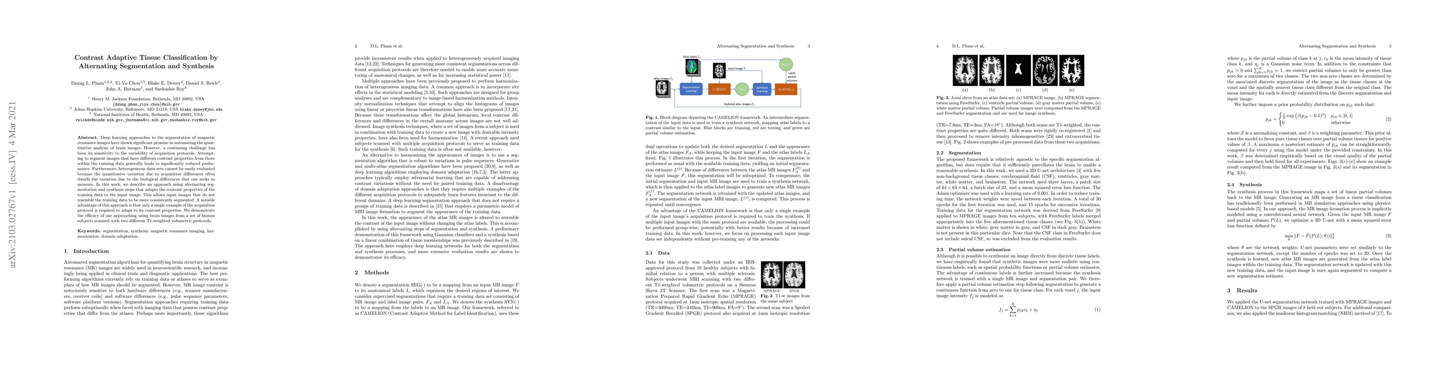
Deep learning approaches to the segmentation of magnetic resonance images have shown significant promise in automating the quantitative analysis of brain images. However, a continuing challenge has ...
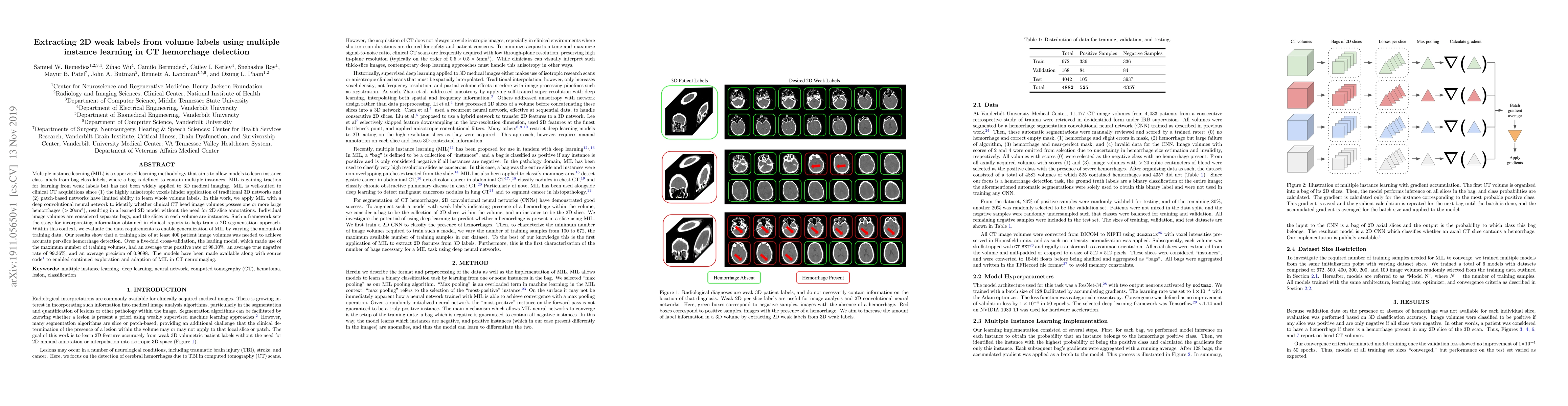
Multiple instance learning (MIL) is a supervised learning methodology that aims to allow models to learn instance class labels from bag class labels, where a bag is defined to contain multiple insta...
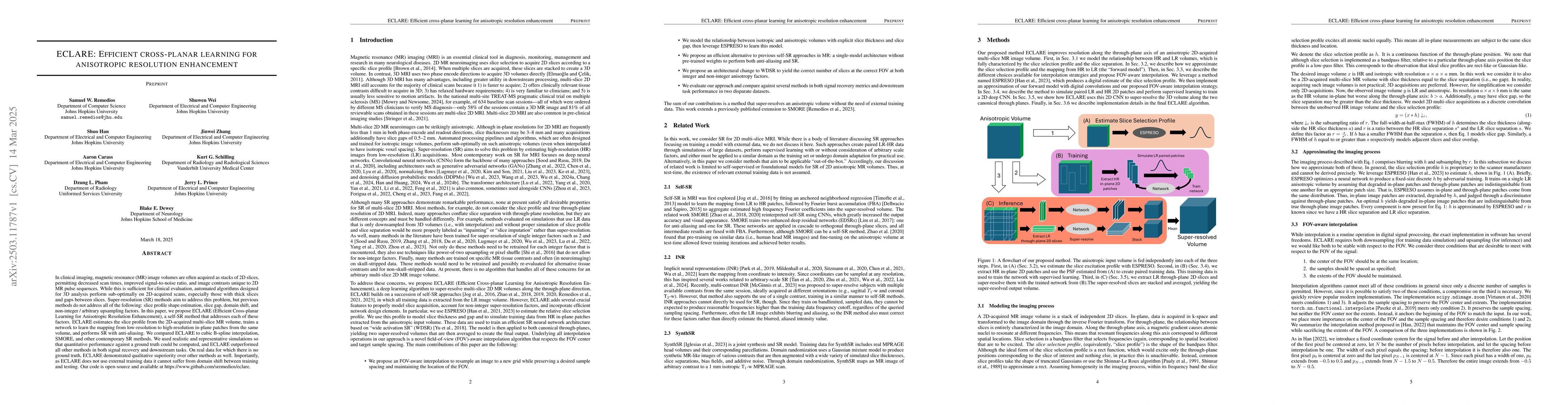
In clinical imaging, magnetic resonance (MR) image volumes are often acquired as stacks of 2D slices, permitting decreased scan times, improved signal-to-noise ratio, and image contrasts unique to 2D ...
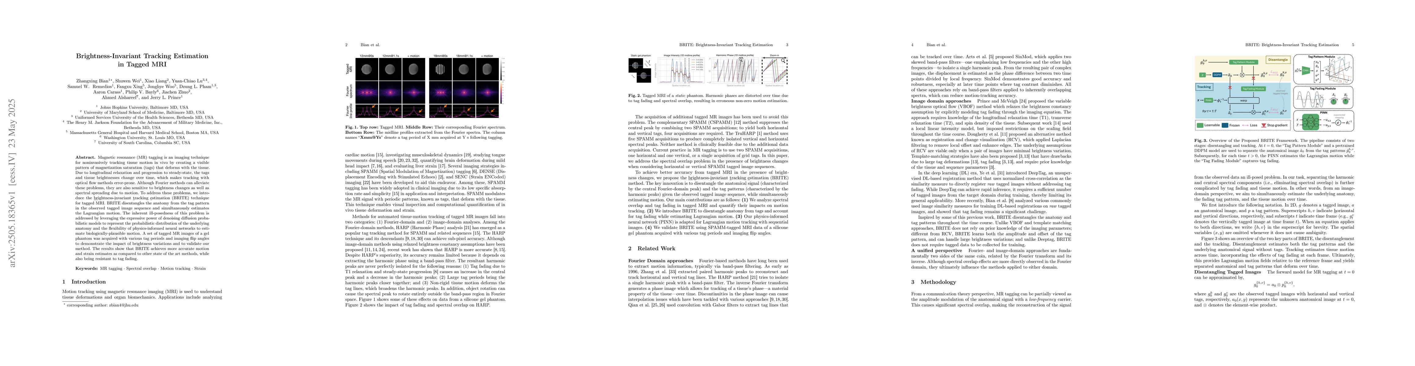
Magnetic resonance (MR) tagging is an imaging technique for noninvasively tracking tissue motion in vivo by creating a visible pattern of magnetization saturation (tags) that deforms with the tissue. ...
Automated segmentation of multiple sclerosis (MS) lesions using multicontrast magnetic resonance (MR) images improves efficiency and reproducibility compared to manual delineation, with deep learning ...
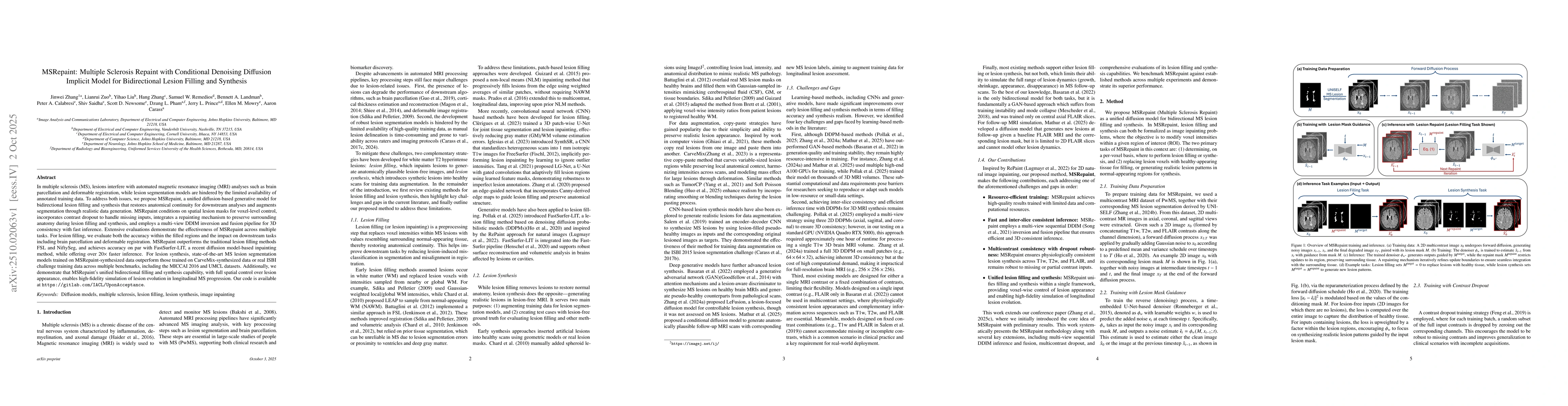
In multiple sclerosis, lesions interfere with automated magnetic resonance imaging analyses such as brain parcellation and deformable registration, while lesion segmentation models are hindered by the...