Academic Profile
Statistics
Similar Authors
Papers on arXiv
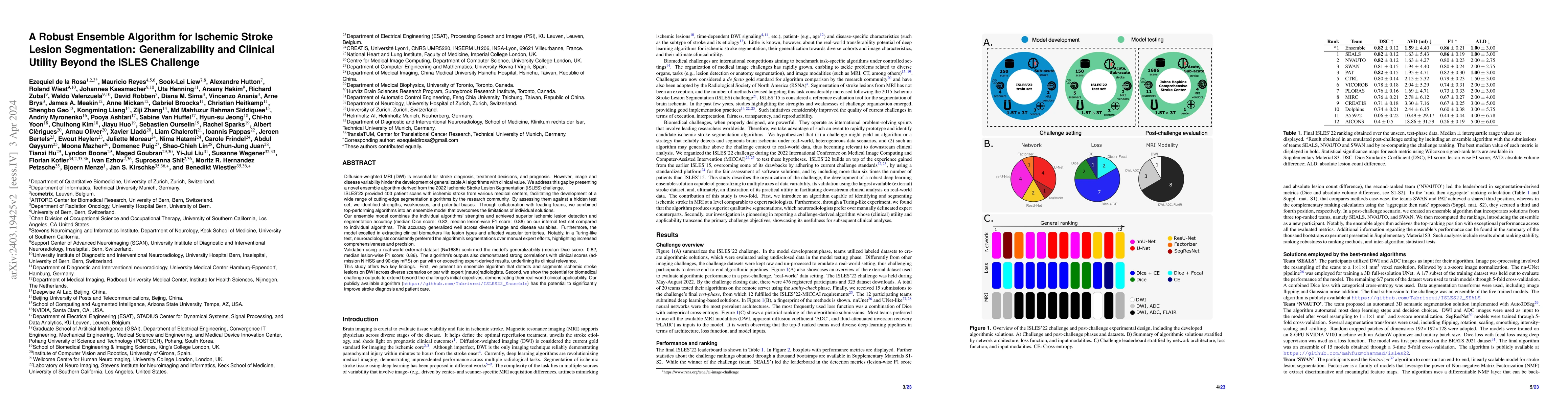
Diffusion-weighted MRI (DWI) is essential for stroke diagnosis, treatment decisions, and prognosis. However, image and disease variability hinder the development of generalizable AI algorithms with ...
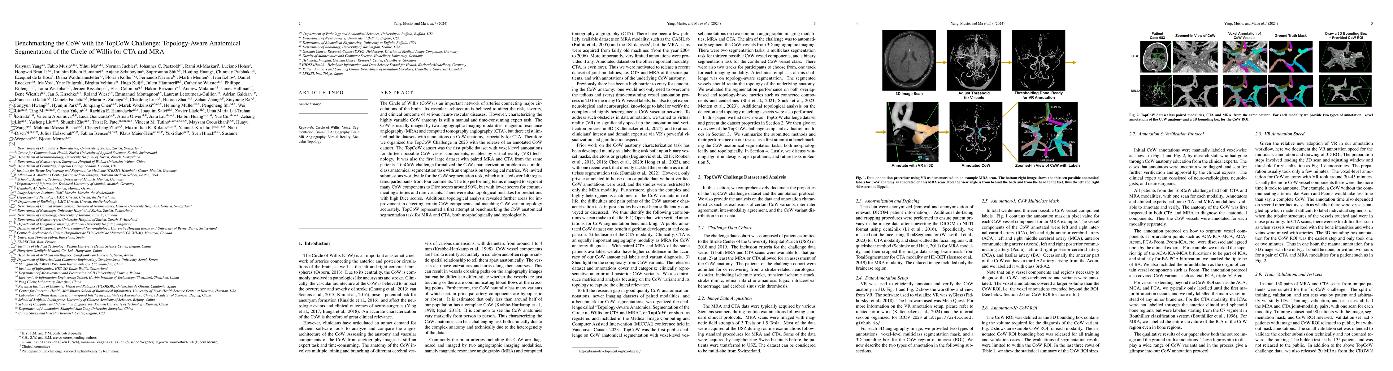
The Circle of Willis (CoW) is an important network of arteries connecting major circulations of the brain. Its vascular architecture is believed to affect the risk, severity, and clinical outcome of...
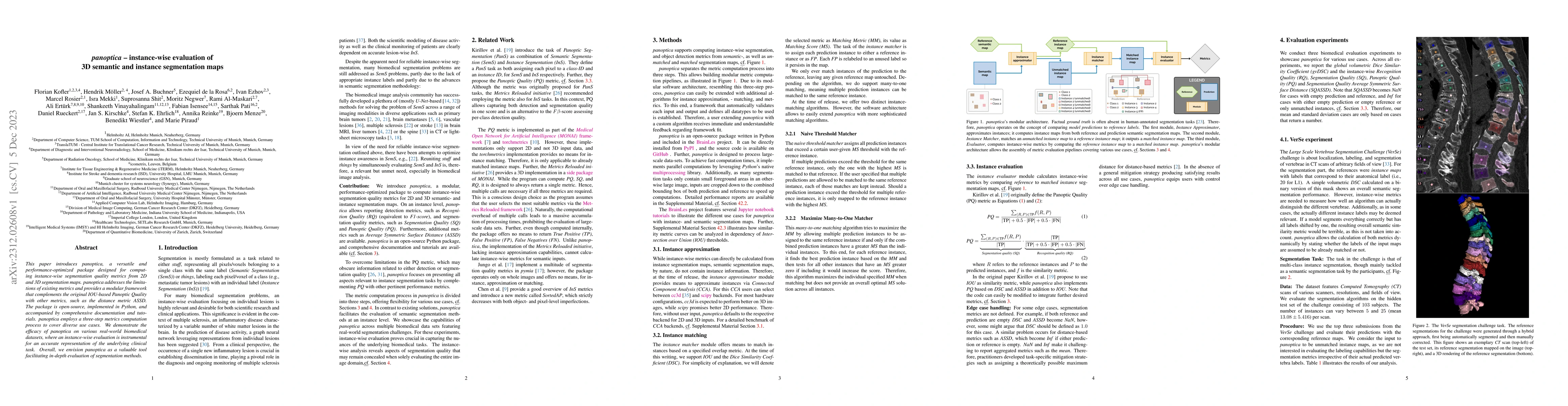
This paper introduces panoptica, a versatile and performance-optimized package designed for computing instance-wise segmentation quality metrics from 2D and 3D segmentation maps. panoptica addresses...
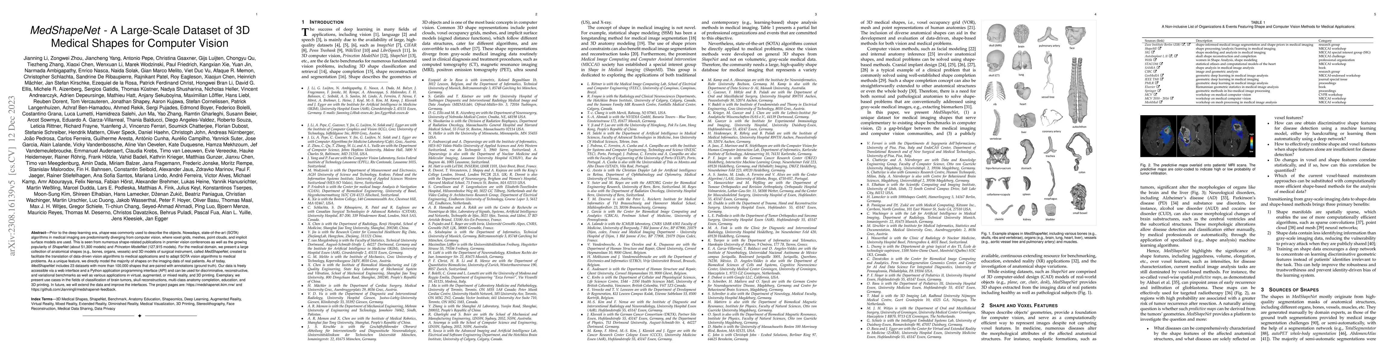
Prior to the deep learning era, shape was commonly used to describe the objects. Nowadays, state-of-the-art (SOTA) algorithms in medical imaging are predominantly diverging from computer vision, whe...
Magnetic resonance imaging (MRI) is a central modality for stroke imaging. It is used upon patient admission to make treatment decisions such as selecting patients for intravenous thrombolysis or en...
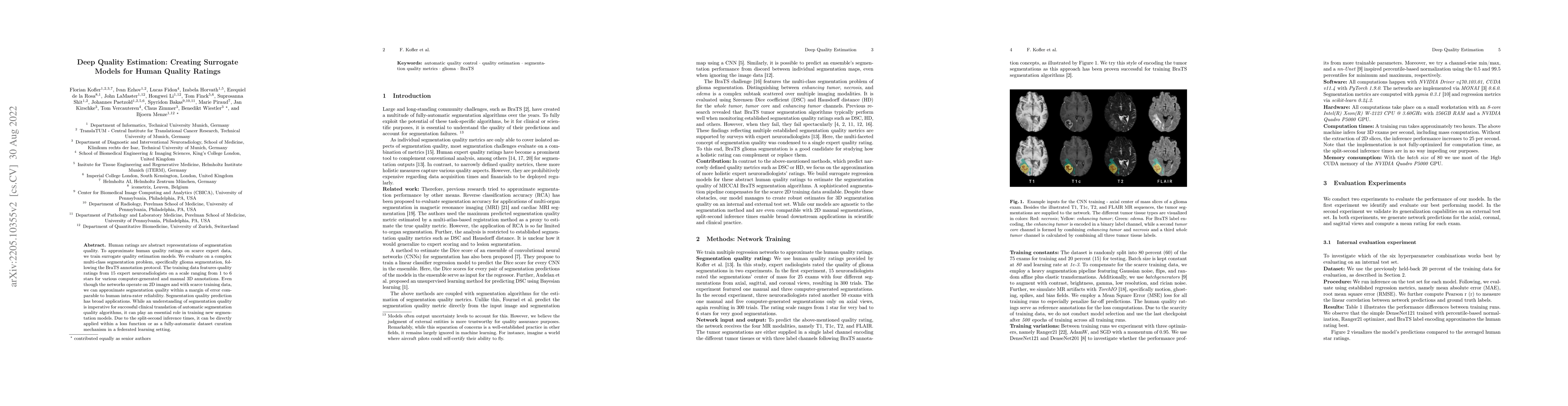
Human ratings are abstract representations of segmentation quality. To approximate human quality ratings on scarce expert data, we train surrogate quality estimation models. We evaluate on a complex...
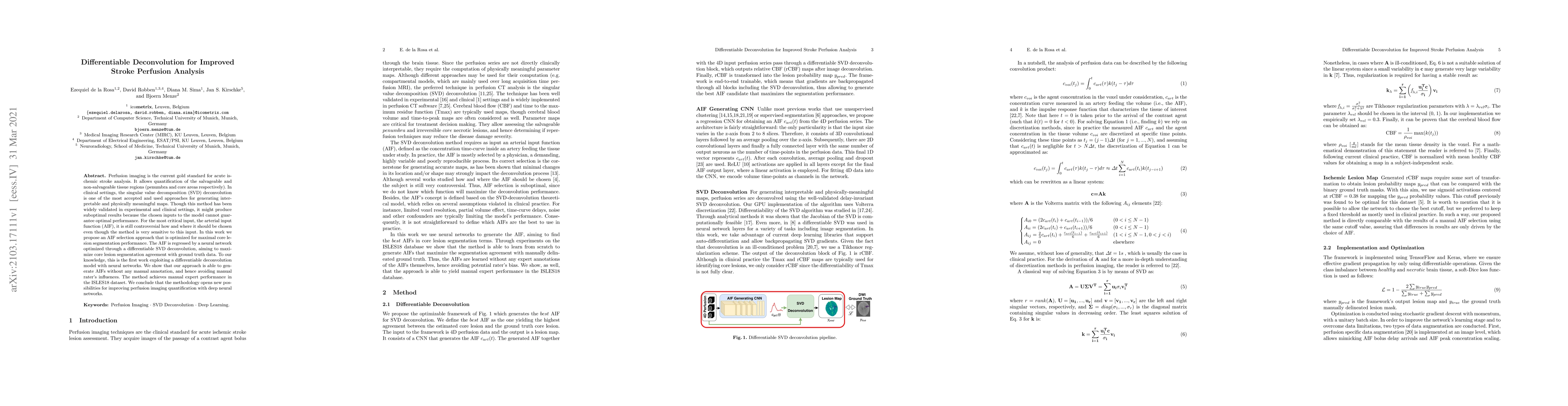
Perfusion imaging is the current gold standard for acute ischemic stroke analysis. It allows quantification of the salvageable and non-salvageable tissue regions (penumbra and core areas respectivel...
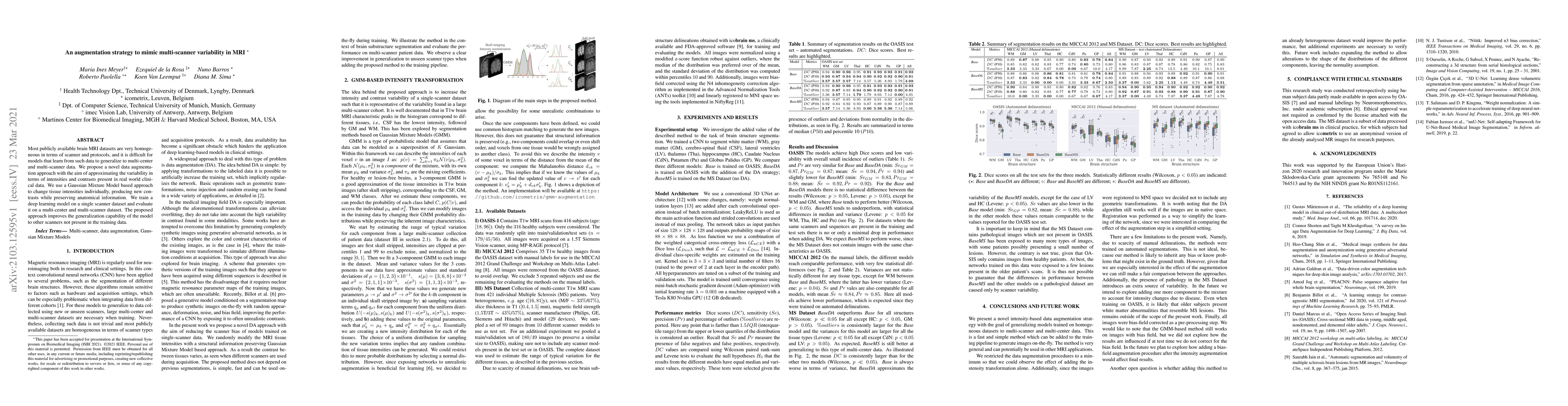
Most publicly available brain MRI datasets are very homogeneous in terms of scanner and protocols, and it is difficult for models that learn from such data to generalize to multi-center and multi-sc...
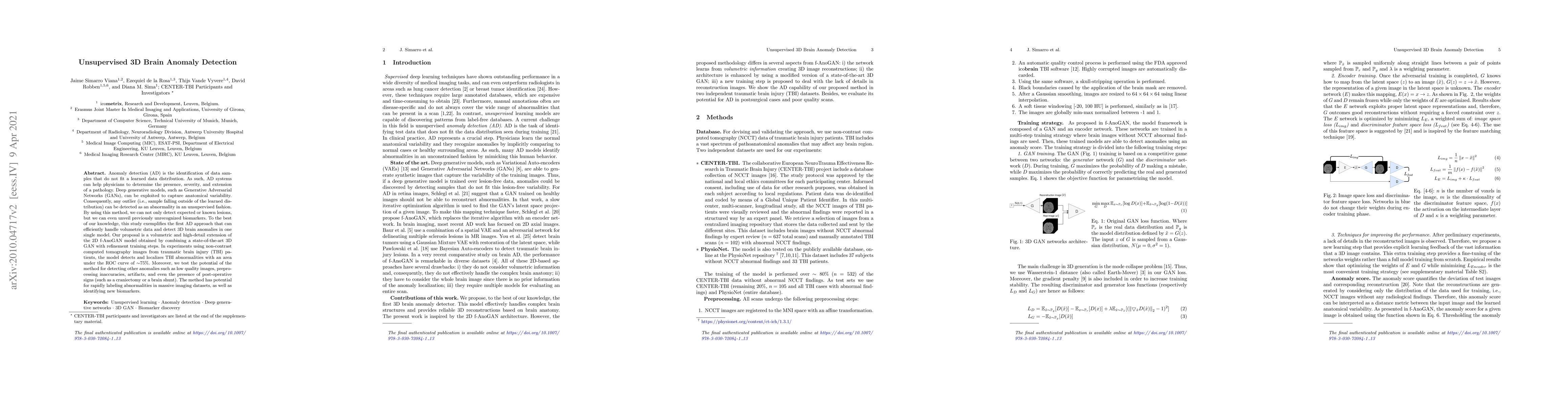
Anomaly detection (AD) is the identification of data samples that do not fit a learned data distribution. As such, AD systems can help physicians to determine the presence, severity, and extension o...
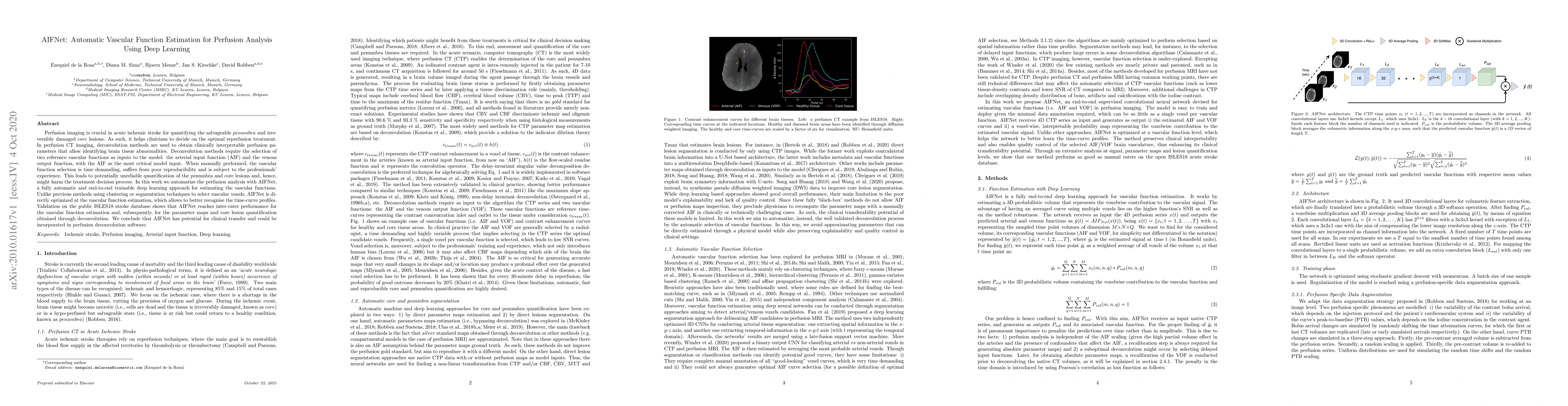
Perfusion imaging is crucial in acute ischemic stroke for quantifying the salvageable penumbra and irreversibly damaged core lesions. As such, it helps clinicians to decide on the optimal reperfusio...
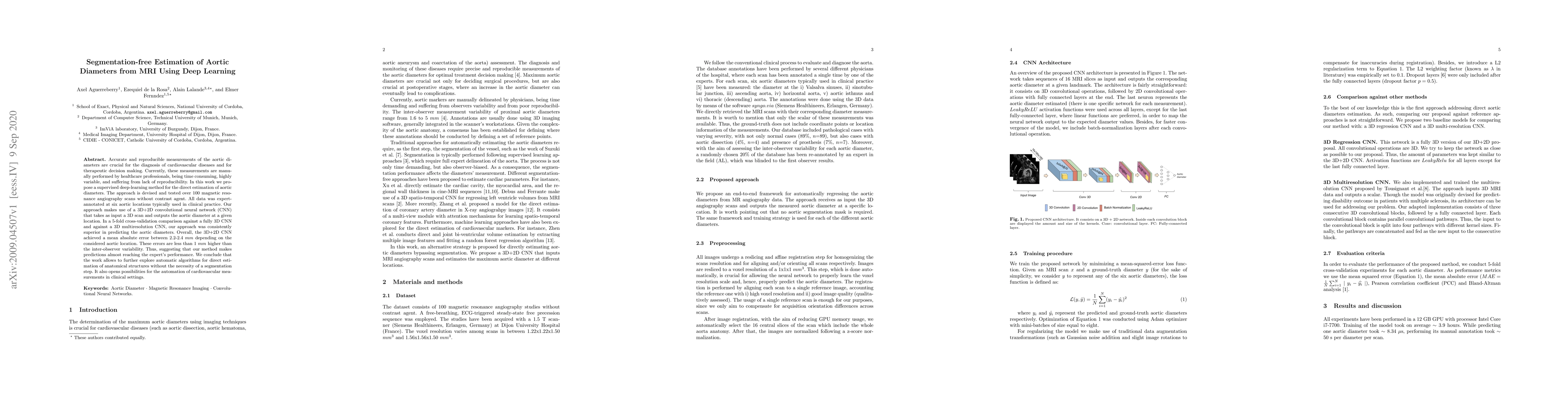
Accurate and reproducible measurements of the aortic diameters are crucial for the diagnosis of cardiovascular diseases and for therapeutic decision making. Currently, these measurements are manuall...
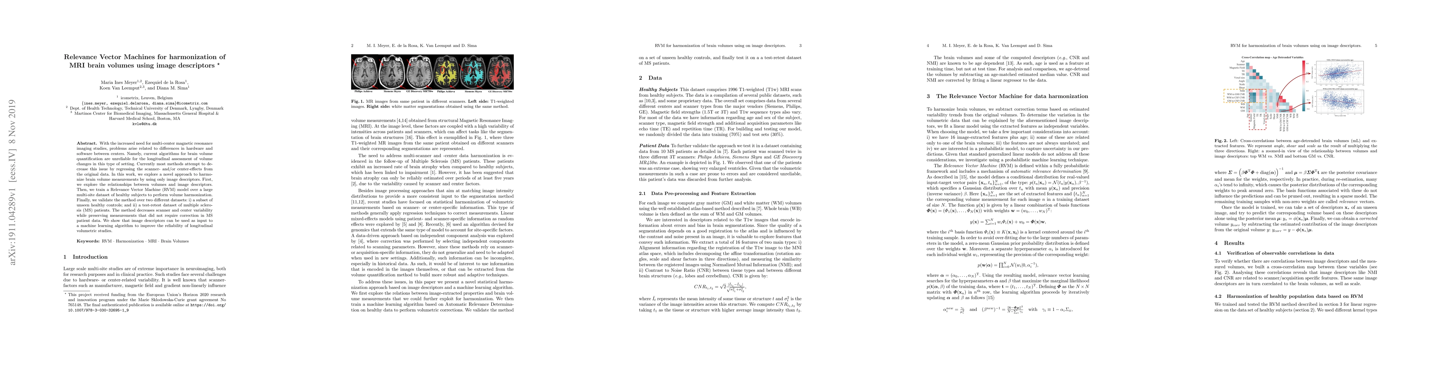
With the increased need for multi-center magnetic resonance imaging studies, problems arise related to differences in hardware and software between centers. Namely, current algorithms for brain volu...
Significance: Late gadolinium enhanced magnetic resonance imaging (LGE-MRI) is the gold standard technique for myocardial viability assessment. Although the technique accurately reflects the damaged...
Computer Tomography (CT) is the gold standard technique for brain damage evaluation after acute Traumatic Brain Injury (TBI). It allows identification of most lesion types and determines the need of...
Accurate estimation of core (irreversibly damaged tissue) and penumbra (salvageable tissue) volumes is essential for ischemic stroke treatment decisions. Perfusion CT, the clinical standard, estimates...
Stroke remains a leading cause of global morbidity and mortality, placing a heavy socioeconomic burden. Over the past decade, advances in endovascular reperfusion therapy and the use of CT and MRI ima...
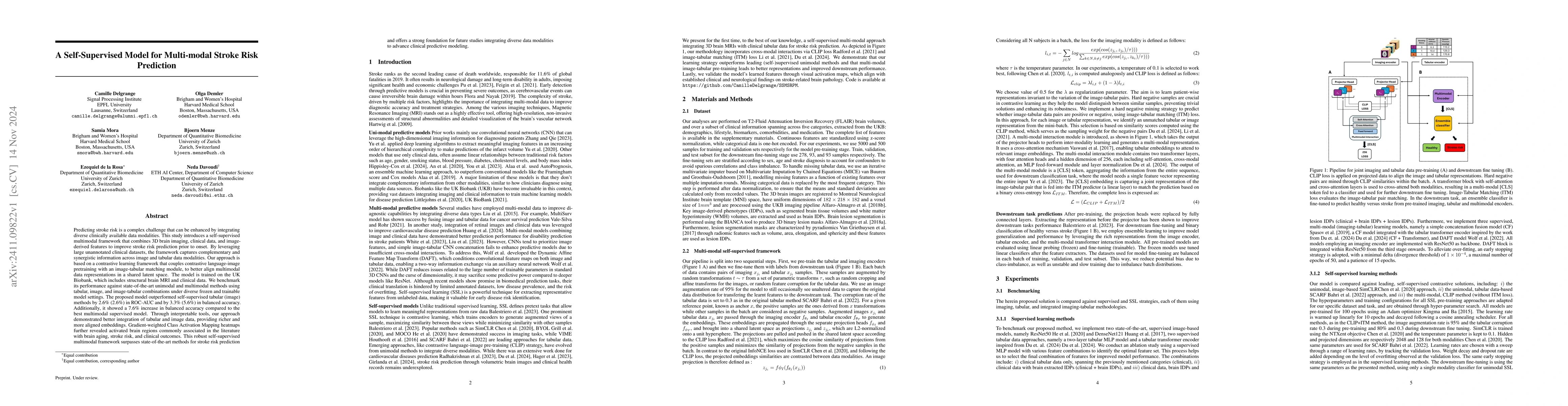
Predicting stroke risk is a complex challenge that can be enhanced by integrating diverse clinically available data modalities. This study introduces a self-supervised multimodal framework that combin...
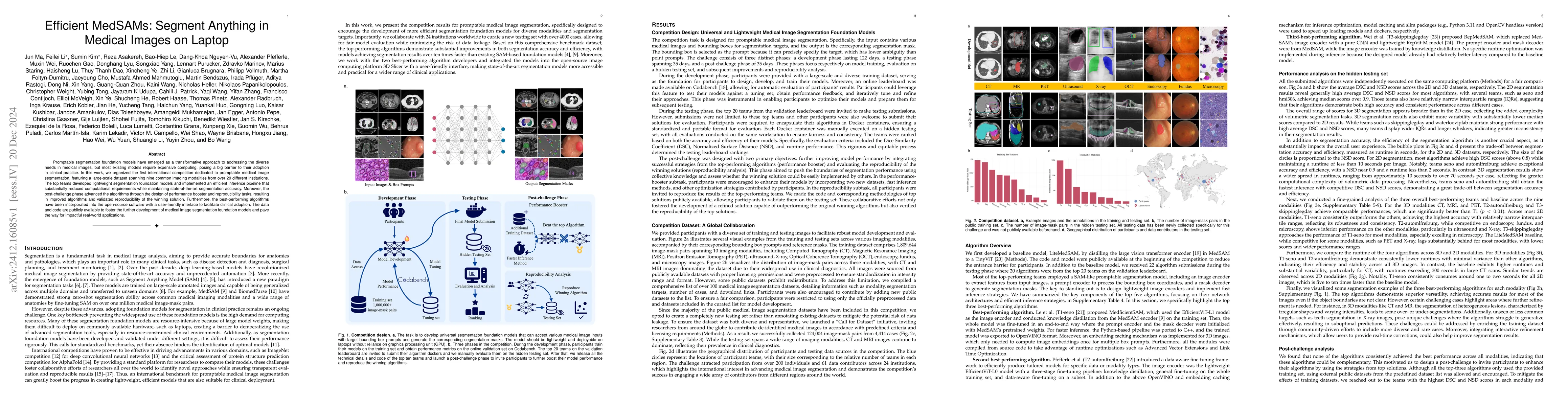
Promptable segmentation foundation models have emerged as a transformative approach to addressing the diverse needs in medical images, but most existing models require expensive computing, posing a bi...
The Brain Tumor Segmentation (BraTS) cluster of challenges has significantly advanced brain tumor image analysis by providing large, curated datasets and addressing clinically relevant tasks. However,...
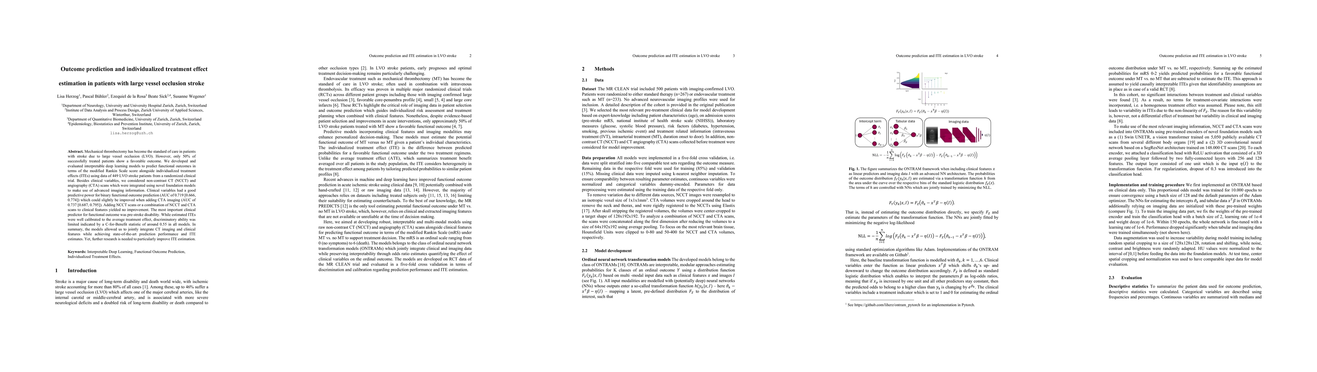
Mechanical thrombectomy has become the standard of care in patients with stroke due to large vessel occlusion (LVO). However, only 50% of successfully treated patients show a favorable outcome. We dev...
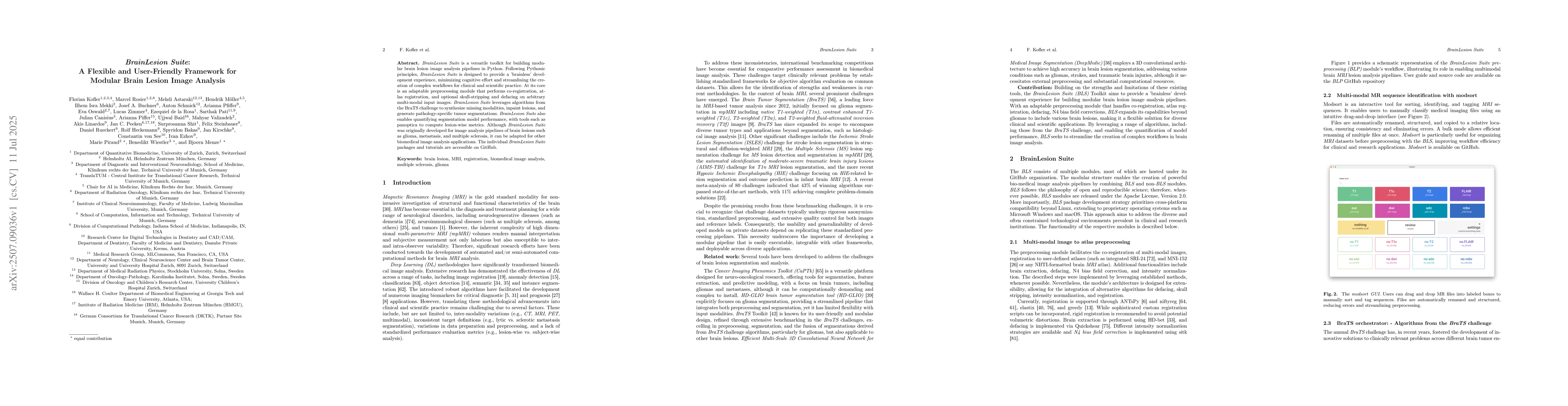
BrainLesion Suite is a versatile toolkit for building modular brain lesion image analysis pipelines in Python. Following Pythonic principles, BrainLesion Suite is designed to provide a 'brainless' dev...
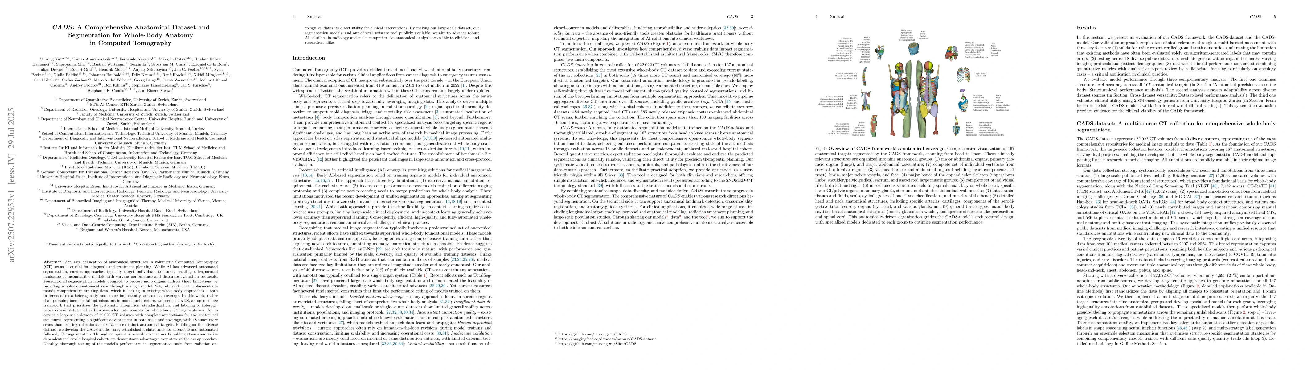
Accurate delineation of anatomical structures in volumetric CT scans is crucial for diagnosis and treatment planning. While AI has advanced automated segmentation, current approaches typically target ...
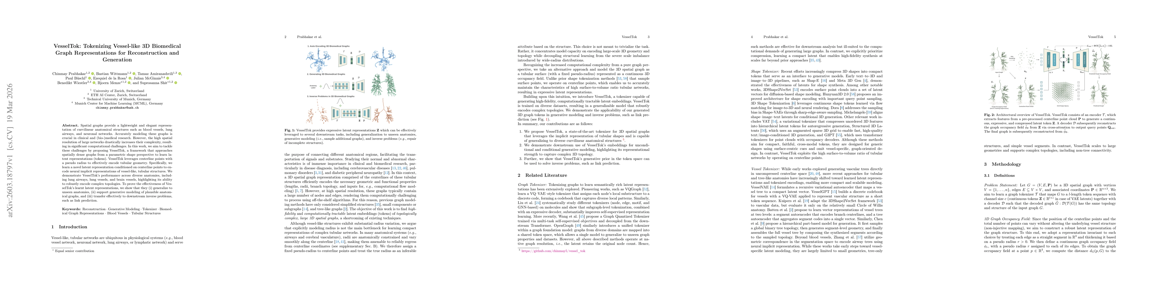
Spatial graphs provide a lightweight and elegant representation of curvilinear anatomical structures such as blood vessels, lung airways, and neuronal networks. Accurately modeling these graphs is cru...
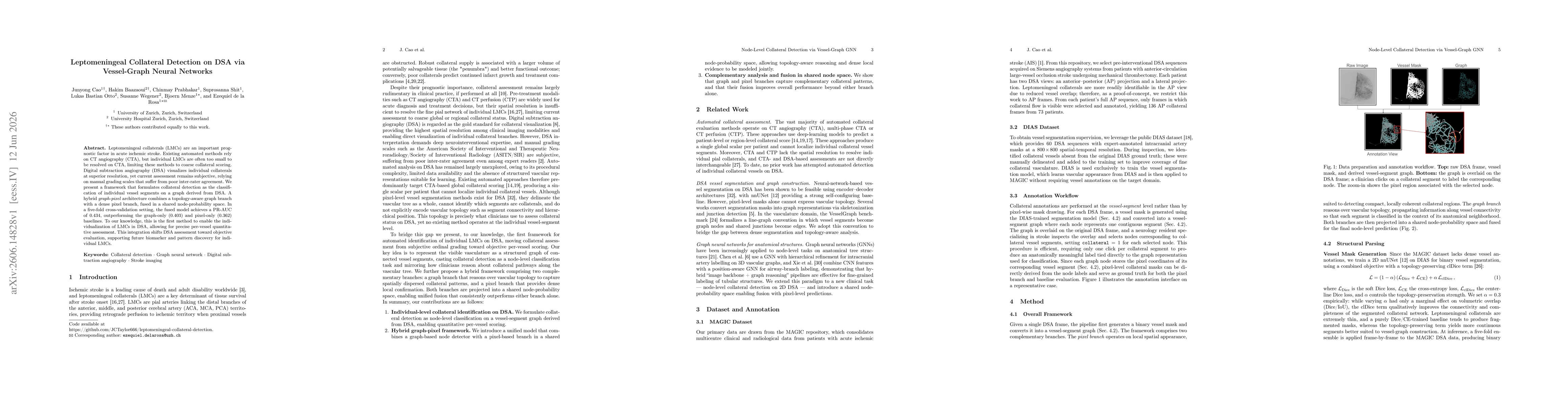
Leptomeningeal collaterals (LMCs) are an important prognostic factor in acute ischemic stroke. Existing automated methods rely on CT angiography (CTA), but individual LMCs are often too small to be re...