Academic Profile
Statistics
Similar Authors
Papers on arXiv
Cardiac magnetic resonance imaging (MRI) has emerged as a clinically gold-standard technique for diagnosing cardiac diseases, thanks to its ability to provide diverse information with multiple modalit...
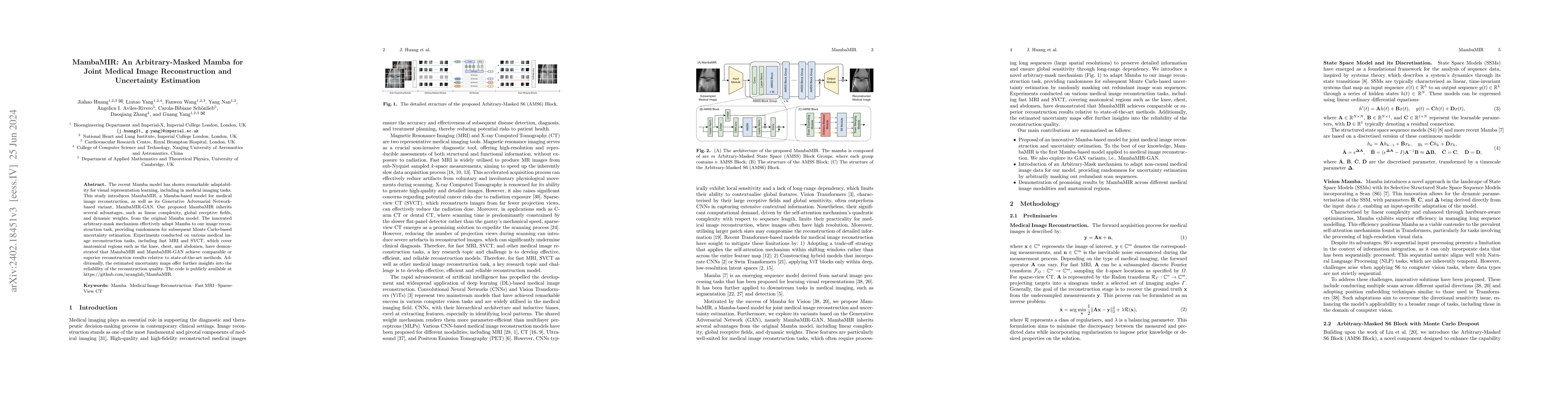
The recent Mamba model has shown remarkable adaptability for visual representation learning, including in medical imaging tasks. This study introduces MambaMIR, a Mamba-based model for medical image r...
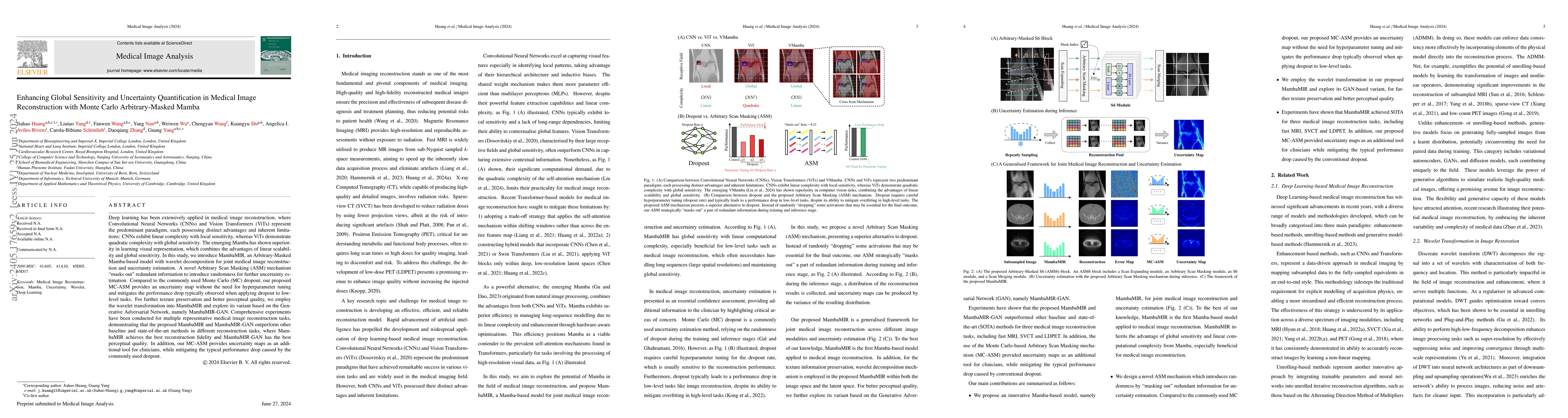
Deep learning has been extensively applied in medical image reconstruction, where Convolutional Neural Networks (CNNs) and Vision Transformers (ViTs) represent the predominant paradigms, each possessi...
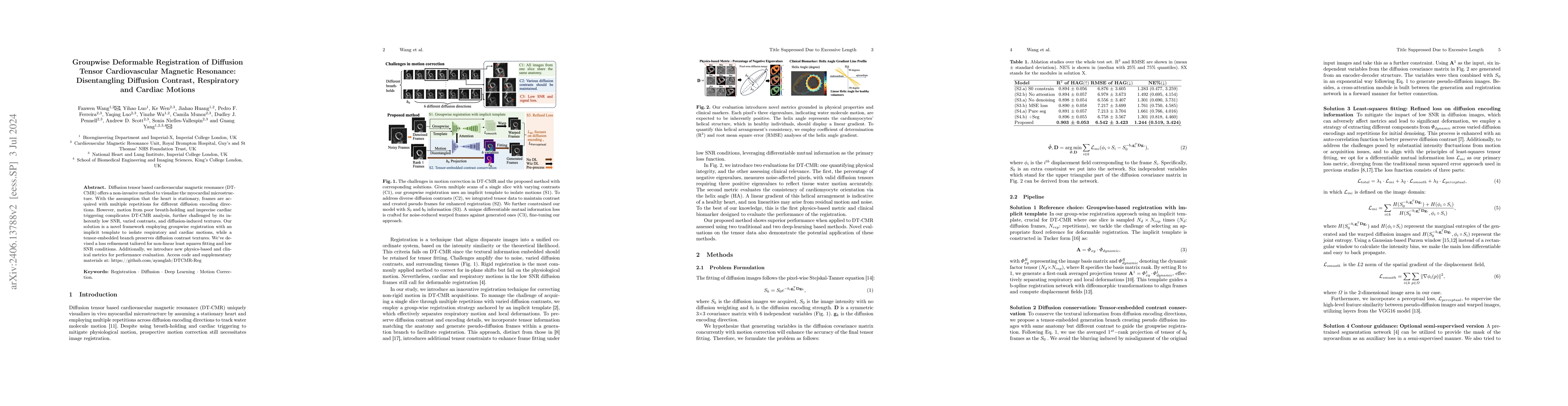
Diffusion tensor based cardiovascular magnetic resonance (DT-CMR) offers a non-invasive method to visualize the myocardial microstructure. With the assumption that the heart is stationary, frames ar...
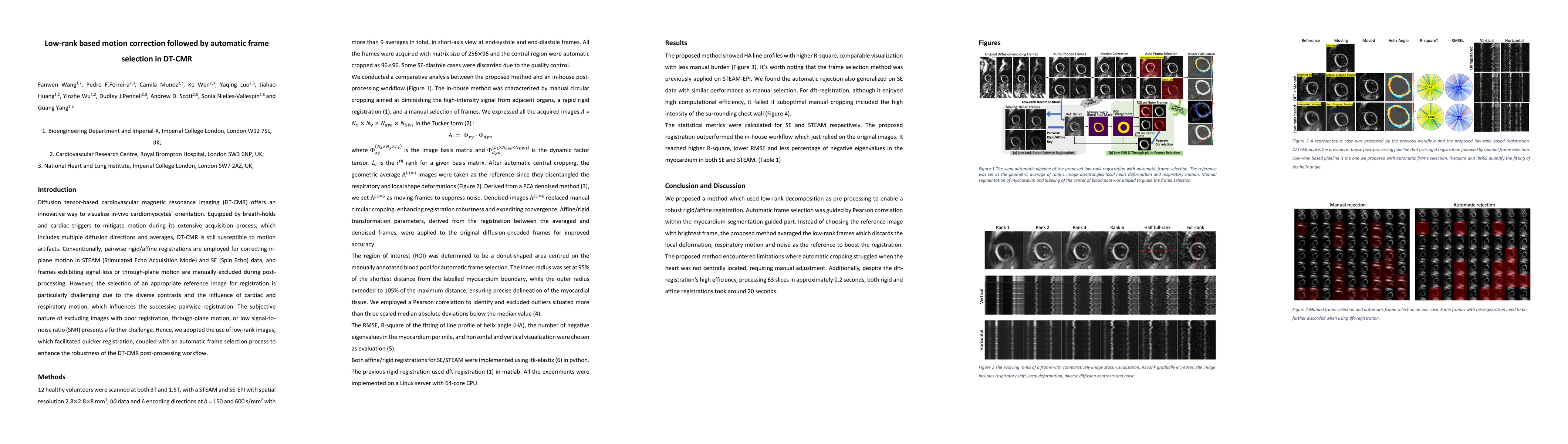
Motivation: Post-processing of in-vivo diffusion tensor CMR (DT-CMR) is challenging due to the low SNR and variation in contrast between frames which makes image registration difficult, and the need...
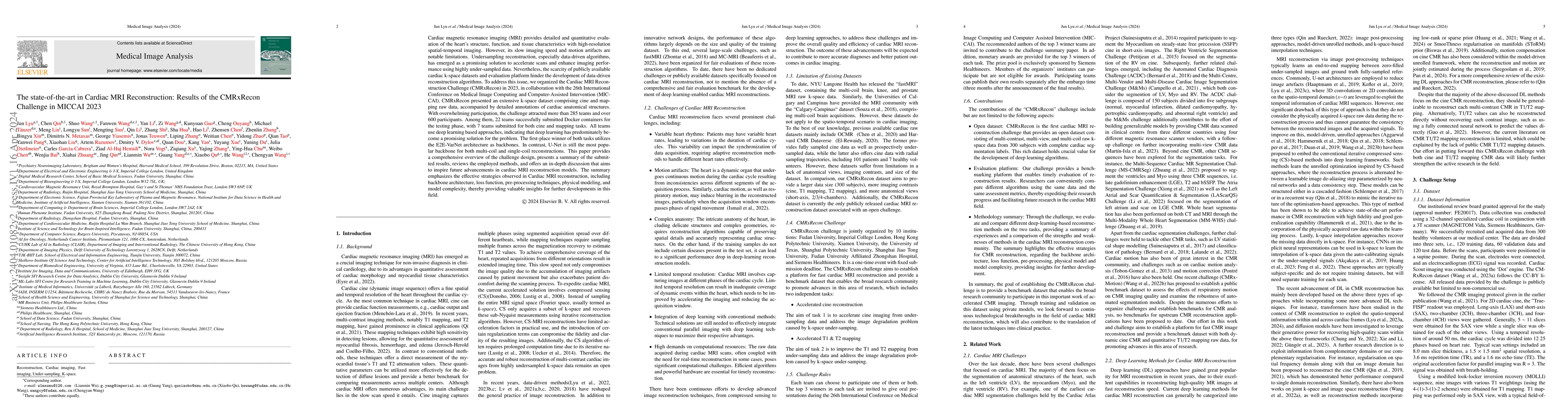
Cardiac MRI, crucial for evaluating heart structure and function, faces limitations like slow imaging and motion artifacts. Undersampling reconstruction, especially data-driven algorithms, has emerg...
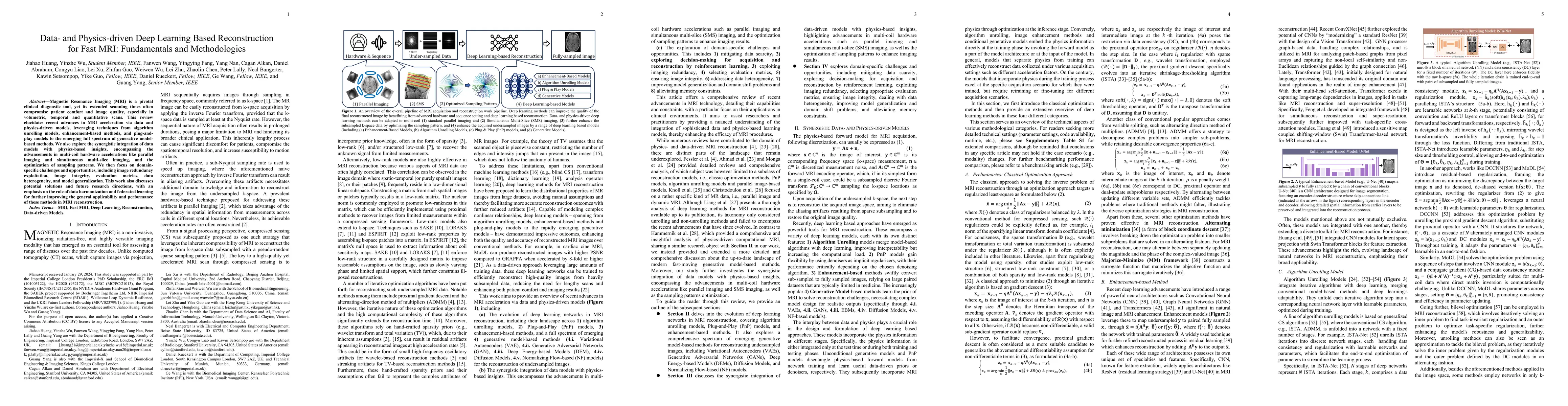
Magnetic Resonance Imaging (MRI) is a pivotal clinical diagnostic tool, yet its extended scanning times often compromise patient comfort and image quality, especially in volumetric, temporal and qua...
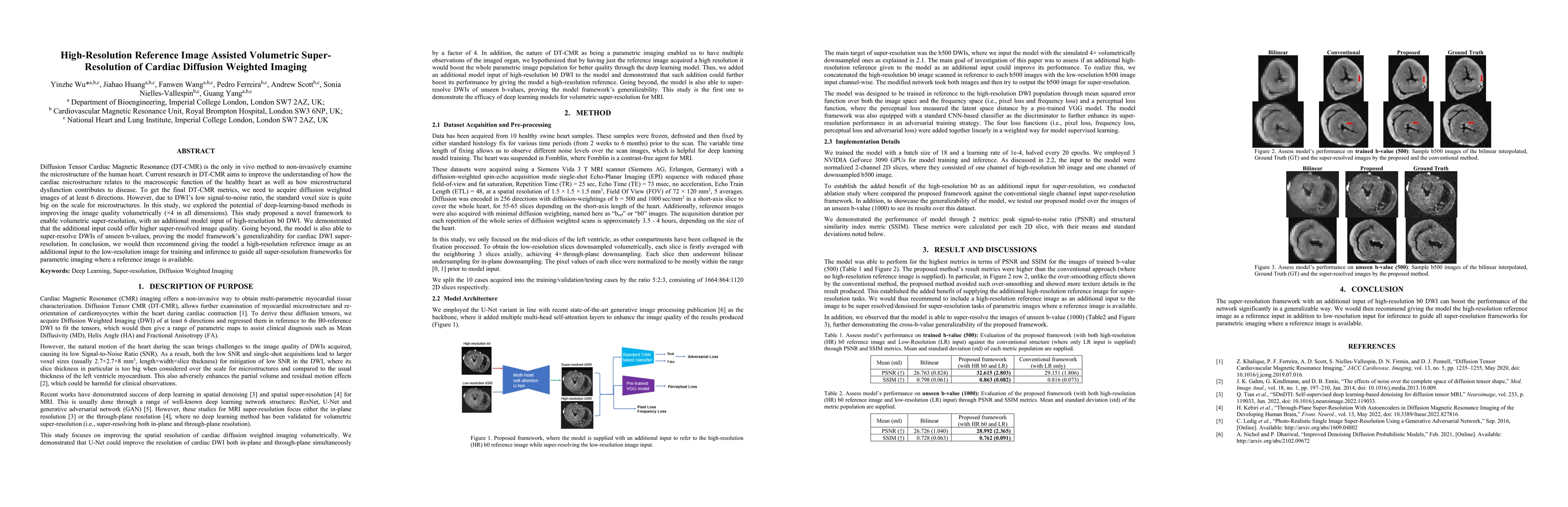
Diffusion Tensor Cardiac Magnetic Resonance (DT-CMR) is the only in vivo method to non-invasively examine the microstructure of the human heart. Current research in DT-CMR aims to improve the unders...
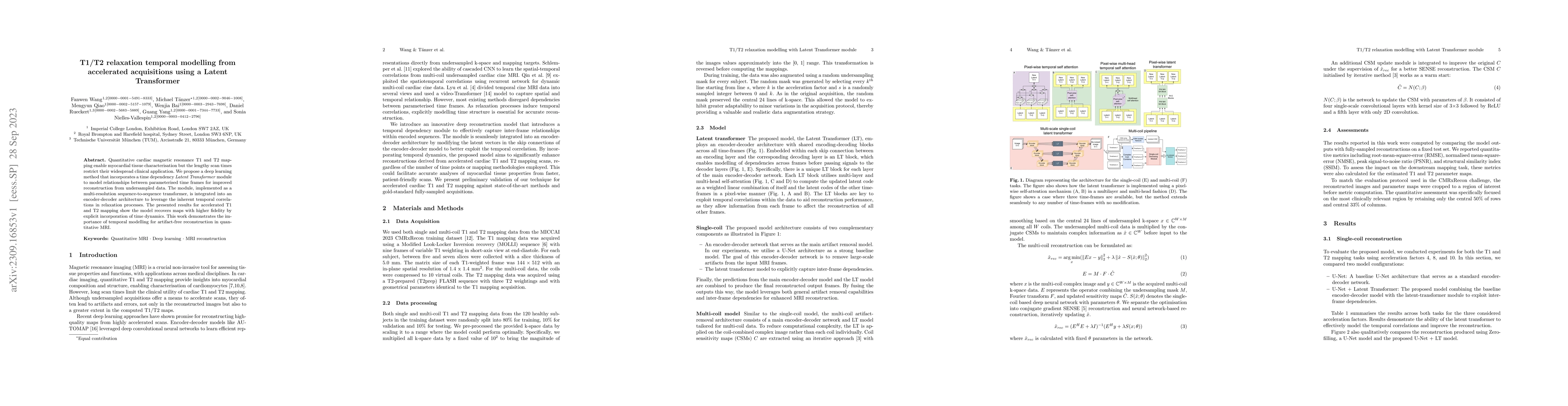
Quantitative cardiac magnetic resonance T1 and T2 mapping enable myocardial tissue characterisation but the lengthy scan times restrict their widespread clinical application. We propose a deep learn...
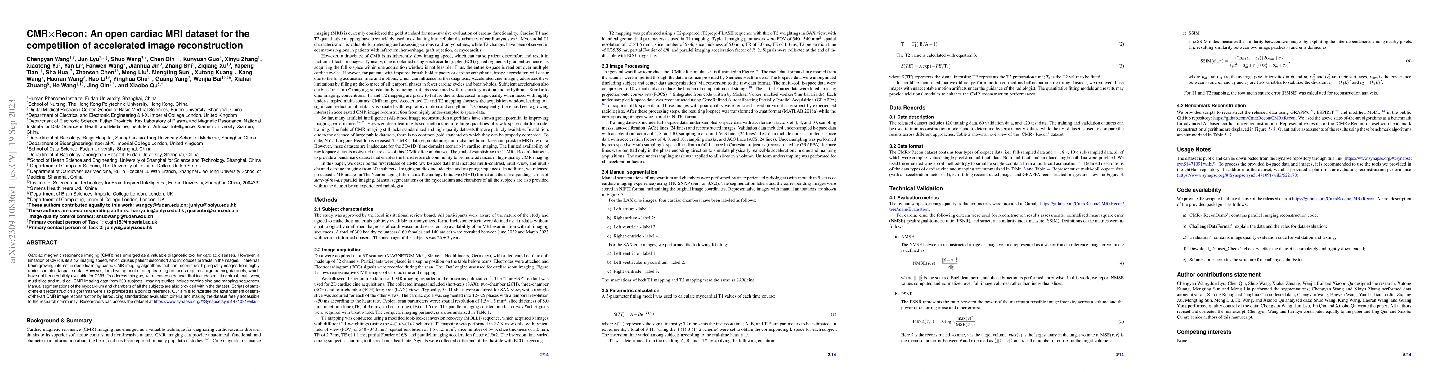
Cardiac magnetic resonance imaging (CMR) has emerged as a valuable diagnostic tool for cardiac diseases. However, a limitation of CMR is its slow imaging speed, which causes patient discomfort and i...
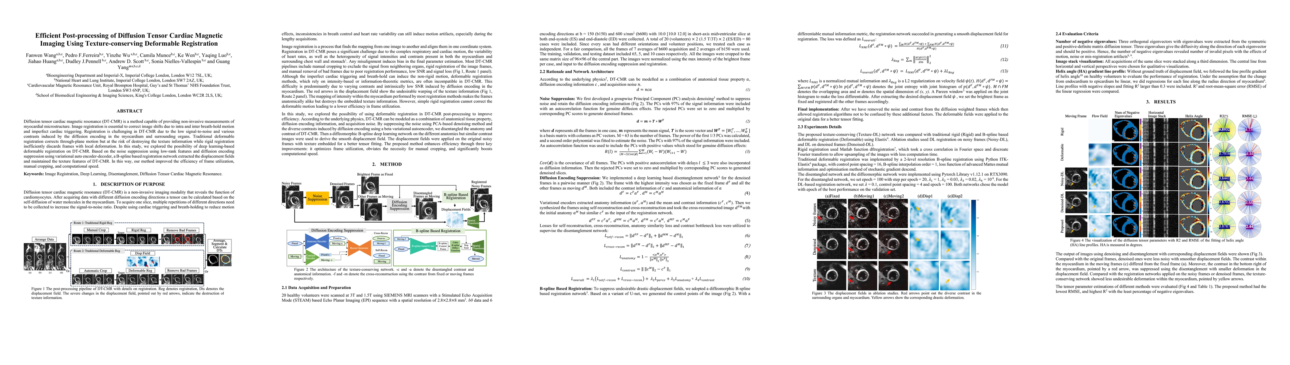
Diffusion tensor cardiac magnetic resonance (DT-CMR) is a method capable of providing non-invasive measurements of myocardial microstructure. Image registration is essential to correct image shifts ...
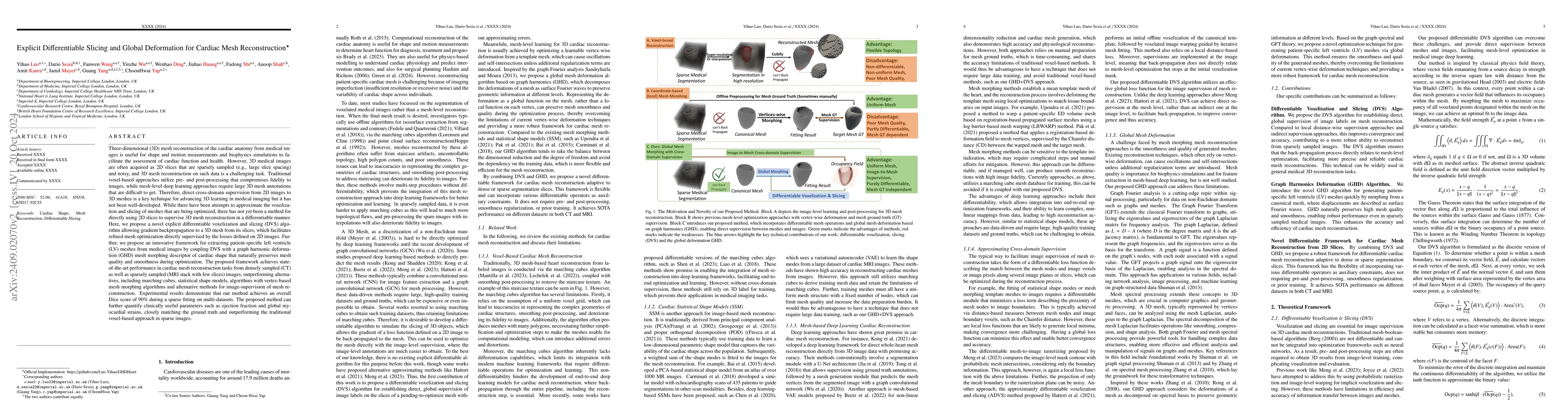
Mesh reconstruction of the cardiac anatomy from medical images is useful for shape and motion measurements and biophysics simulations to facilitate the assessment of cardiac function and health. Howev...
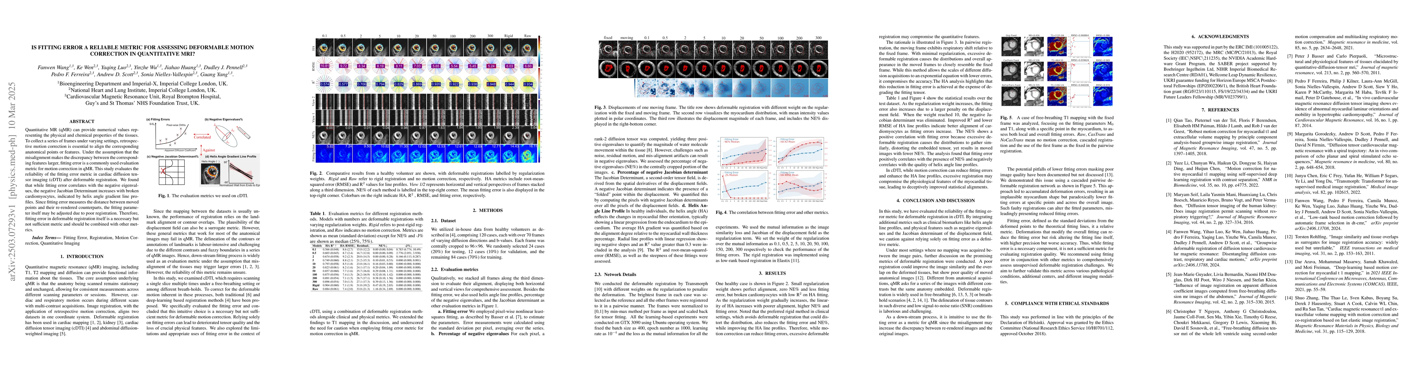
Quantitative MR (qMR) can provide numerical values representing the physical and chemical properties of the tissues. To collect a series of frames under varying settings, retrospective motion correcti...
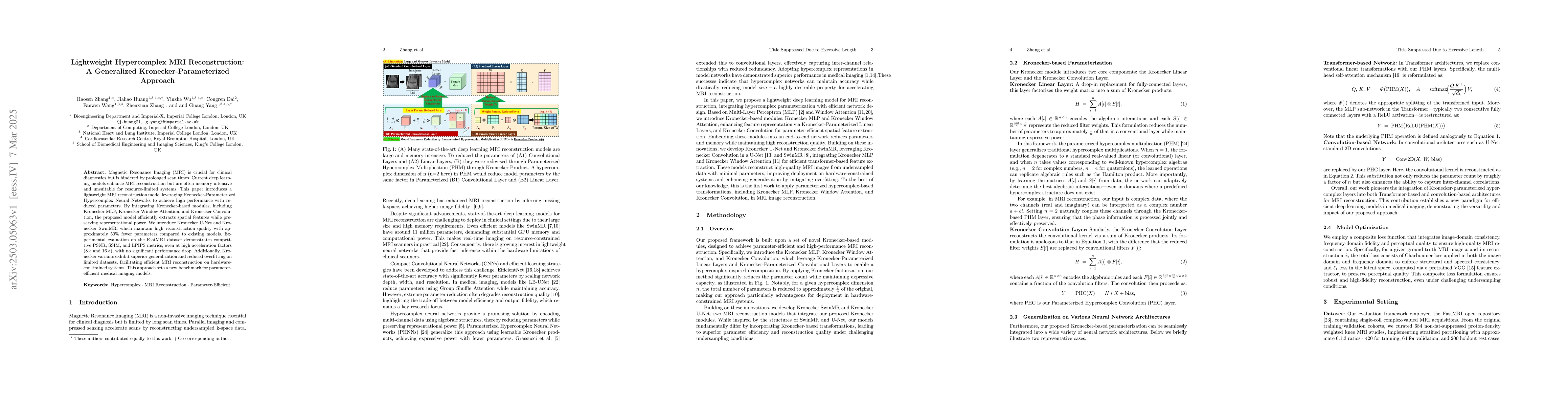
Magnetic Resonance Imaging (MRI) is crucial for clinical diagnostics but is hindered by prolonged scan times. Current deep learning models enhance MRI reconstruction but are often memory-intensive and...
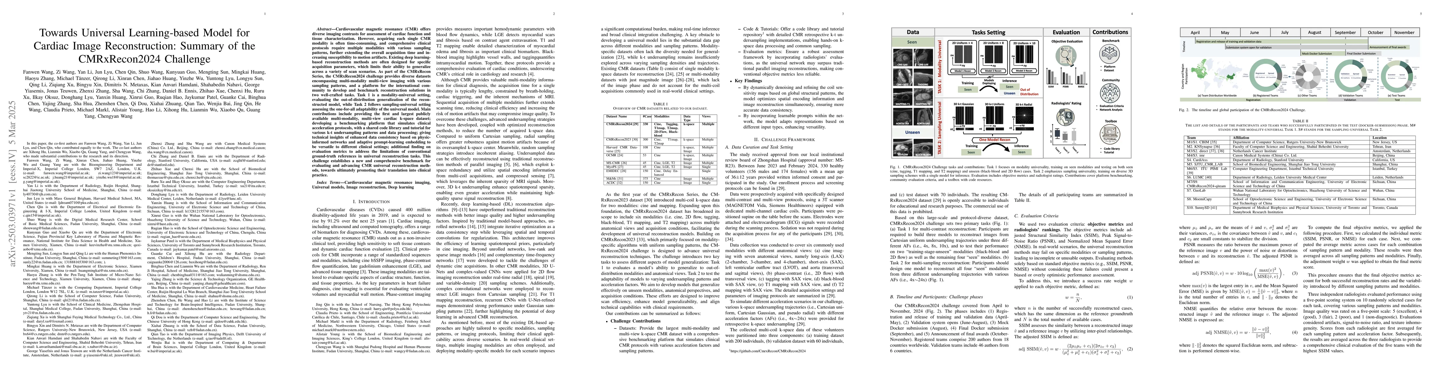
Cardiovascular magnetic resonance (CMR) offers diverse imaging contrasts for assessment of cardiac function and tissue characterization. However, acquiring each single CMR modality is often time-consu...
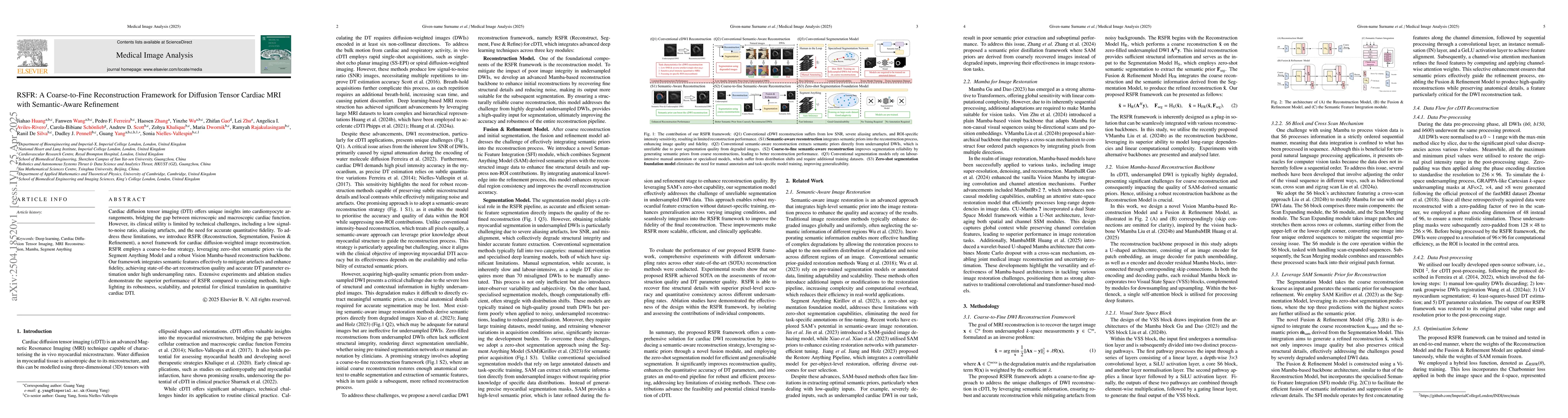
Cardiac diffusion tensor imaging (DTI) offers unique insights into cardiomyocyte arrangements, bridging the gap between microscopic and macroscopic cardiac function. However, its clinical utility is l...
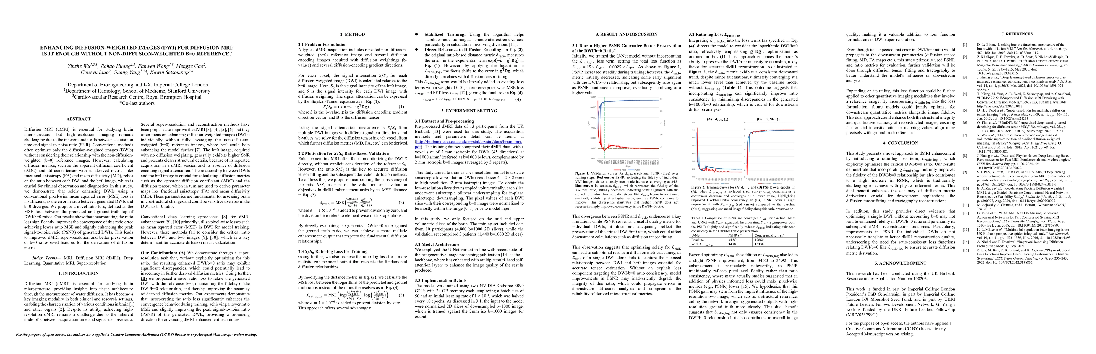
Diffusion MRI (dMRI) is essential for studying brain microstructure, but high-resolution imaging remains challenging due to the inherent trade-offs between acquisition time and signal-to-noise ratio (...
Magnetic Resonance Imaging (MRI) is critical for clinical diagnostics but is often limited by long acquisition times and low signal-to-noise ratios, especially in modalities like diffusion and functio...
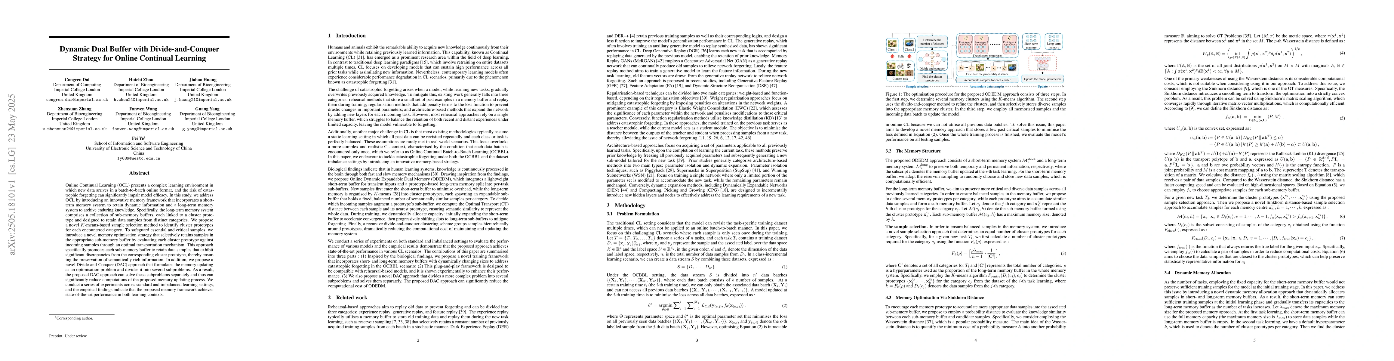
Online Continual Learning (OCL) presents a complex learning environment in which new data arrives in a batch-to-batch online format, and the risk of catastrophic forgetting can significantly impair mo...
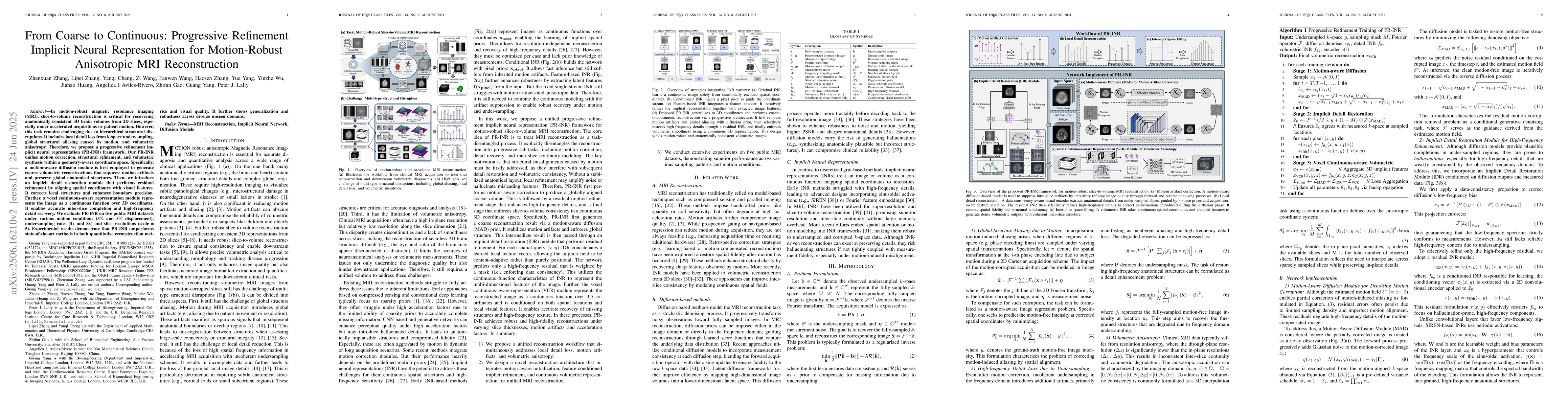
In motion-robust magnetic resonance imaging (MRI), slice-to-volume reconstruction is critical for recovering anatomically consistent 3D brain volumes from 2D slices, especially under accelerated acqui...
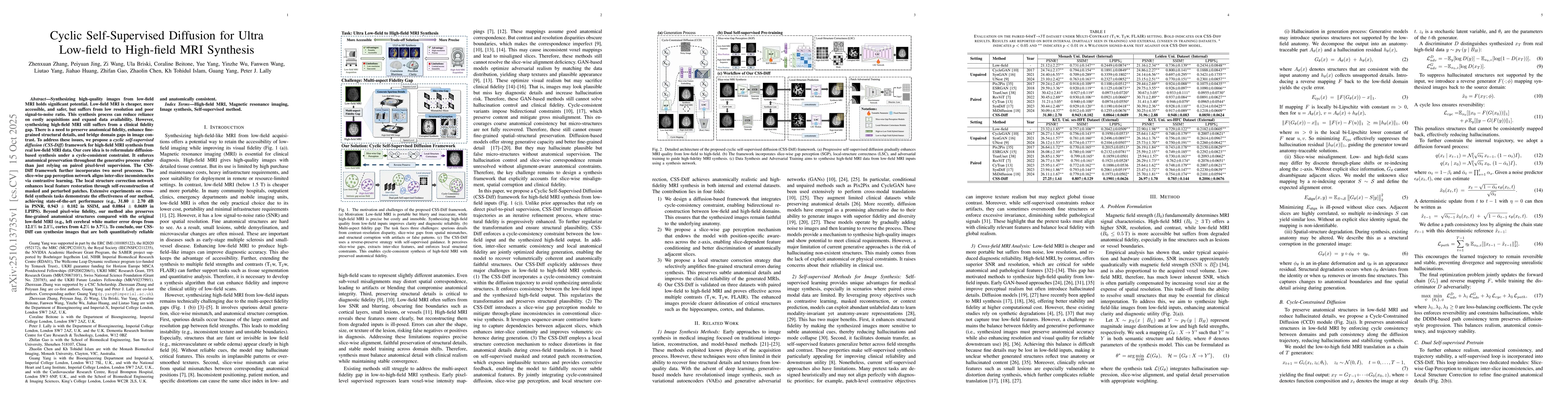
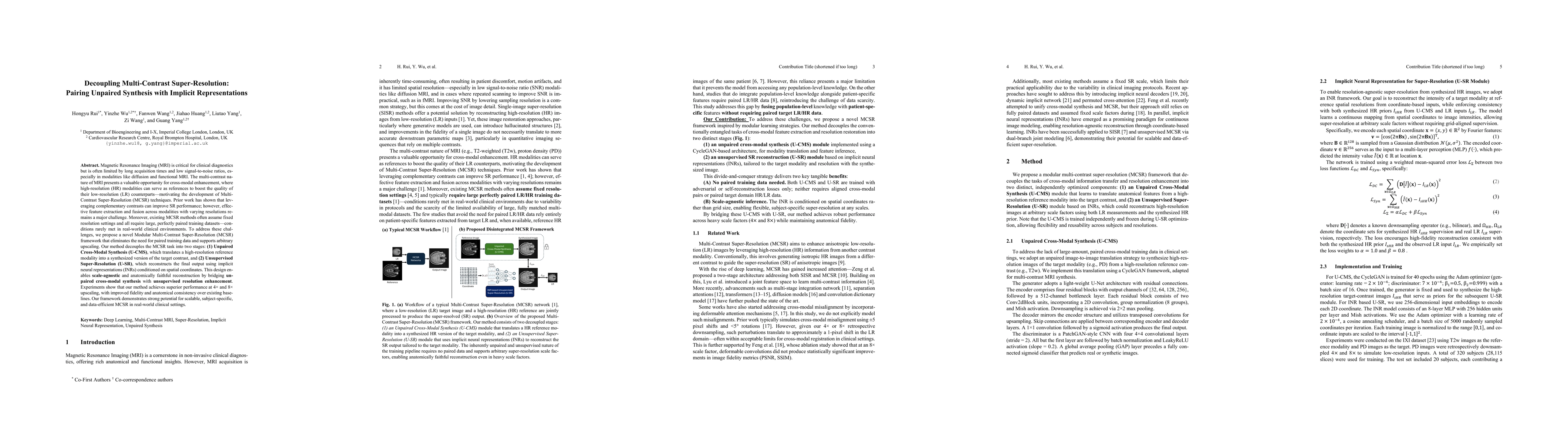
Synthesizing high-quality images from low-field MRI holds significant potential. Low-field MRI is cheaper, more accessible, and safer, but suffers from low resolution and poor signal-to-noise ratio. T...
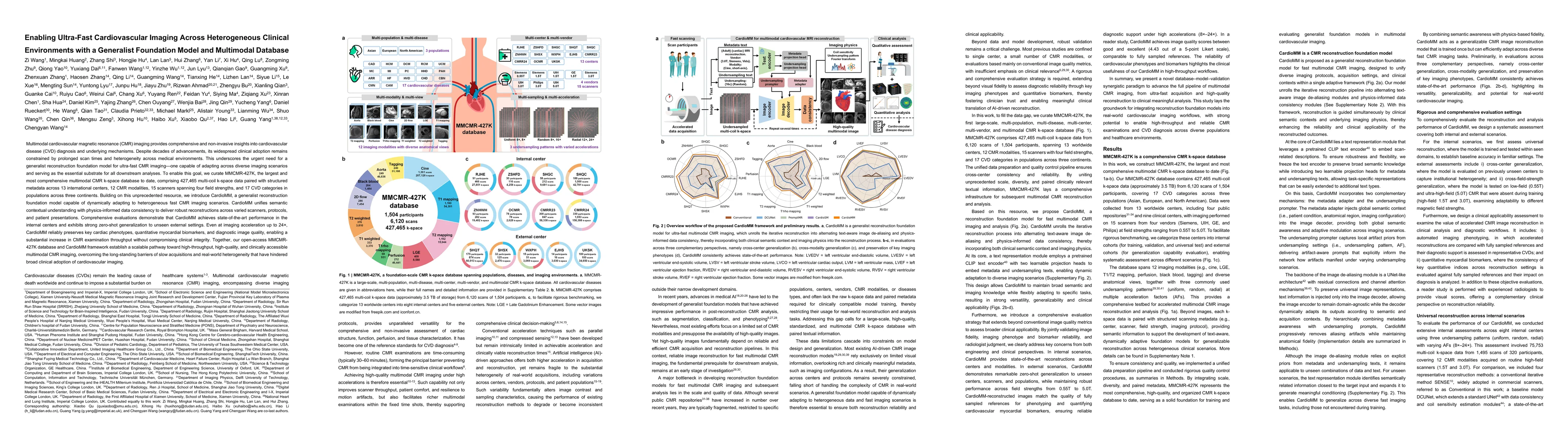
Multimodal cardiovascular magnetic resonance (CMR) imaging provides comprehensive and non-invasive insights into cardiovascular disease (CVD) diagnosis and underlying mechanisms. Despite decades of ad...
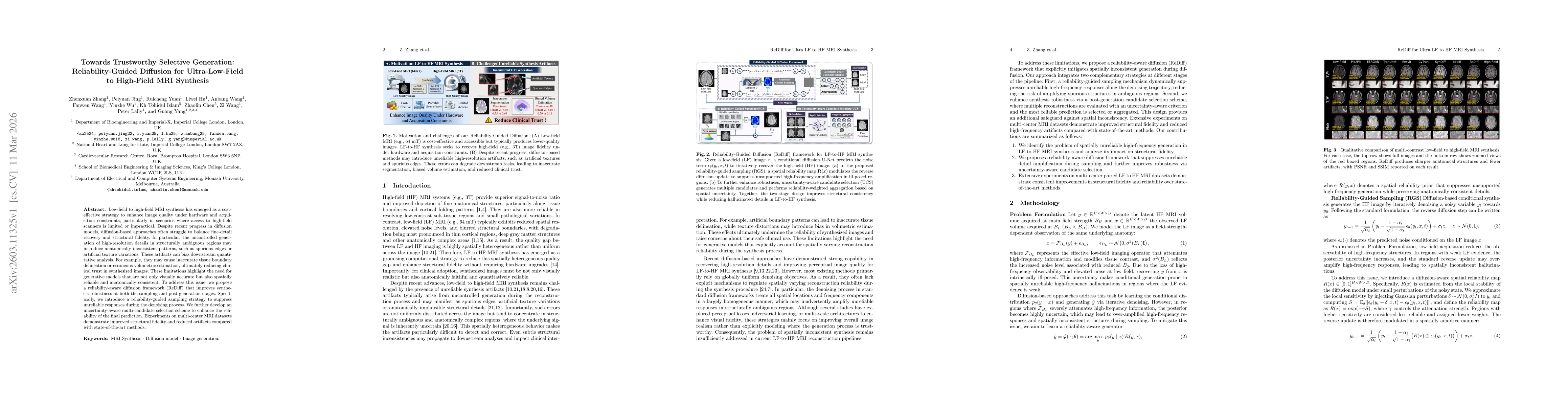
Low-field to high-field MRI synthesis has emerged as a cost-effective strategy to enhance image quality under hardware and acquisition constraints, particularly in scenarios where access to high-field...
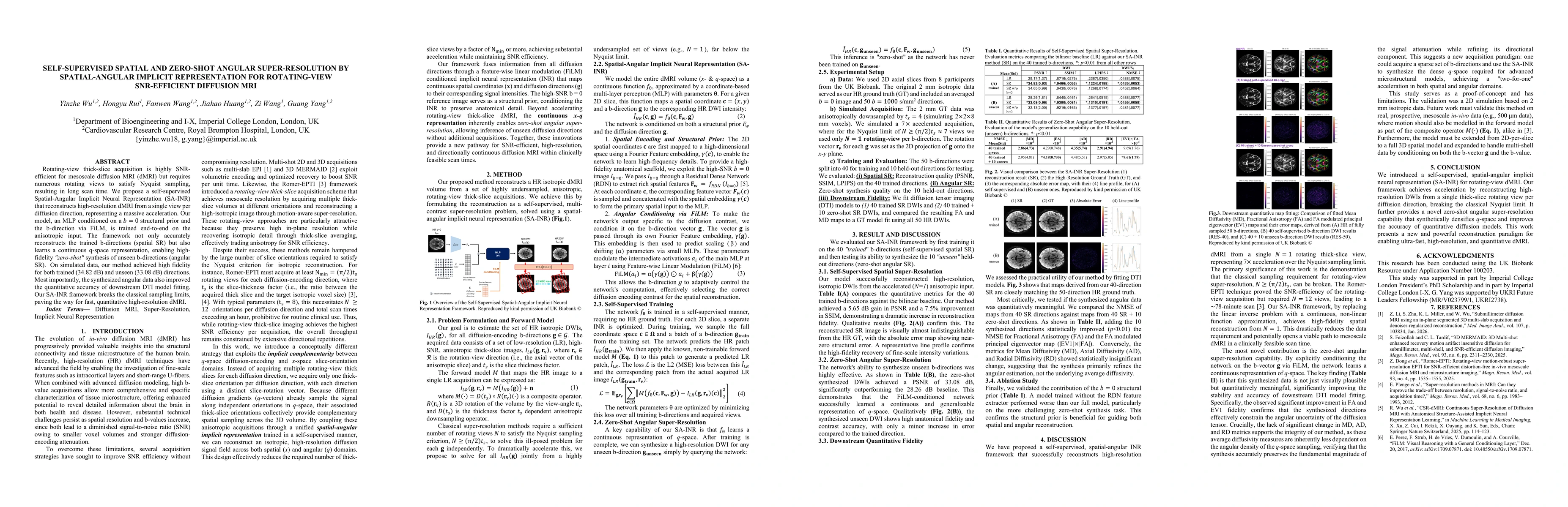
Rotating-view thick-slice acquisition is highly SNR-efficient for mesoscale diffusion MRI (dMRI) but requires numerous rotating views to satisfy Nyquist sampling, resulting in long scan time. We propo...
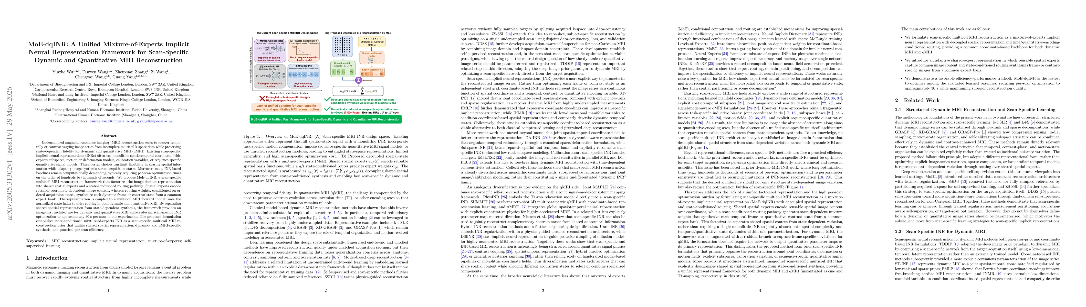
Undersampled magnetic resonance imaging (MRI) reconstruction seeks to recover temporally or contrast-varying image series from incomplete multicoil k-space data while preserving state-dependent fideli...