Academic Profile
Statistics
Similar Authors
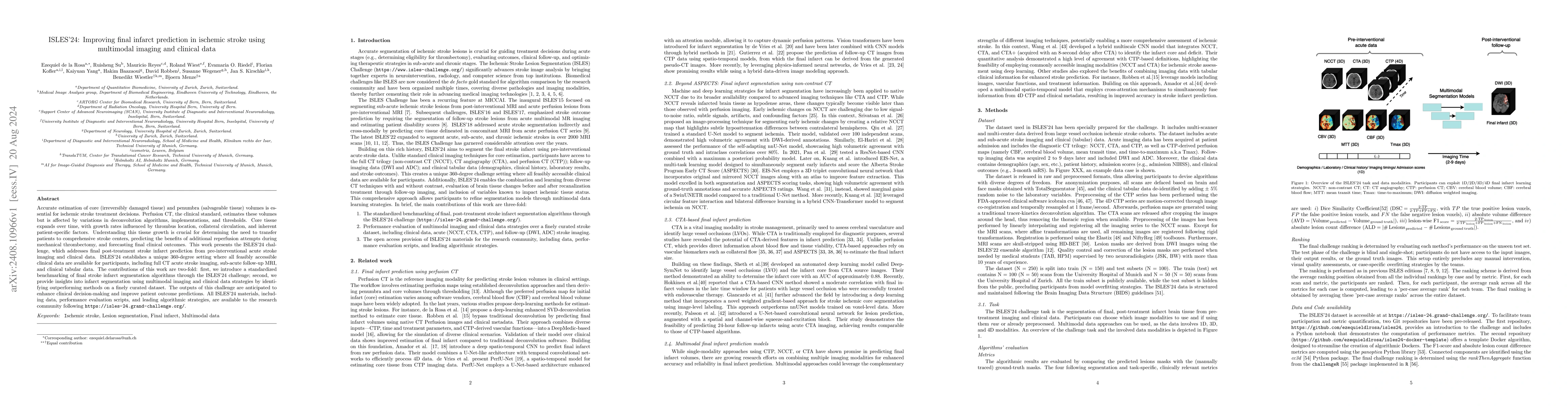
Papers on arXiv
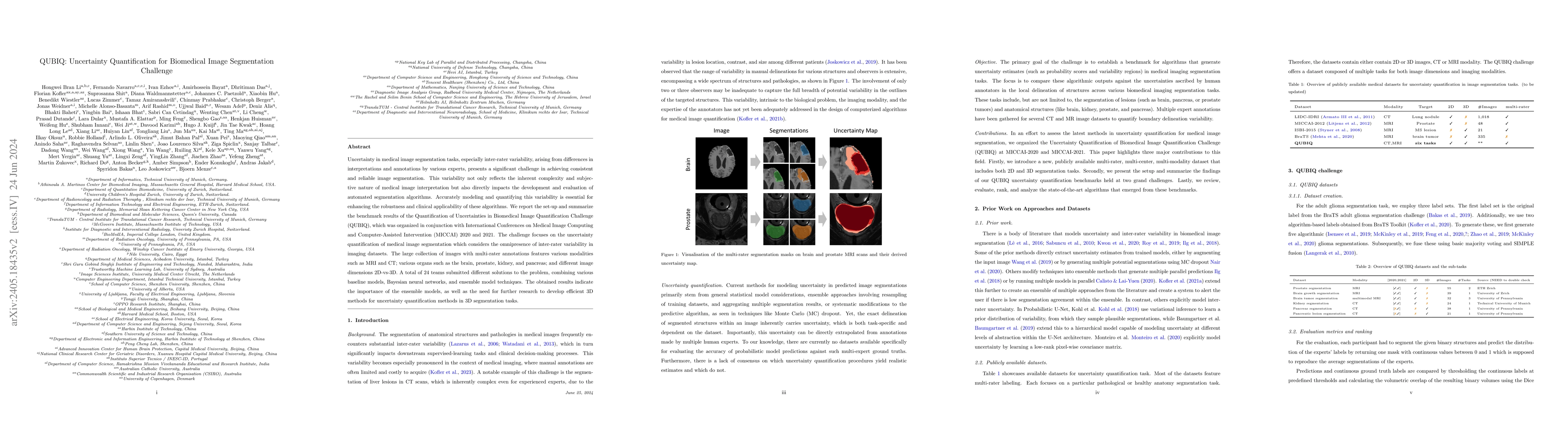
Uncertainty in medical image segmentation tasks, especially inter-rater variability, arising from differences in interpretations and annotations by various experts, presents a significant challenge in...
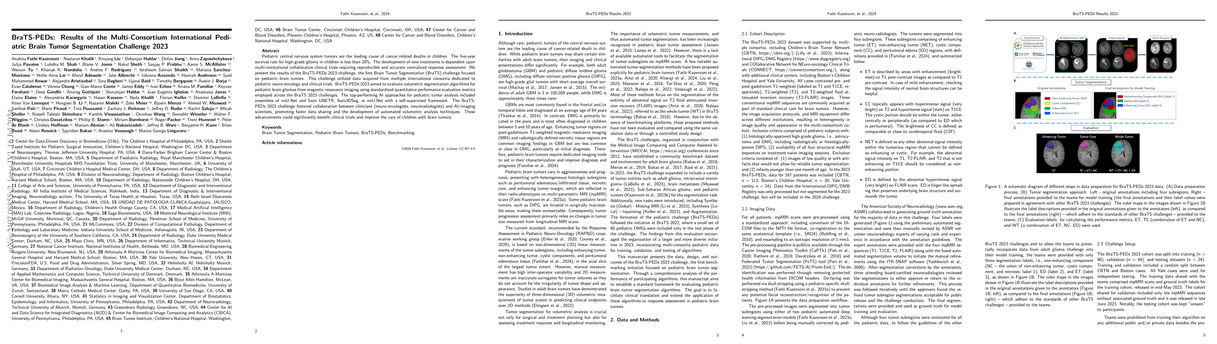
Pediatric central nervous system tumors are the leading cause of cancer-related deaths in children. The five-year survival rate for high-grade glioma in children is less than 20%. The development of n...
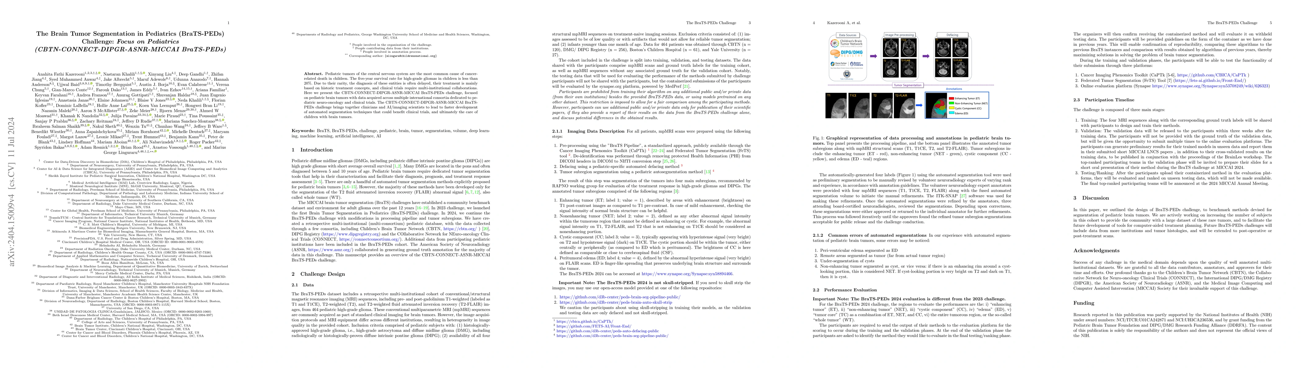
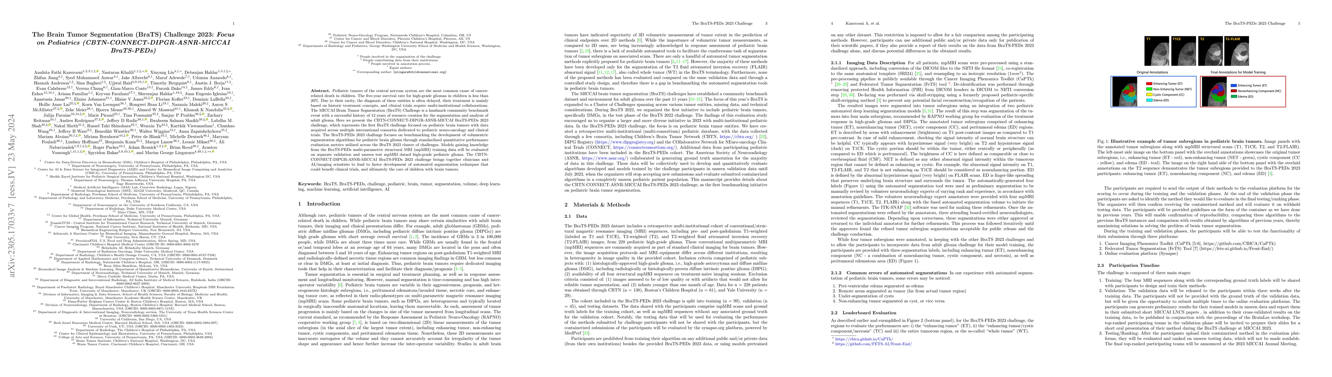
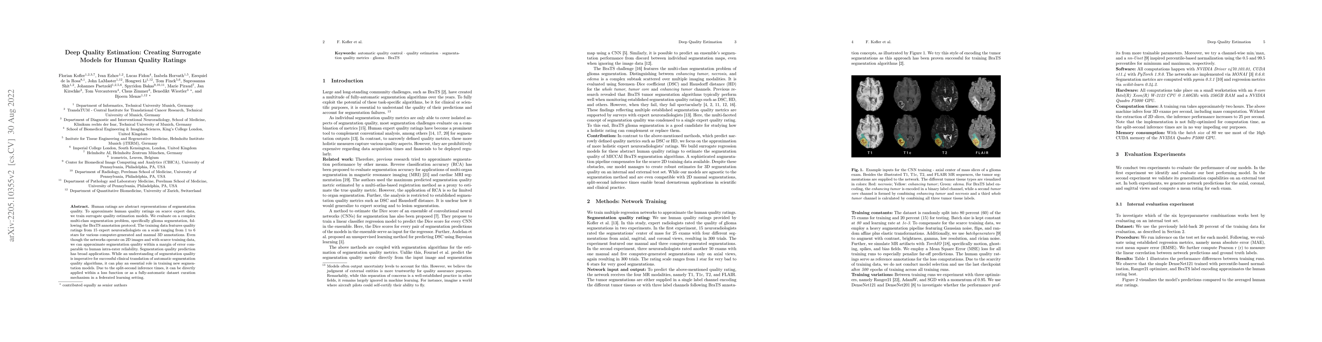
Pediatric tumors of the central nervous system are the most common cause of cancer-related death in children. The five-year survival rate for high-grade gliomas in children is less than 20%. Due to th...
Accurate image registration is pivotal in biomedical image analysis, where selecting suitable registration algorithms demands careful consideration. While numerous algorithms are available, the evalua...
The 2024 Brain Tumor Segmentation Meningioma Radiotherapy (BraTS-MEN-RT) challenge aims to advance automated segmentation algorithms using the largest known multi-institutional dataset of radiothera...
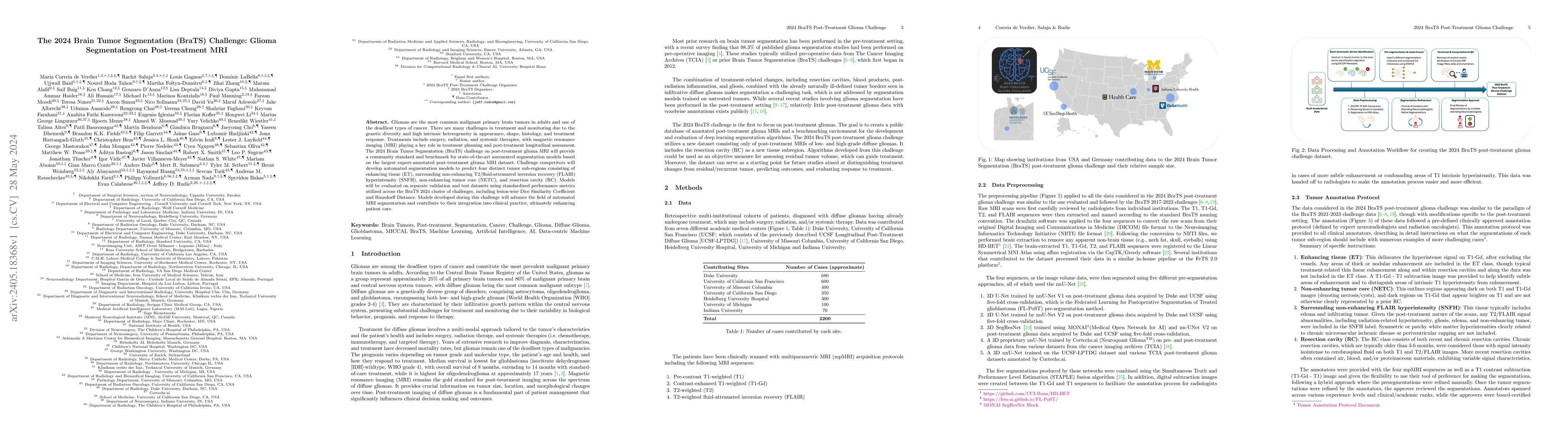
Gliomas are the most common malignant primary brain tumors in adults and one of the deadliest types of cancer. There are many challenges in treatment and monitoring due to the genetic diversity and ...
We describe the design and results from the BraTS 2023 Intracranial Meningioma Segmentation Challenge. The BraTS Meningioma Challenge differed from prior BraTS Glioma challenges in that it focused o...
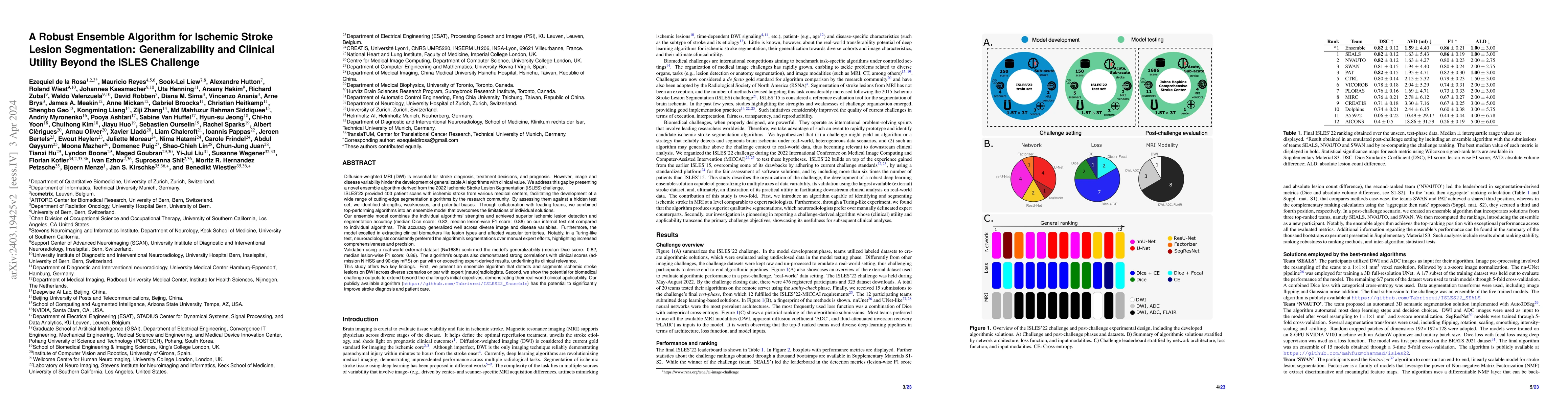
Diffusion-weighted MRI (DWI) is essential for stroke diagnosis, treatment decisions, and prognosis. However, image and disease variability hinder the development of generalizable AI algorithms with ...
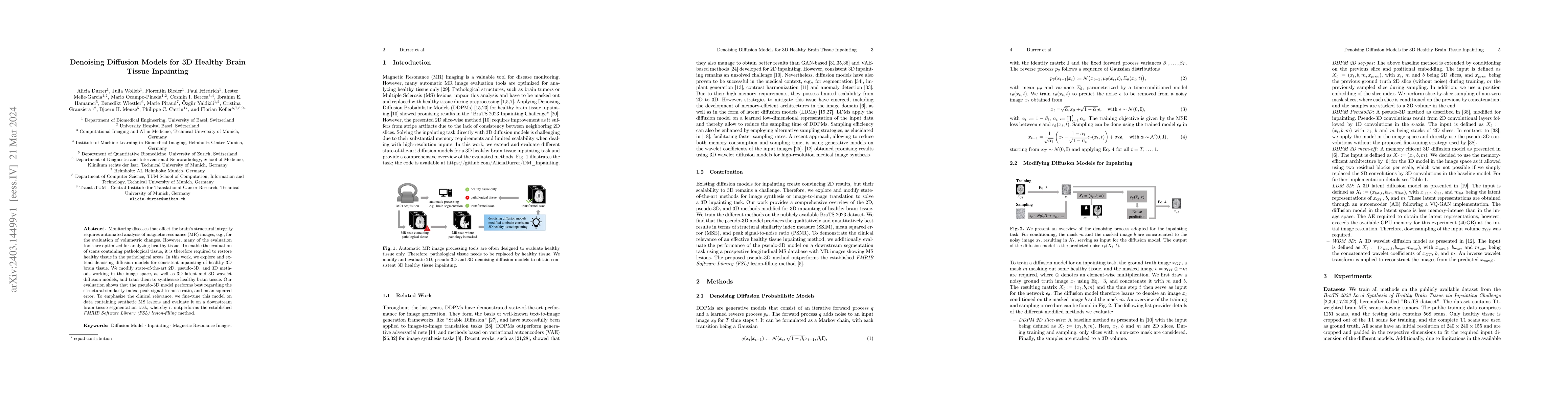
Monitoring diseases that affect the brain's structural integrity requires automated analysis of magnetic resonance (MR) images, e.g., for the evaluation of volumetric changes. However, many of the e...
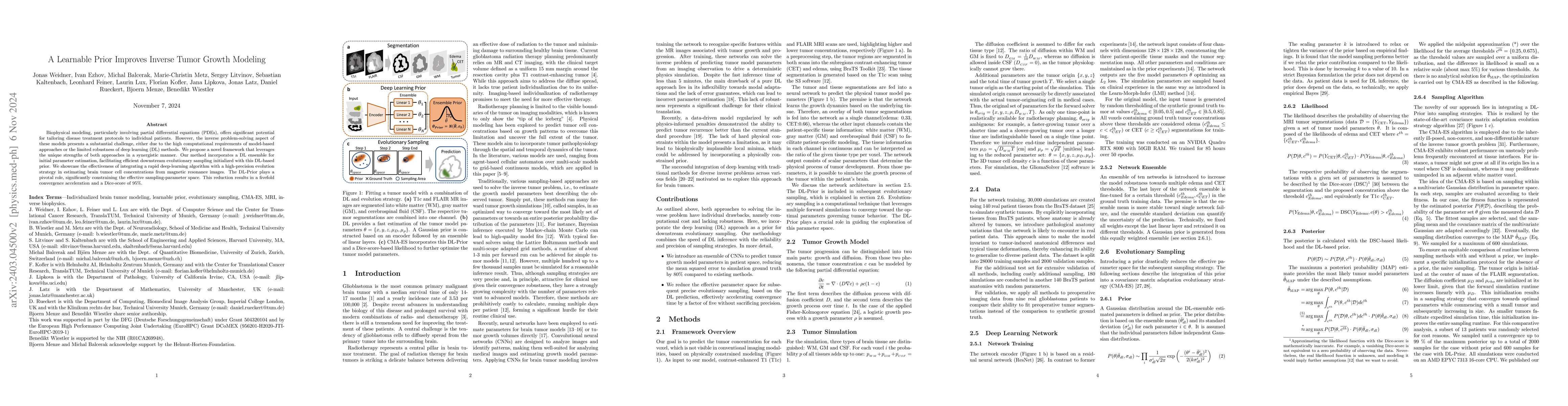
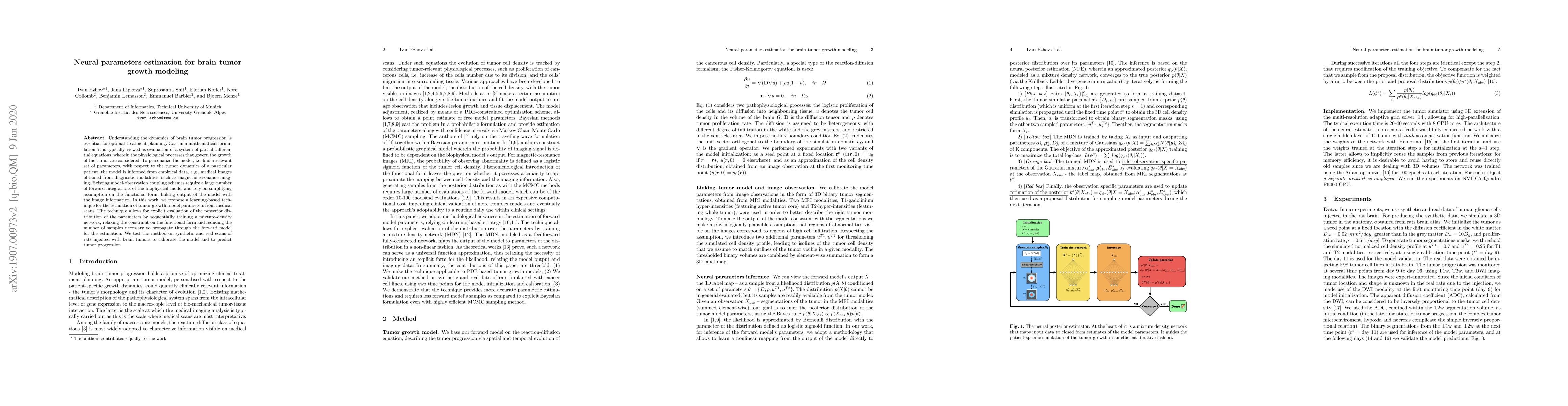
Biophysical modeling, particularly involving partial differential equations (PDEs), offers significant potential for tailoring disease treatment protocols to individual patients. However, the invers...
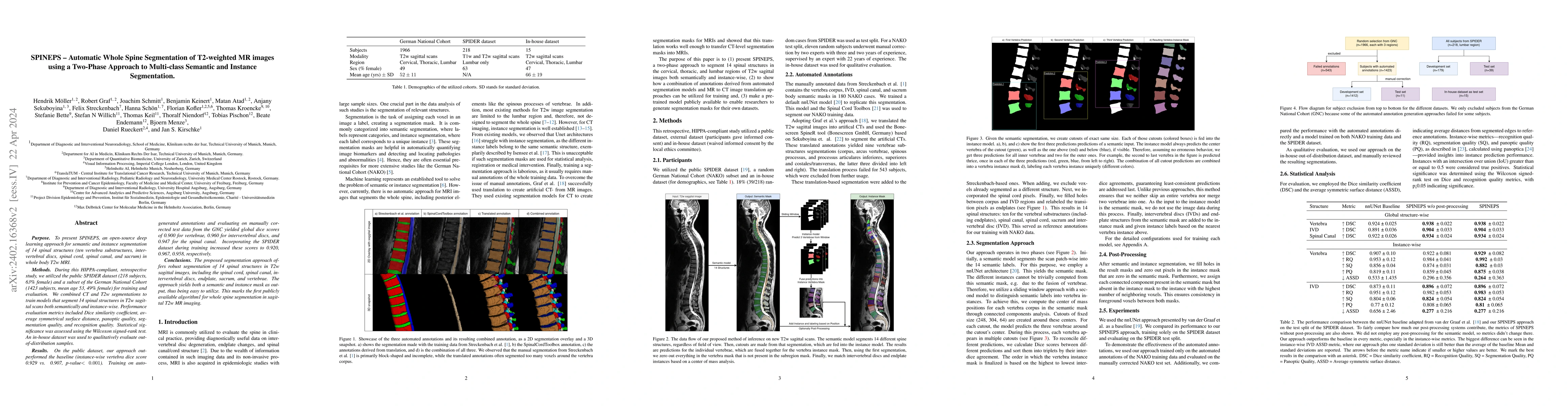
Purpose. To present SPINEPS, an open-source deep learning approach for semantic and instance segmentation of 14 spinal structures (ten vertebra substructures, intervertebral discs, spinal cord, spin...
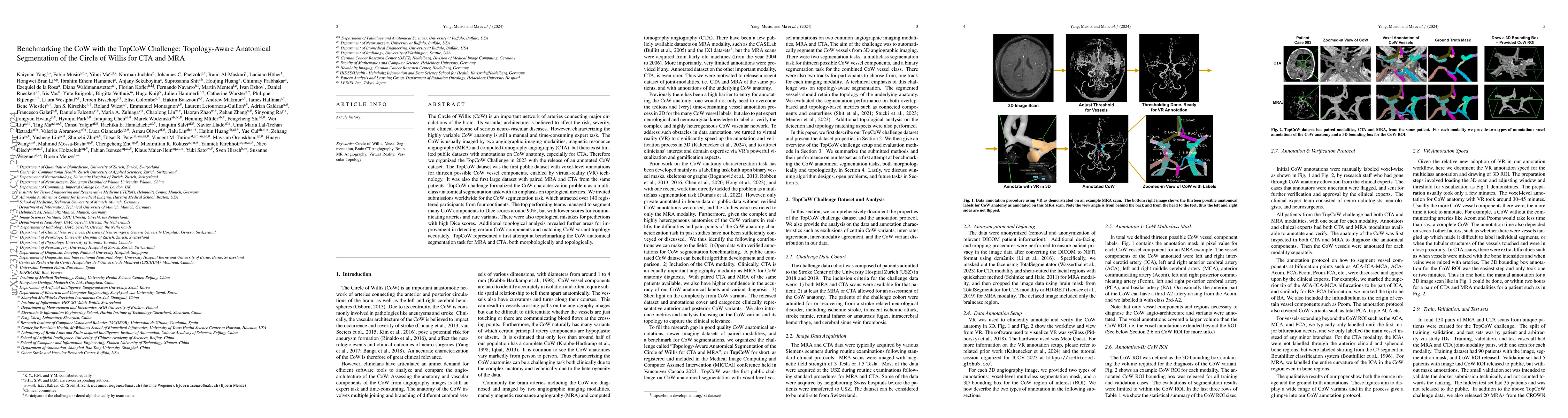
The Circle of Willis (CoW) is an important network of arteries connecting major circulations of the brain. Its vascular architecture is believed to affect the risk, severity, and clinical outcome of...
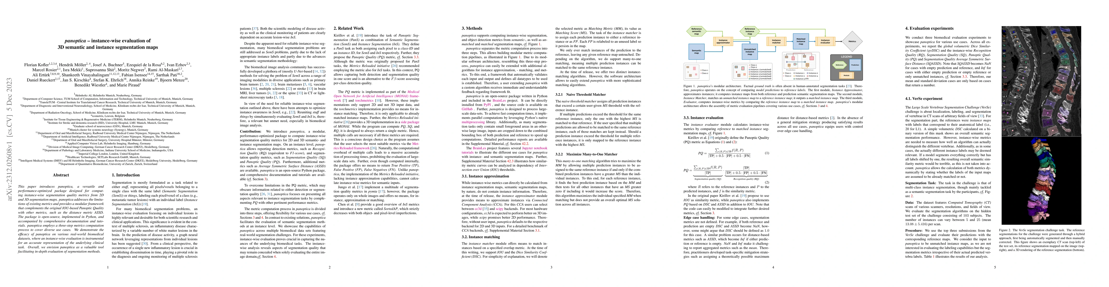
This paper introduces panoptica, a versatile and performance-optimized package designed for computing instance-wise segmentation quality metrics from 2D and 3D segmentation maps. panoptica addresses...
The translation of AI-generated brain metastases (BM) segmentation into clinical practice relies heavily on diverse, high-quality annotated medical imaging datasets. The BraTS-METS 2023 challenge ha...
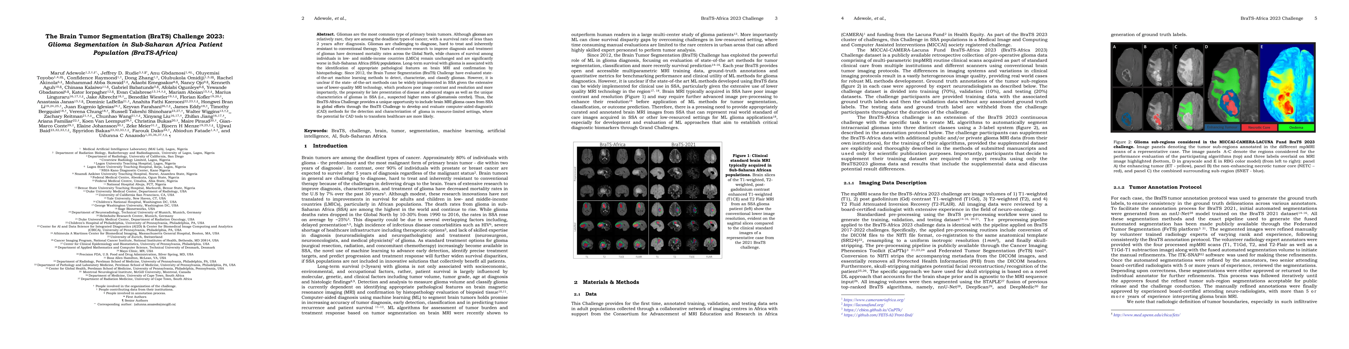
Gliomas are the most common type of primary brain tumors. Although gliomas are relatively rare, they are among the deadliest types of cancer, with a survival rate of less than 2 years after diagnosi...
Pediatric tumors of the central nervous system are the most common cause of cancer-related death in children. The five-year survival rate for high-grade gliomas in children is less than 20\%. Due to...
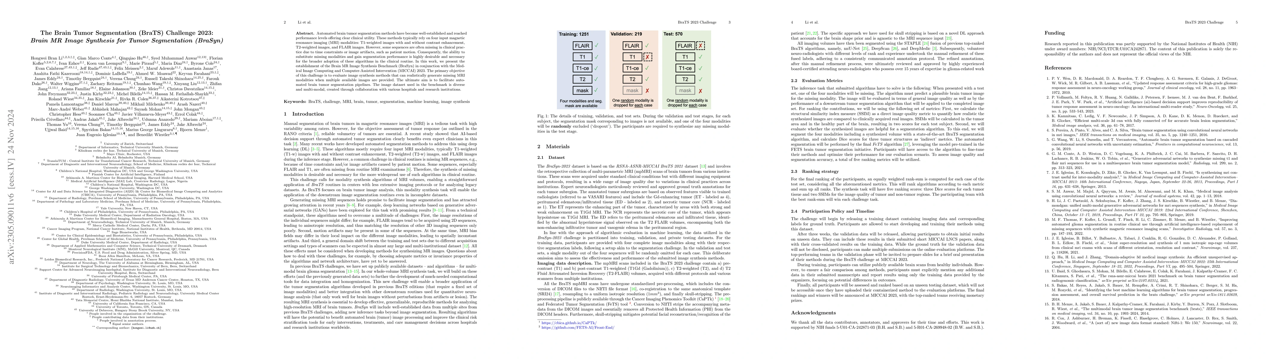
Automated brain tumor segmentation methods have become well-established and reached performance levels offering clear clinical utility. These methods typically rely on four input magnetic resonance ...
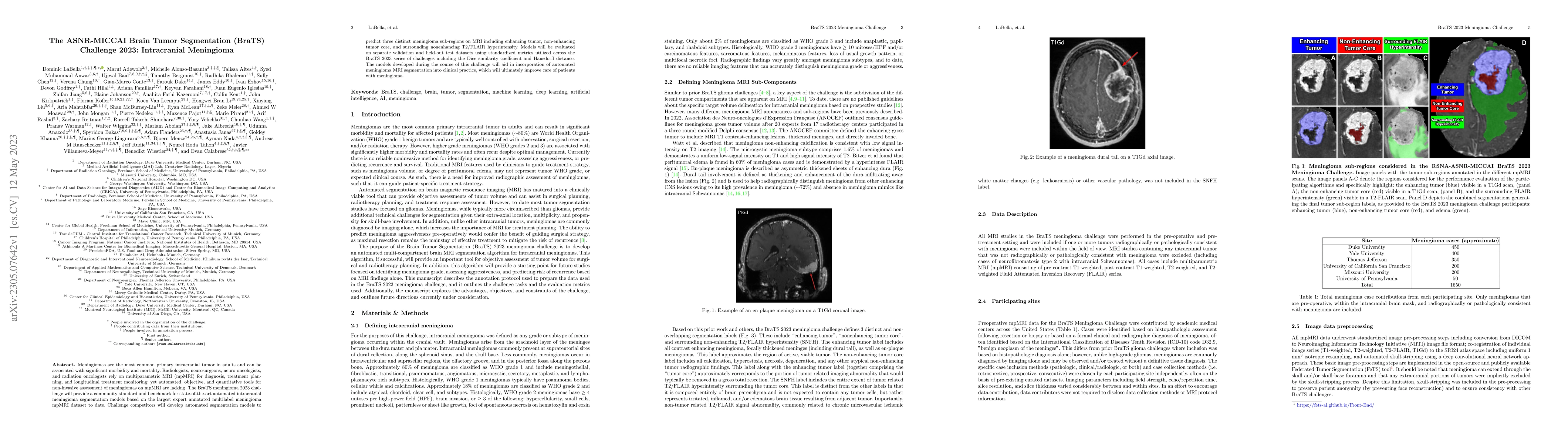
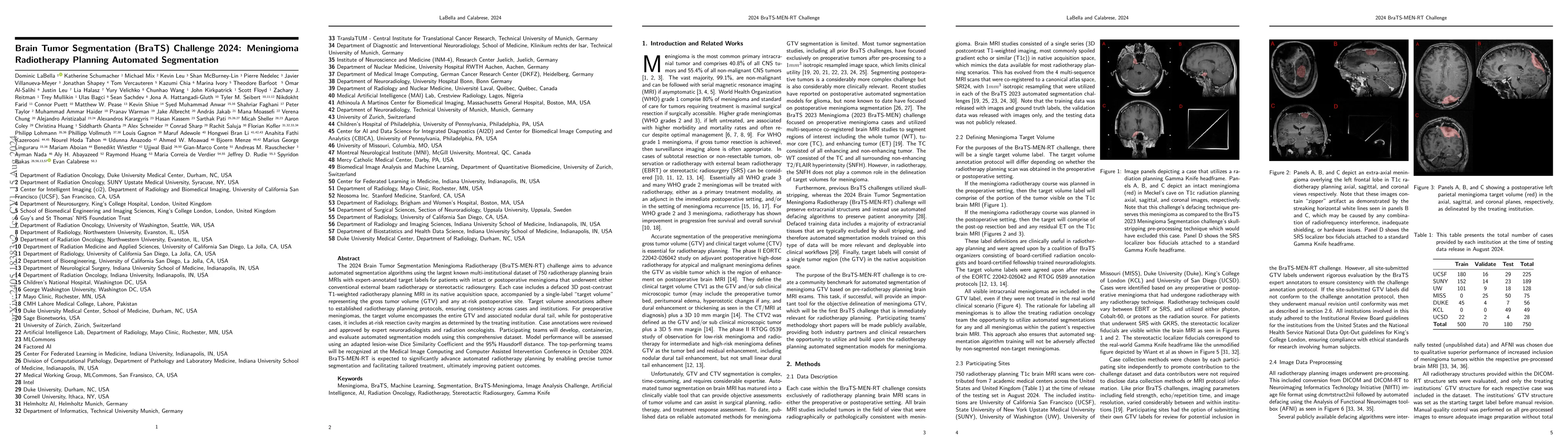
Meningiomas are the most common primary intracranial tumor in adults and can be associated with significant morbidity and mortality. Radiologists, neurosurgeons, neuro-oncologists, and radiation onc...
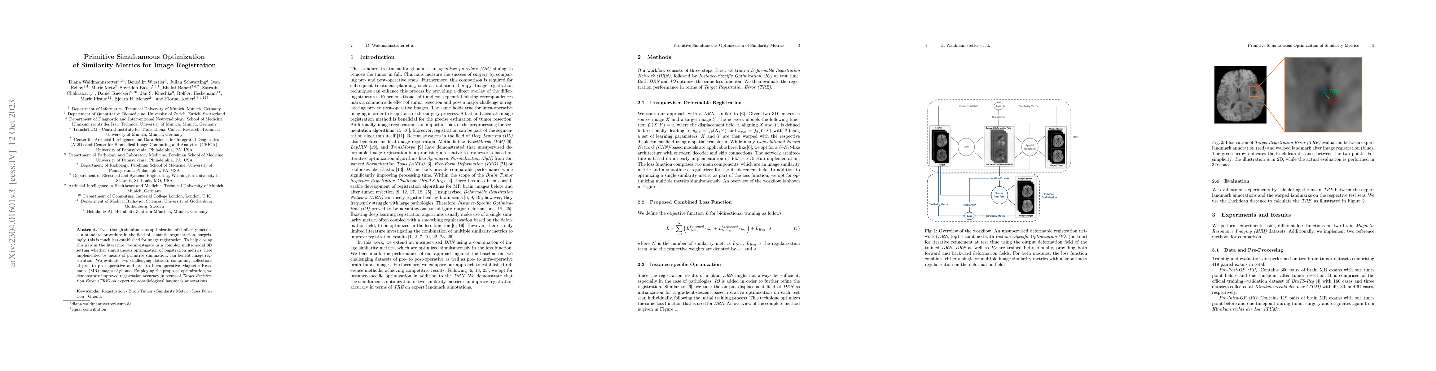
Even though simultaneous optimization of similarity metrics is a standard procedure in the field of semantic segmentation, surprisingly, this is much less established for image registration. To help...
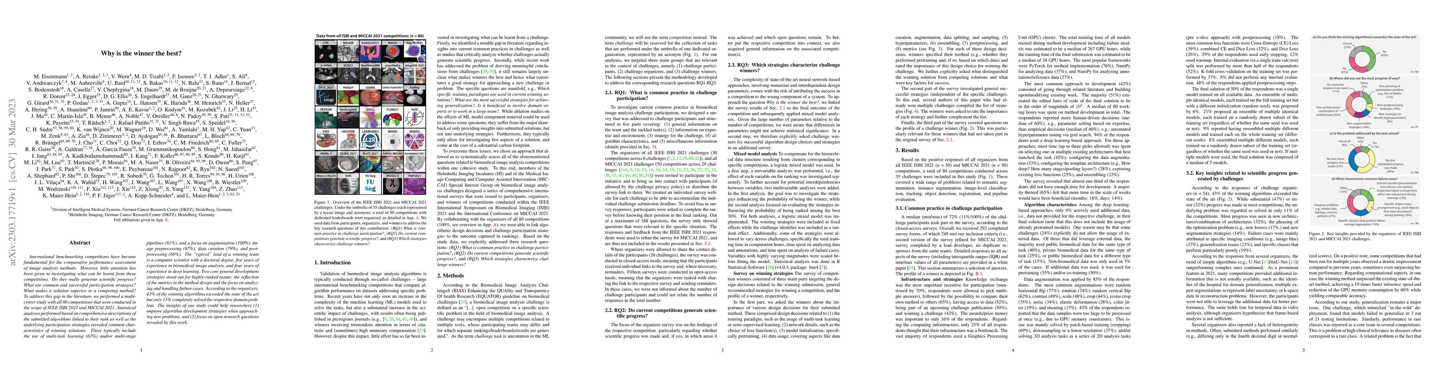
International benchmarking competitions have become fundamental for the comparative performance assessment of image analysis methods. However, little attention has been given to investigating what c...
Validation metrics are key for the reliable tracking of scientific progress and for bridging the current chasm between artificial intelligence (AI) research and its translation into practice. Howeve...
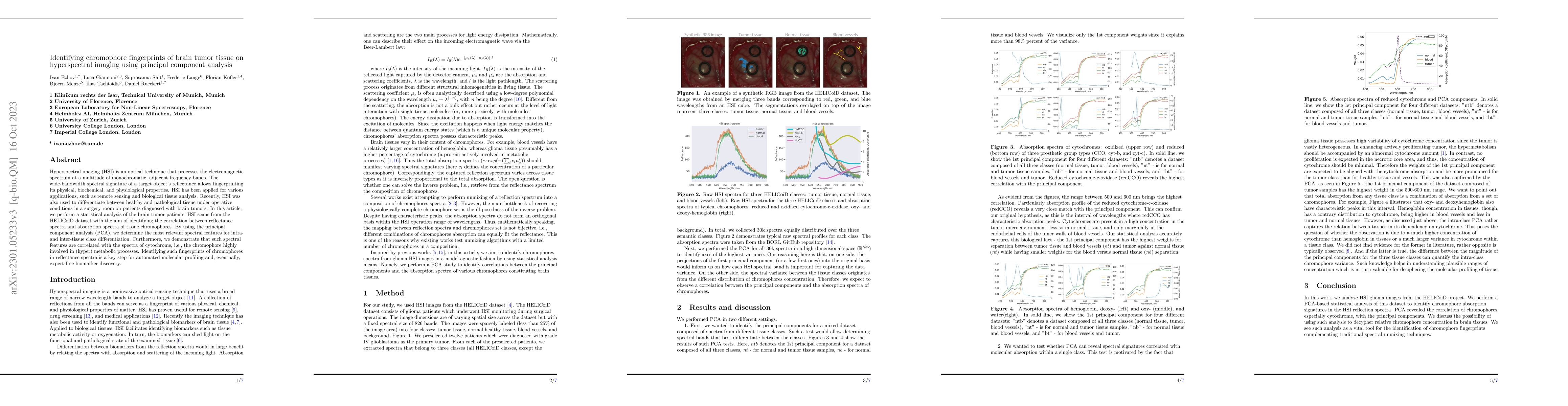
Hyperspectral imaging (HSI) is an optical technique that processes the electromagnetic spectrum at a multitude of monochromatic, adjacent frequency bands. The wide-bandwidth spectral signature of a ...
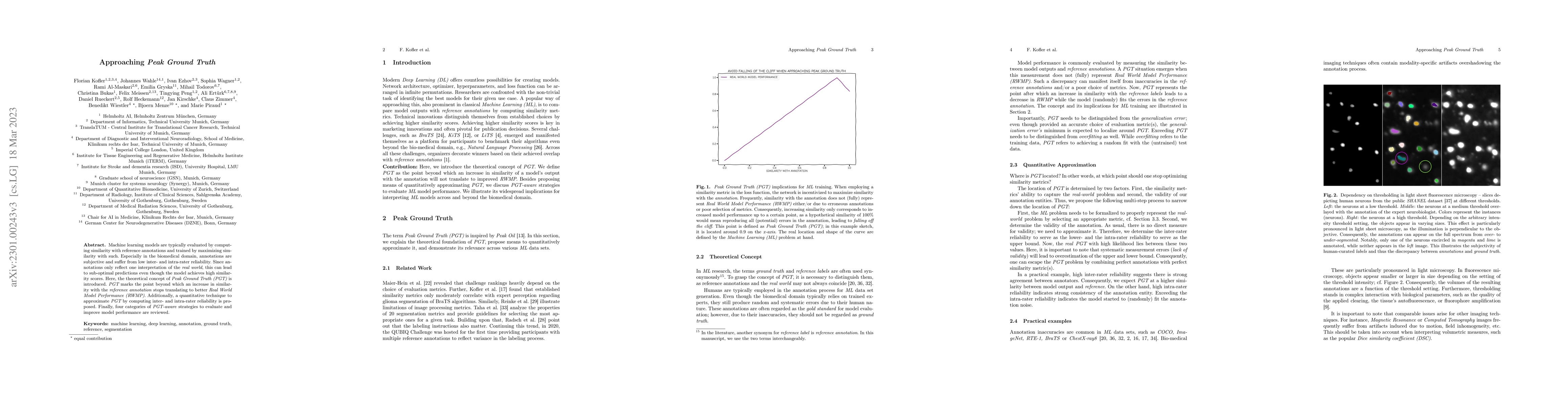
Machine learning models are typically evaluated by computing similarity with reference annotations and trained by maximizing similarity with such. Especially in the biomedical domain, annotations ar...
Imaging markers of cerebral small vessel disease provide valuable information on brain health, but their manual assessment is time-consuming and hampered by substantial intra- and interrater variabi...
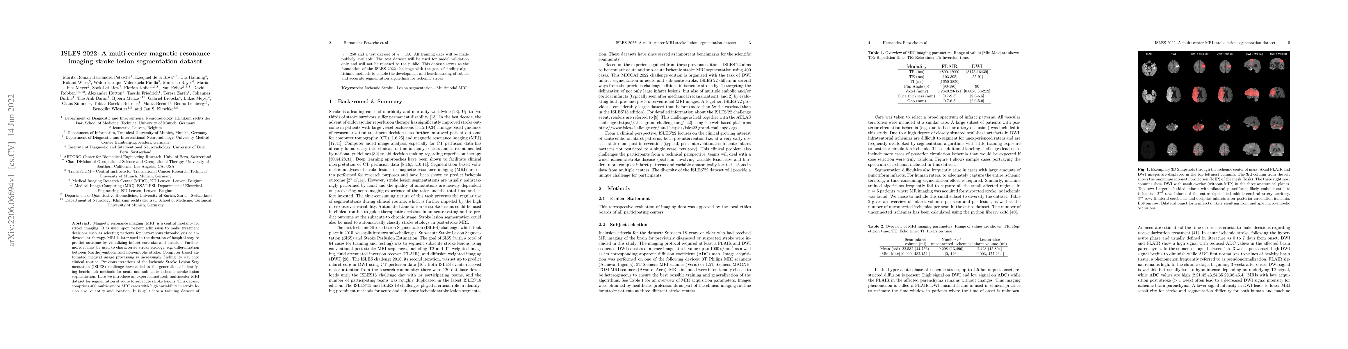
Magnetic resonance imaging (MRI) is a central modality for stroke imaging. It is used upon patient admission to make treatment decisions such as selecting patients for intravenous thrombolysis or en...
Increasing evidence shows that flaws in machine learning (ML) algorithm validation are an underestimated global problem. Particularly in automatic biomedical image analysis, chosen performance metri...
Human ratings are abstract representations of segmentation quality. To approximate human quality ratings on scarce expert data, we train surrogate quality estimation models. We evaluate on a complex...
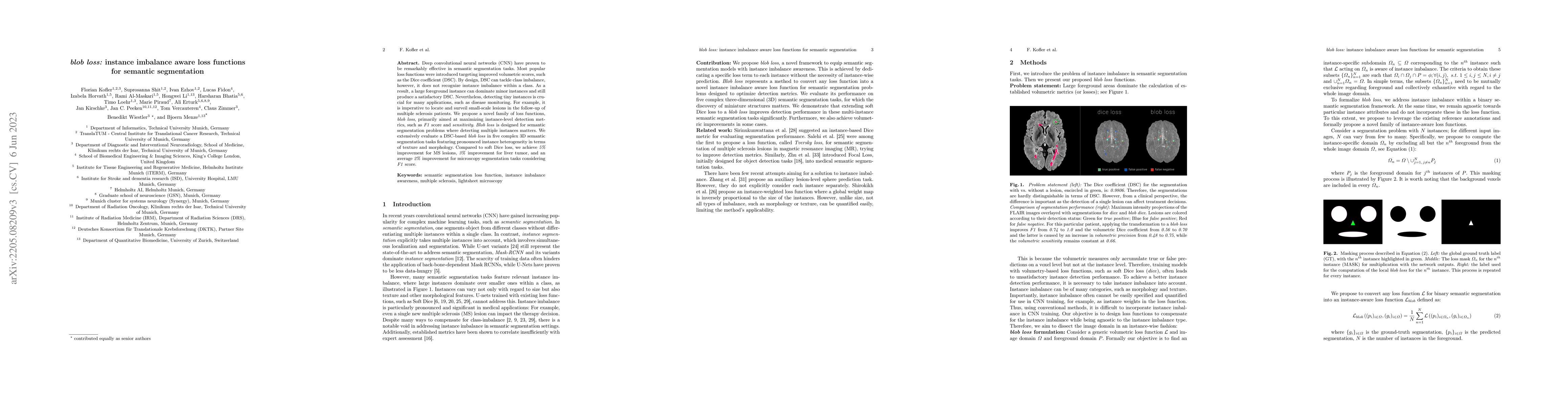
Deep convolutional neural networks (CNN) have proven to be remarkably effective in semantic segmentation tasks. Most popular loss functions were introduced targeting improved volumetric scores, such...
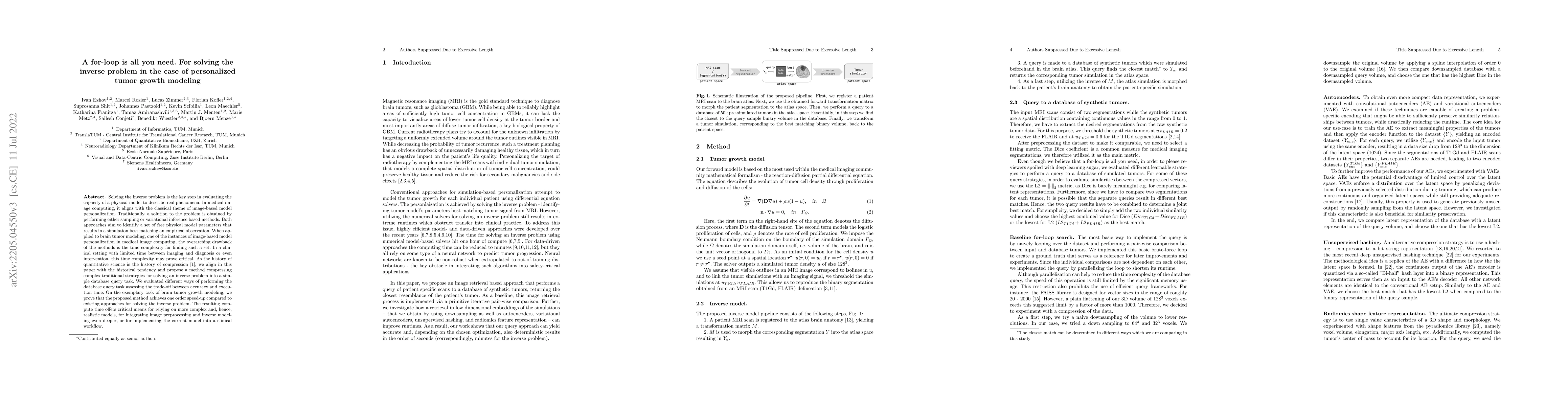
Solving the inverse problem is the key step in evaluating the capacity of a physical model to describe real phenomena. In medical image computing, it aligns with the classical theme of image-based m...
Although machine learning (ML) has shown promise in numerous domains, there are concerns about generalizability to out-of-sample data. This is currently addressed by centrally sharing ample, and imp...
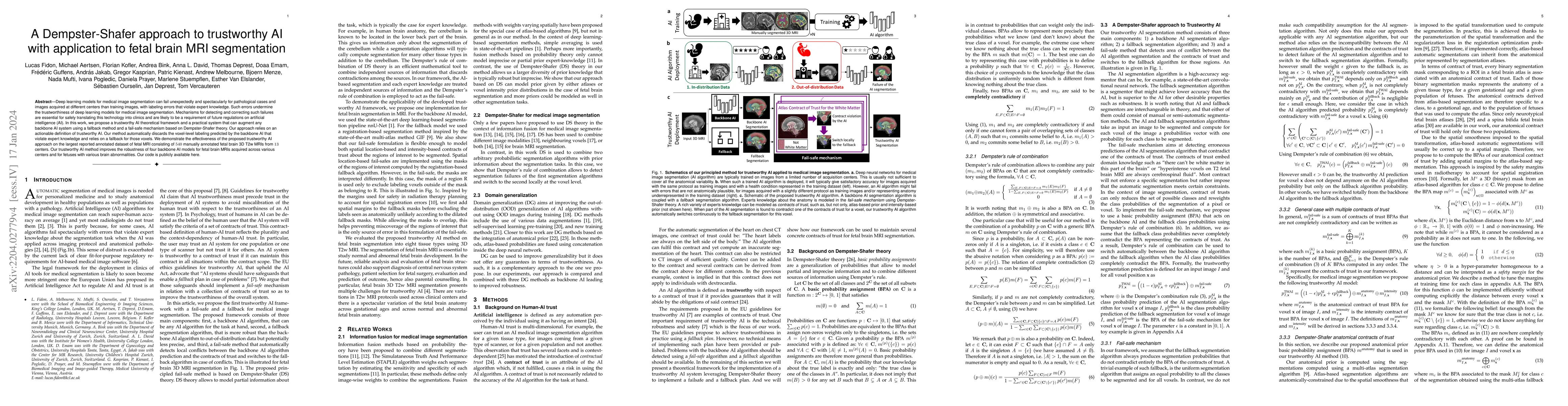
Deep learning models for medical image segmentation can fail unexpectedly and spectacularly for pathological cases and images acquired at different centers than training images, with labeling errors...
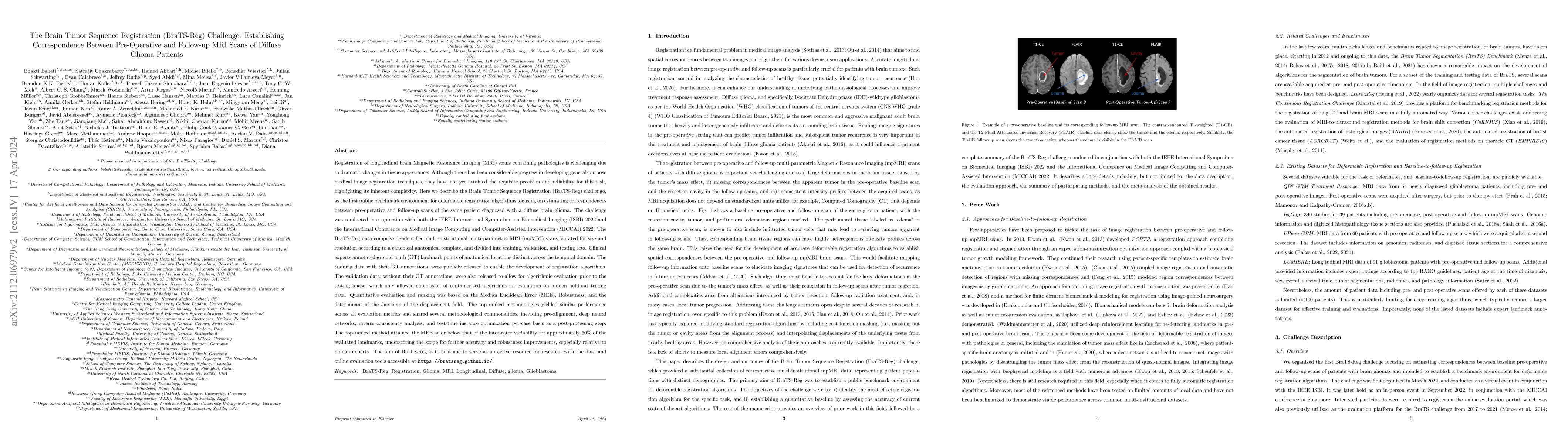
Registration of longitudinal brain MRI scans containing pathologies is challenging due to dramatic changes in tissue appearance. Although there has been progress in developing general-purpose medica...
We propose a simple new aggregation strategy for federated learning that won the MICCAI Federated Tumor Segmentation Challenge 2021 (FETS), the first ever challenge on Federated Learning in the Mach...
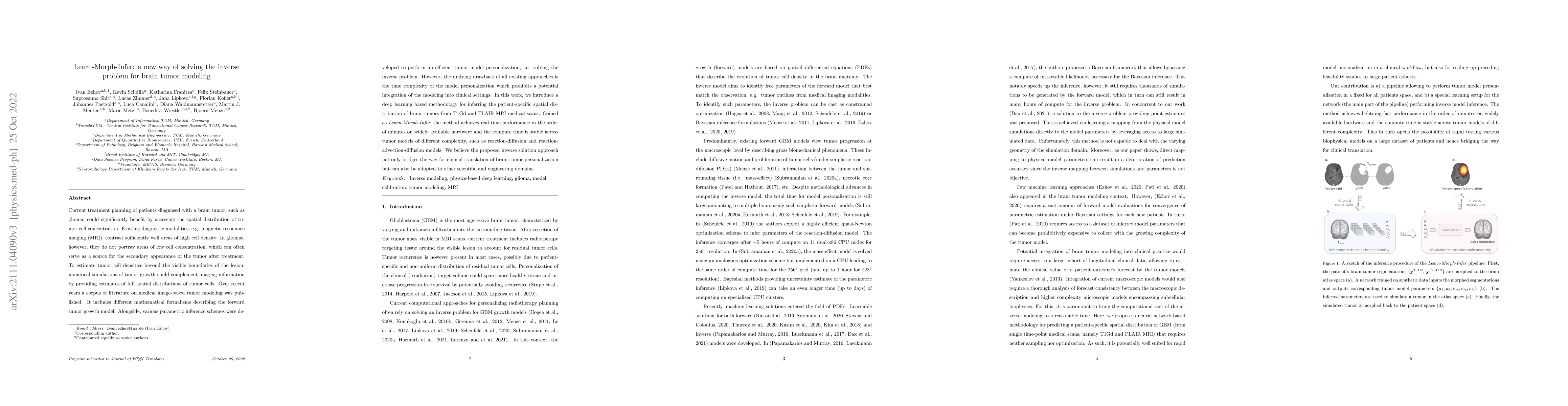
Current treatment planning of patients diagnosed with a brain tumor, such as glioma, could significantly benefit by accessing the spatial distribution of tumor cell concentration. Existing diagnosti...
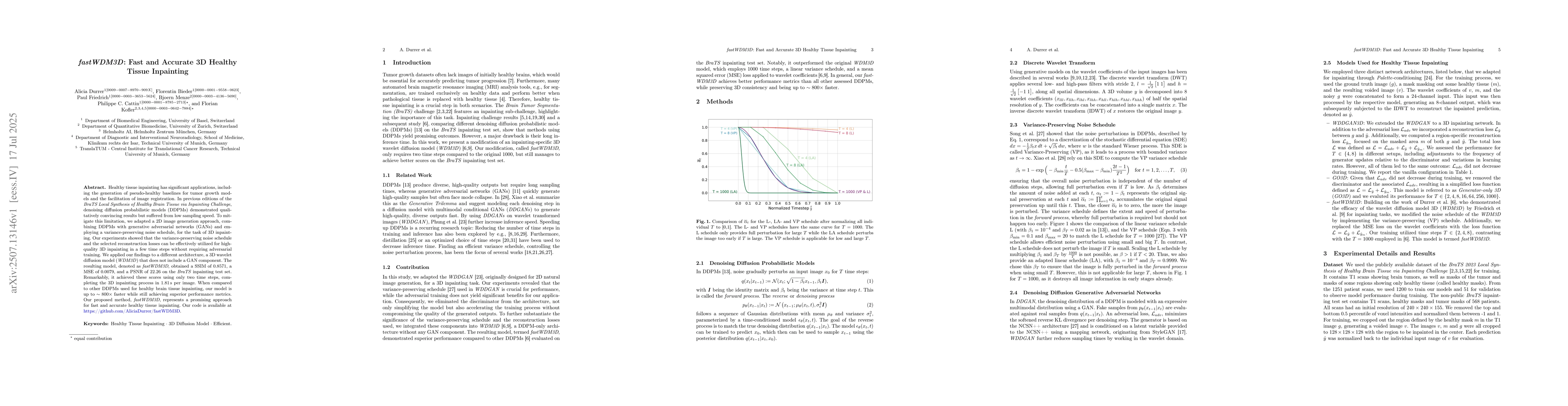
Fast and accurate solutions of time-dependent partial differential equations (PDEs) are of pivotal interest to many research fields, including physics, engineering, and biology. Generally, implicit/...
While the importance of automatic image analysis is continuously increasing, recent meta-research revealed major flaws with respect to algorithm validation. Performance metrics are particularly key ...
Metrics optimized in complex machine learning tasks are often selected in an ad-hoc manner. It is unknown how they align with human expert perception. We explore the correlations between established...
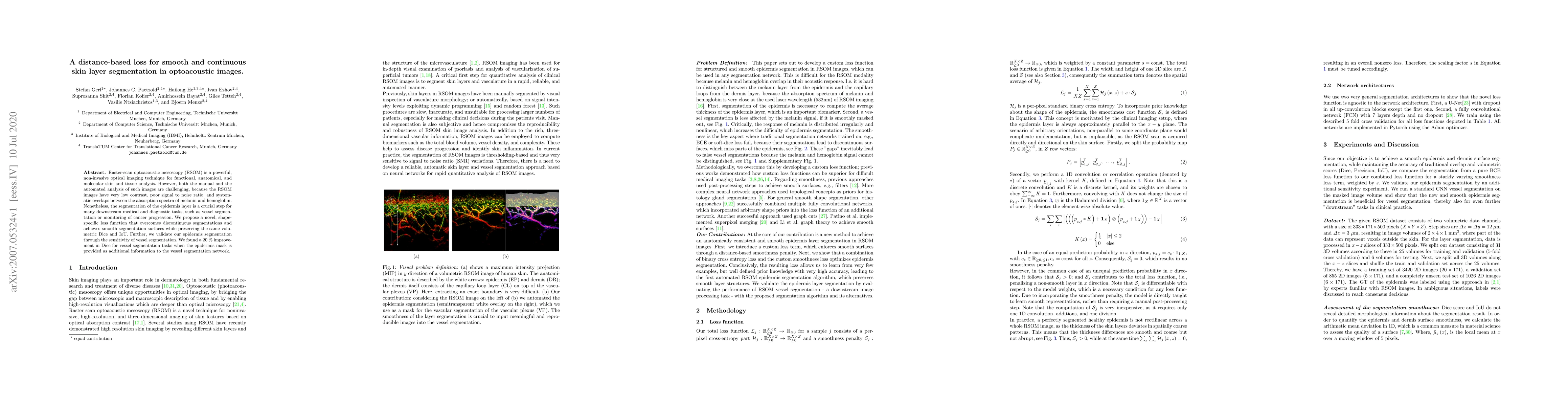
Raster-scan optoacoustic mesoscopy (RSOM) is a powerful, non-invasive optical imaging technique for functional, anatomical, and molecular skin and tissue analysis. However, both the manual and the a...
Understanding the dynamics of brain tumor progression is essential for optimal treatment planning. Cast in a mathematical formulation, it is typically viewed as evaluation of a system of partial dif...
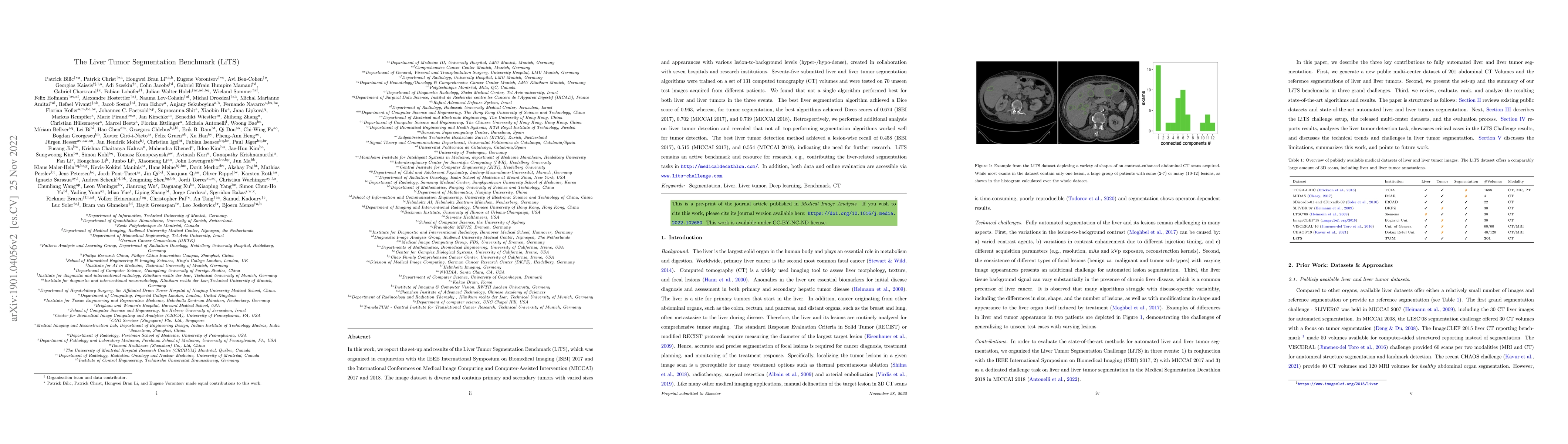
In this work, we report the set-up and results of the Liver Tumor Segmentation Benchmark (LiTS), which was organized in conjunction with the IEEE International Symposium on Biomedical Imaging (ISBI)...
Accurate estimation of core (irreversibly damaged tissue) and penumbra (salvageable tissue) volumes is essential for ischemic stroke treatment decisions. Perfusion CT, the clinical standard, estimates...
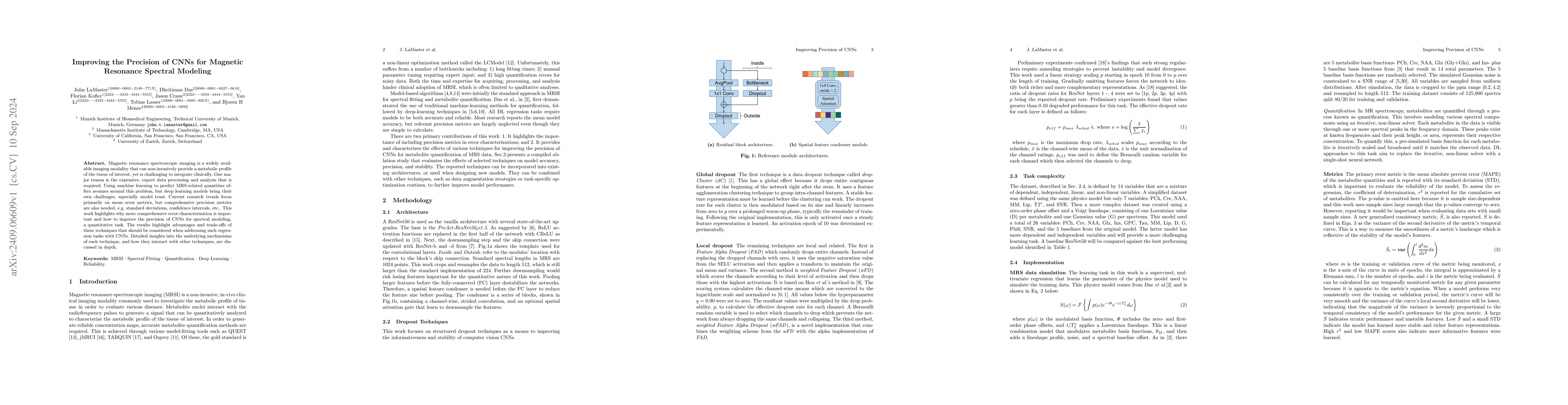
Magnetic resonance spectroscopic imaging is a widely available imaging modality that can non-invasively provide a metabolic profile of the tissue of interest, yet is challenging to integrate clinicall...
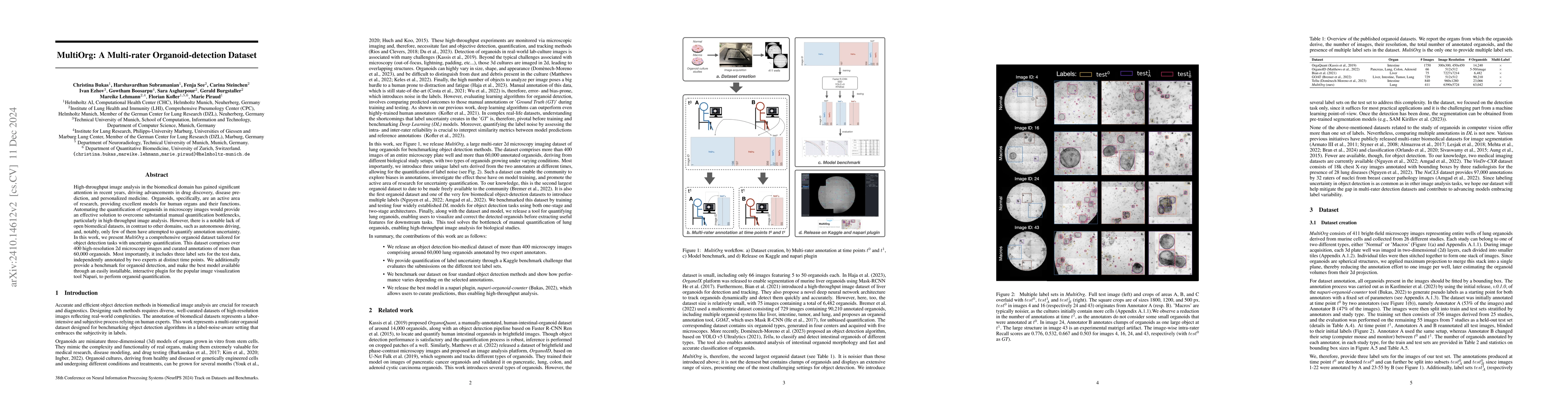
High-throughput image analysis in the biomedical domain has gained significant attention in recent years, driving advancements in drug discovery, disease prediction, and personalized medicine. Organoi...
In this paper, we propose a methodology for extracting molecular tumor biomarkers from hyperspectral imaging (HSI), an emerging technology for intraoperative tissue assessment. To achieve this, we emp...
Despite continuous advancements in cancer treatment, brain metastatic disease remains a significant complication of primary cancer and is associated with an unfavorable prognosis. One approach for imp...
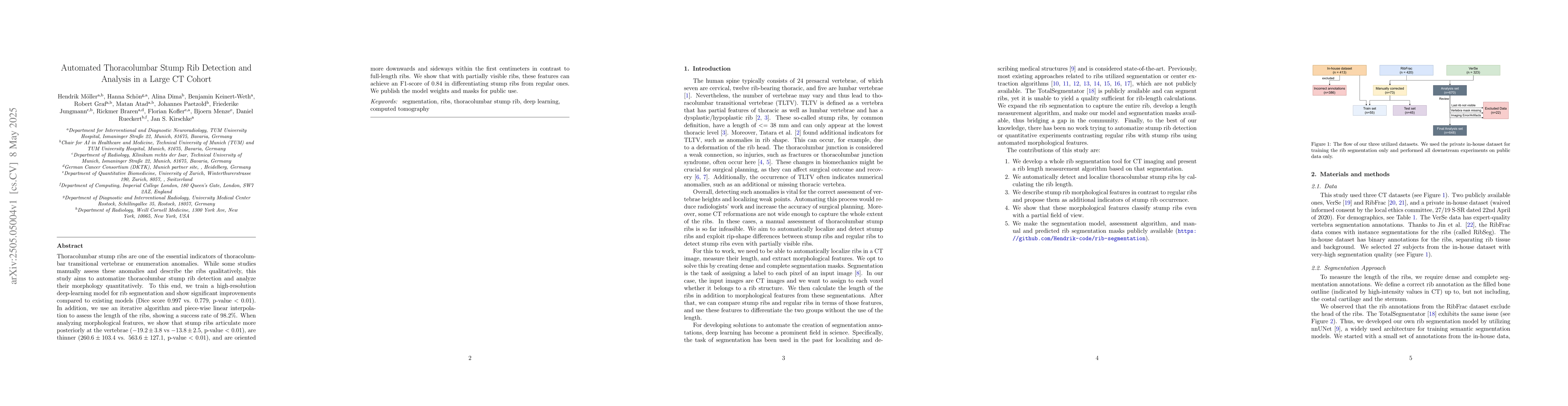
Thoracolumbar stump ribs are one of the essential indicators of thoracolumbar transitional vertebrae or enumeration anomalies. While some studies manually assess these anomalies and describe the ribs ...
The Brain Tumor Segmentation (BraTS) cluster of challenges has significantly advanced brain tumor image analysis by providing large, curated datasets and addressing clinically relevant tasks. However,...
Healthy tissue inpainting has significant applications, including the generation of pseudo-healthy baselines for tumor growth models and the facilitation of image registration. In previous editions of...
BrainLesion Suite is a versatile toolkit for building modular brain lesion image analysis pipelines in Python. Following Pythonic principles, BrainLesion Suite is designed to provide a 'brainless' dev...
Instance-sensitive losses for semantic segmentation such as blob loss and CC loss were designed to address instance imbalance, ensuring small lesions generate the same gradient as large ones, but oper...
LLM-based Software Engineering agents face a critical bottleneck: context length limitations cause failures on complex, long-horizon tasks. One promising solution is to encode context as continuous em...
The Panoptic Quality (PQ) metric is the standard for jointly evaluating instance and semantic segmentation. However, its original definition relies on a One-to-One matching between predicted and groun...