Academic Profile
Statistics
Similar Authors
Papers on arXiv
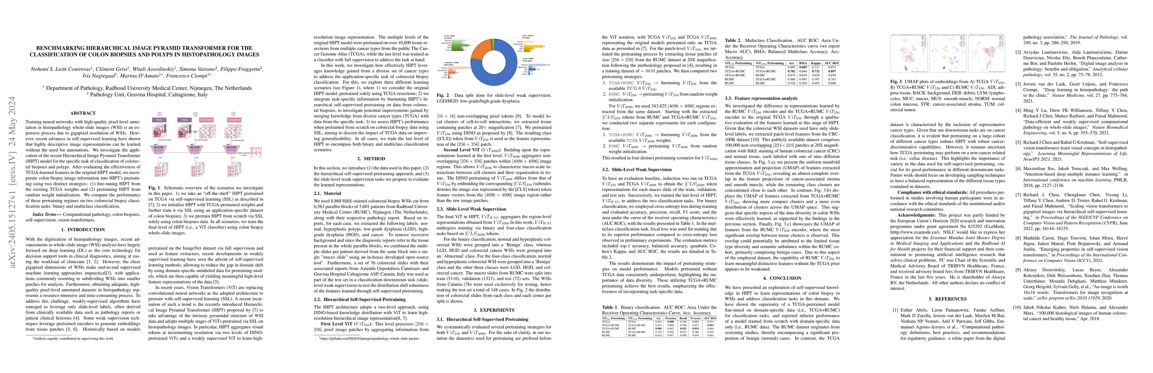
Training neural networks with high-quality pixel-level annotation in histopathology whole-slide images (WSI) is an expensive process due to gigapixel resolution of WSIs. However, recent advances in ...
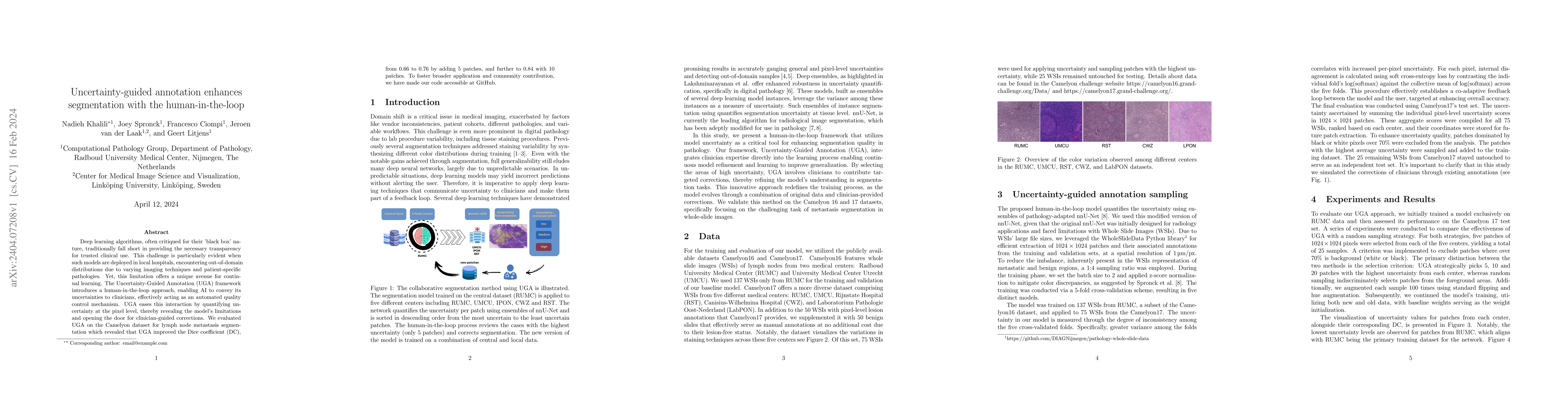
Deep learning algorithms, often critiqued for their 'black box' nature, traditionally fall short in providing the necessary transparency for trusted clinical use. This challenge is particularly evid...
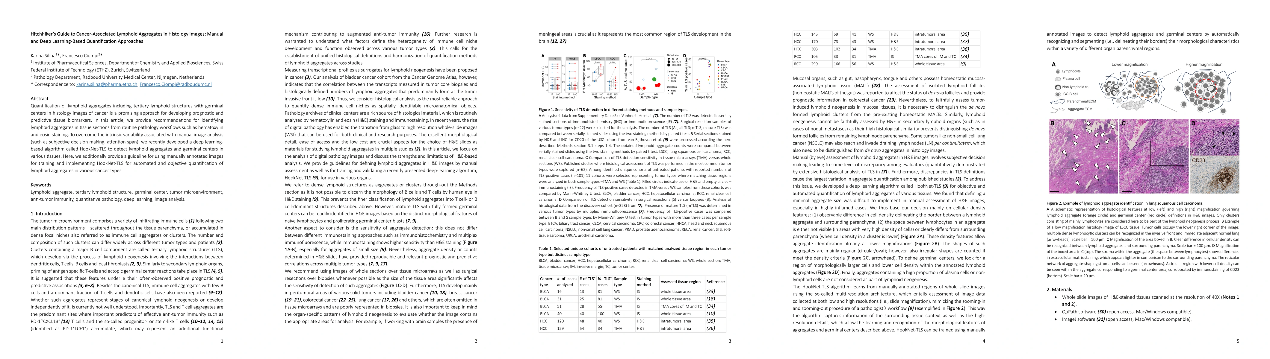
Quantification of lymphoid aggregates including tertiary lymphoid structures with germinal centers in histology images of cancer is a promising approach for developing prognostic and predictive tiss...
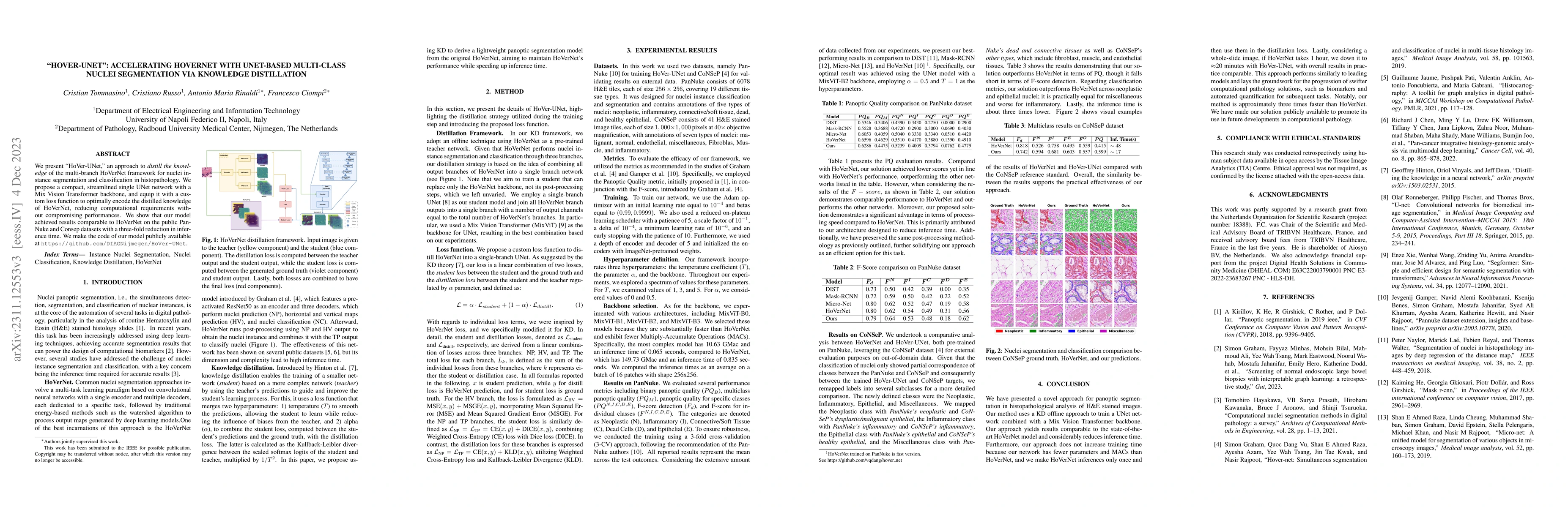
We present HoVer-UNet, an approach to distill the knowledge of the multi-branch HoVerNet framework for nuclei instance segmentation and classification in histopathology. We propose a compact, stream...
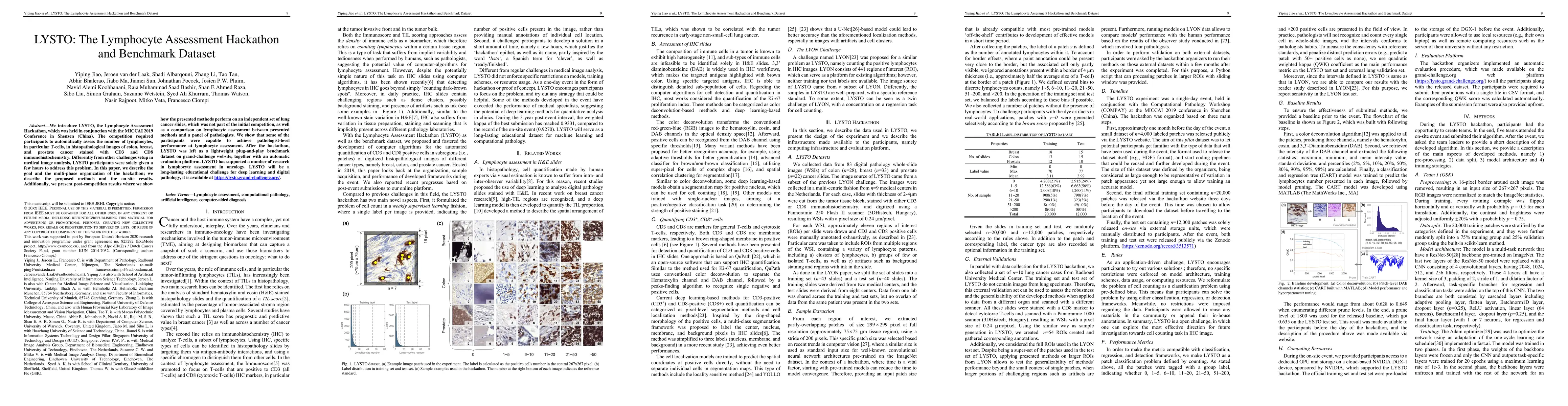
We introduce LYSTO, the Lymphocyte Assessment Hackathon, which was held in conjunction with the MICCAI 2019 Conference in Shenzen (China). The competition required participants to automatically asse...
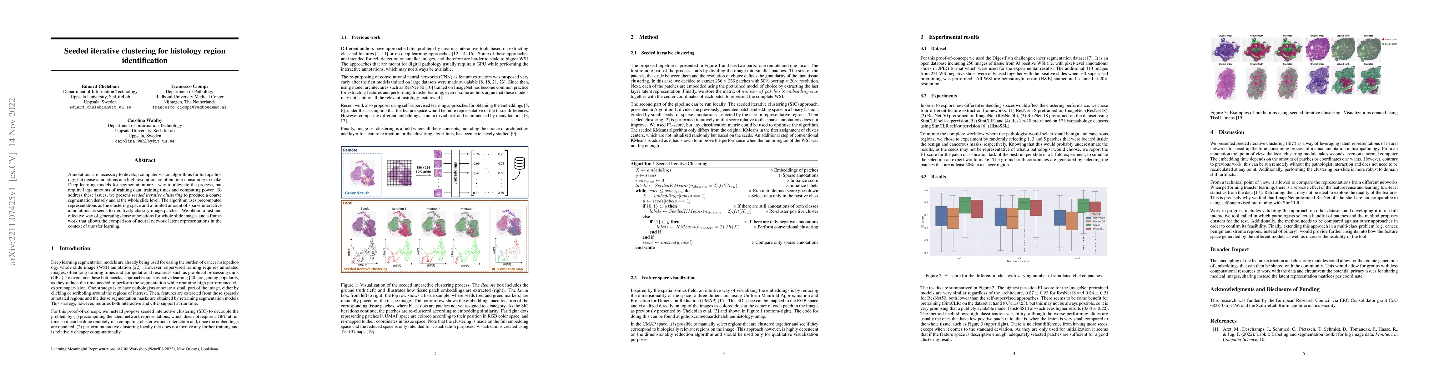
Annotations are necessary to develop computer vision algorithms for histopathology, but dense annotations at a high resolution are often time-consuming to make. Deep learning models for segmentation...
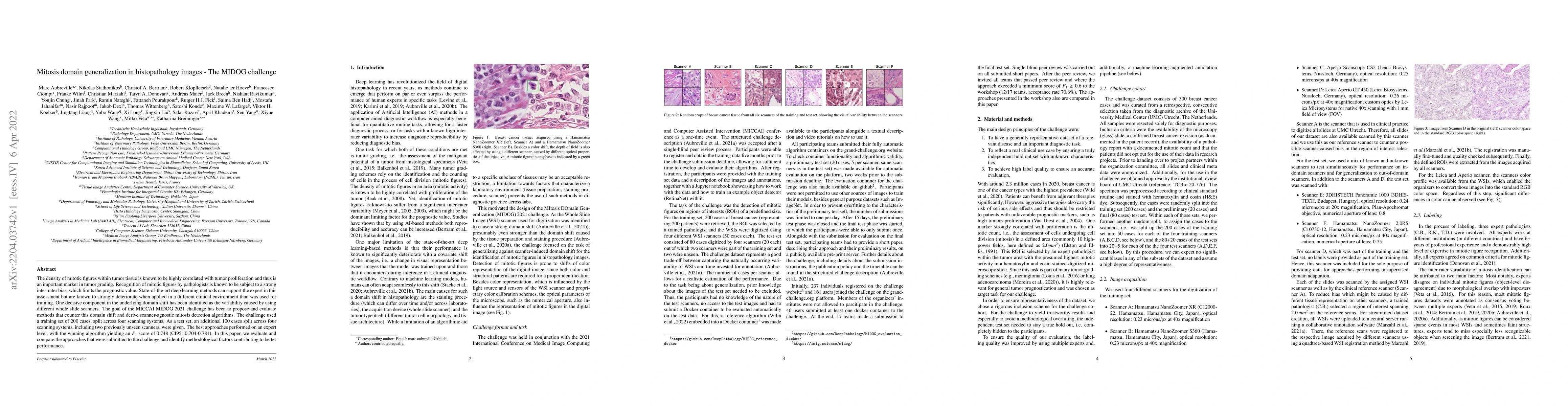
The density of mitotic figures within tumor tissue is known to be highly correlated with tumor proliferation and thus is an important marker in tumor grading. Recognition of mitotic figures by patho...
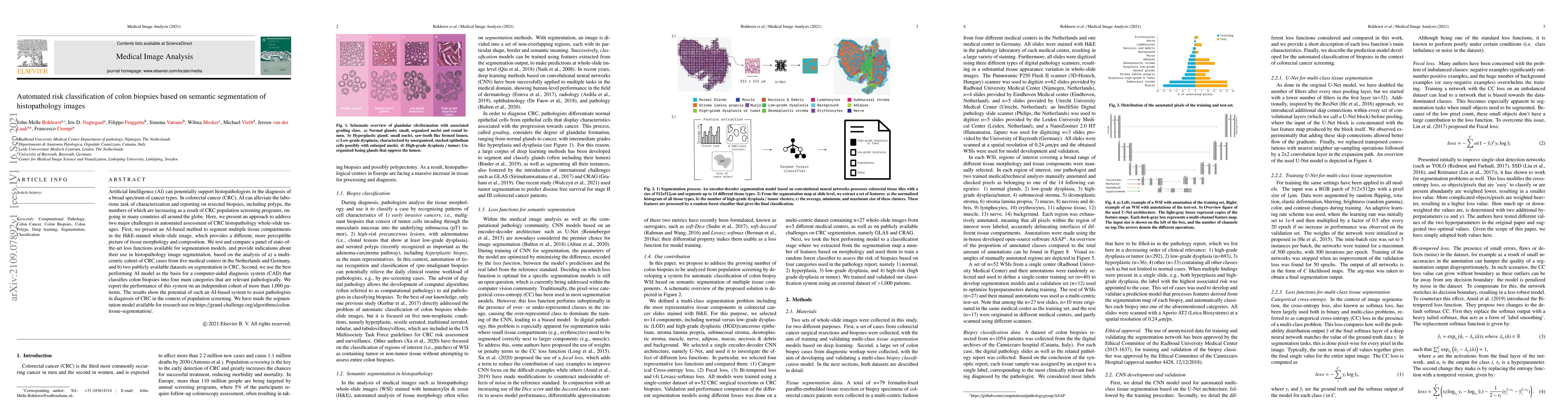
Artificial Intelligence (AI) can potentially support histopathologists in the diagnosis of a broad spectrum of cancer types. In colorectal cancer (CRC), AI can alleviate the laborious task of charac...
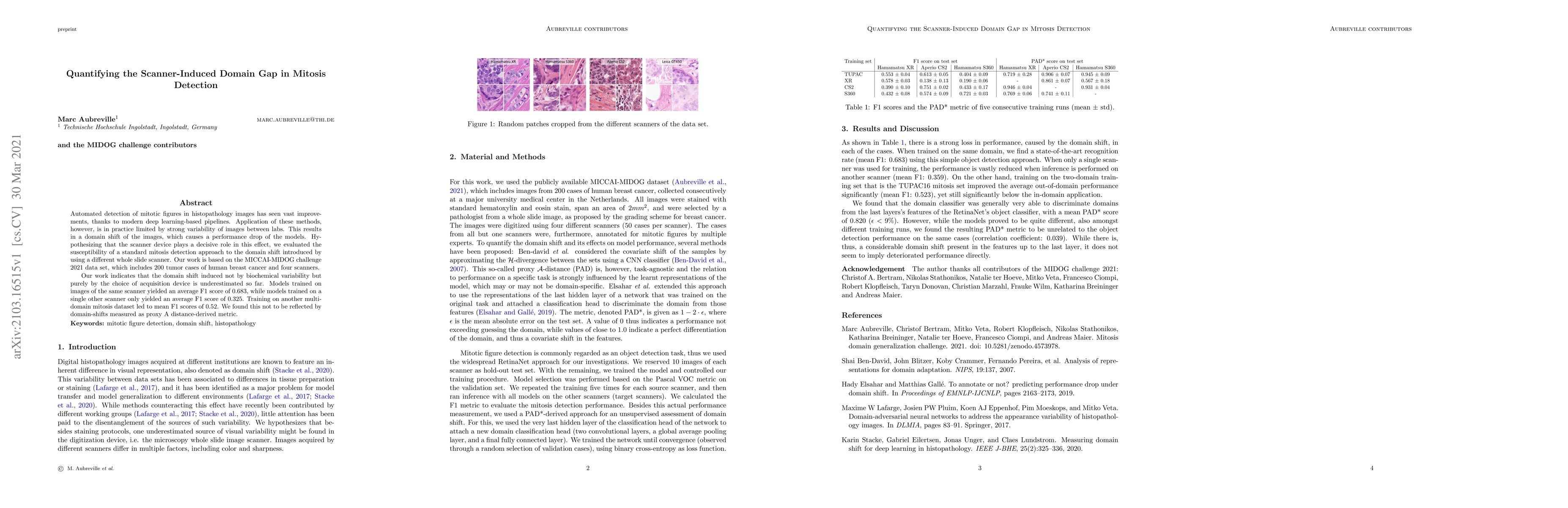
Automated detection of mitotic figures in histopathology images has seen vast improvements, thanks to modern deep learning-based pipelines. Application of these methods, however, is in practice limi...
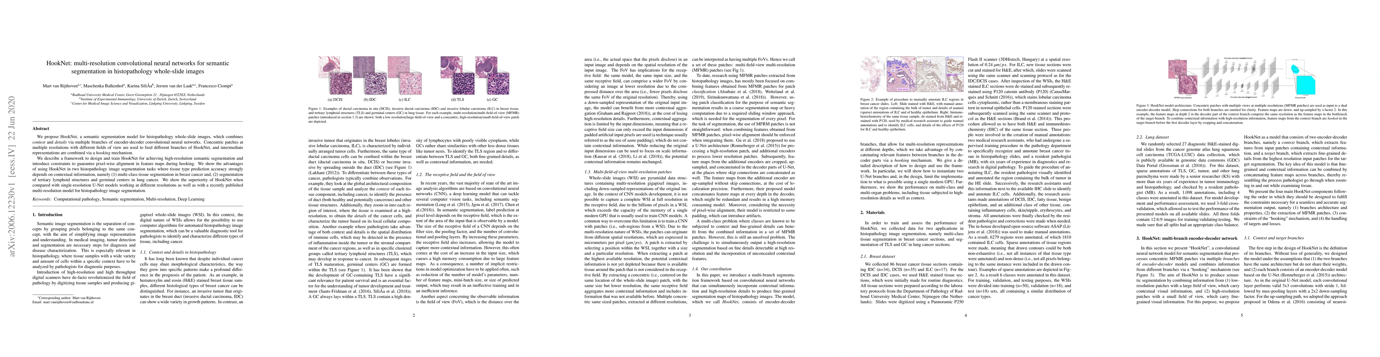
We propose HookNet, a semantic segmentation model for histopathology whole-slide images, which combines context and details via multiple branches of encoder-decoder convolutional neural networks. Co...
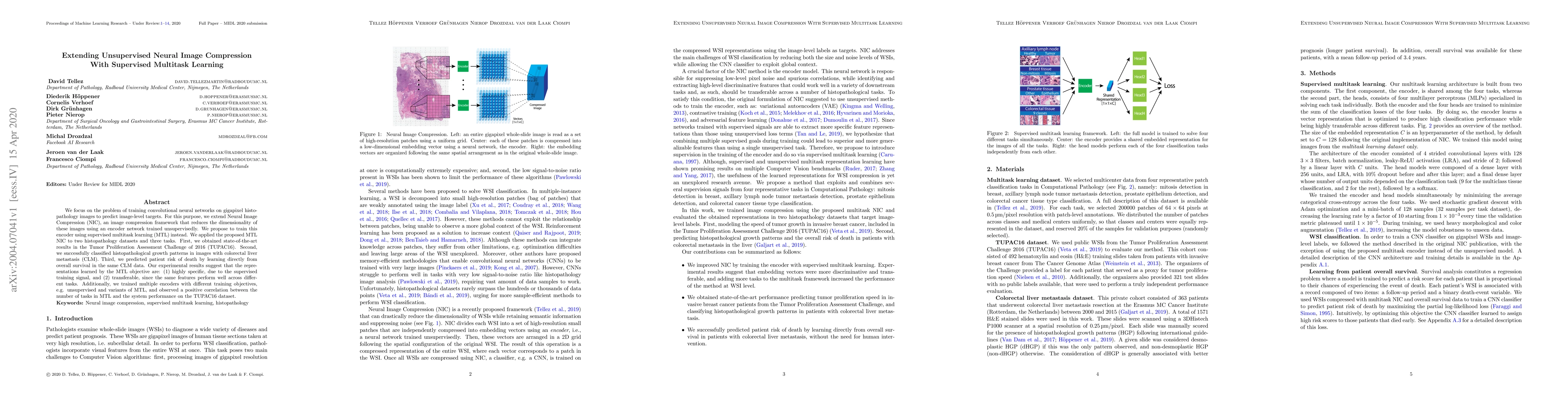
We focus on the problem of training convolutional neural networks on gigapixel histopathology images to predict image-level targets. For this purpose, we extend Neural Image Compression (NIC), an im...
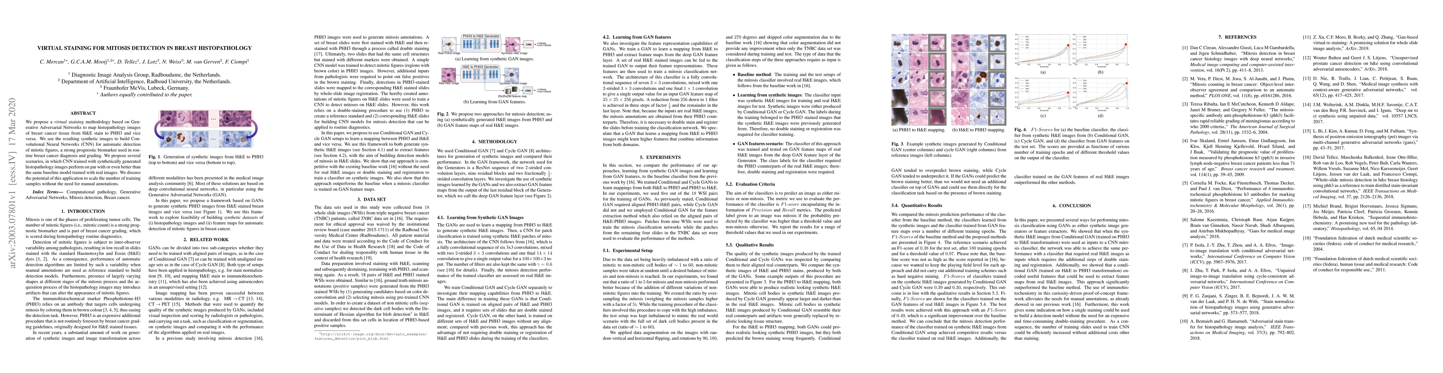
We propose a virtual staining methodology based on Generative Adversarial Networks to map histopathology images of breast cancer tissue from H&E stain to PHH3 and vice versa. We use the resulting sy...
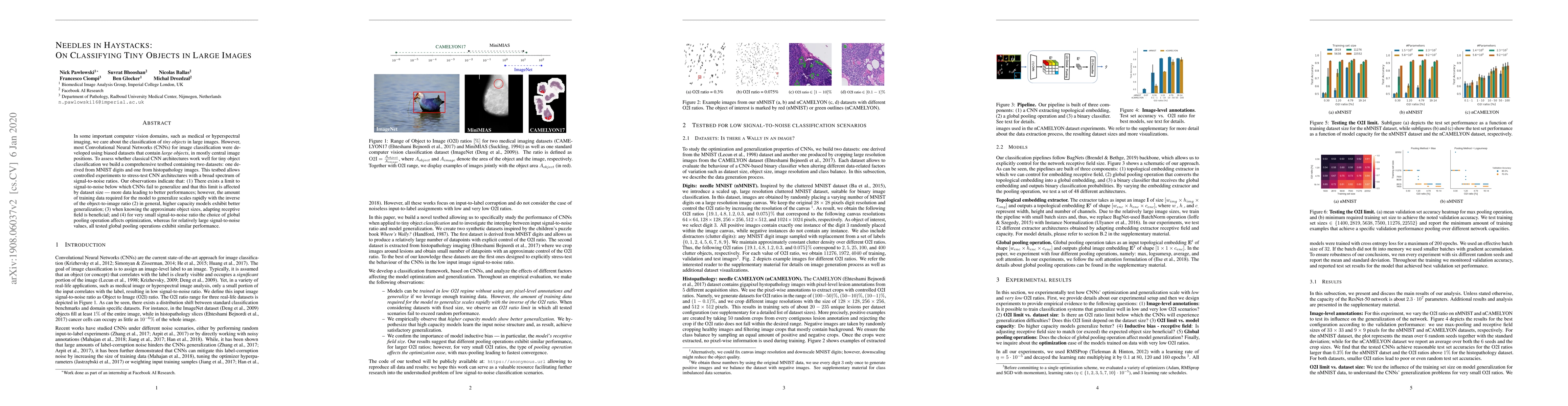
In some important computer vision domains, such as medical or hyperspectral imaging, we care about the classification of tiny objects in large images. However, most Convolutional Neural Networks (CN...
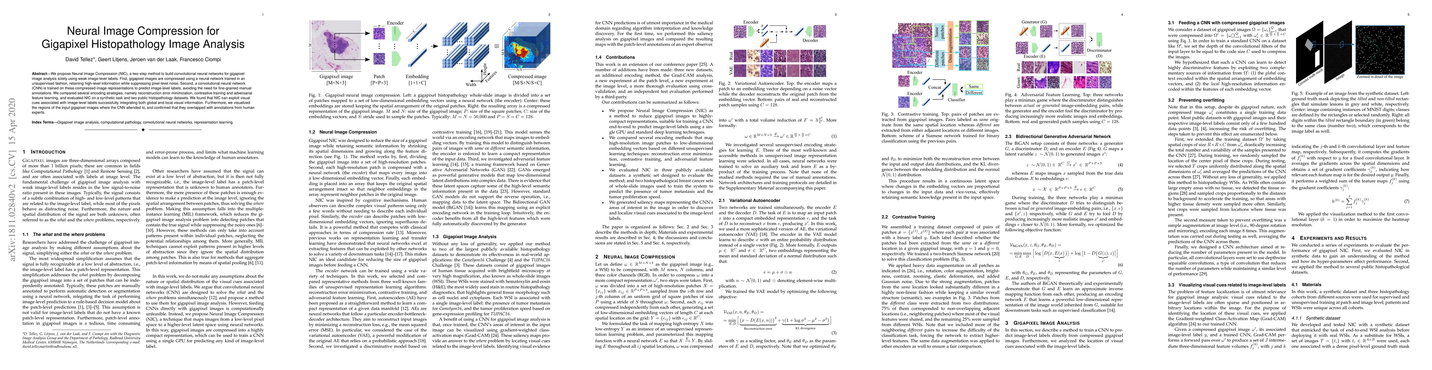
We propose Neural Image Compression (NIC), a two-step method to build convolutional neural networks for gigapixel image analysis solely using weak image-level labels. First, gigapixel images are com...
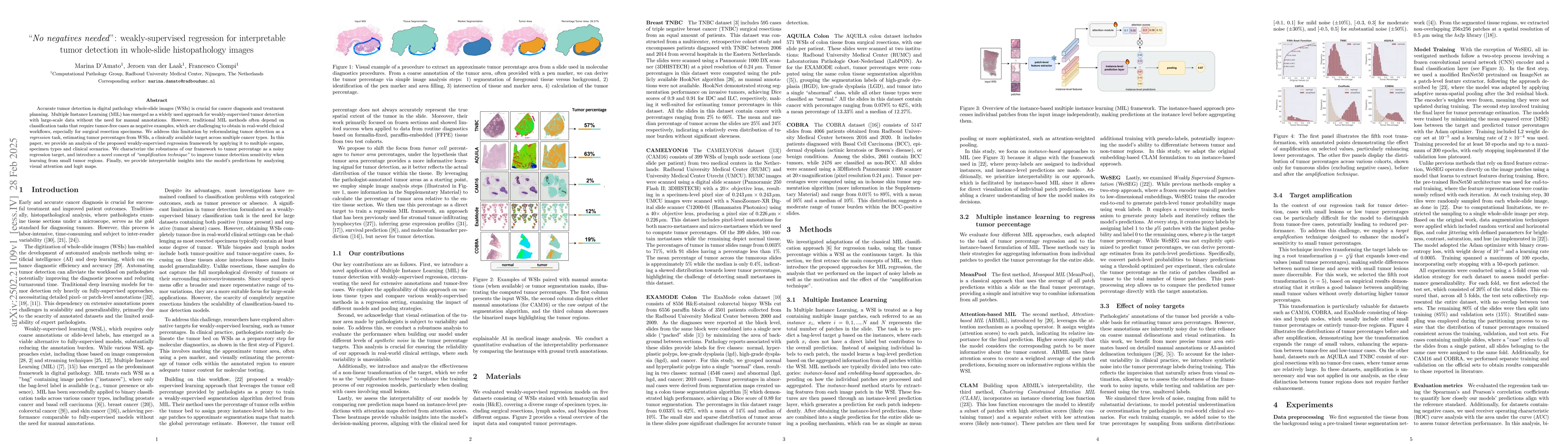
Accurate tumor detection in digital pathology whole-slide images (WSIs) is crucial for cancer diagnosis and treatment planning. Multiple Instance Learning (MIL) has emerged as a widely used approach f...
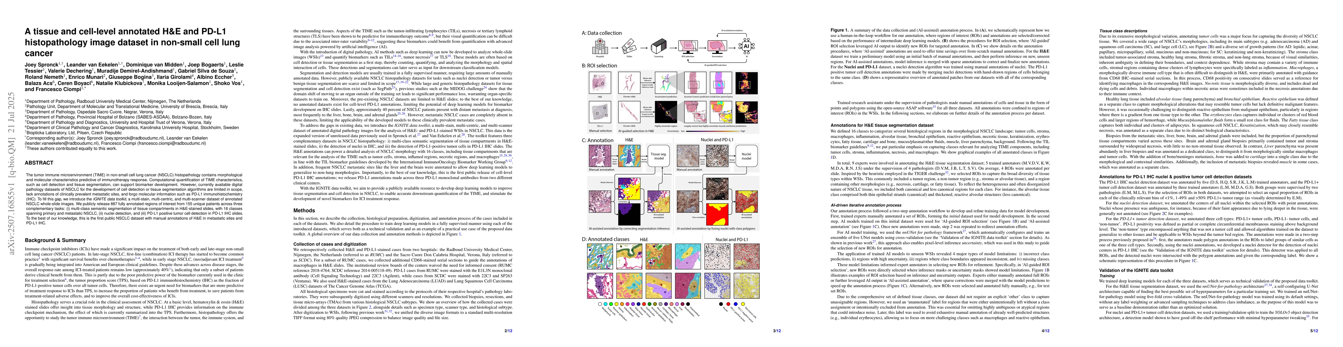
The tumor immune microenvironment (TIME) in non-small cell lung cancer (NSCLC) histopathology contains morphological and molecular characteristics predictive of immunotherapy response. Computational q...
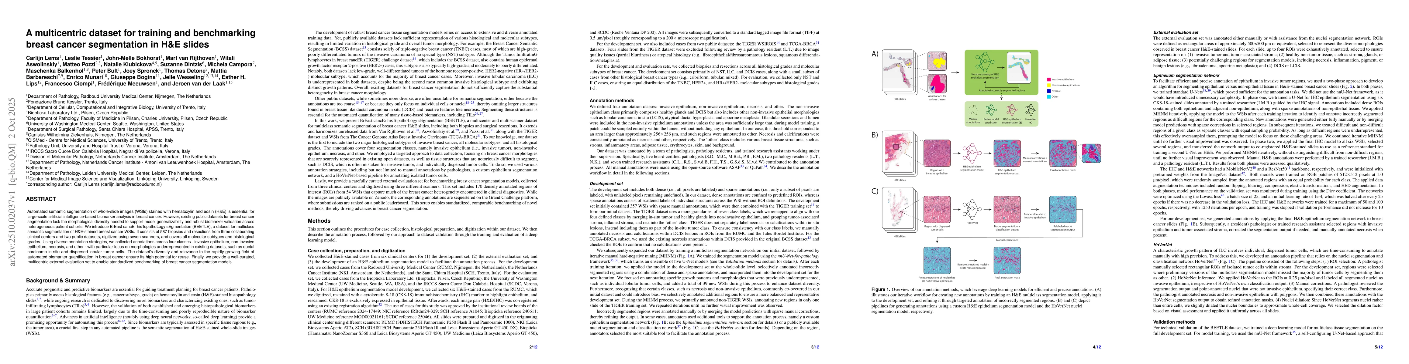
Automated semantic segmentation of whole-slide images (WSIs) stained with hematoxylin and eosin (H&E) is essential for large-scale artificial intelligence-based biomarker analysis in breast cancer. Ho...
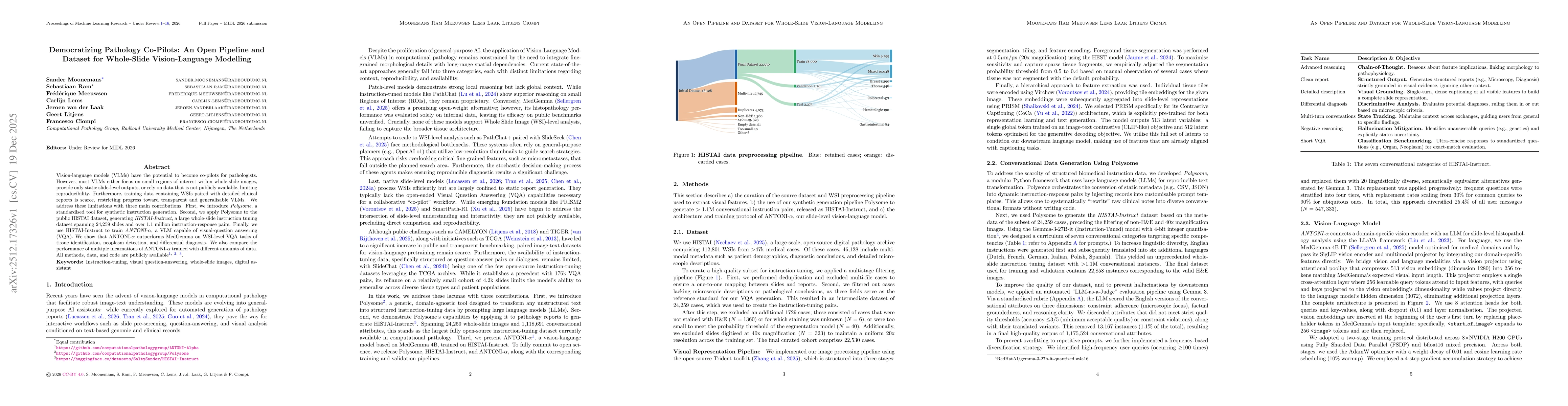
Vision-language models (VLMs) have the potential to become co-pilots for pathologists. However, most VLMs either focus on small regions of interest within whole-slide images, provide only static slide...

Medical foundation models show promise to learn broadly generalizable features from large, diverse datasets. This could be the base for reliable cross-modality generalization and rapid adaptation to n...
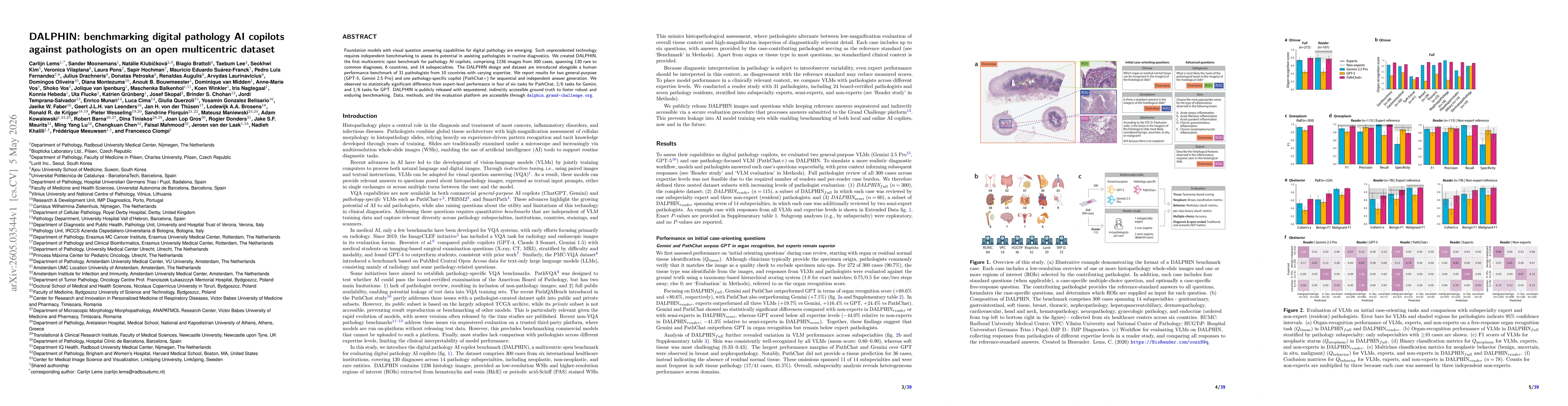
Foundation models with visual question answering capabilities for digital pathology are emerging. Such unprecedented technology requires independent benchmarking to assess its potential in assisting p...