Academic Profile
Statistics
Similar Authors
Papers on arXiv
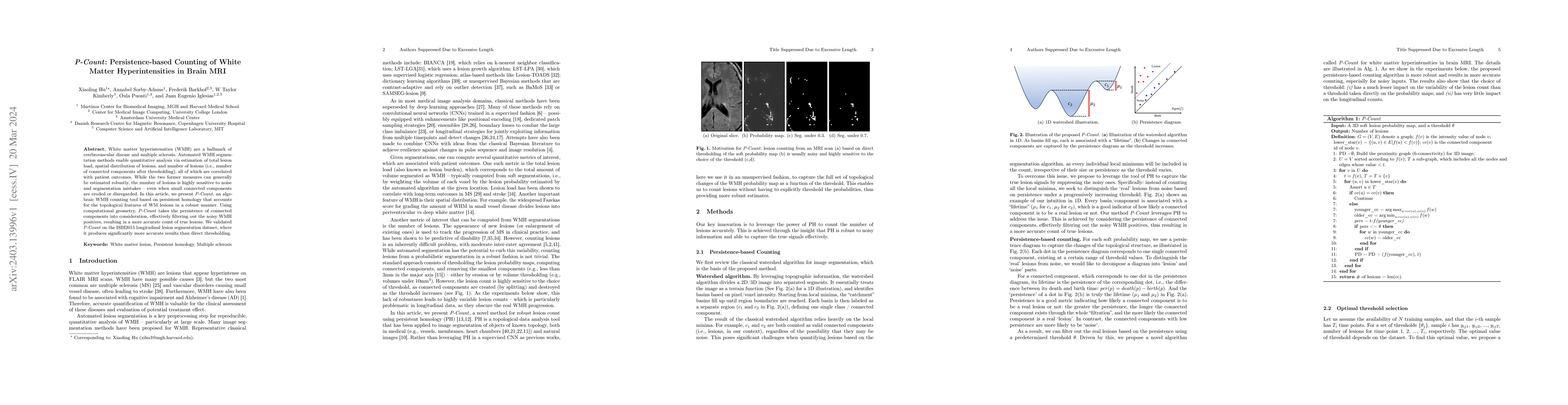
White matter hyperintensities (WMH) are a hallmark of cerebrovascular disease and multiple sclerosis. Automated WMH segmentation methods enable quantitative analysis via estimation of total lesion l...
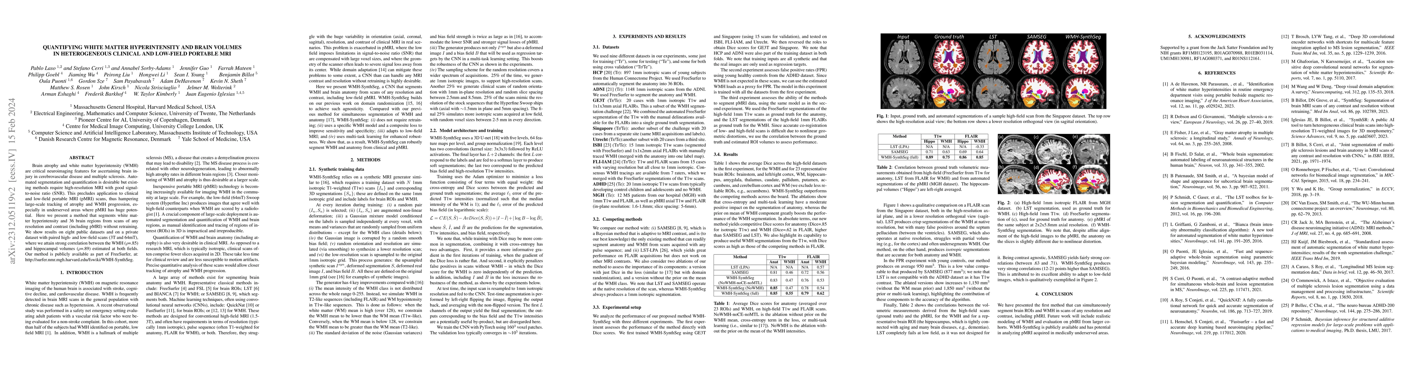
Brain atrophy and white matter hyperintensity (WMH) are critical neuroimaging features for ascertaining brain injury in cerebrovascular disease and multiple sclerosis. Automated segmentation and qua...
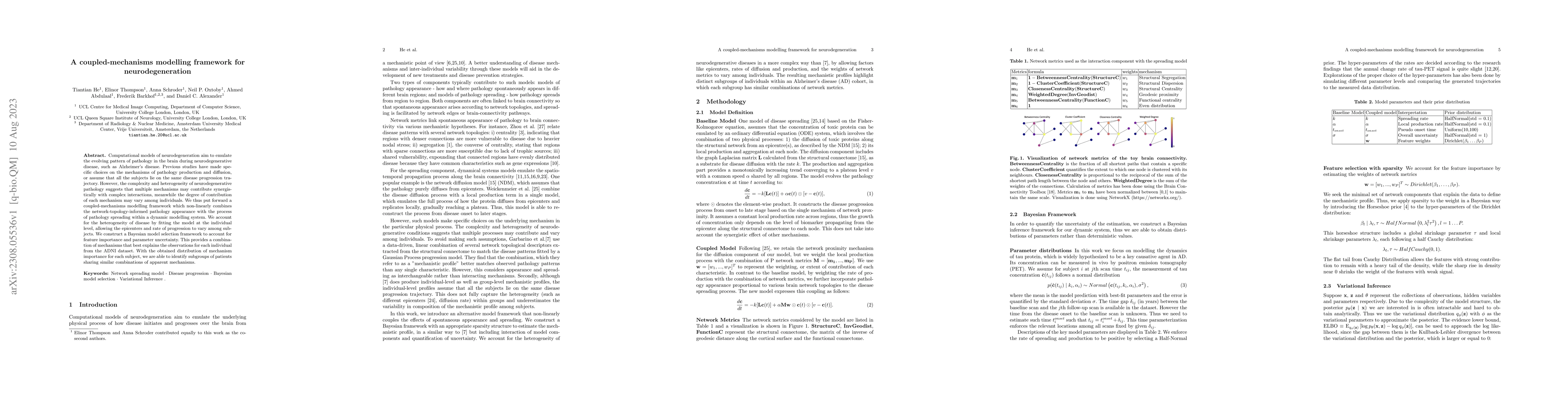
Computational models of neurodegeneration aim to emulate the evolving pattern of pathology in the brain during neurodegenerative disease, such as Alzheimer's disease. Previous studies have made spec...
The translation of AI-generated brain metastases (BM) segmentation into clinical practice relies heavily on diverse, high-quality annotated medical imaging datasets. The BraTS-METS 2023 challenge ha...
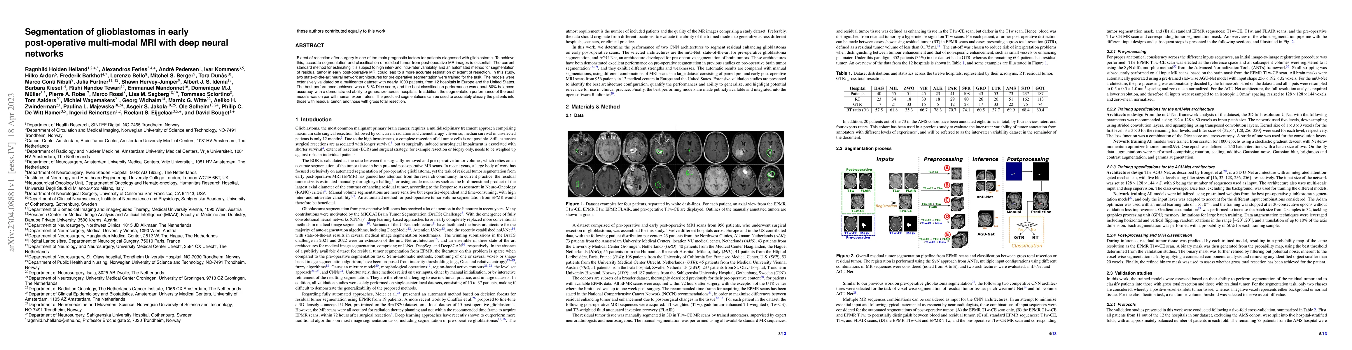
Extent of resection after surgery is one of the main prognostic factors for patients diagnosed with glioblastoma. To achieve this, accurate segmentation and classification of residual tumor from pos...
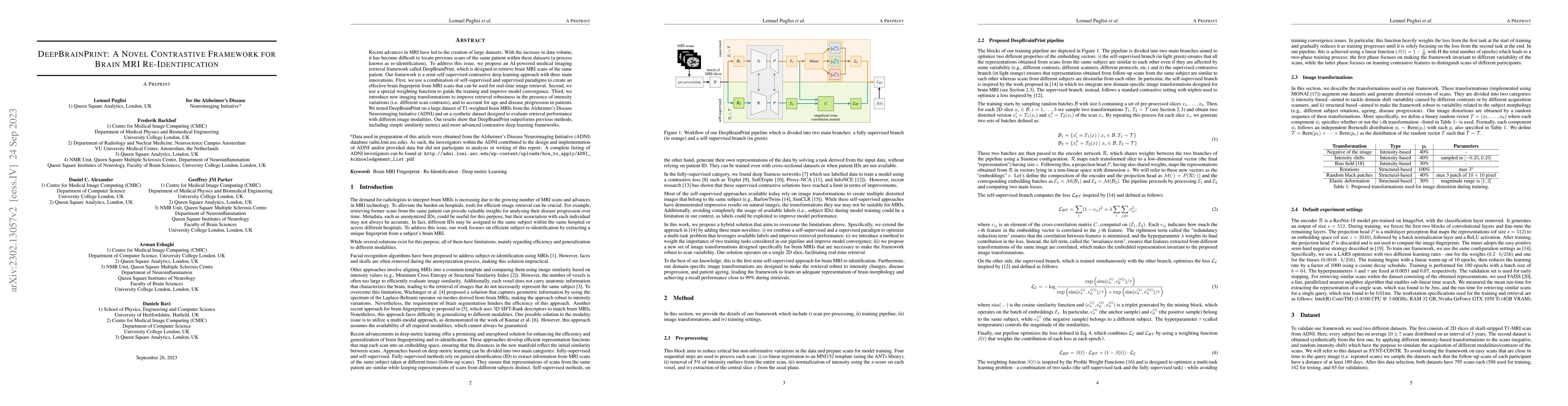
Recent advances in MRI have led to the creation of large datasets. With the increase in data volume, it has become difficult to locate previous scans of the same patient within these datasets (a pro...
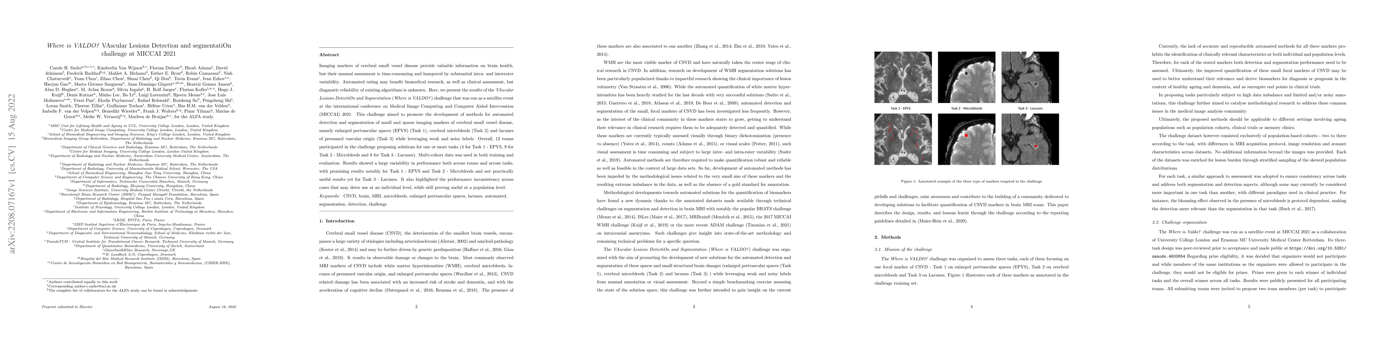
Imaging markers of cerebral small vessel disease provide valuable information on brain health, but their manual assessment is time-consuming and hampered by substantial intra- and interrater variabi...
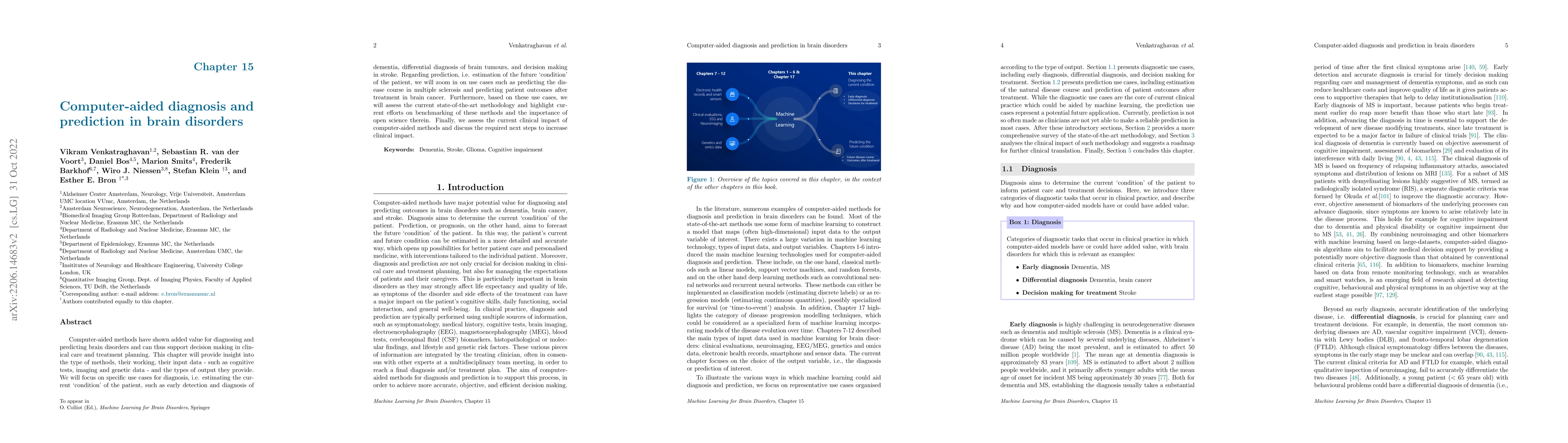
Computer-aided methods have shown added value for diagnosing and predicting brain disorders and can thus support decision making in clinical care and treatment planning. This chapter will provide in...
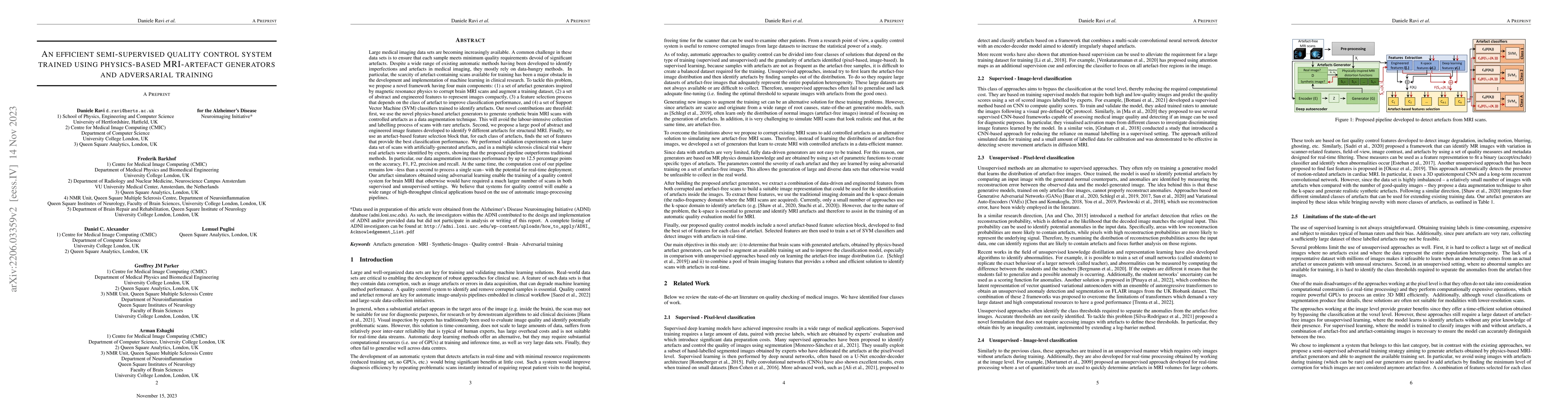
Large medical imaging data sets are becoming increasingly available, but ensuring sample quality without significant artefacts is challenging. Existing methods for identifying imperfections in medic...
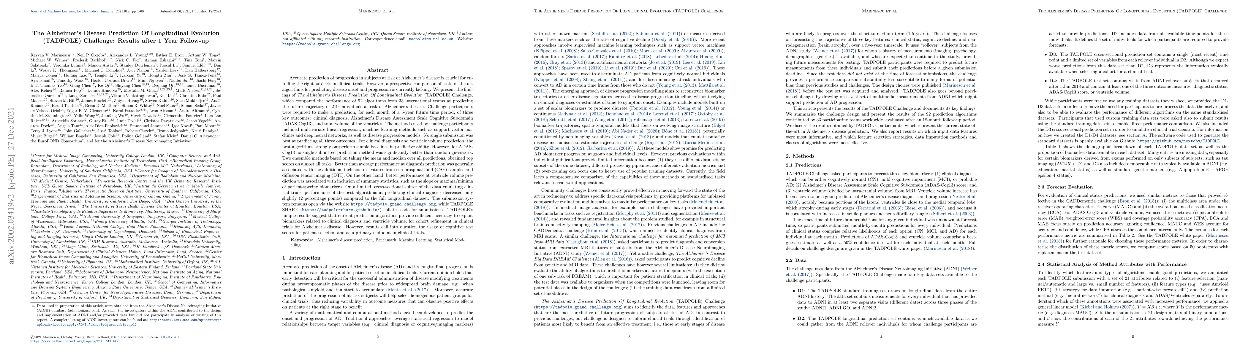
We present the findings of "The Alzheimer's Disease Prediction Of Longitudinal Evolution" (TADPOLE) Challenge, which compared the performance of 92 algorithms from 33 international teams at predicti...
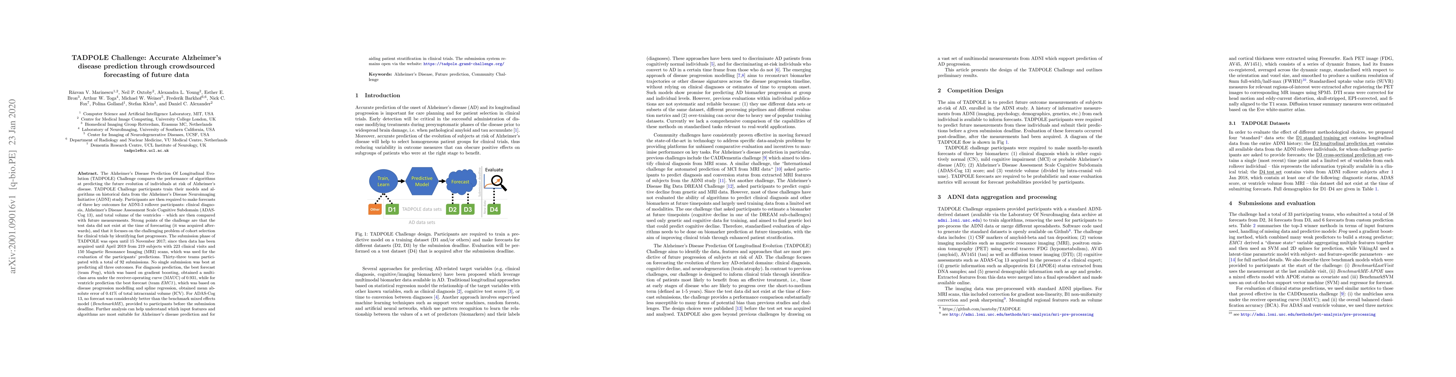
The TADPOLE Challenge compares the performance of algorithms at predicting the future evolution of individuals at risk of Alzheimer's disease. TADPOLE Challenge participants train their models and a...
Despite continuous advancements in cancer treatment, brain metastatic disease remains a significant complication of primary cancer and is associated with an unfavorable prognosis. One approach for imp...
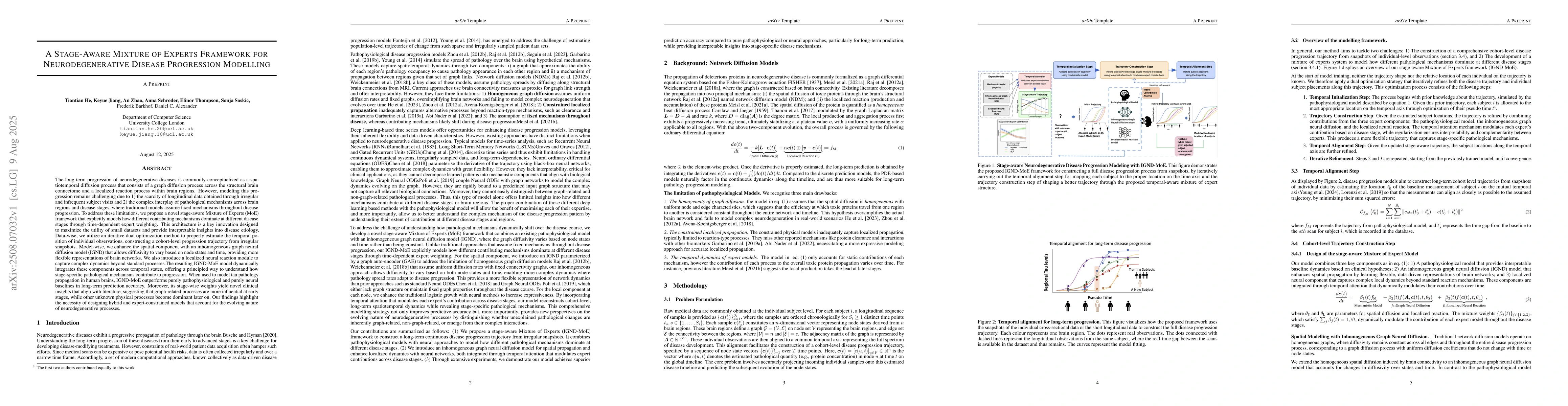
The long-term progression of neurodegenerative diseases is commonly conceptualized as a spatiotemporal diffusion process that consists of a graph diffusion process across the structural brain connecto...
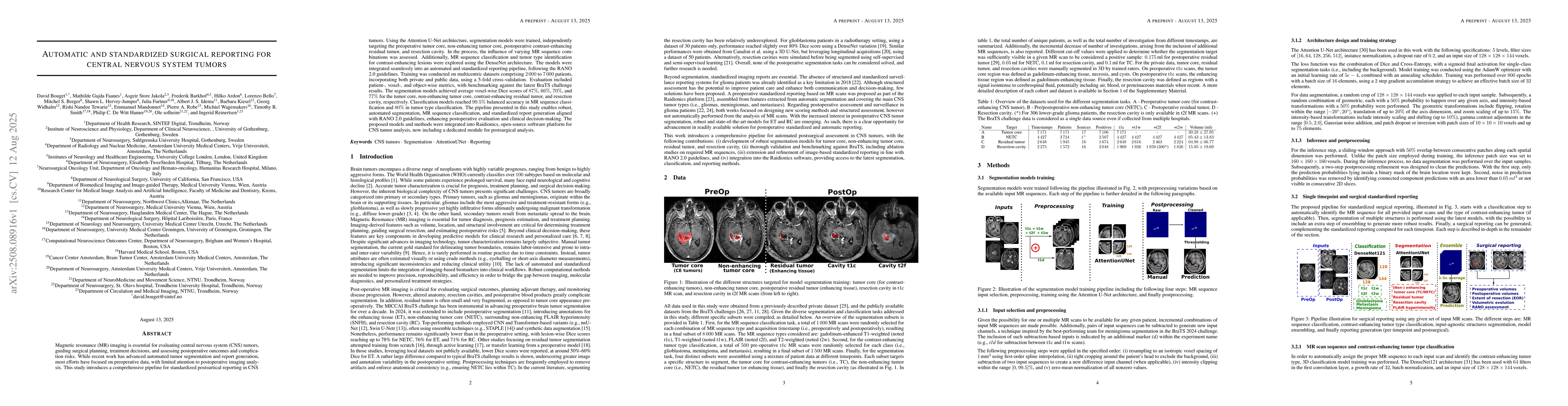
Magnetic resonance (MR) imaging is essential for evaluating central nervous system (CNS) tumors, guiding surgical planning, treatment decisions, and assessing postoperative outcomes and complication r...
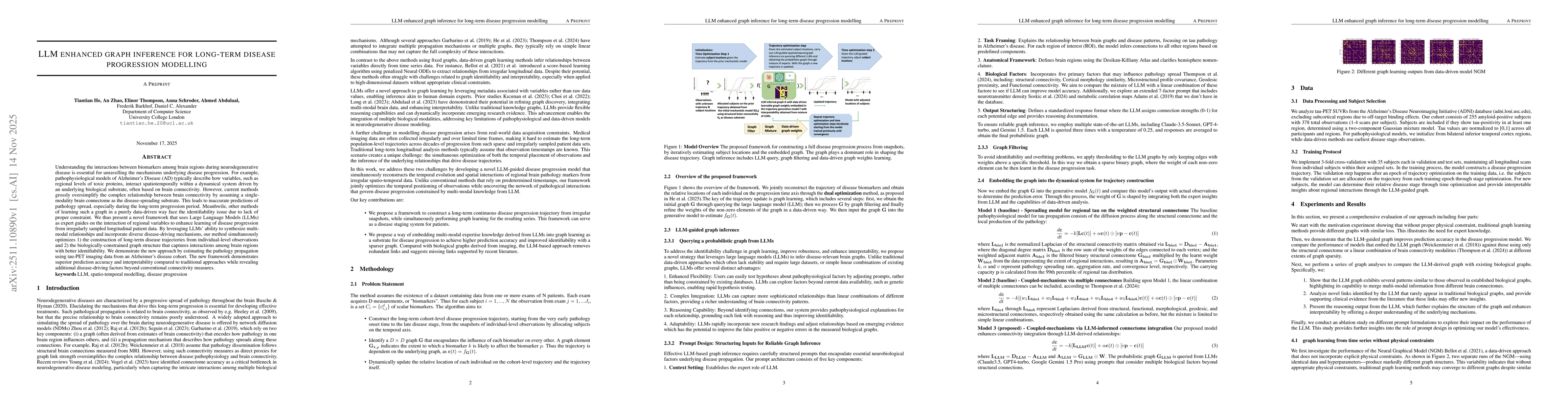
Understanding the interactions between biomarkers among brain regions during neurodegenerative disease is essential for unravelling the mechanisms underlying disease progression. For example, pathophy...
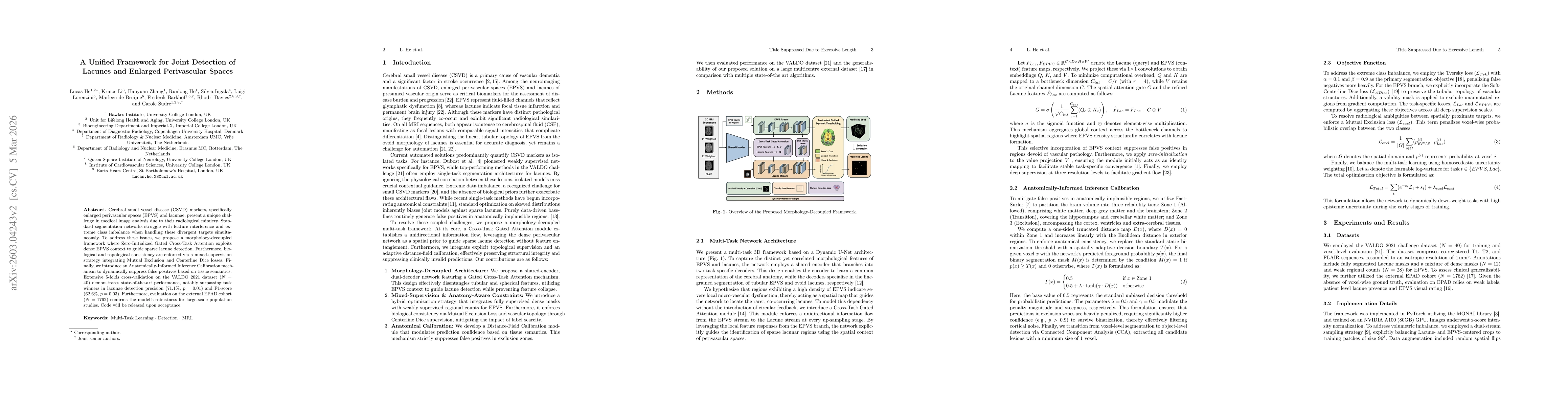
Cerebral small vessel disease (CSVD) markers, specifically enlarged perivascular spaces (EPVS) and lacunae, present a unique challenge in medical image analysis due to their radiological mimicry. Stan...
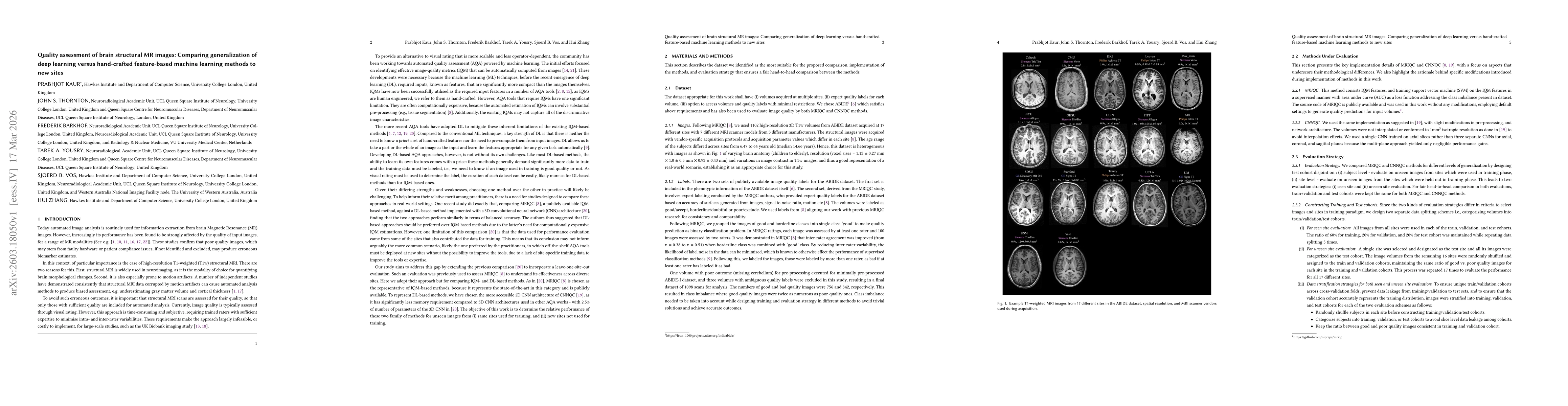
Quality assessment of brain structural MR images is critical for large-scale neuroimaging studies, where motion artifacts can significantly bias clinical estimates. While visual rating remains the gol...
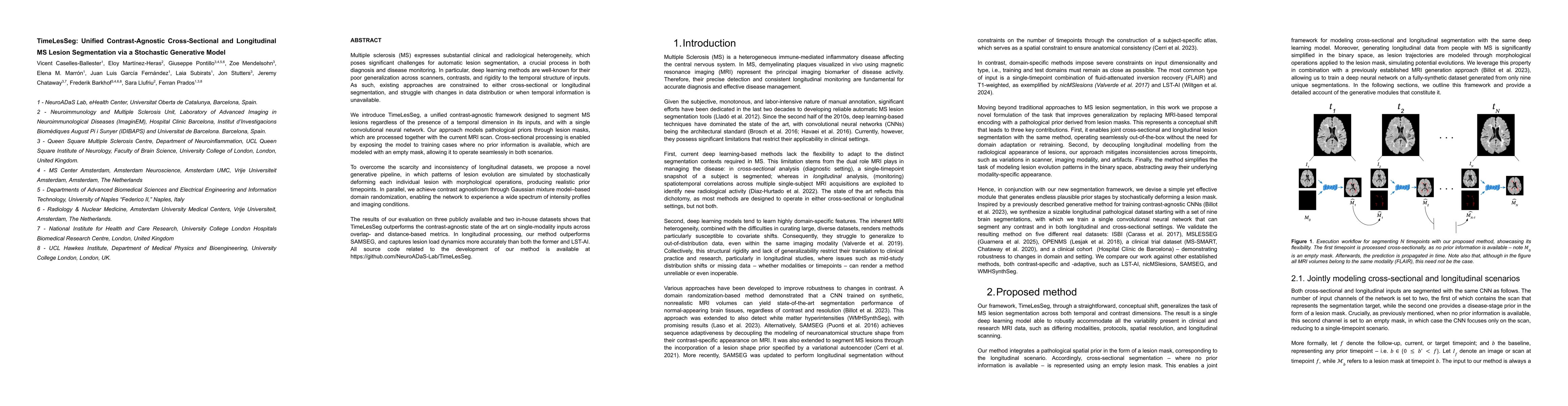
Multiple sclerosis (MS) expresses substantial clinical and radiological heterogeneity, which poses significant challenges for automatic lesion segmentation. The current deep learning-based SOTA is hig...
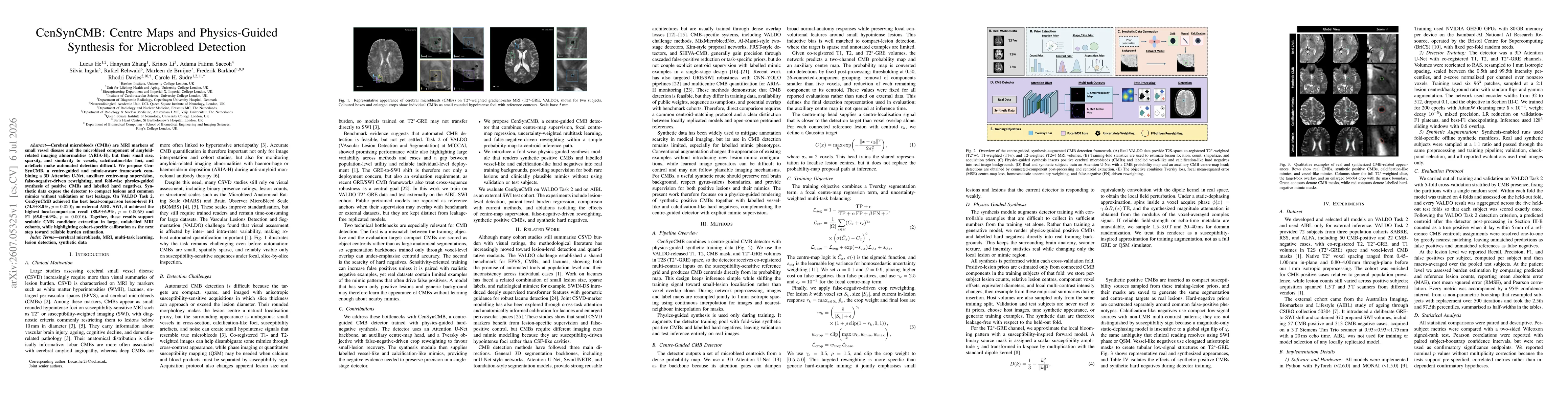
Cerebral microbleeds (CMBs) are MRI markers of small vessel disease and the microbleed component of amyloid related imaging abnormalities (ARIA-H), but their small size, sparsity, and similarity to ve...