Academic Profile
Statistics
Similar Authors
Papers on arXiv
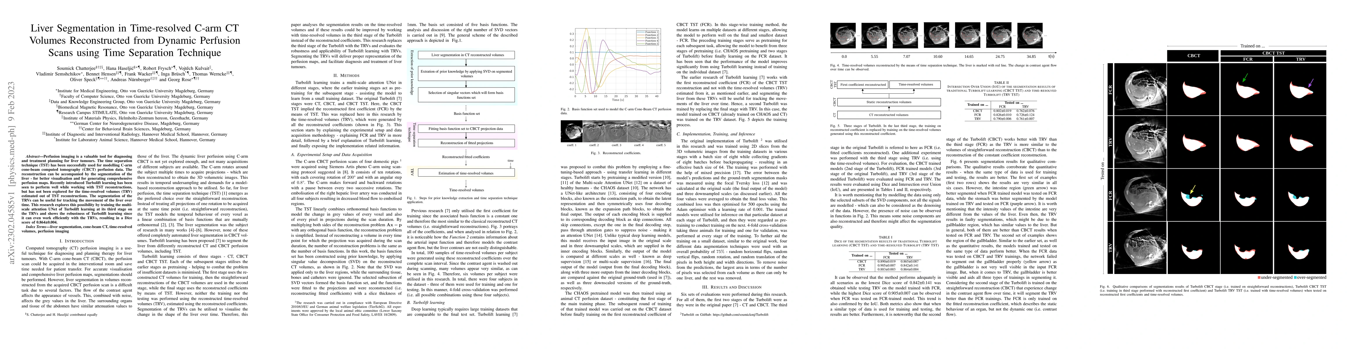
Perfusion imaging is a valuable tool for diagnosing and treatment planning for liver tumours. The time separation technique (TST) has been successfully used for modelling C-arm cone-beam computed to...
Model-based reconstruction employing the time separation technique (TST) was found to improve dynamic perfusion imaging of the liver using C-arm cone-beam computed tomography (CBCT). To apply TST us...
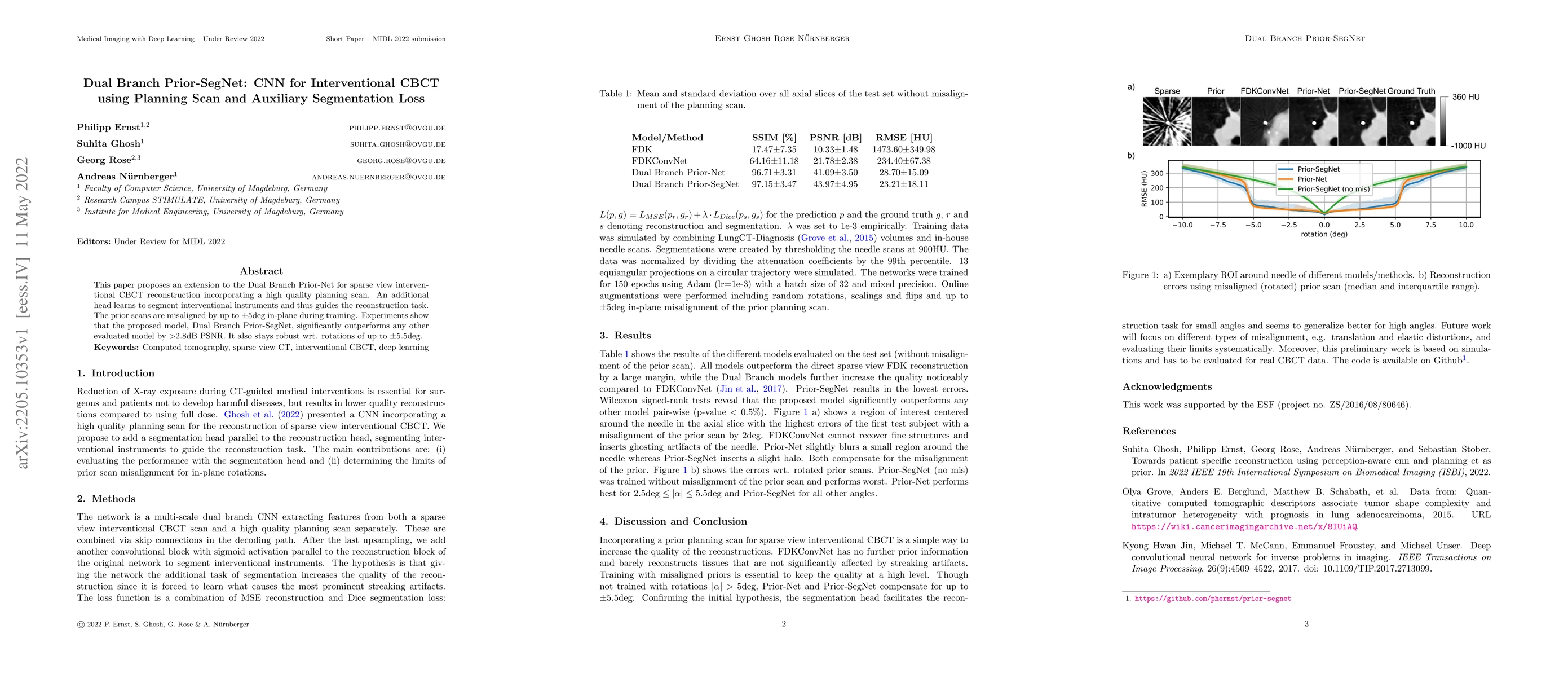
This paper proposes an extension to the Dual Branch Prior-Net for sparse view interventional CBCT reconstruction incorporating a high quality planning scan. An additional head learns to segment inte...
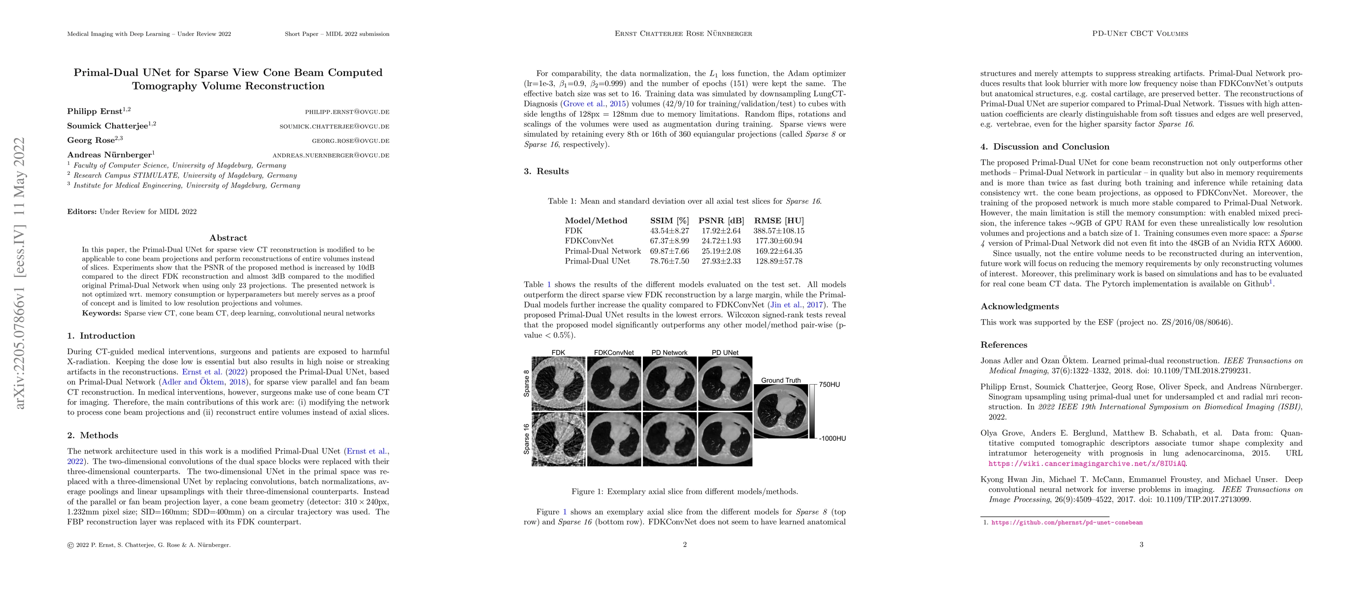
In this paper, the Primal-Dual UNet for sparse view CT reconstruction is modified to be applicable to cone beam projections and perform reconstructions of entire volumes instead of slices. Experimen...
Magnetic resonance imaging (MRI) provides high spatial resolution and excellent soft-tissue contrast without using harmful ionising radiation. Dynamic MRI is an essential tool for interventions to v...
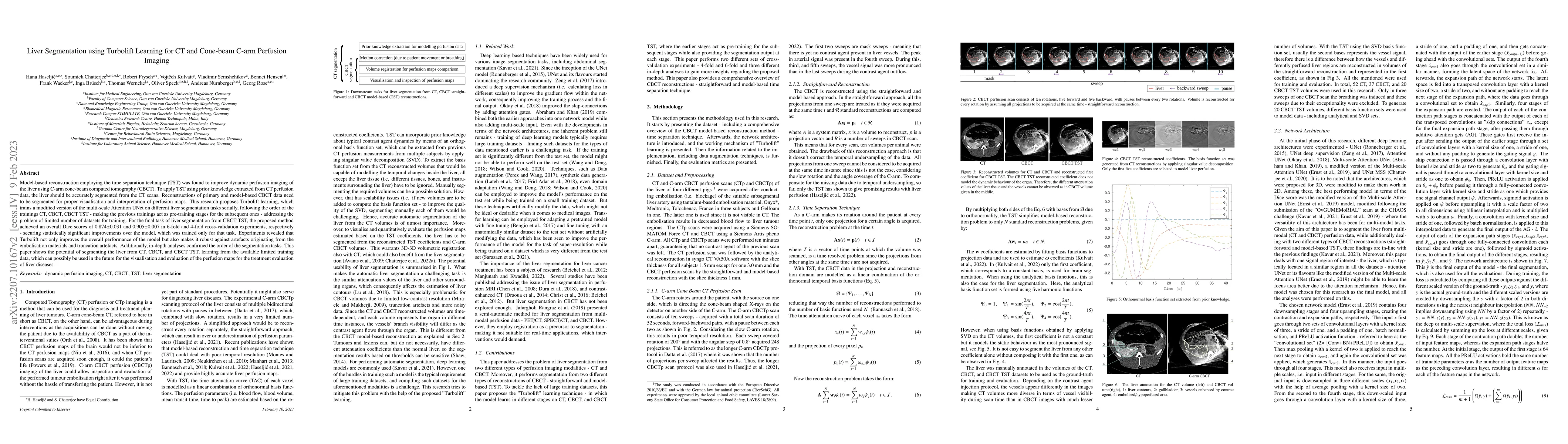
Computed tomography and magnetic resonance imaging are two widely used clinical imaging modalities for non-invasive diagnosis. However, both of these modalities come with certain problems. CT uses h...
CT perfusion imaging (CTP) plays an important role in decision making for the treatment of acute ischemic stroke with large vessel occlusion. Since the CT perfusion scan time is approximately one mi...
Perfusion imaging is an interesting new modality for evaluation and assessment of the liver cancer treatment. C-Arm CT provides a possibility to perform perfusion imaging scans intra-operatively for...
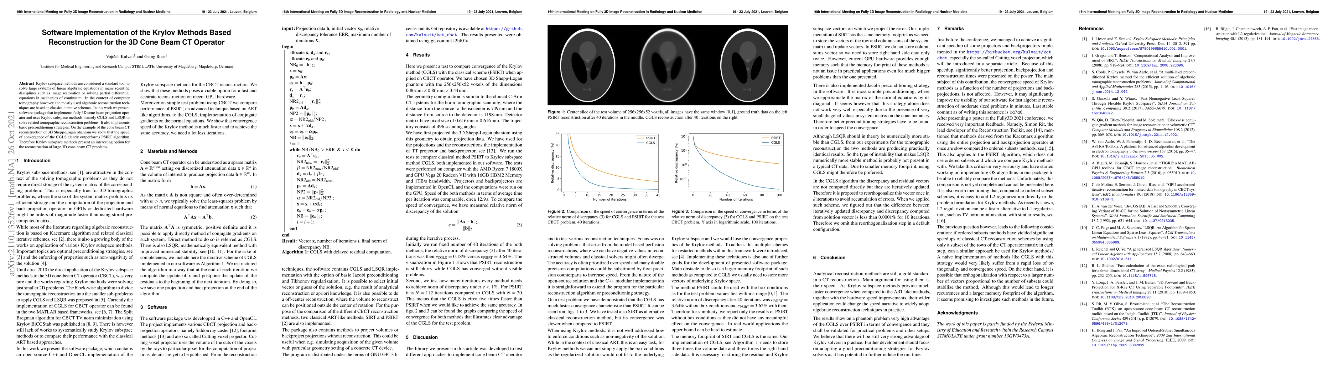
Krylov subspace methods are considered a standard tool to solve large systems of linear algebraic equations in many scientific disciplines such as image restoration or solving partial differential e...
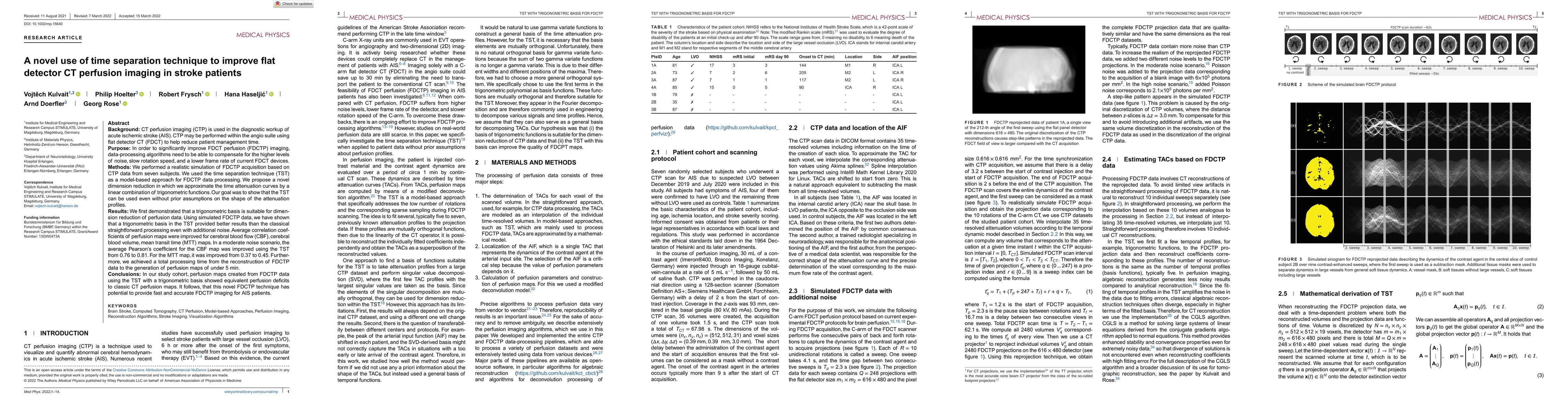
CT perfusion imaging (CTP) is used in the diagnostic workup of acute ischemic stroke (AIS). CTP may be performed within the angio suite using flat detector CT (FDCT) to help reduce patient managemen...
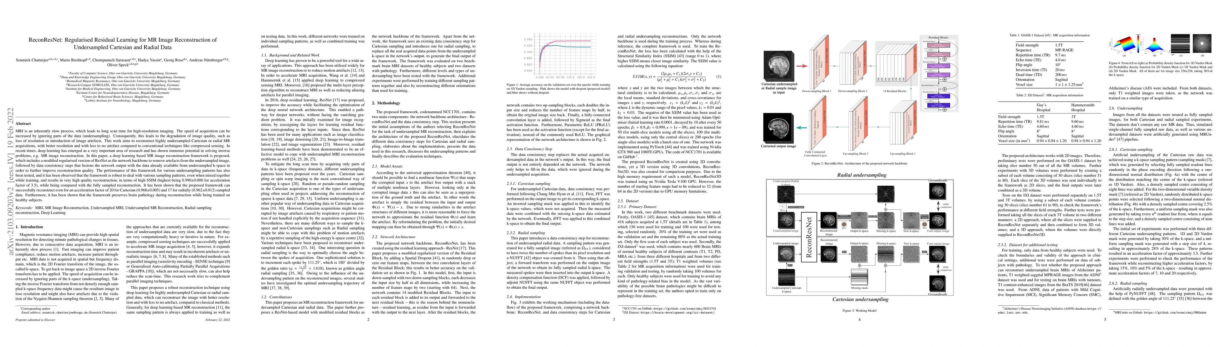
MRI is an inherently slow process, which leads to long scan time for high-resolution imaging. The speed of acquisition can be increased by ignoring parts of the data (undersampling). Consequently, t...
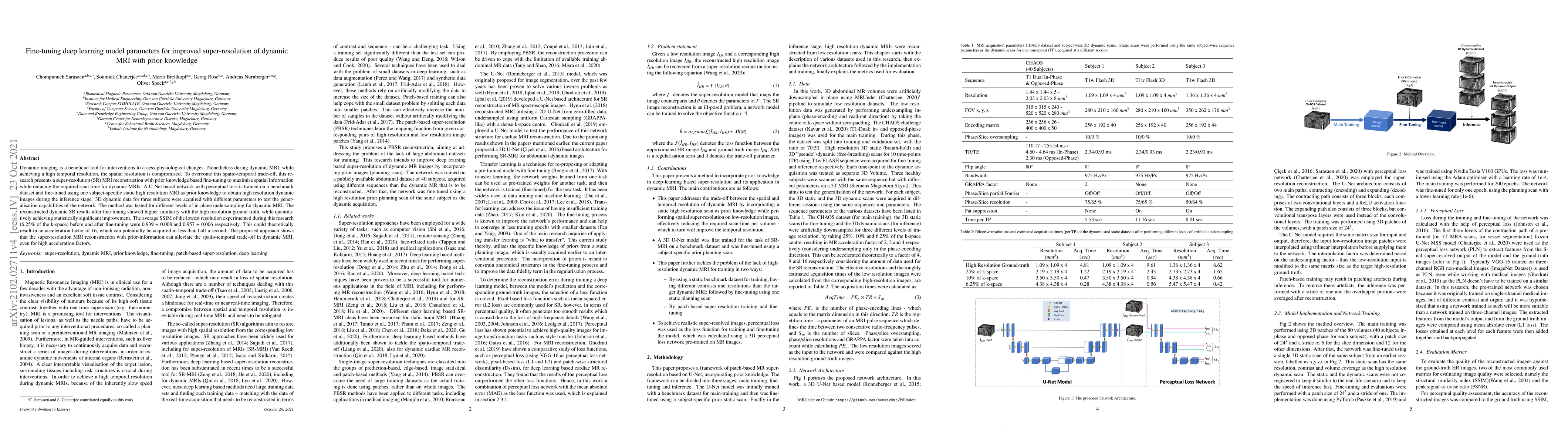
Dynamic imaging is a beneficial tool for interventions to assess physiological changes. Nonetheless during dynamic MRI, while achieving a high temporal resolution, the spatial resolution is compromi...
The outbreak of COVID-19 has shocked the entire world with its fairly rapid spread and has challenged different sectors. One of the most effective ways to limit its spread is the early and accurate ...
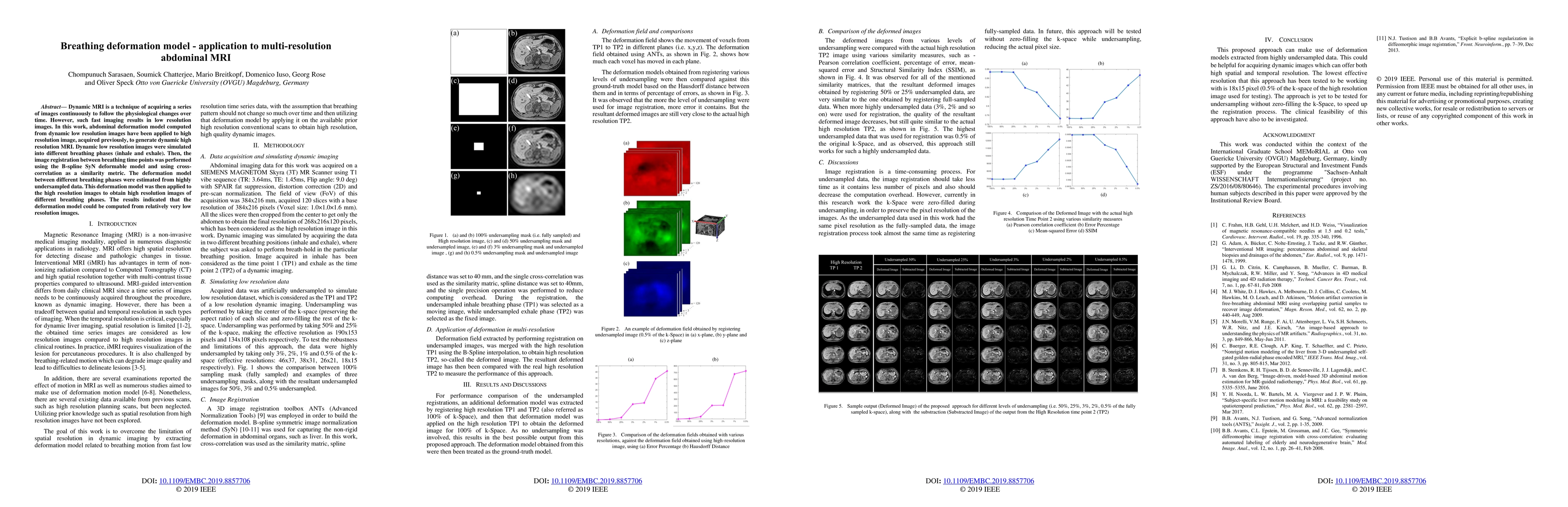
Dynamic MRI is a technique of acquiring a series of images continuously to follow the physiological changes over time. However, such fast imaging results in low resolution images. In this work, abdo...
The success of embolisation, a minimally invasive treatment of liver cancer, could be evaluated in the operational room with cone-beam CT by acquiring a dynamic perfusion scan. The reconstruction algo...