Academic Profile
Statistics
Similar Authors
Papers on arXiv
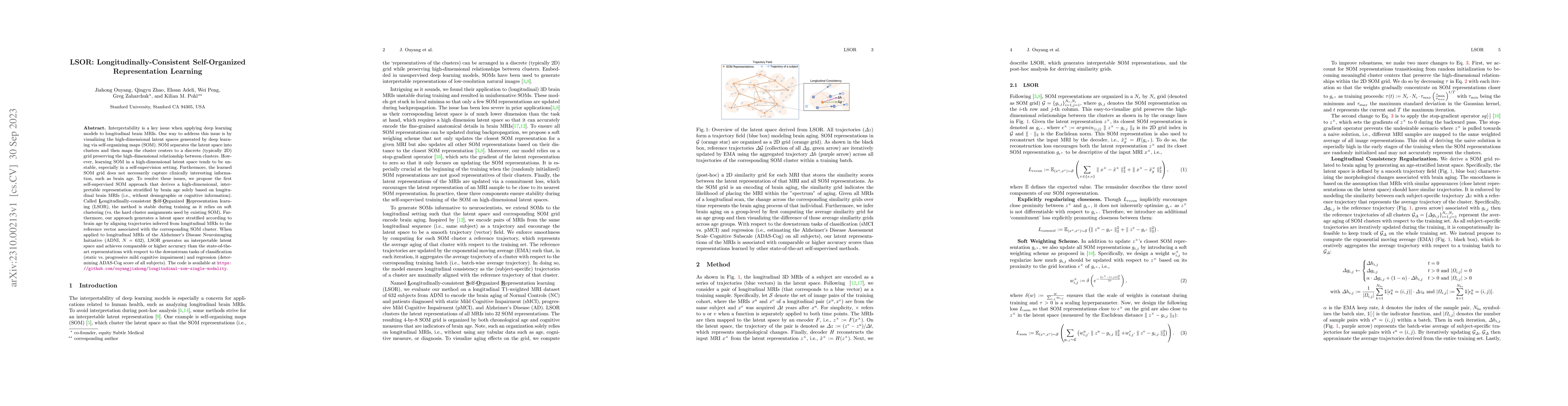
Interpretability is a key issue when applying deep learning models to longitudinal brain MRIs. One way to address this issue is by visualizing the high-dimensional latent spaces generated by deep le...
Purpose: Multi-expert deep learning training methods to automatically quantify ischemic brain tissue on Non-Contrast CT Materials and Methods: The data set consisted of 260 Non-Contrast CTs from 233...
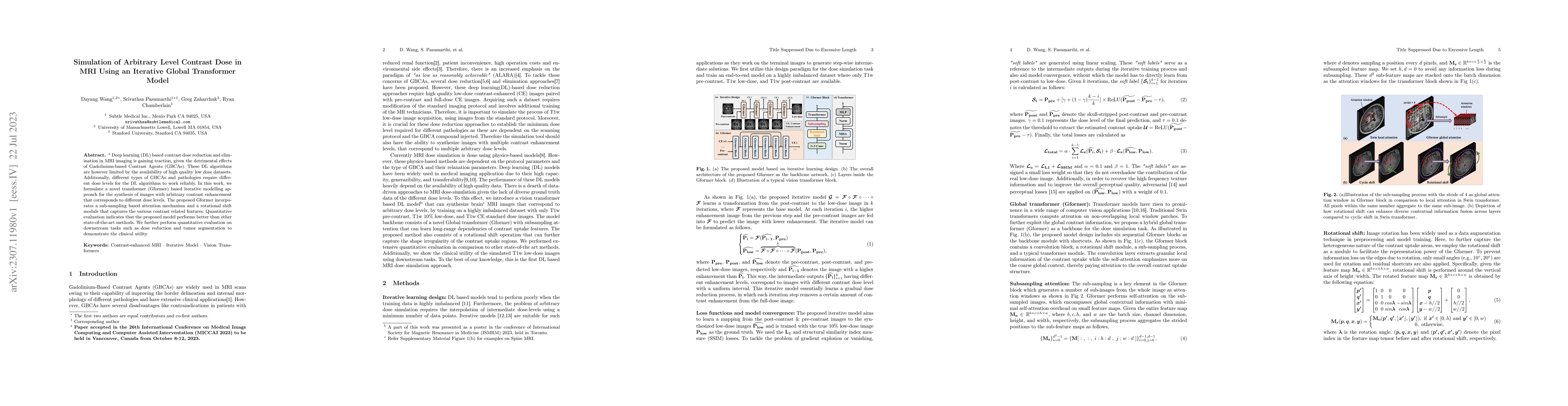
Deep learning (DL) based contrast dose reduction and elimination in MRI imaging is gaining traction, given the detrimental effects of Gadolinium-based Contrast Agents (GBCAs). These DL algorithms ar...
To determine if a convolutional neural network (CNN) deep learning model can accurately segment acute ischemic changes on non-contrast CT compared to neuroradiologists. Non-contrast CT (NCCT) examin...
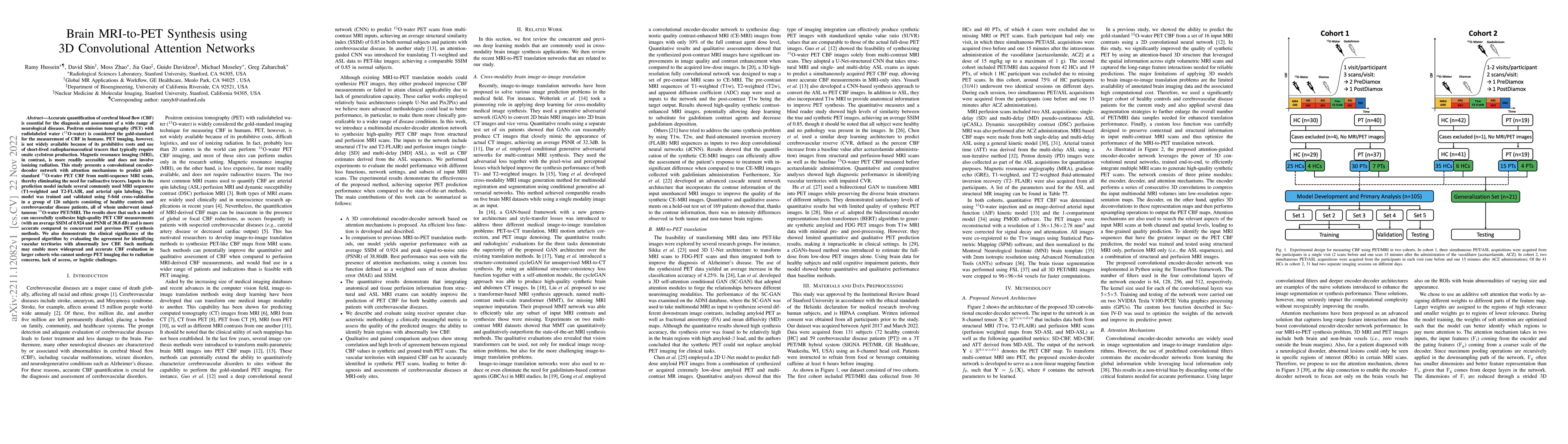
Accurate quantification of cerebral blood flow (CBF) is essential for the diagnosis and assessment of a wide range of neurological diseases. Positron emission tomography (PET) with radiolabeled wate...
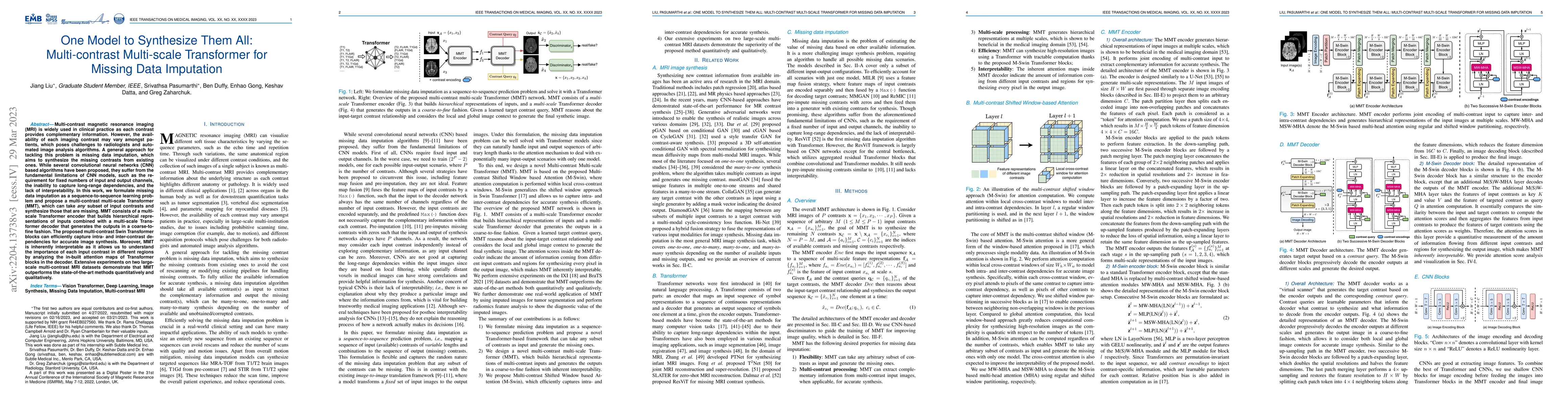
Multi-contrast magnetic resonance imaging (MRI) is widely used in clinical practice as each contrast provides complementary information. However, the availability of each imaging contrast may vary a...
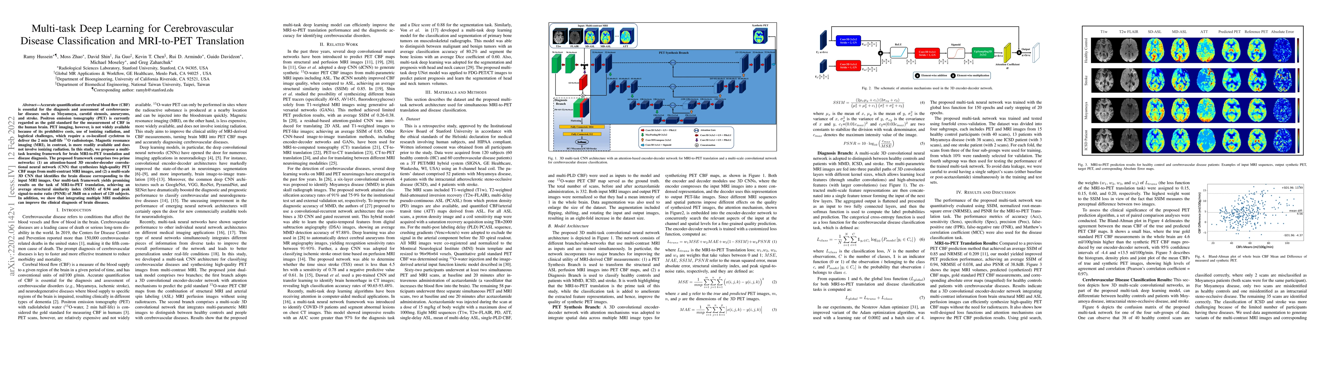
Accurate quantification of cerebral blood flow (CBF) is essential for the diagnosis and assessment of cerebrovascular diseases such as Moyamoya, carotid stenosis, aneurysms, and stroke. Positron emi...
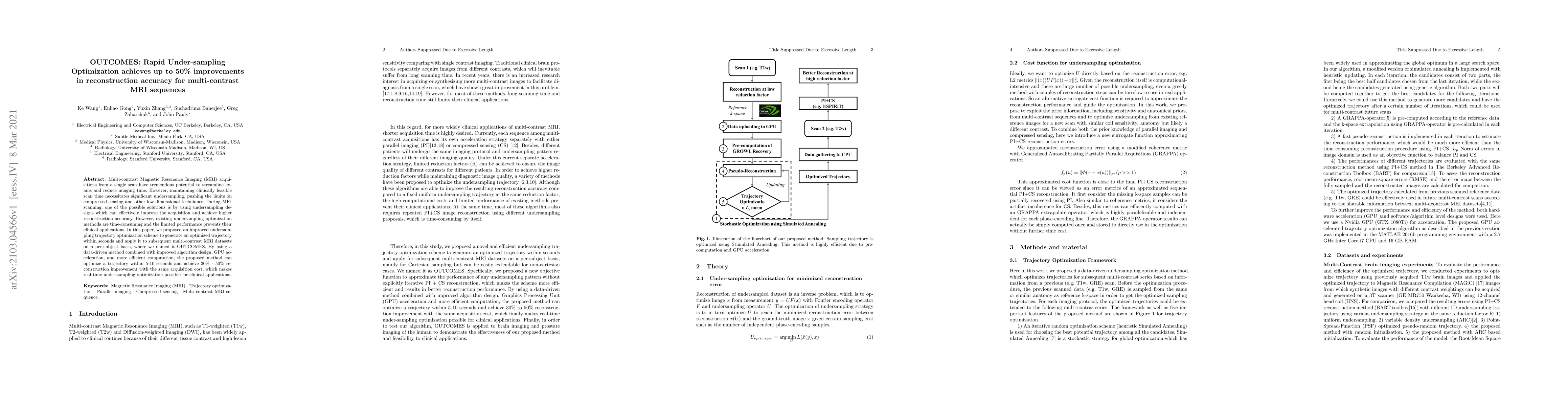
Multi-contrast Magnetic Resonance Imaging (MRI) acquisitions from a single scan have tremendous potential to streamline exams and reduce imaging time. However, maintaining clinically feasible scan t...
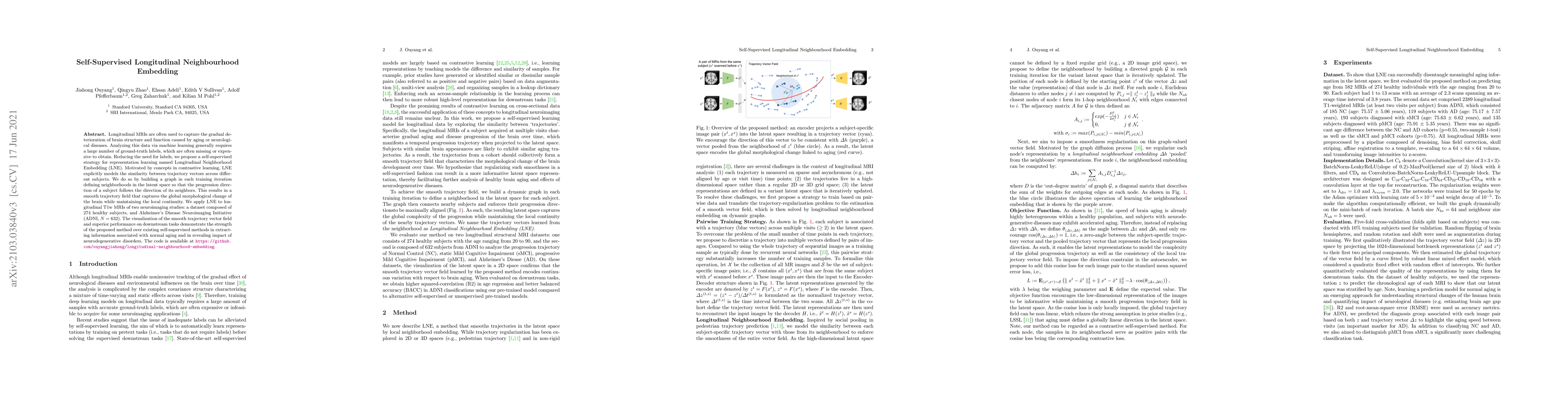
Longitudinal MRIs are often used to capture the gradual deterioration of brain structure and function caused by aging or neurological diseases. Analyzing this data via machine learning generally req...
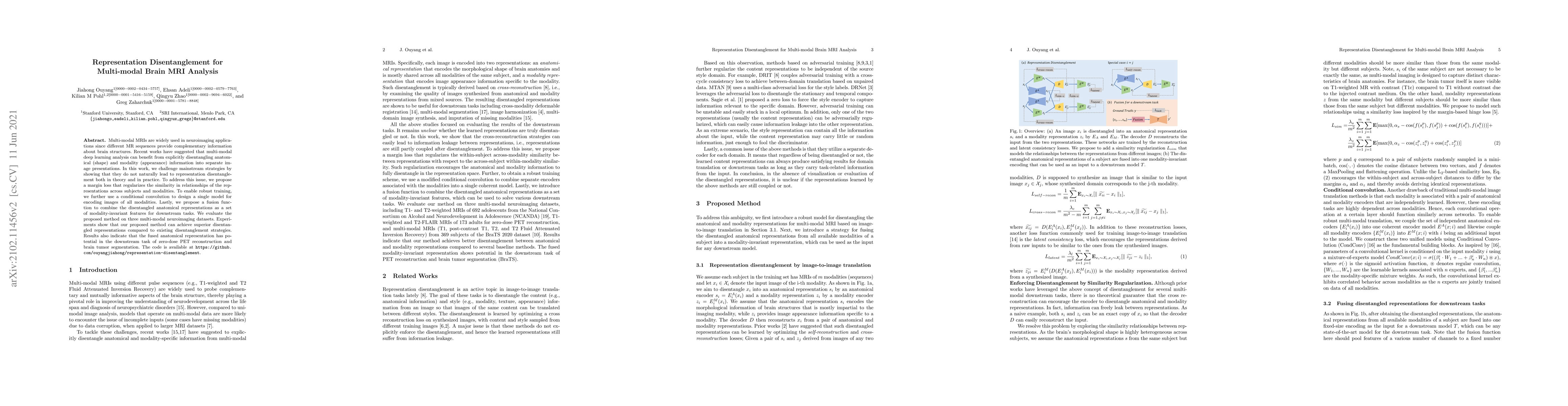
Multi-modal MRIs are widely used in neuroimaging applications since different MR sequences provide complementary information about brain structures. Recent works have suggested that multi-modal deep...
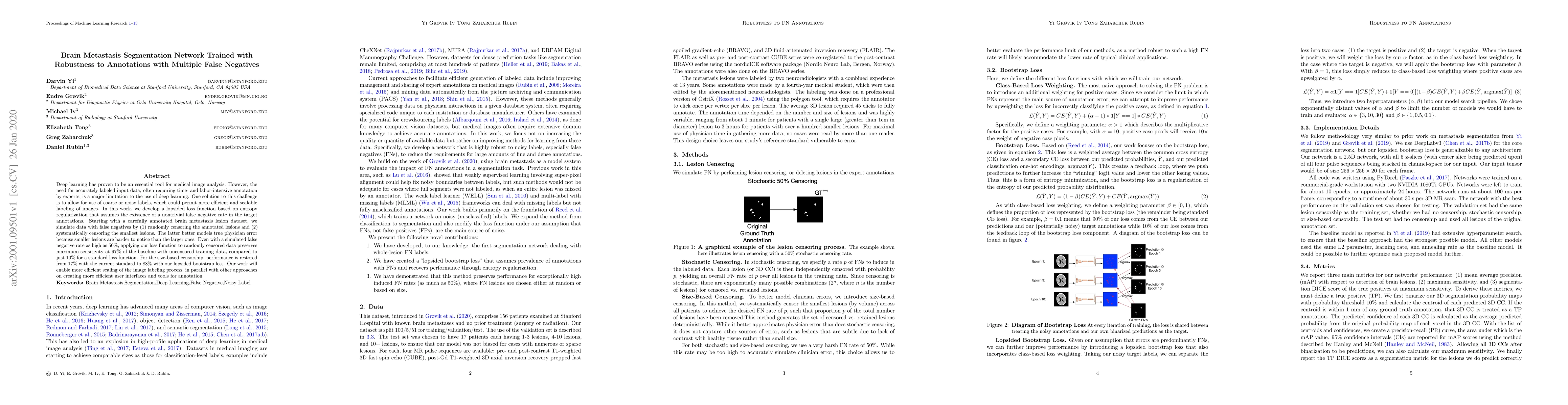
Deep learning has proven to be an essential tool for medical image analysis. However, the need for accurately labeled input data, often requiring time- and labor-intensive annotation by experts, is ...
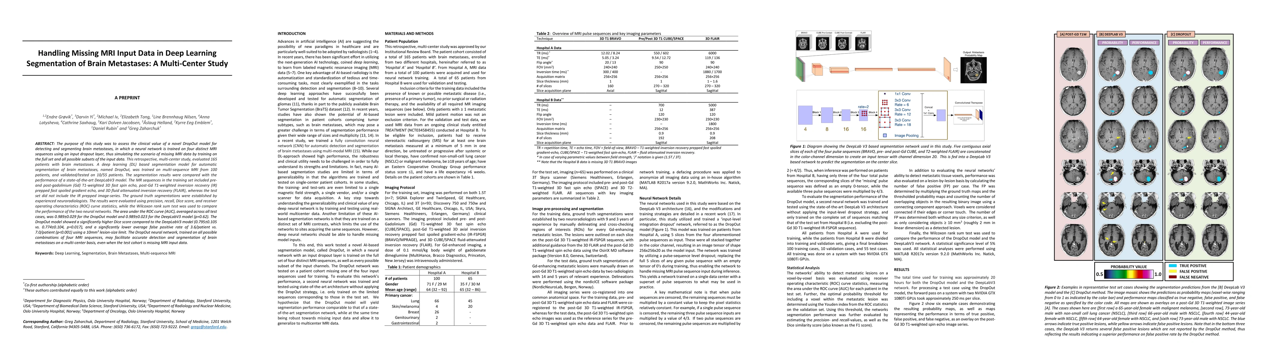
The purpose was to assess the clinical value of a novel DropOut model for detecting and segmenting brain metastases, in which a neural network is trained on four distinct MRI sequences using an inpu...
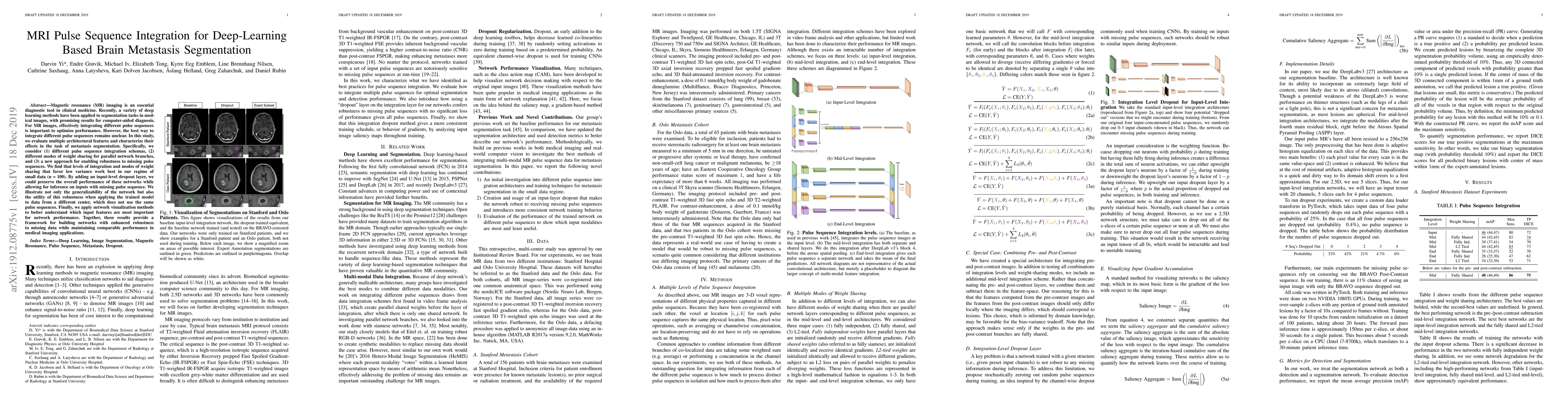
Magnetic resonance (MR) imaging is an essential diagnostic tool in clinical medicine. Recently, a variety of deep learning methods have been applied to segmentation tasks in medical images, with pro...
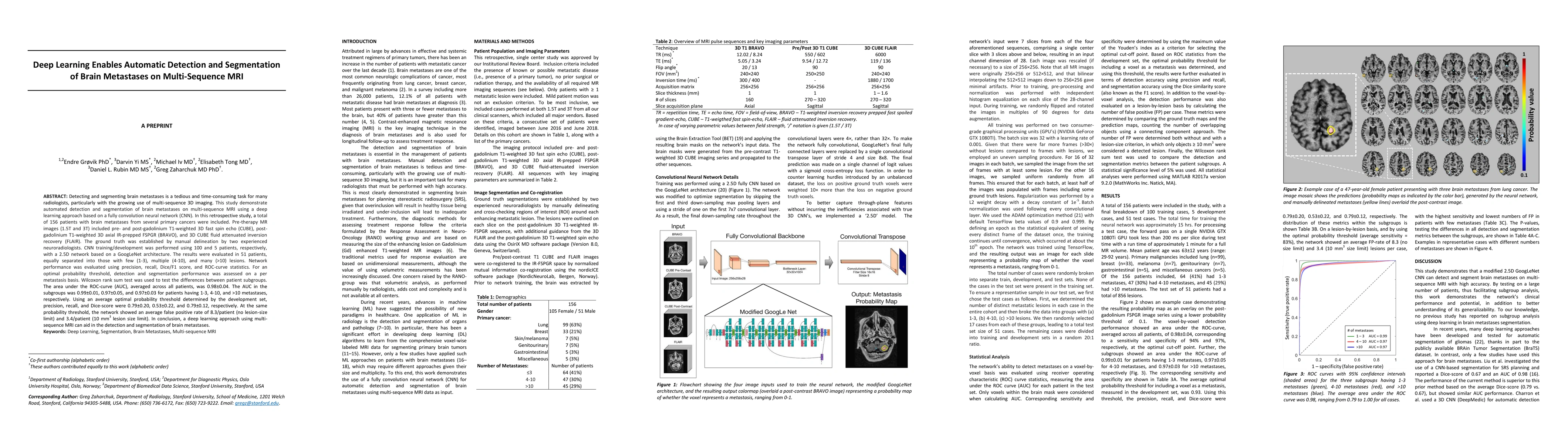
Detecting and segmenting brain metastases is a tedious and time-consuming task for many radiologists, particularly with the growing use of multi-sequence 3D imaging. This study demonstrates automate...
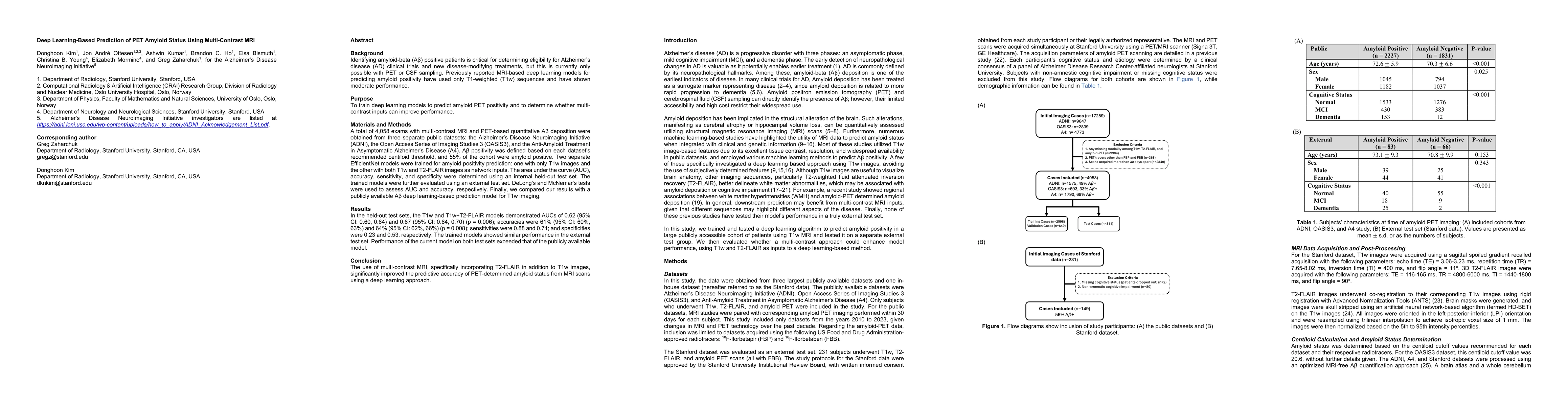
Identifying amyloid-beta positive patients is crucial for determining eligibility for Alzheimer's disease (AD) clinical trials and new disease-modifying treatments, but currently requires PET or CSF s...
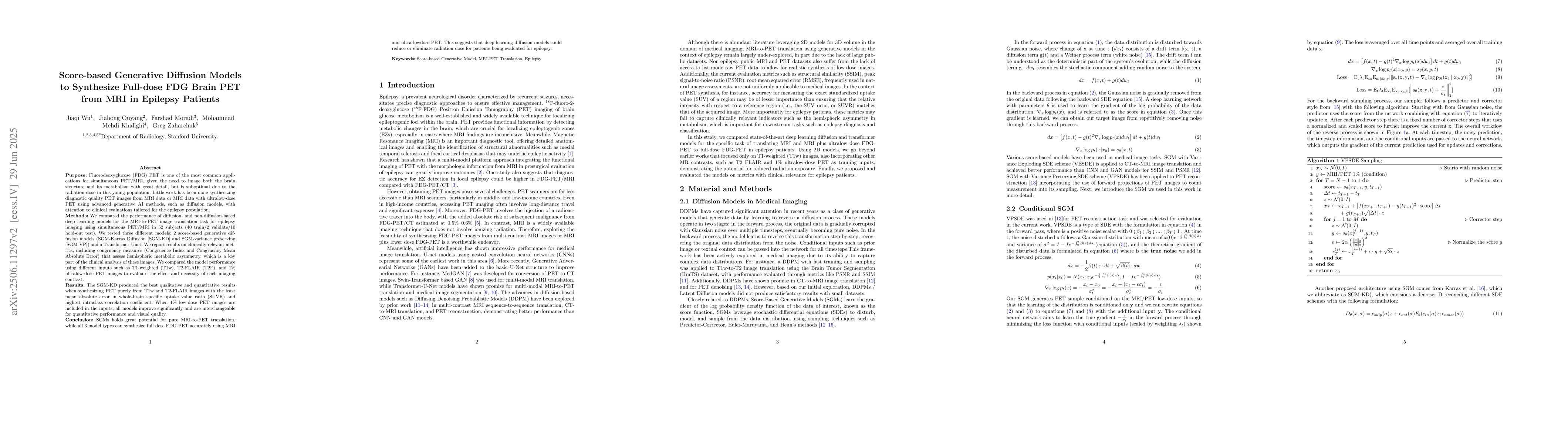
Fluorodeoxyglucose (FDG) PET to evaluate patients with epilepsy is one of the most common applications for simultaneous PET/MRI, given the need to image both brain structure and metabolism, but is sub...
Predicting outcomes in acute ischemic stroke (AIS) guides clinical decision-making, patient counseling, and resource allocation. Clinical notes contain rich contextual information, but their unstructu...
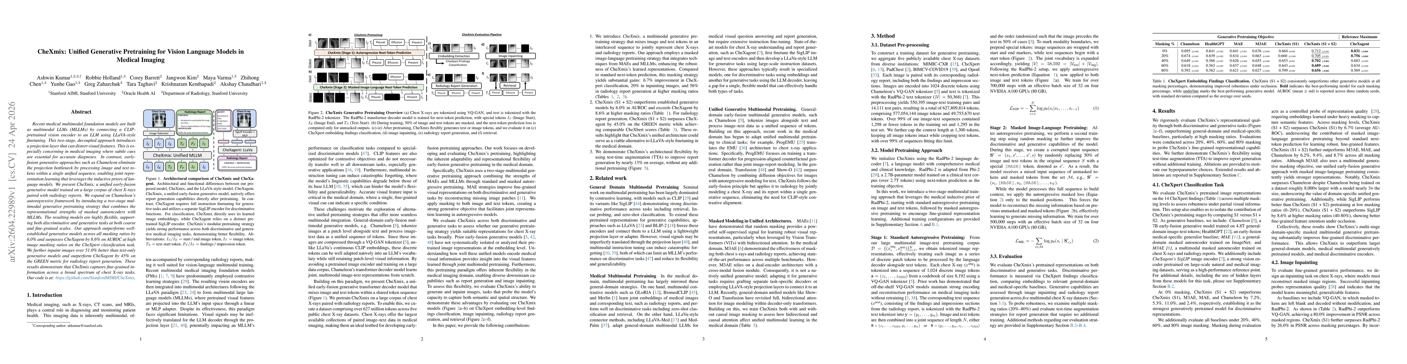
Recent medical multimodal foundation models are built as multimodal LLMs (MLLMs) by connecting a CLIP-pretrained vision encoder to an LLM using LLaVA-style finetuning. This two-stage, decoupled approa...