Academic Profile
Statistics
Similar Authors
Papers on arXiv
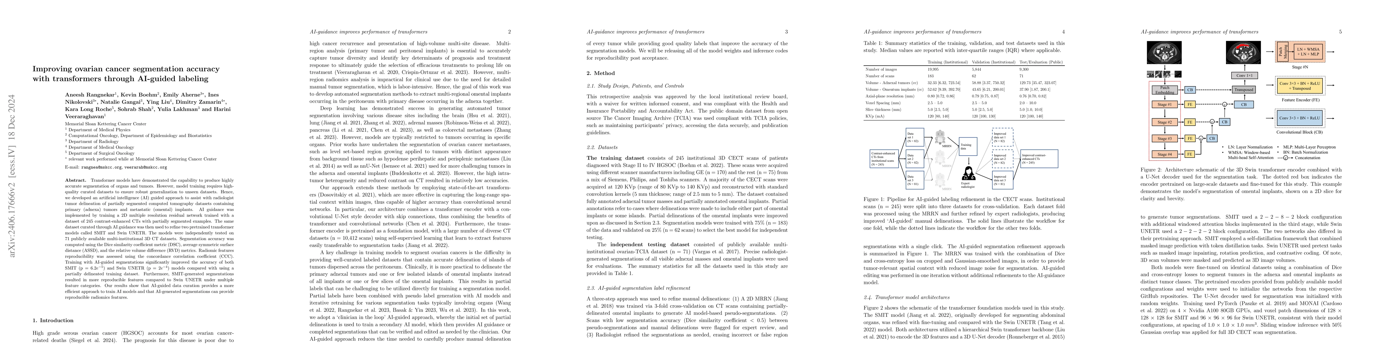
Two self-supervised pretrained transformer-based segmentation models (SMIT and Swin UNETR) fine-tuned on a dataset of ovarian cancer CT images provided reasonably accurate delineations of the tumors...
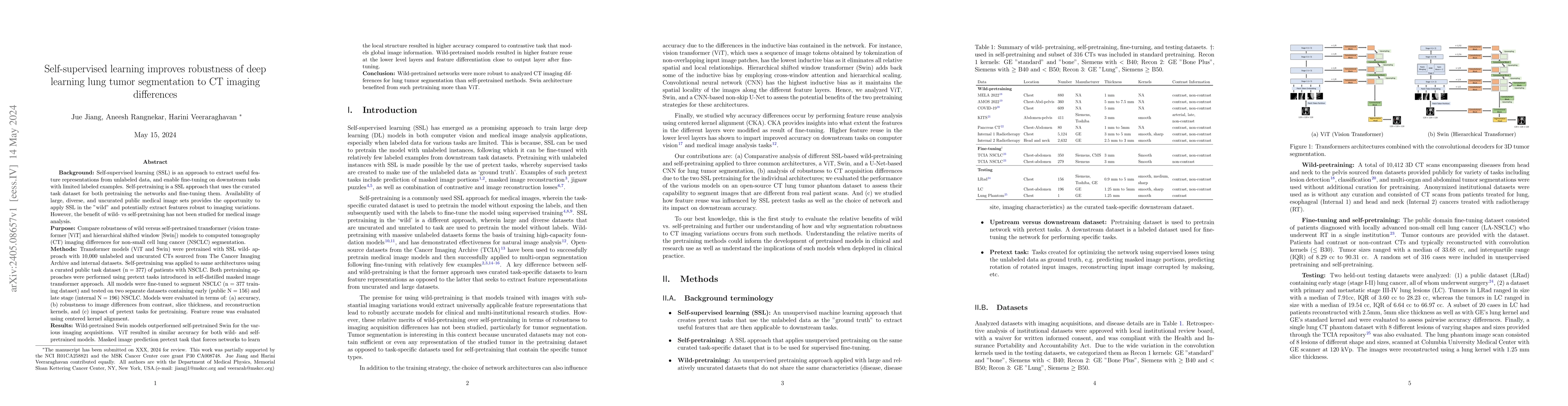
Self-supervised learning (SSL) is an approach to extract useful feature representations from unlabeled data, and enable fine-tuning on downstream tasks with limited labeled examples. Self-pretrainin...
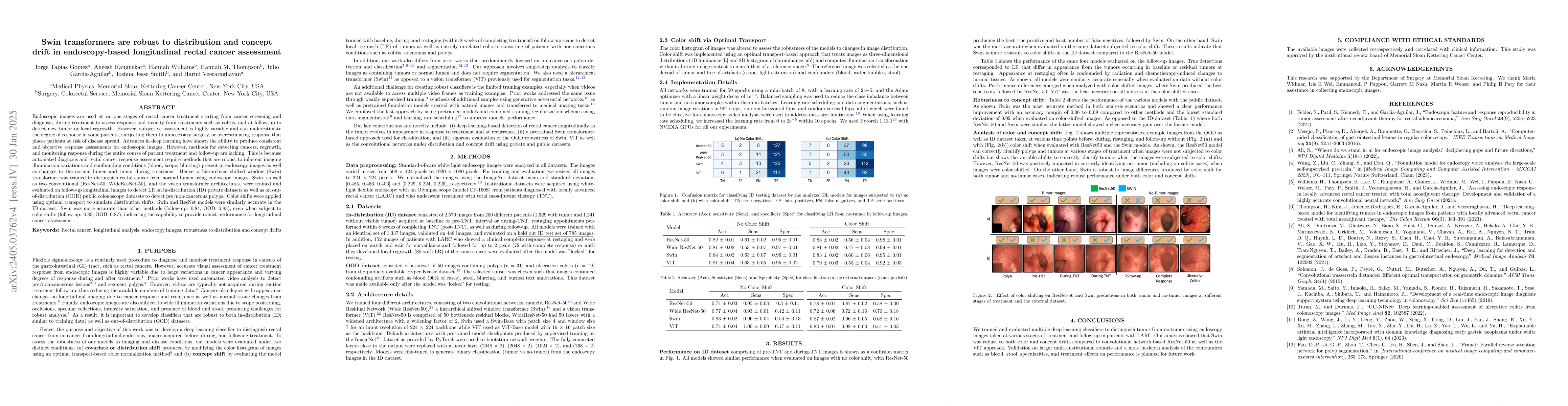
We developed a deep learning classifier of rectal cancer response (tumor vs. no-tumor) to total neoadjuvant treatment (TNT) from endoscopic images acquired before, during, and following TNT. We furt...
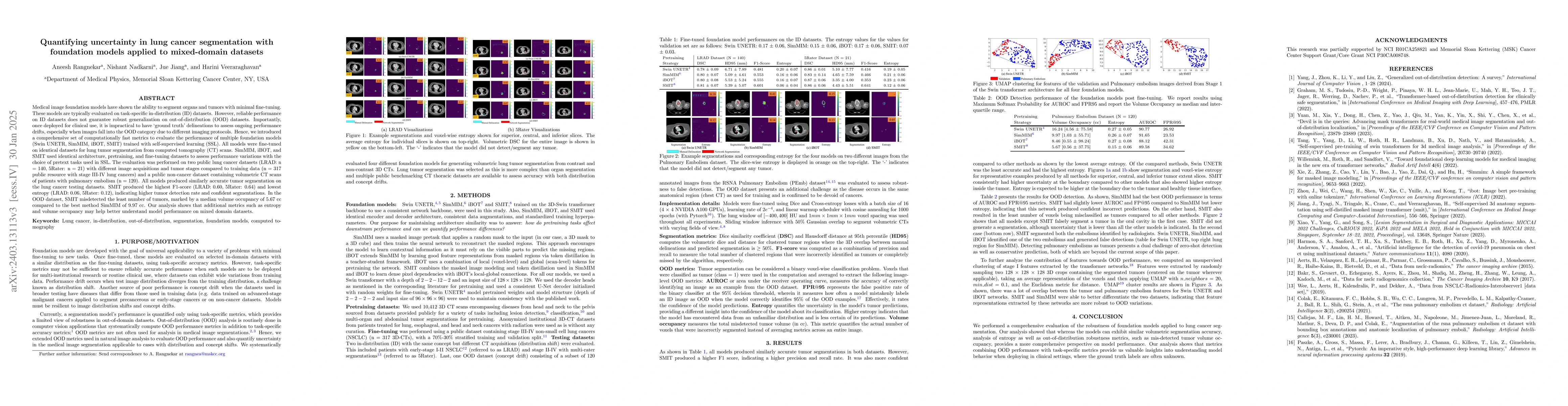
We assessed the trustworthiness of two self-supervision pretrained transformer models, Swin UNETR and SMIT, for fine-tuned lung (LC) tumor segmentation using 670 CT and MRI scans. We measured segmen...
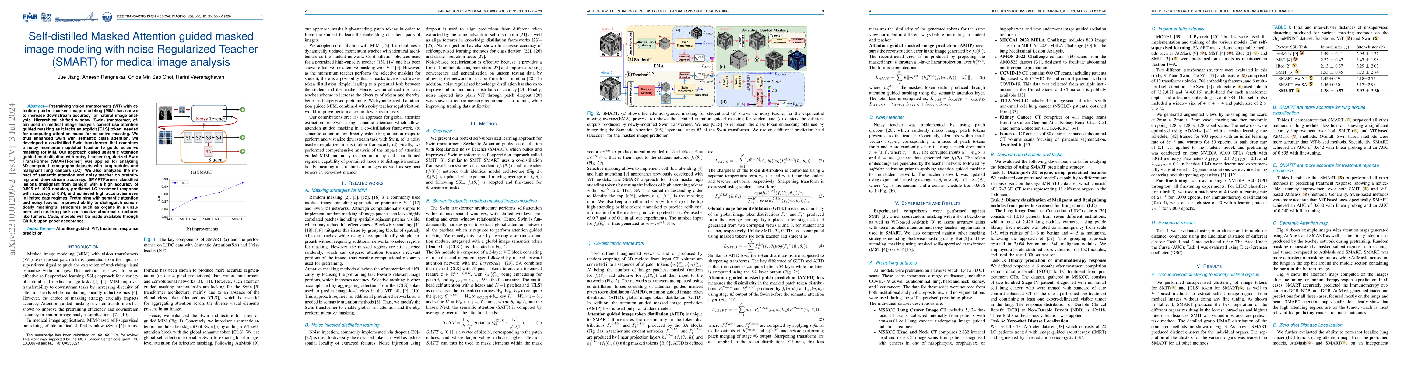
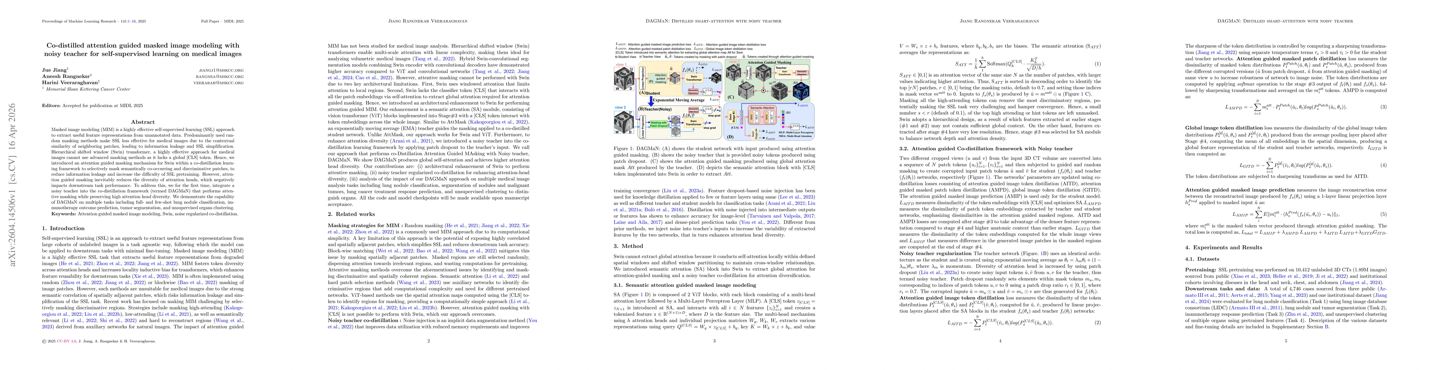
Pretraining vision transformers (ViT) with attention guided masked image modeling (MIM) has shown to increase downstream accuracy for natural image analysis. Hierarchical shifted window (Swin) trans...
Dose escalation radiotherapy allows increased control of prostate cancer (PCa) but requires segmentation of dominant index lesions (DIL), motivating the development of automated methods for fast, ac...
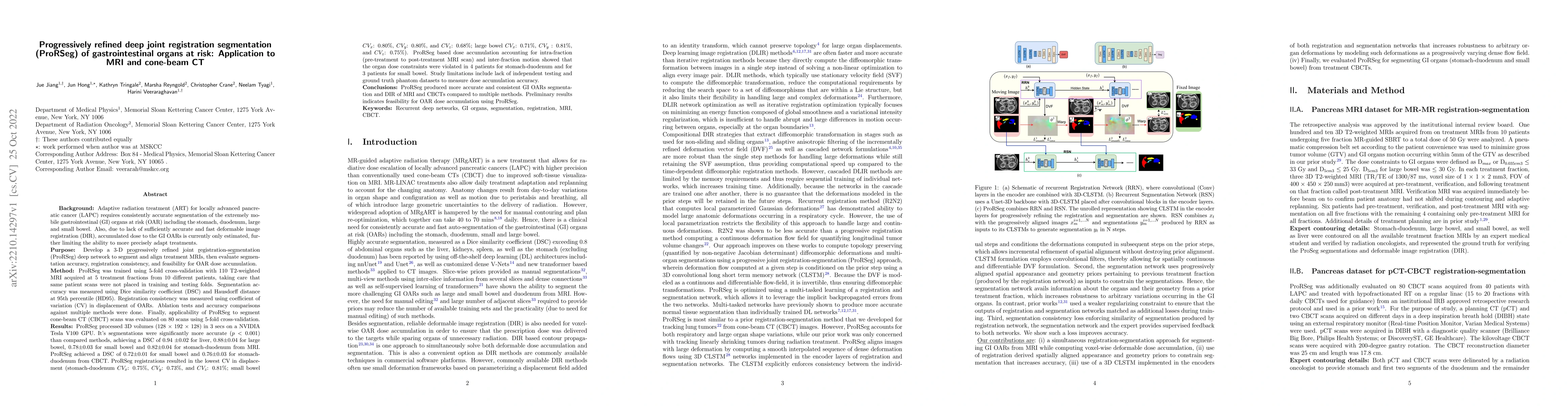
Method: ProRSeg was trained using 5-fold cross-validation with 110 T2-weighted MRI acquired at 5 treatment fractions from 10 different patients, taking care that same patient scans were not placed i...
Vision transformers, with their ability to more efficiently model long-range context, have demonstrated impressive accuracy gains in several computer vision and medical image analysis tasks includin...
The Histogram of Oriented Gradient is a widely used image feature, which describes local image directionality based on numerical differentiation. Due to its ill-posed nature, small noise may lead to...
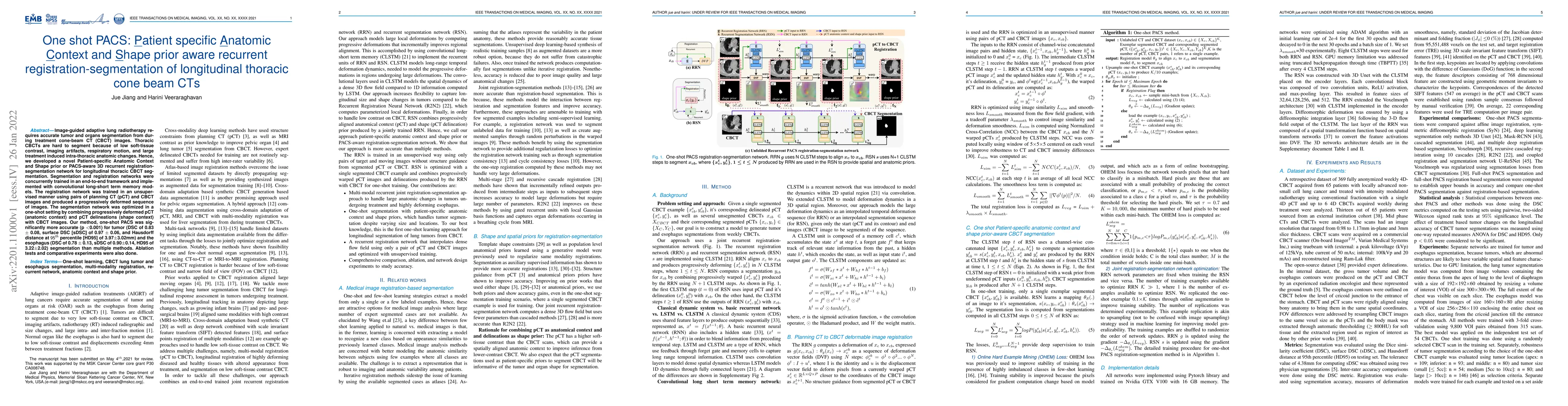
Image-guided adaptive lung radiotherapy requires accurate tumor and organs segmentation from during treatment cone-beam CT (CBCT) images. Thoracic CBCTs are hard to segment because of low soft-tissu...
Accurate and robust segmentation of lung cancers from CT, even those located close to mediastinum, is needed to more accurately plan and deliver radiotherapy and to measure treatment response. There...
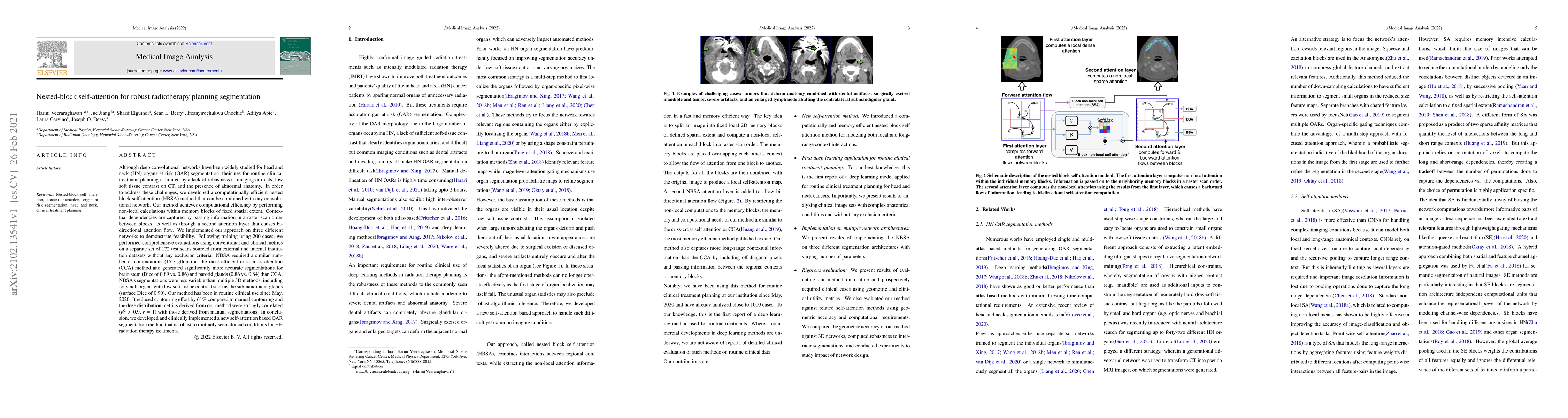
Although deep convolutional networks have been widely studied for head and neck (HN) organs at risk (OAR) segmentation, their use for routine clinical treatment planning is limited by a lack of robu...
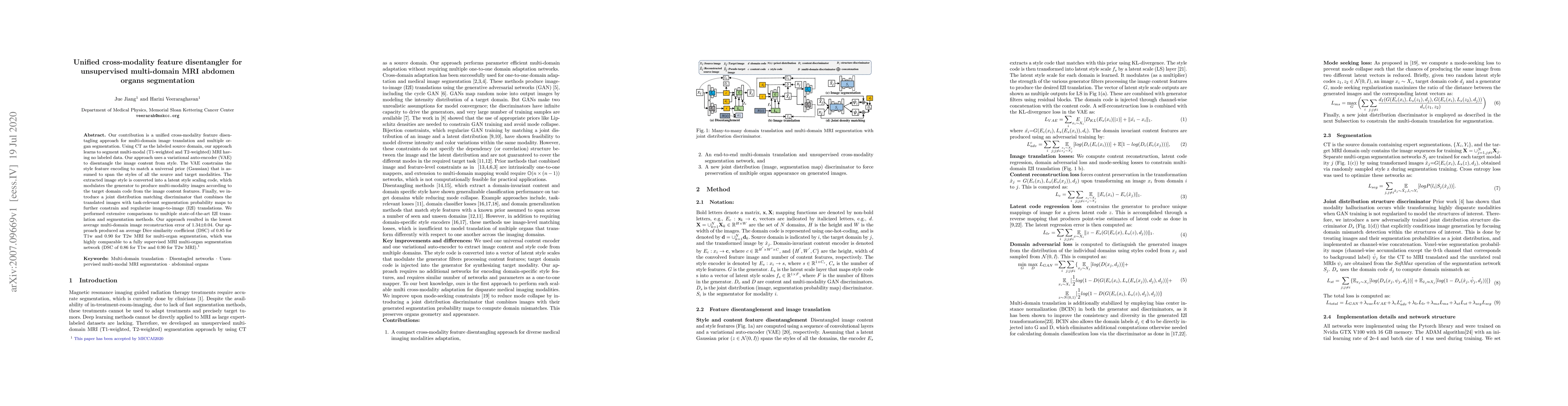
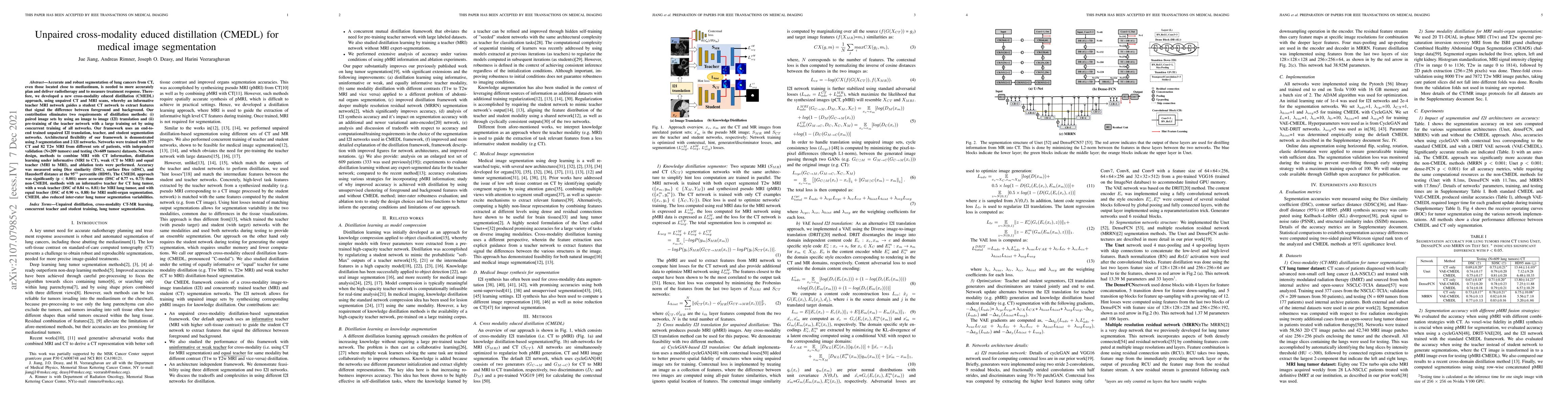
Our contribution is a unified cross-modality feature disentagling approach for multi-domain image translation and multiple organ segmentation. Using CT as the labeled source domain, our approach lea...
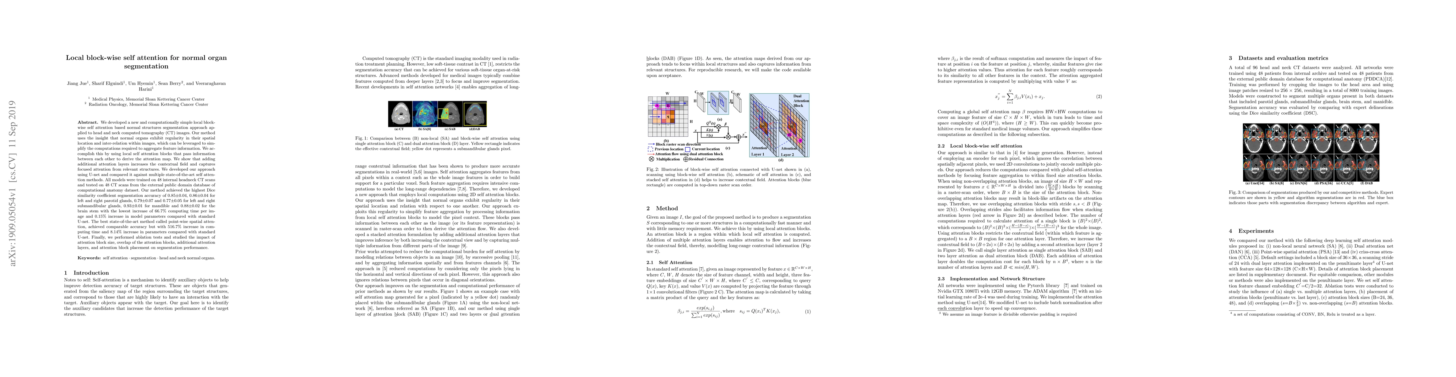
We developed a new and computationally simple local block-wise self attention based normal structures segmentation approach applied to head and neck computed tomography (CT) images. Our method uses ...
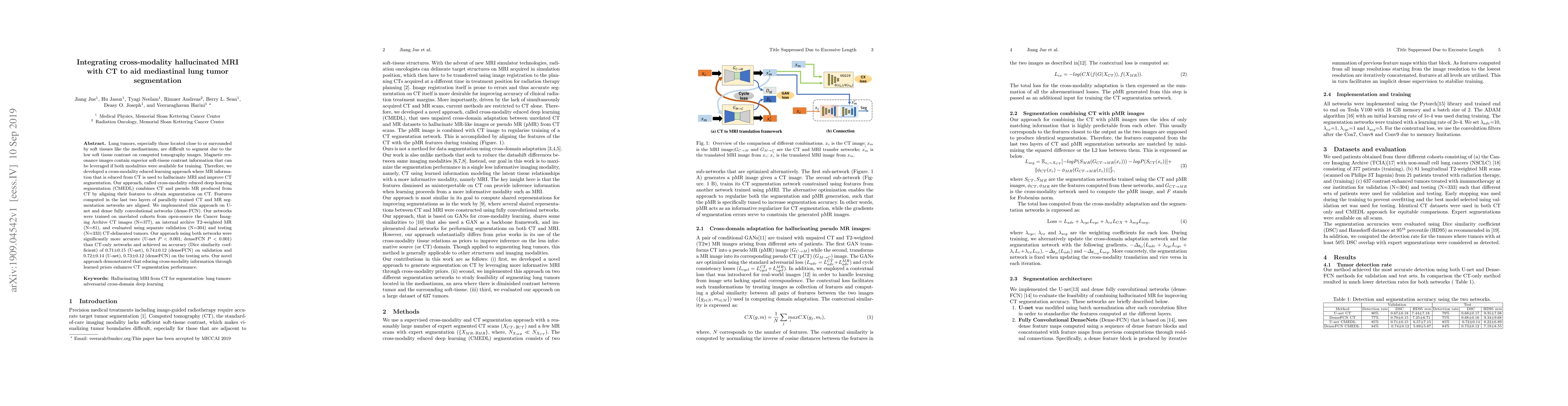
Lung tumors, especially those located close to or surrounded by soft tissues like the mediastinum, are difficult to segment due to the low soft tissue contrast on computed tomography images. Magneti...
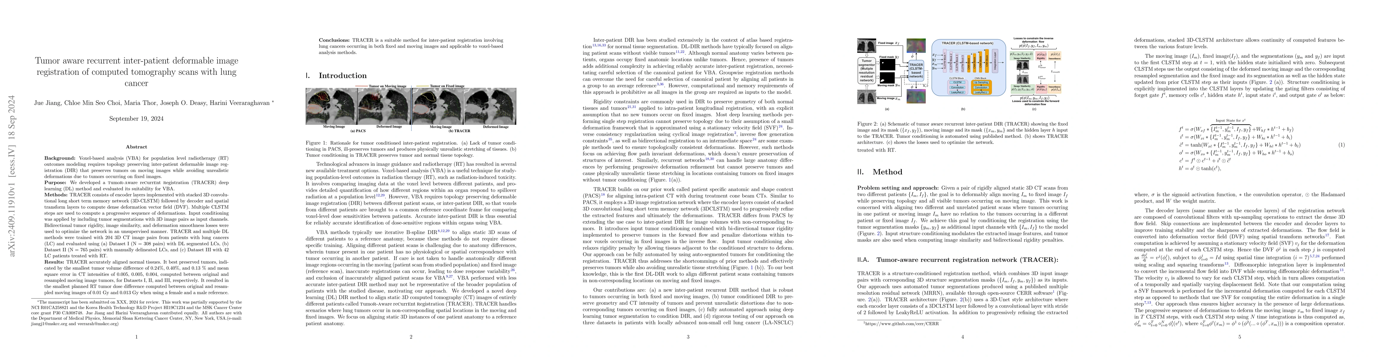
Background: Voxel-based analysis (VBA) for population level radiotherapy (RT) outcomes modeling requires topology preserving inter-patient deformable image registration (DIR) that preserves tumors on ...
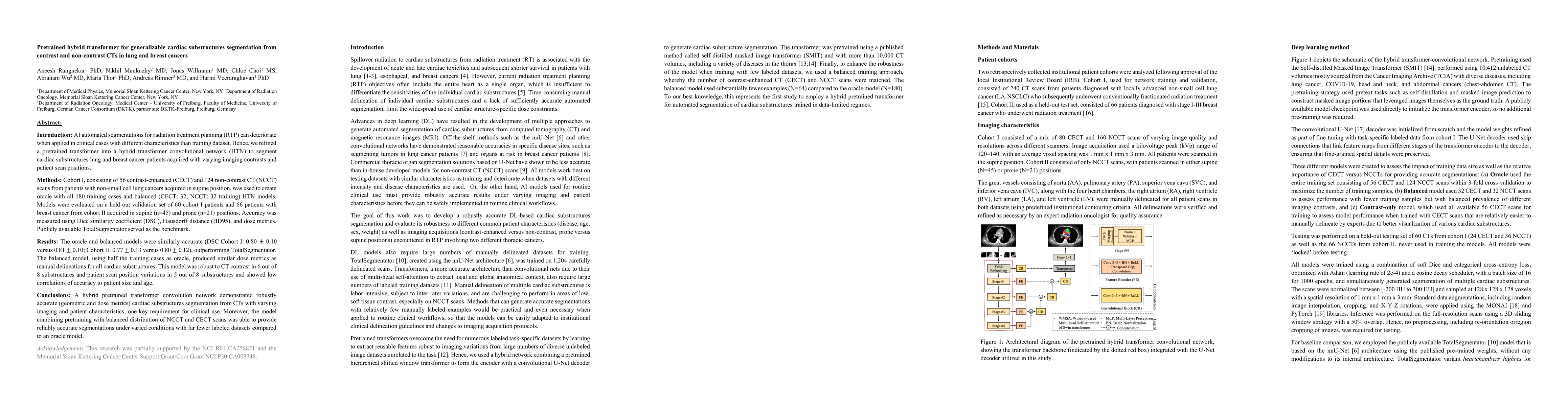
AI automated segmentations for radiation treatment planning (RTP) can deteriorate when applied in clinical cases with different characteristics than training dataset. Hence, we refined a pretrained tr...
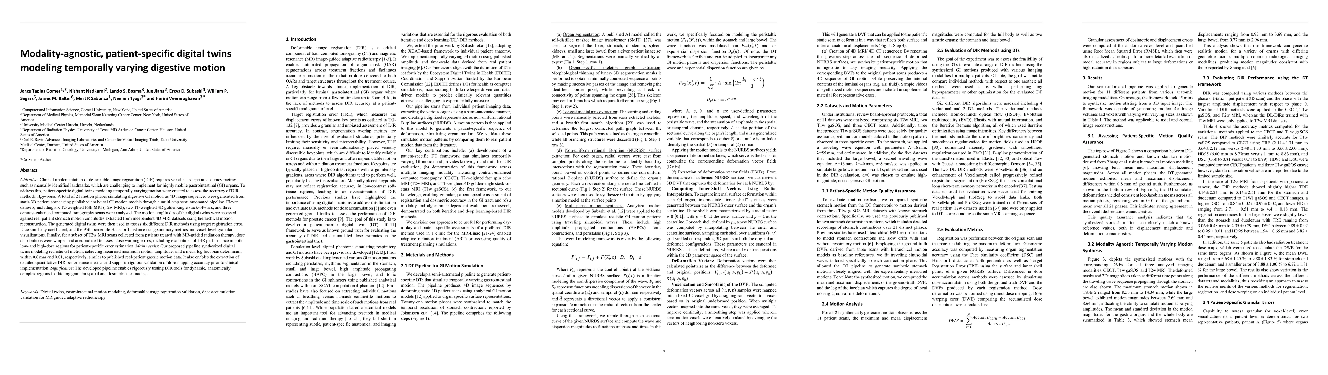
Objective: Clinical implementation of deformable image registration (DIR) requires voxel-based spatial accuracy metrics such as manually identified landmarks, which are challenging to implement for hi...
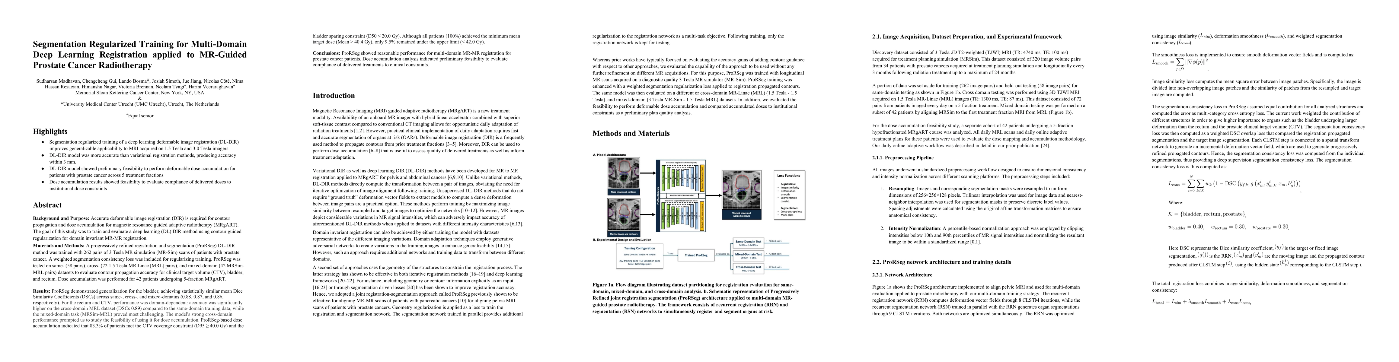
Background: Accurate deformable image registration (DIR) is required for contour propagation and dose accumulation in MR-guided adaptive radiotherapy (MRgART). This study trained and evaluated a deep ...
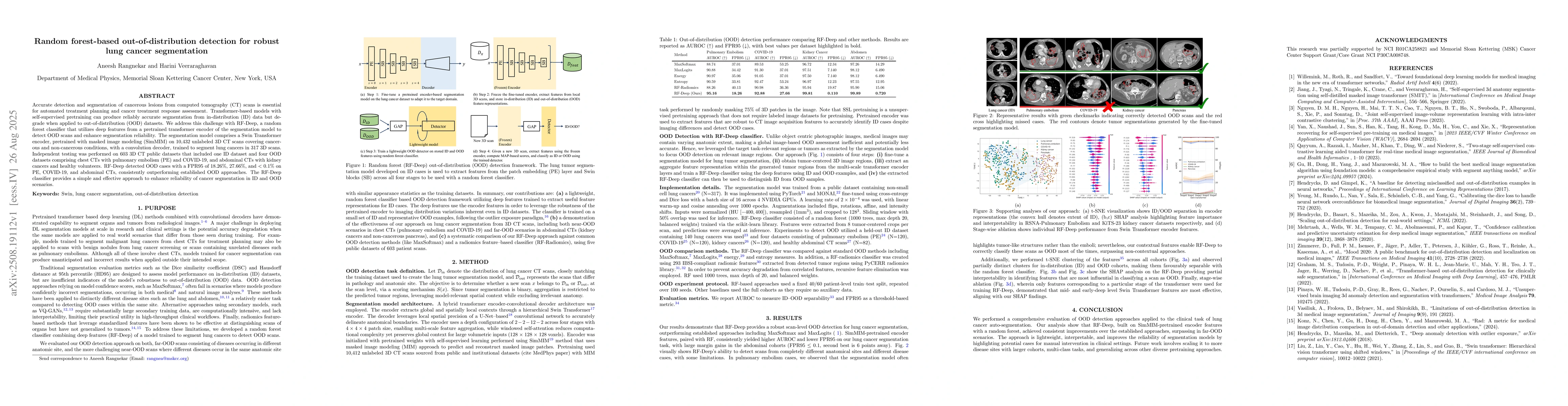
Accurate detection and segmentation of cancerous lesions from computed tomography (CT) scans is essential for automated treatment planning and cancer treatment response assessment. Transformer-based m...
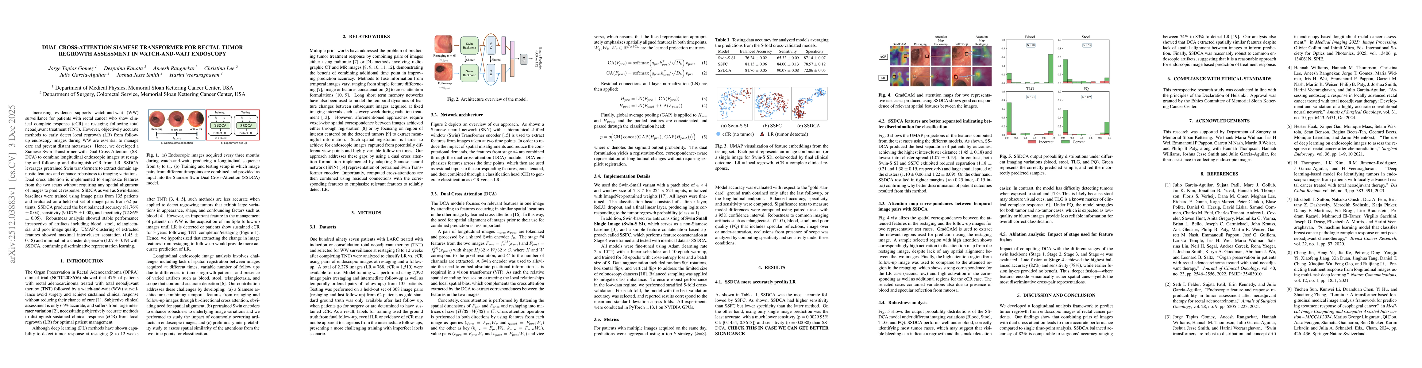
Increasing evidence supports watch-and-wait (WW) surveillance for patients with rectal cancer who show clinical complete response (cCR) at restaging following total neoadjuvant treatment (TNT). Howeve...
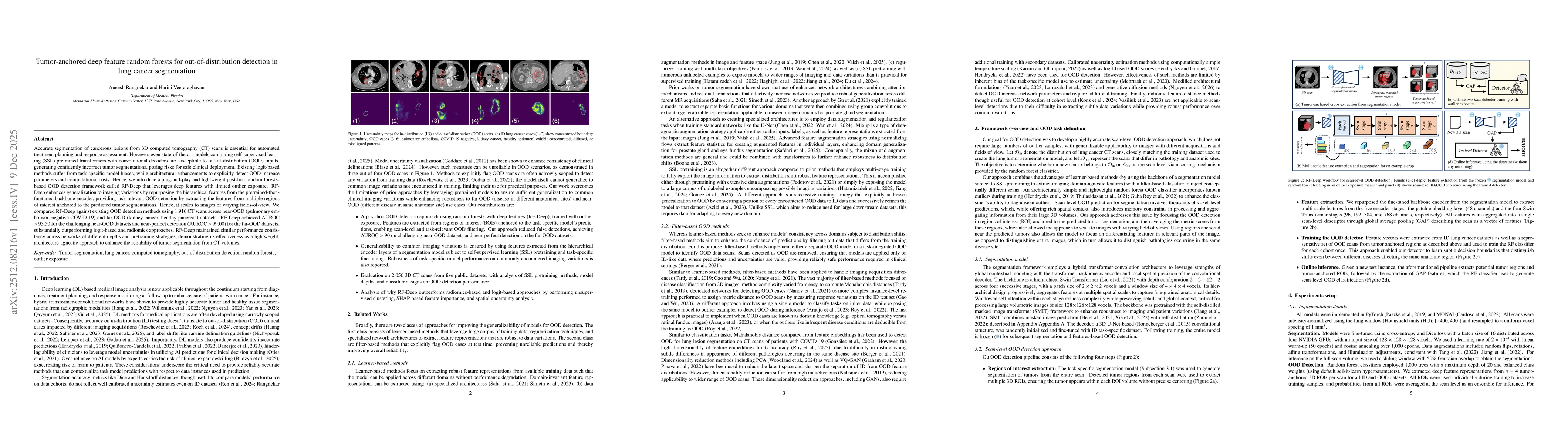
Accurate segmentation of cancerous lesions from 3D computed tomography (CT) scans is essential for automated treatment planning and response assessment. However, even state-of-the-art models combining...
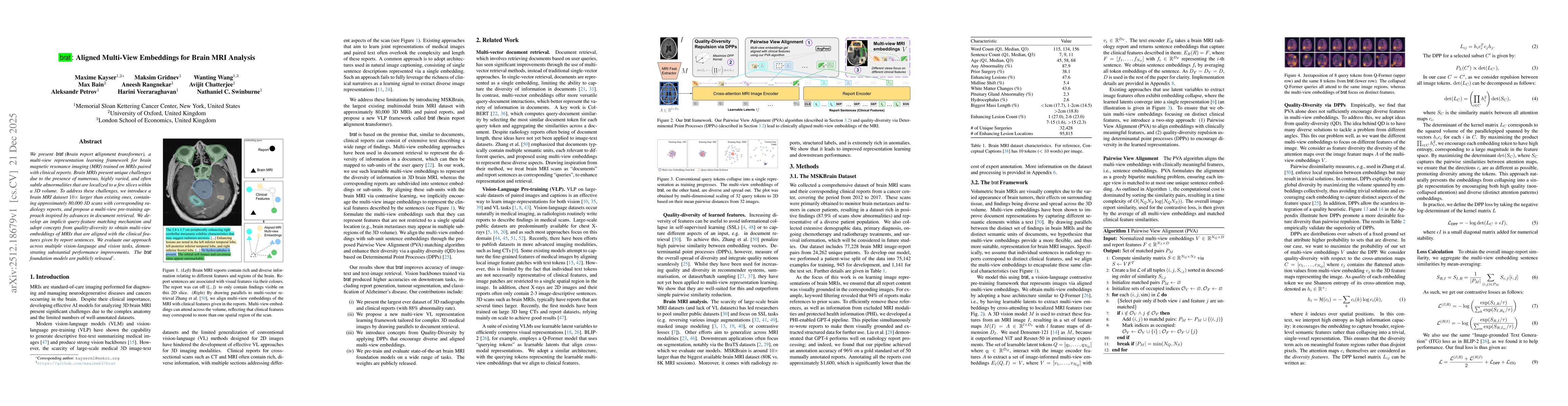
We present brat (brain report alignment transformer), a multi-view representation learning framework for brain magnetic resonance imaging (MRI) trained on MRIs paired with clinical reports. Brain MRIs...
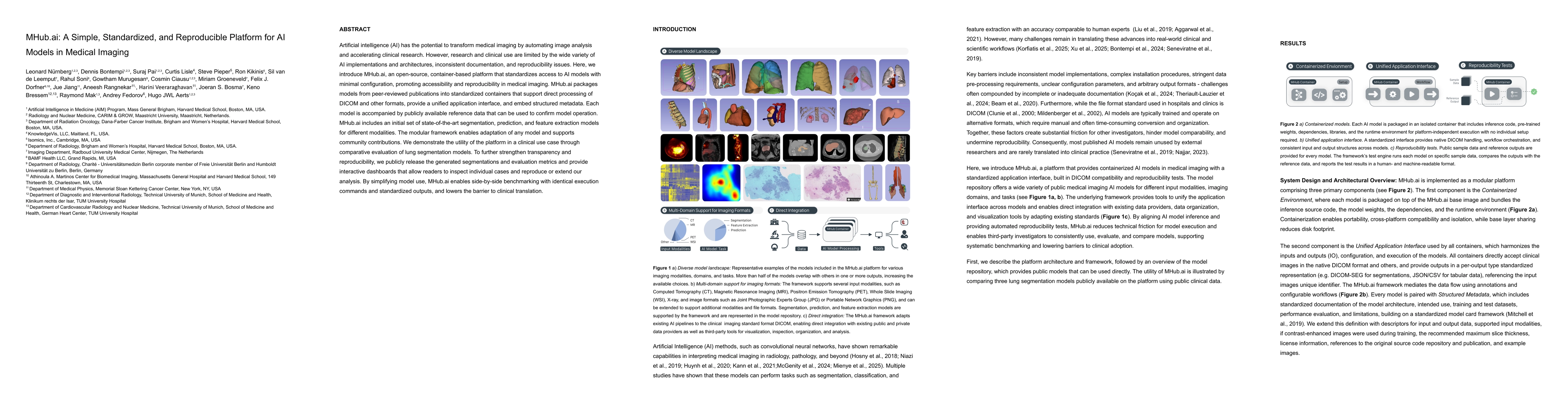
Artificial intelligence (AI) has the potential to transform medical imaging by automating image analysis and accelerating clinical research. However, research and clinical use are limited by the wide ...
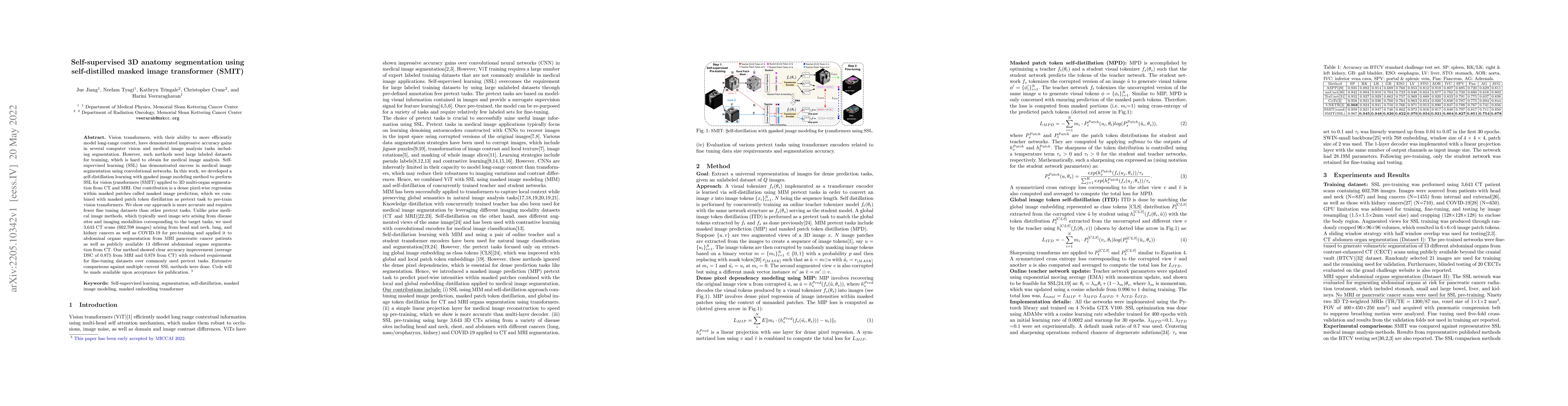
Masked image modeling (MIM) is a highly effective self-supervised learning (SSL) approach to extract useful feature representations from unannotated data. Predominantly used random masking methods mak...
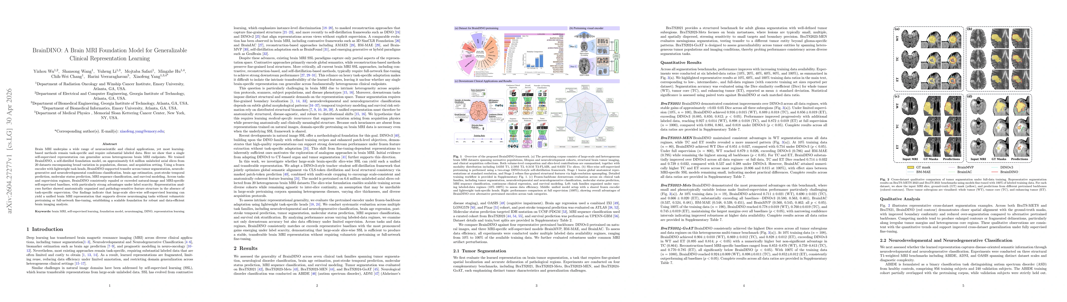
Brain MRI underpins a wide range of neuroscientific and clinical applications, yet most learning-based methods remain task-specific and require substantial labeled data. Here we show that a single sel...
Pretraining on large-scale datasets has been shown to improve transformer generalizability, even for out-of-domain (OOD) modalities and tasks. However, two common assumptions often fail under OOD tran...

Clinical trial studies indicate benefit of watch-and-wait (WW) surveillance for patients with rectal cancer showing a complete or near clinical response (CR) directly after treatment (restaging). Howe...
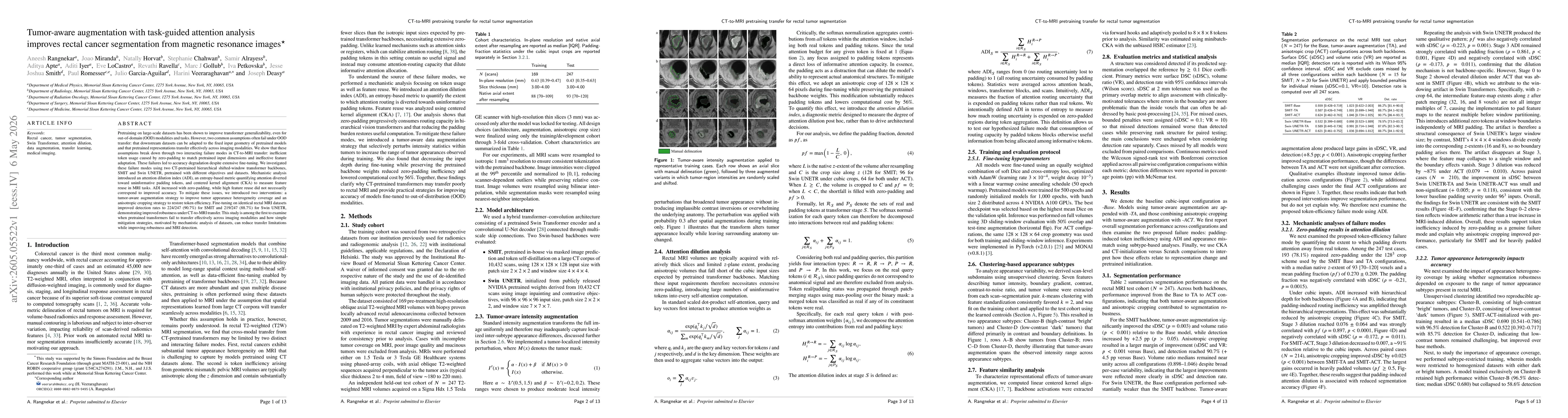
Methods: Nine SSL methods spanning four pretext-task families were pretrained from scratch using the same 10{,}412 3D CT scans (1.89~M 2D axial slices) covering varied disease sites. The pretrained Sw...
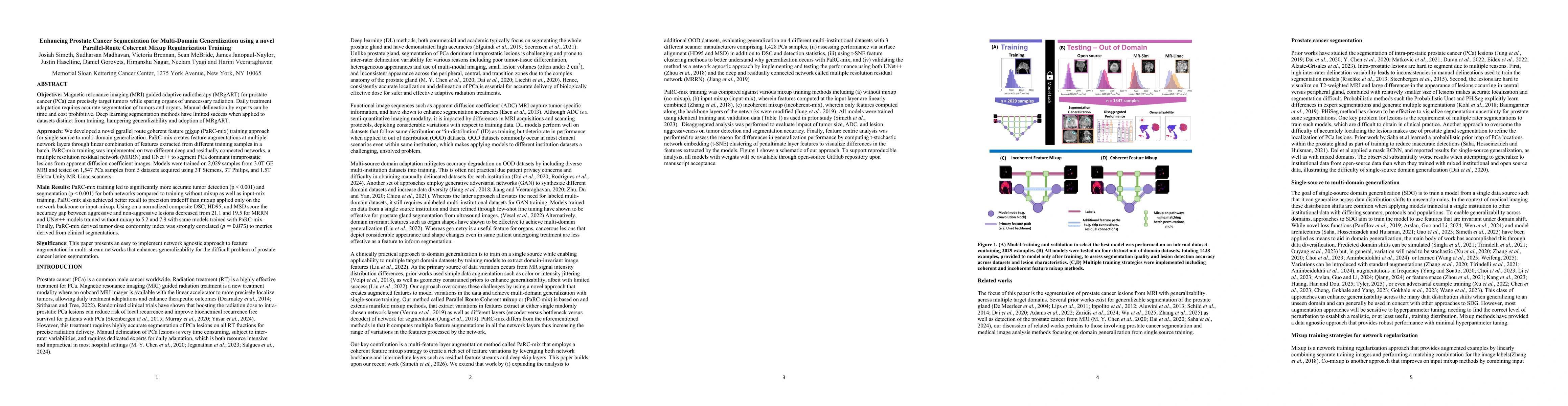
MRI guided adaptive radiotherapy (MRgART) for prostate cancer (PCa) targets tumors while sparing organs from unnecessary radiation. Daily treatment adaptation requires accurate segmentation of tumors ...