Academic Profile
Statistics
Similar Authors
Papers on arXiv
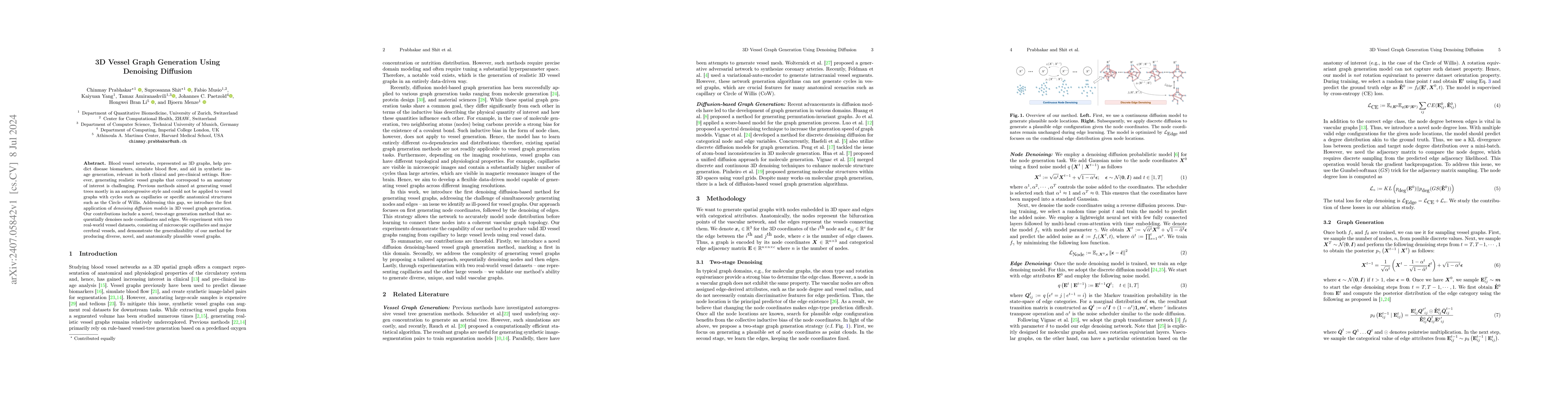
Blood vessel networks, represented as 3D graphs, help predict disease biomarkers, simulate blood flow, and aid in synthetic image generation, relevant in both clinical and pre-clinical settings. Howev...
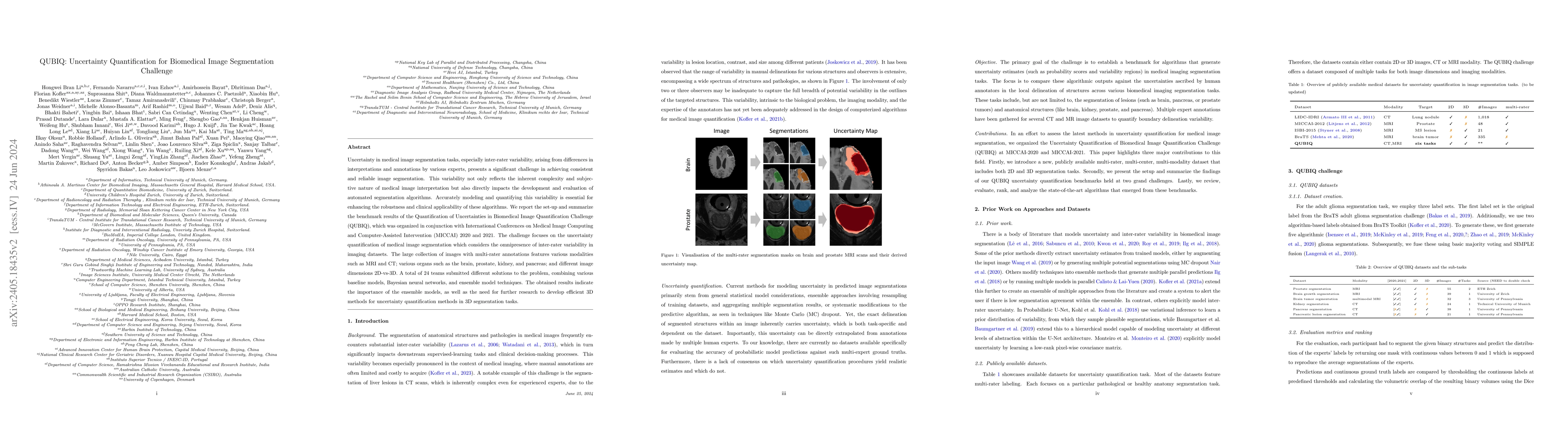
Uncertainty in medical image segmentation tasks, especially inter-rater variability, arising from differences in interpretations and annotations by various experts, presents a significant challenge in...
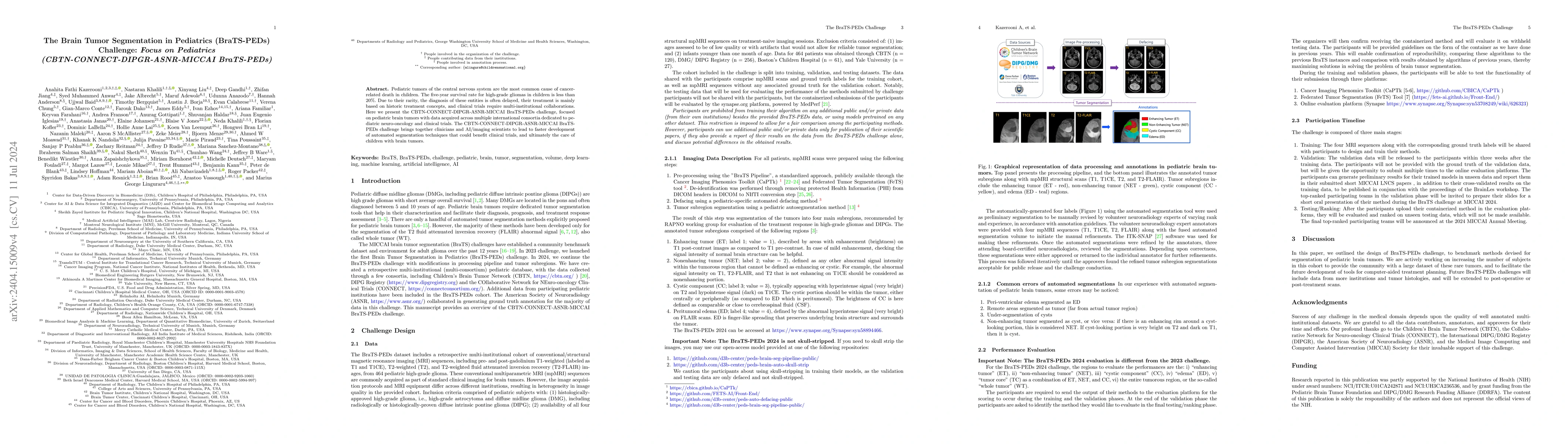
Pediatric tumors of the central nervous system are the most common cause of cancer-related death in children. The five-year survival rate for high-grade gliomas in children is less than 20%. Due to th...
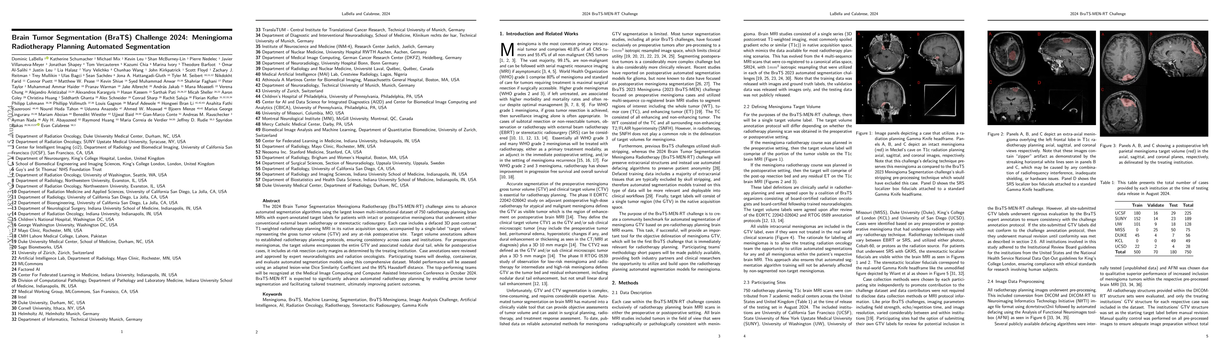
The 2024 Brain Tumor Segmentation Meningioma Radiotherapy (BraTS-MEN-RT) challenge aims to advance automated segmentation algorithms using the largest known multi-institutional dataset of radiothera...
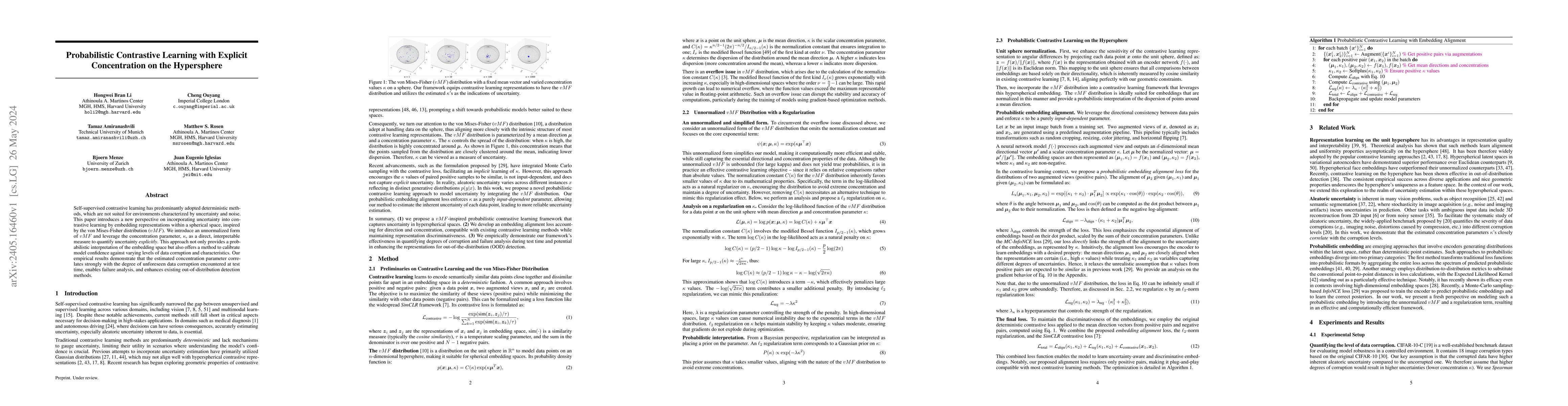
Self-supervised contrastive learning has predominantly adopted deterministic methods, which are not suited for environments characterized by uncertainty and noise. This paper introduces a new perspe...
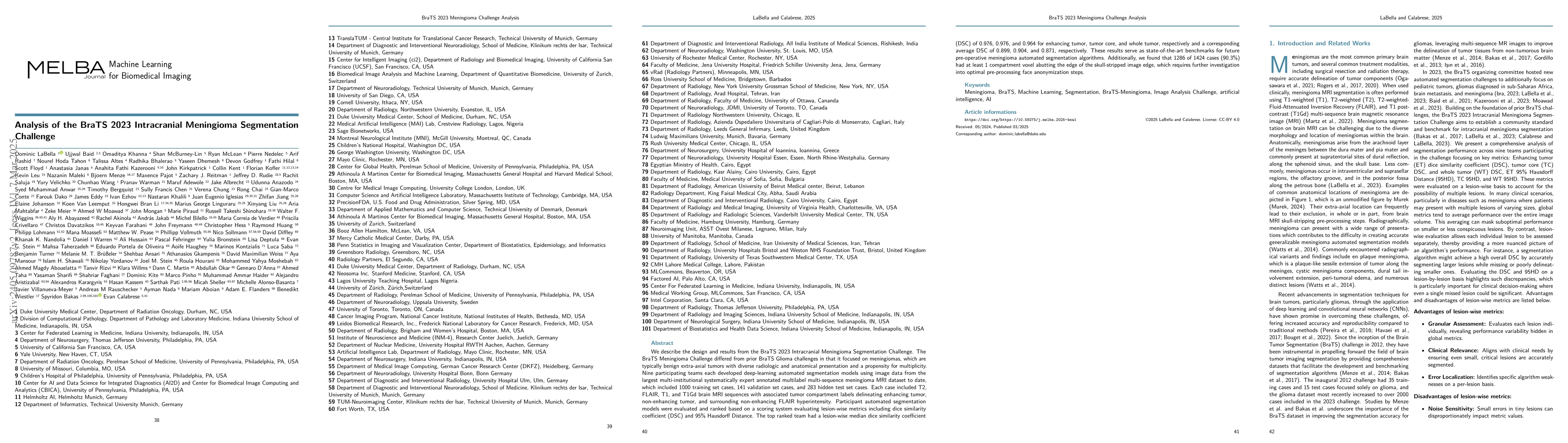
We describe the design and results from the BraTS 2023 Intracranial Meningioma Segmentation Challenge. The BraTS Meningioma Challenge differed from prior BraTS Glioma challenges in that it focused o...
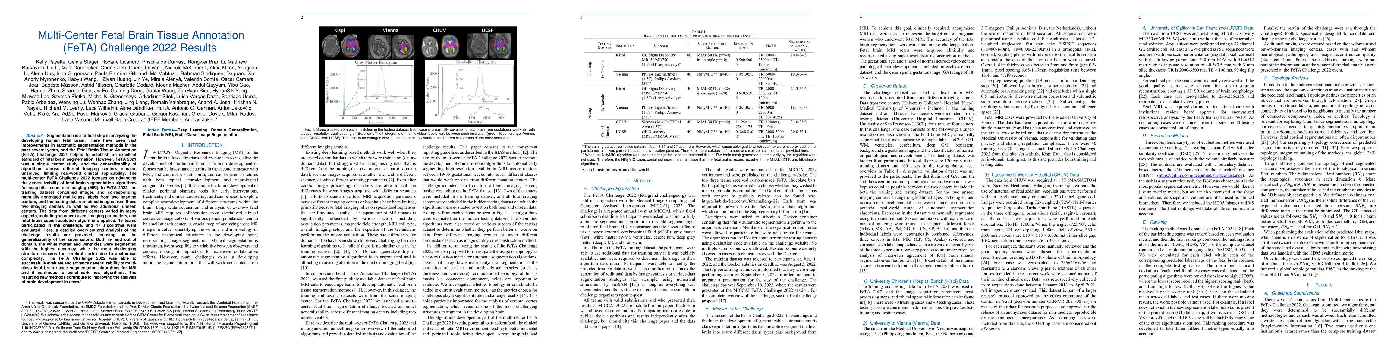
Segmentation is a critical step in analyzing the developing human fetal brain. There have been vast improvements in automatic segmentation methods in the past several years, and the Fetal Brain Tiss...
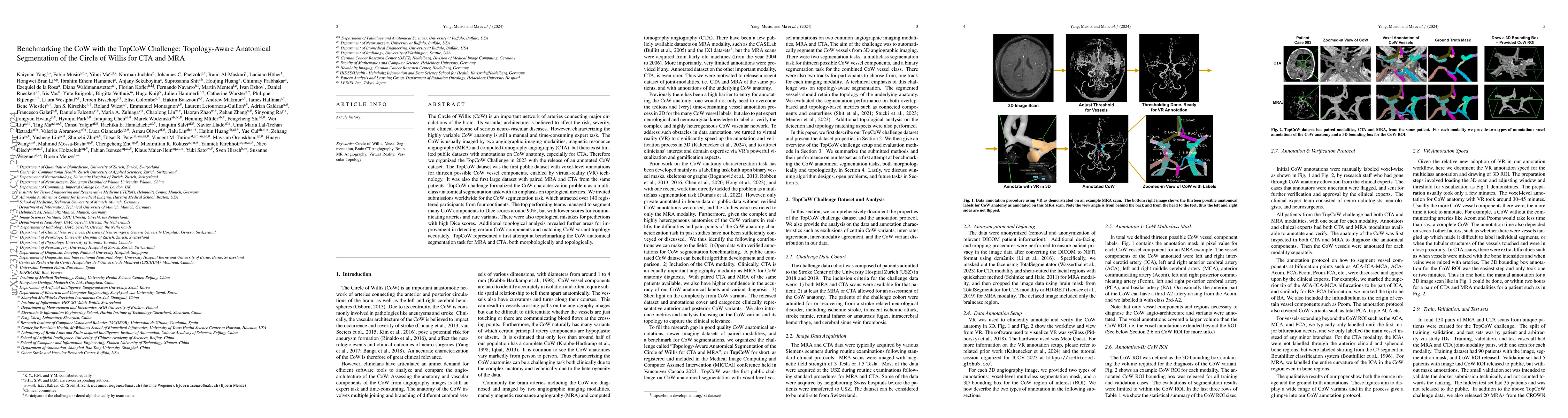
The Circle of Willis (CoW) is an important network of arteries connecting major circulations of the brain. Its vascular architecture is believed to affect the risk, severity, and clinical outcome of...
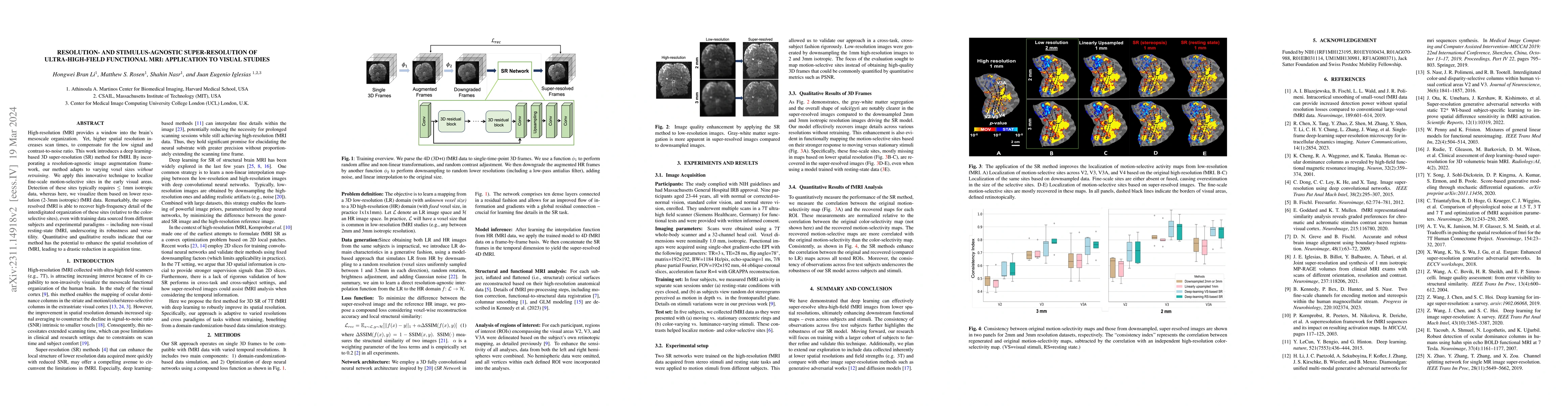
High-resolution fMRI provides a window into the brain's mesoscale organization. Yet, higher spatial resolution increases scan times, to compensate for the low signal and contrast-to-noise ratio. Thi...
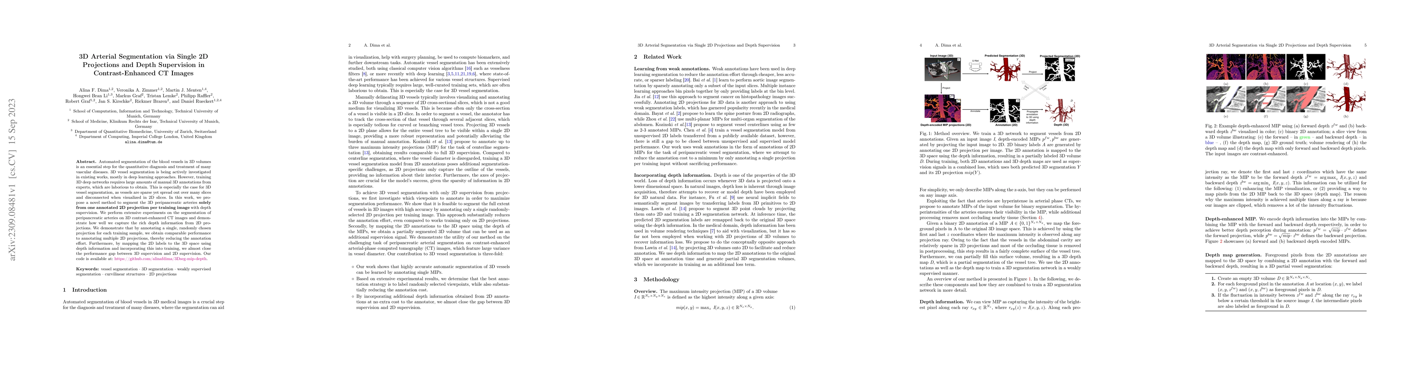
Automated segmentation of the blood vessels in 3D volumes is an essential step for the quantitative diagnosis and treatment of many vascular diseases. 3D vessel segmentation is being actively invest...
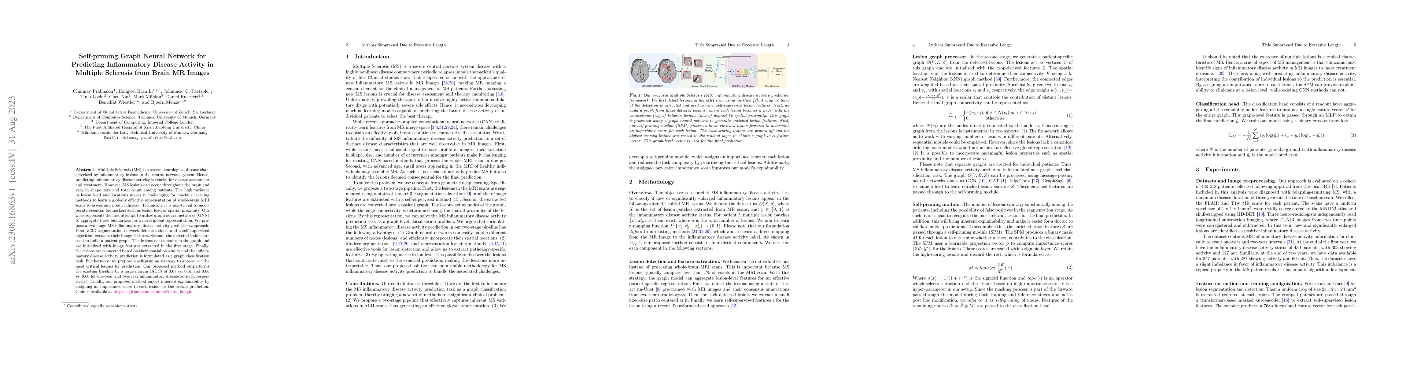
Multiple Sclerosis (MS) is a severe neurological disease characterized by inflammatory lesions in the central nervous system. Hence, predicting inflammatory disease activity is crucial for disease a...
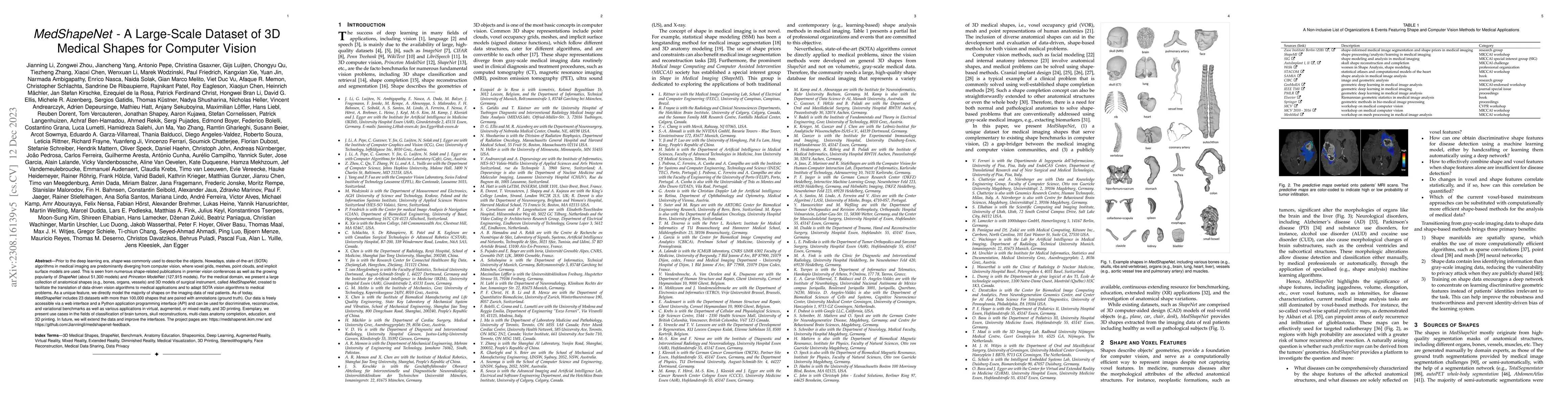
Prior to the deep learning era, shape was commonly used to describe the objects. Nowadays, state-of-the-art (SOTA) algorithms in medical imaging are predominantly diverging from computer vision, whe...
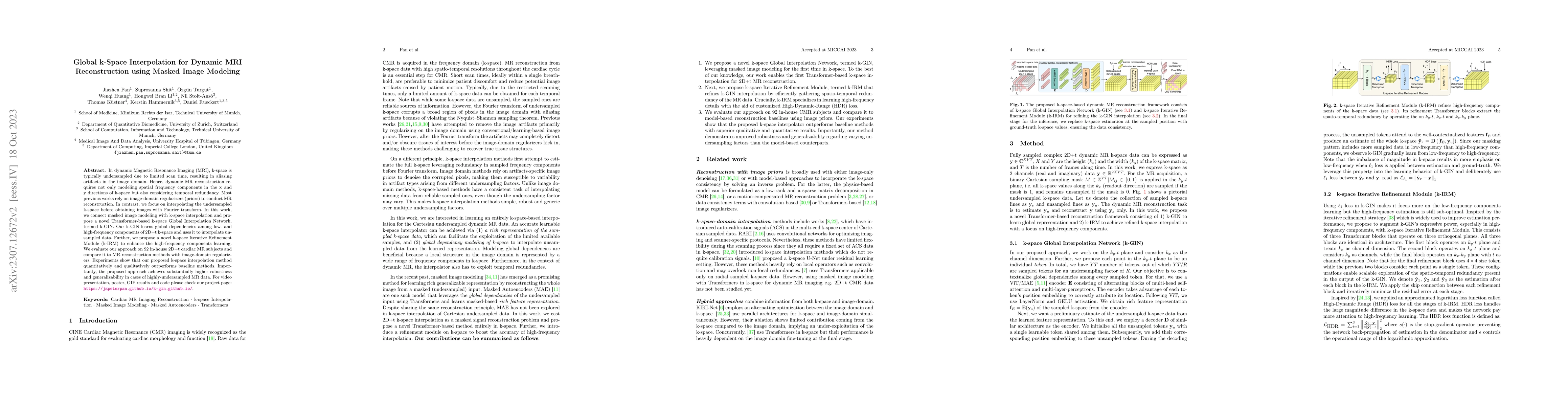
In dynamic Magnetic Resonance Imaging (MRI), k-space is typically undersampled due to limited scan time, resulting in aliasing artifacts in the image domain. Hence, dynamic MR reconstruction require...
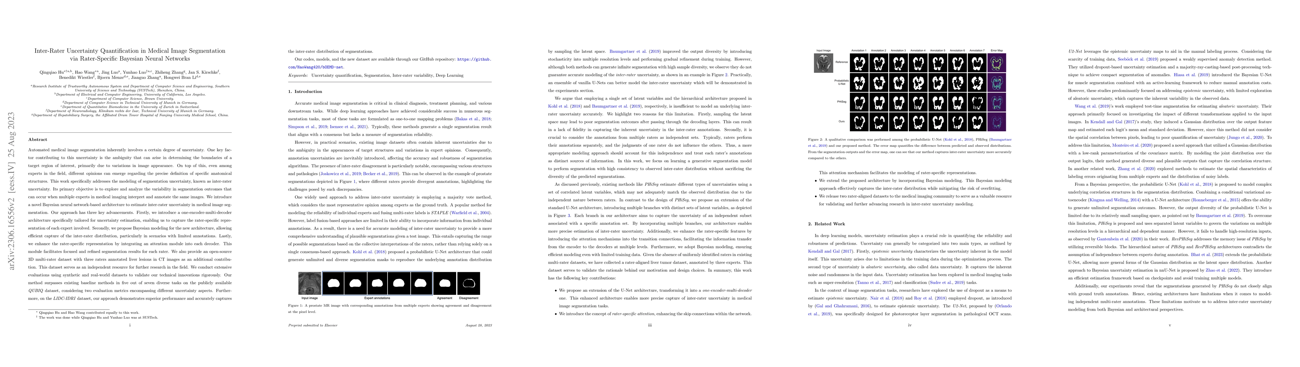
Automated medical image segmentation inherently involves a certain degree of uncertainty. One key factor contributing to this uncertainty is the ambiguity that can arise in determining the boundarie...
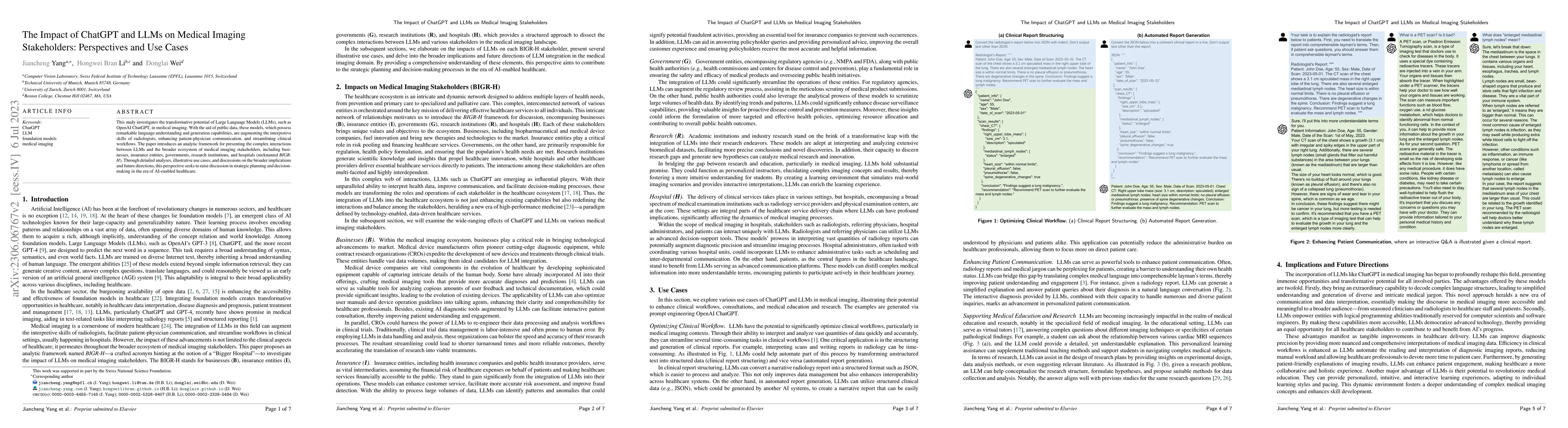
This study investigates the transformative potential of Large Language Models (LLMs), such as OpenAI ChatGPT, in medical imaging. With the aid of public data, these models, which possess remarkable ...
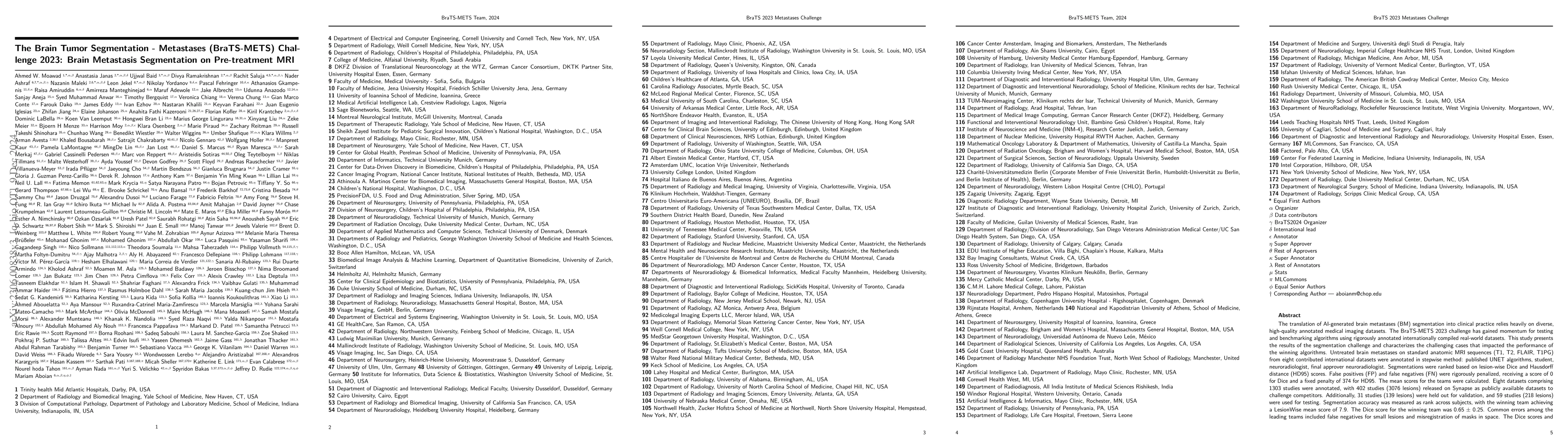
The translation of AI-generated brain metastases (BM) segmentation into clinical practice relies heavily on diverse, high-quality annotated medical imaging datasets. The BraTS-METS 2023 challenge ha...
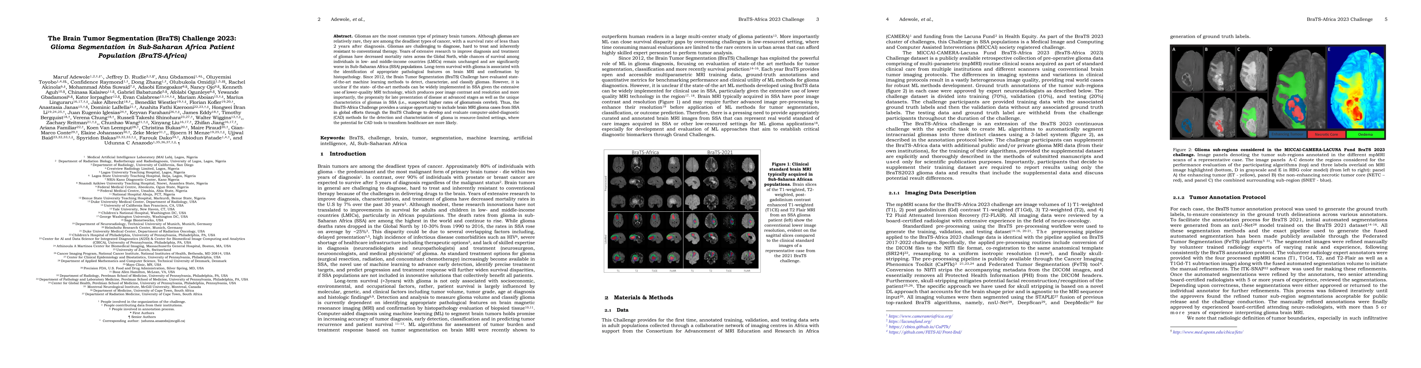
Gliomas are the most common type of primary brain tumors. Although gliomas are relatively rare, they are among the deadliest types of cancer, with a survival rate of less than 2 years after diagnosi...
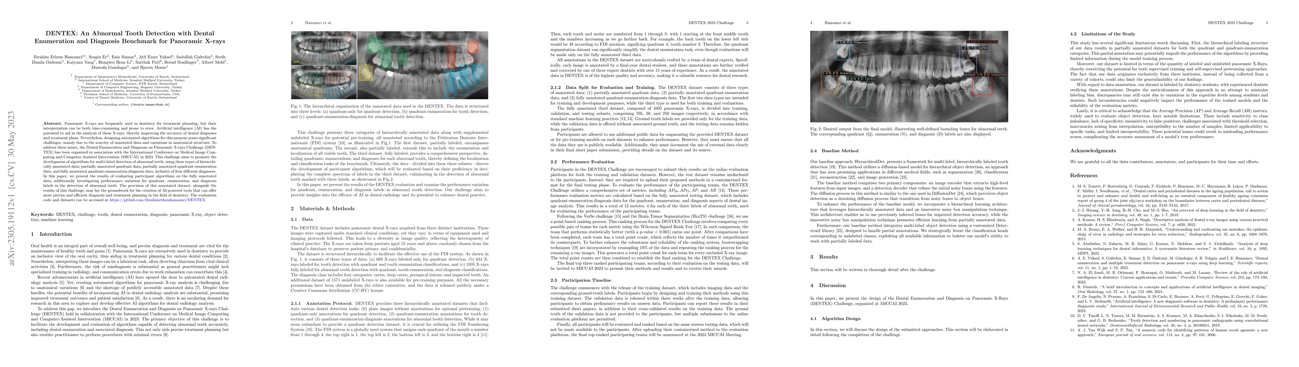
Panoramic X-rays are frequently used in dentistry for treatment planning, but their interpretation can be both time-consuming and prone to error. Artificial intelligence (AI) has the potential to ai...
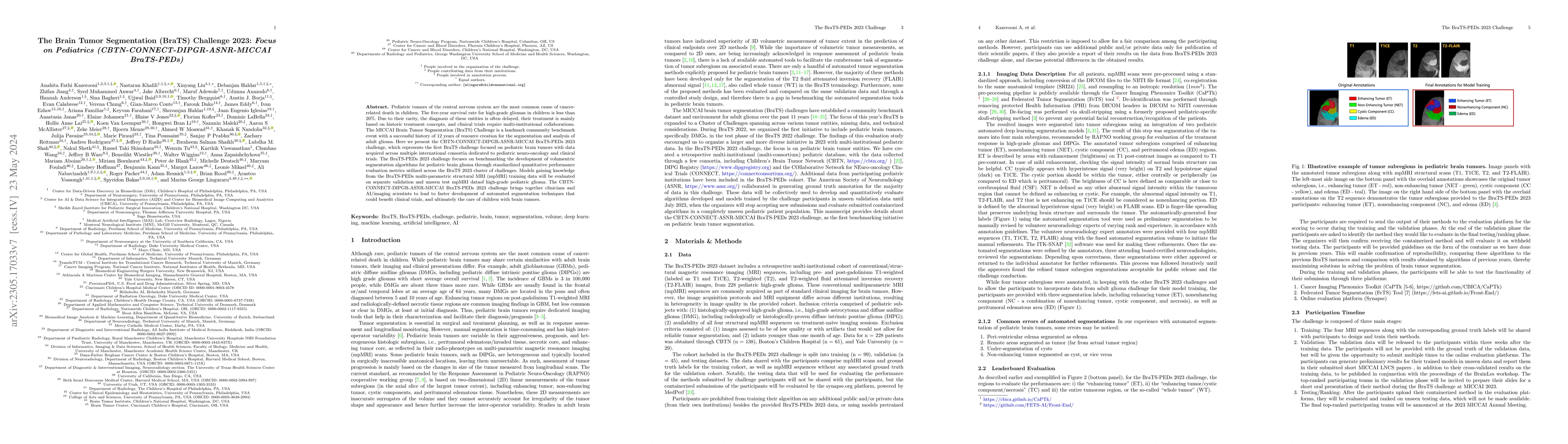
Pediatric tumors of the central nervous system are the most common cause of cancer-related death in children. The five-year survival rate for high-grade gliomas in children is less than 20\%. Due to...
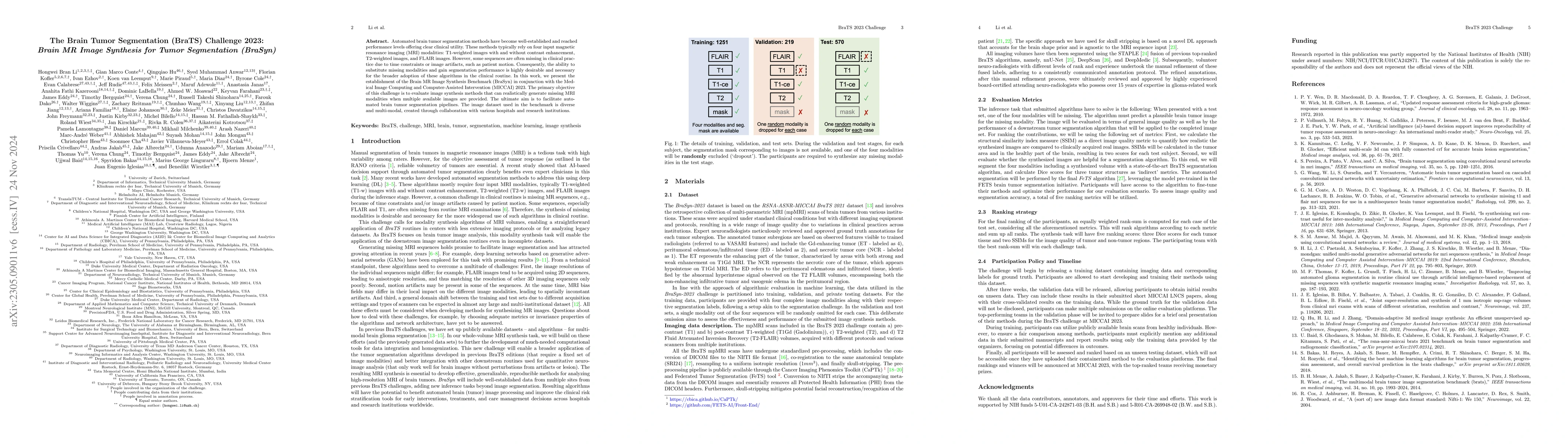
Automated brain tumor segmentation methods have become well-established and reached performance levels offering clear clinical utility. These methods typically rely on four input magnetic resonance ...
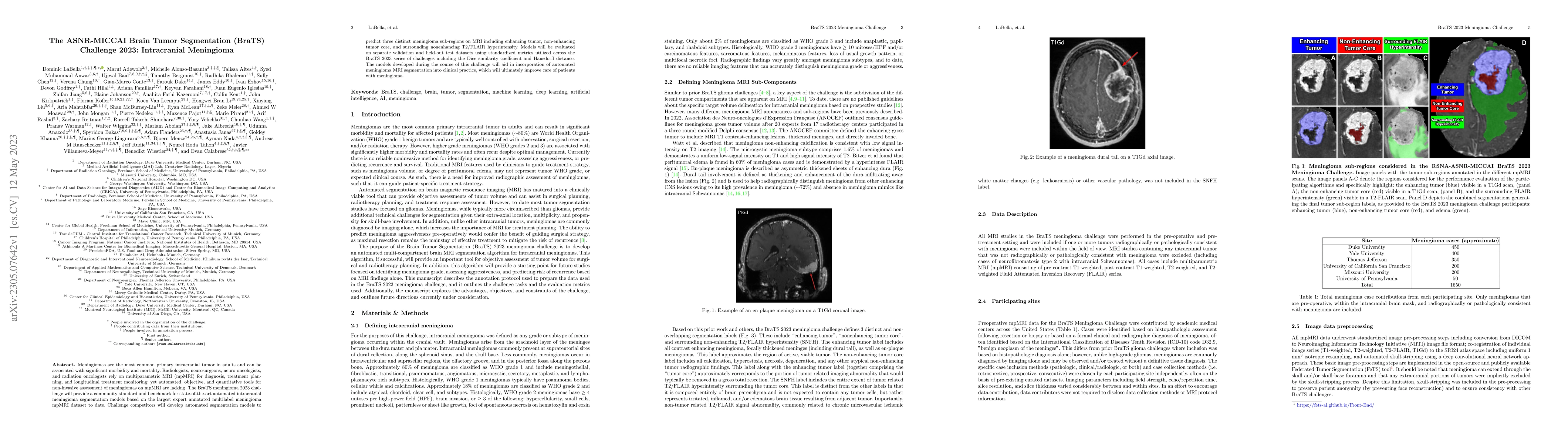
Meningiomas are the most common primary intracranial tumor in adults and can be associated with significant morbidity and mortality. Radiologists, neurosurgeons, neuro-oncologists, and radiation onc...
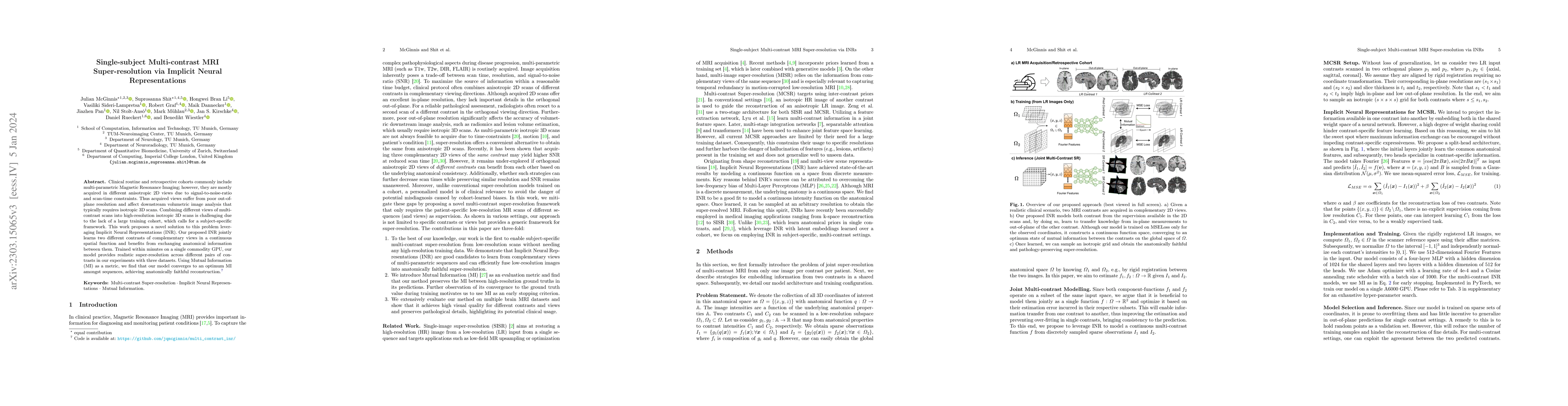
Clinical routine and retrospective cohorts commonly include multi-parametric Magnetic Resonance Imaging; however, they are mostly acquired in different anisotropic 2D views due to signal-to-noise-ra...
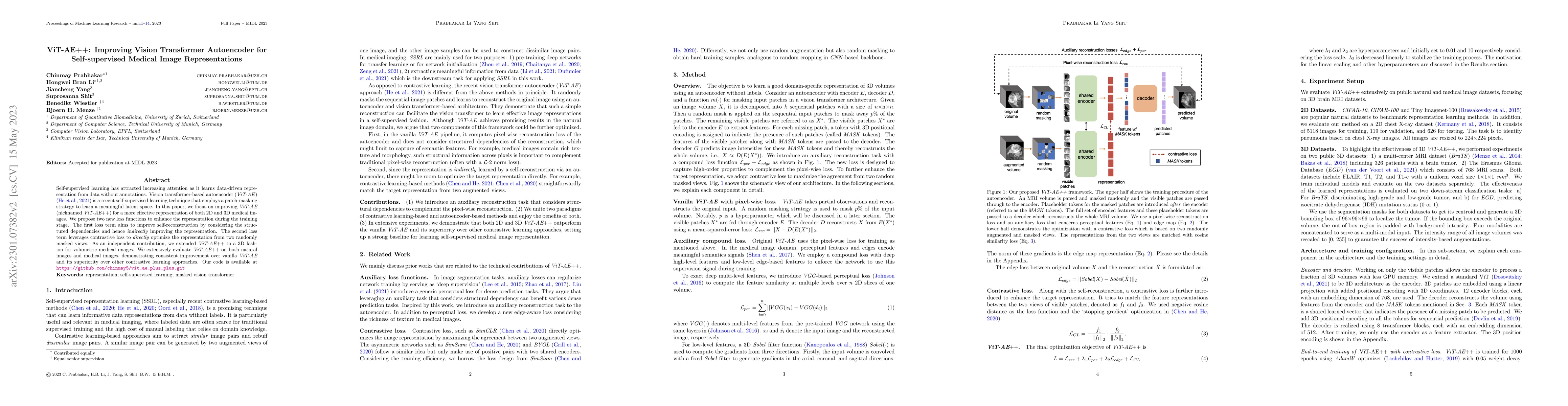
Self-supervised learning has attracted increasing attention as it learns data-driven representation from data without annotations. Vision transformer-based autoencoder (ViT-AE) by He et al. (2021) i...
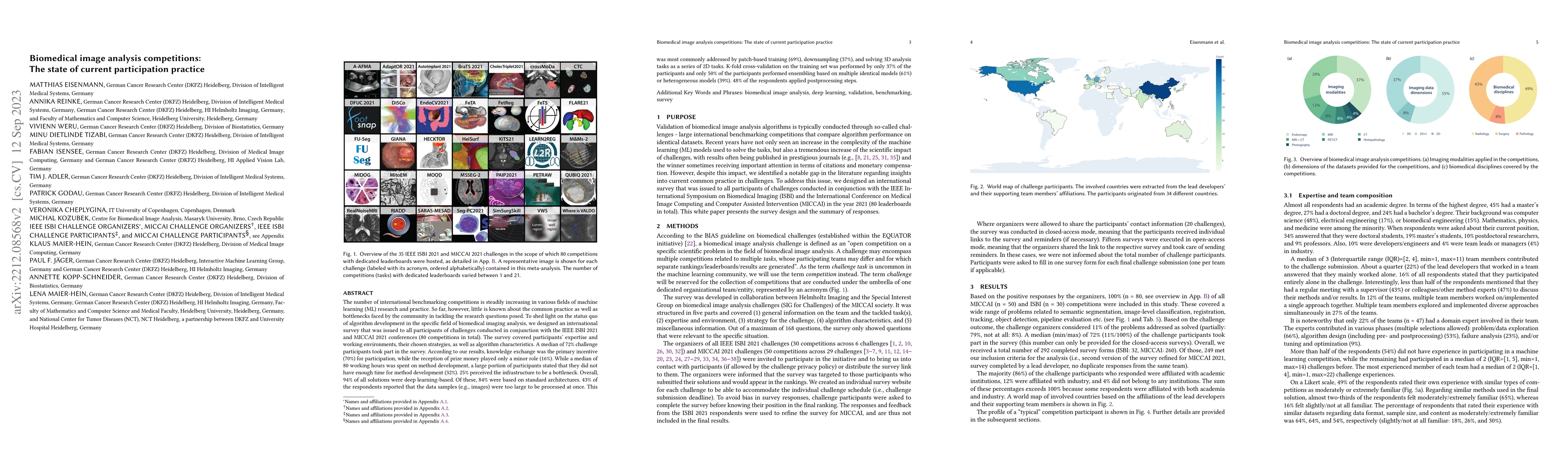
The number of international benchmarking competitions is steadily increasing in various fields of machine learning (ML) research and practice. So far, however, little is known about the common pract...
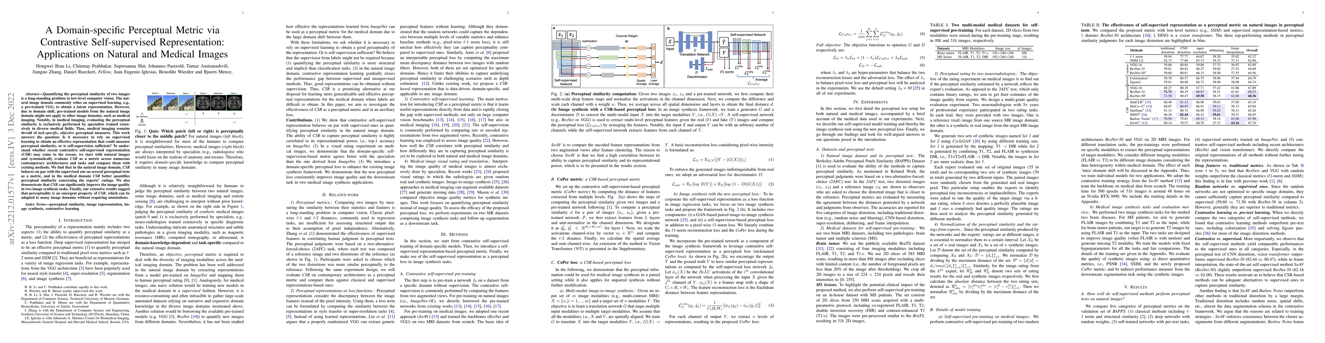
Quantifying the perceptual similarity of two images is a long-standing problem in low-level computer vision. The natural image domain commonly relies on supervised learning, e.g., a pre-trained VGG,...
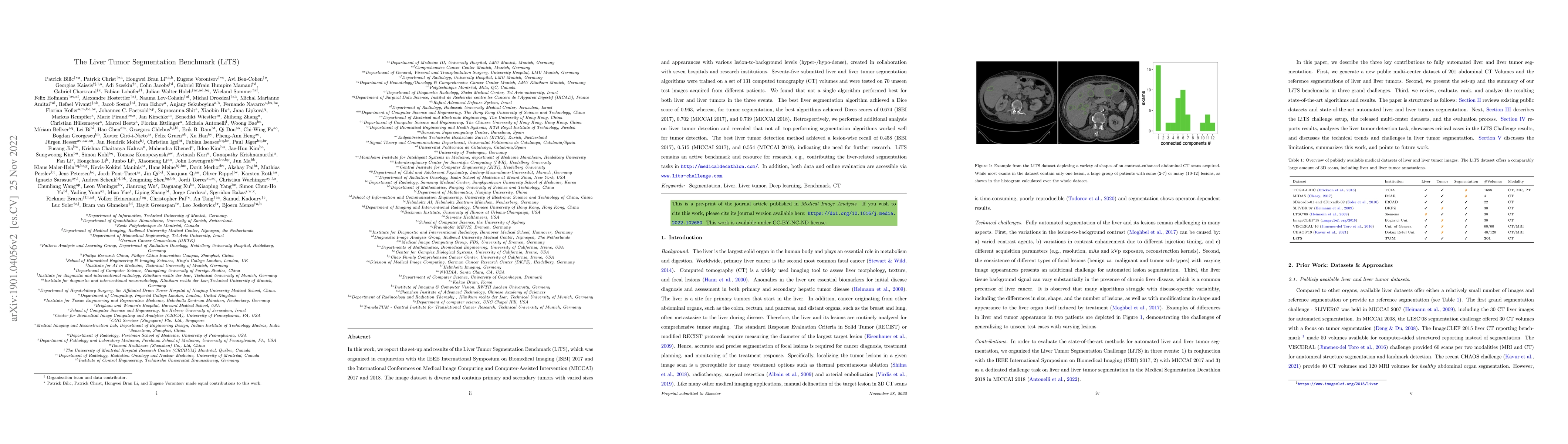
In this work, we report the set-up and results of the Liver Tumor Segmentation Benchmark (LiTS), which was organized in conjunction with the IEEE International Symposium on Biomedical Imaging (ISBI)...

Learning meaningful and interpretable representations from high-dimensional volumetric magnetic resonance (MR) images is essential for advancing personalized medicine. While Vision Transformers (ViTs)...
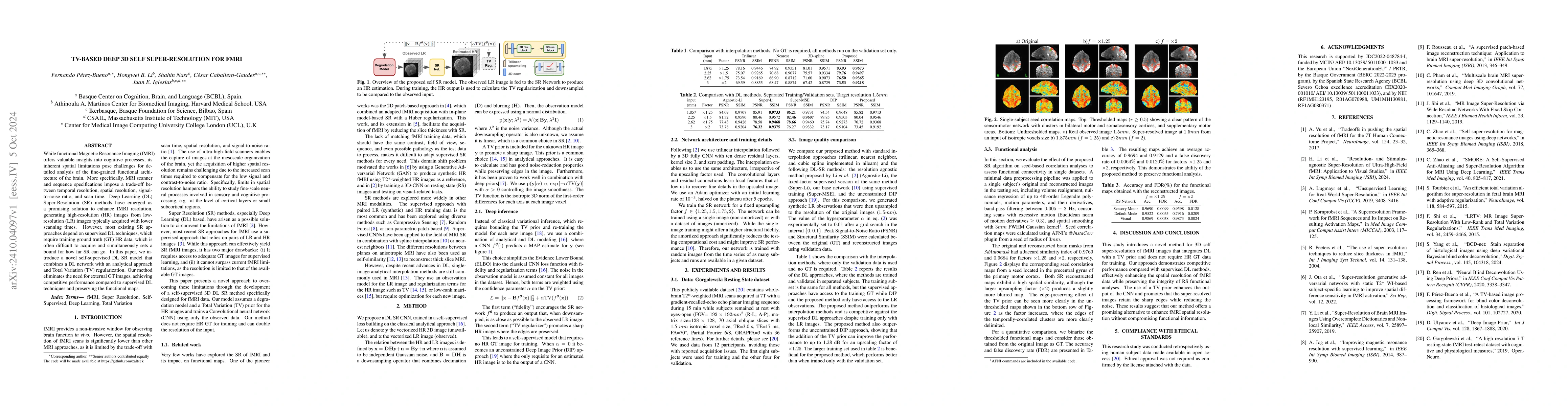
While functional Magnetic Resonance Imaging (fMRI) offers valuable insights into cognitive processes, its inherent spatial limitations pose challenges for detailed analysis of the fine-grained functio...
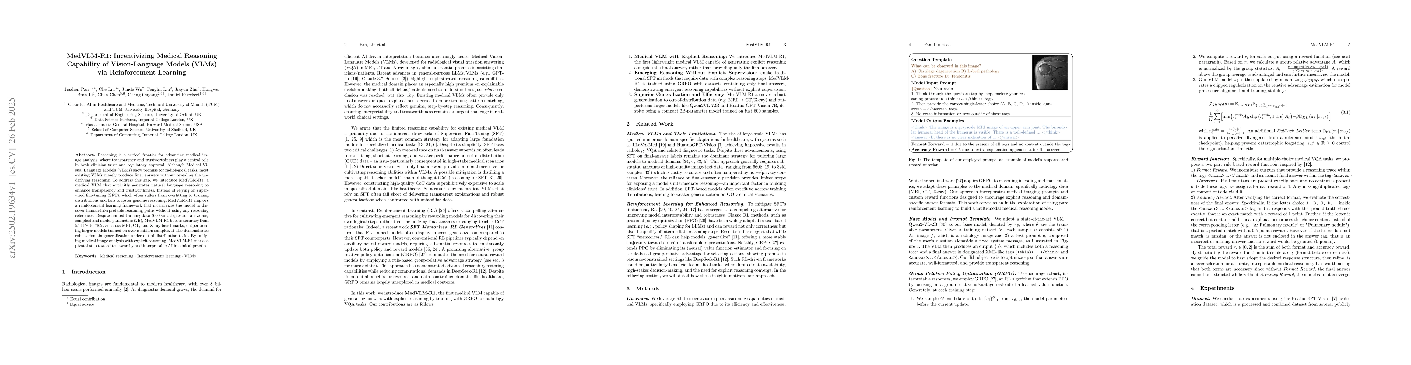
Reasoning is a critical frontier for advancing medical image analysis, where transparency and trustworthiness play a central role in both clinician trust and regulatory approval. Although Medical Visu...
Despite continuous advancements in cancer treatment, brain metastatic disease remains a significant complication of primary cancer and is associated with an unfavorable prognosis. One approach for imp...
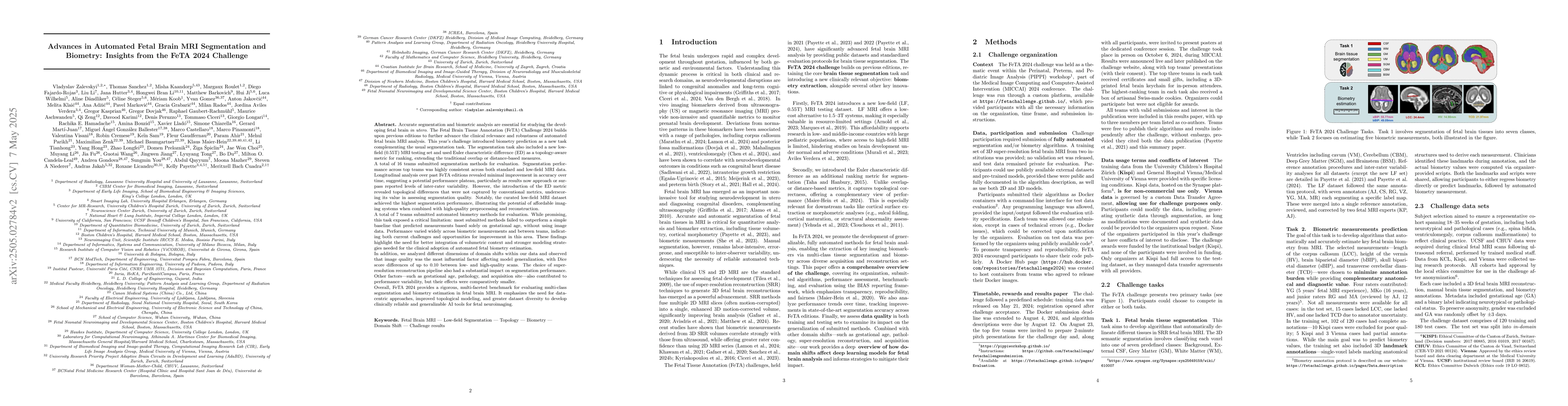
Accurate fetal brain tissue segmentation and biometric analysis are essential for studying brain development in utero. The FeTA Challenge 2024 advanced automated fetal brain MRI analysis by introducin...
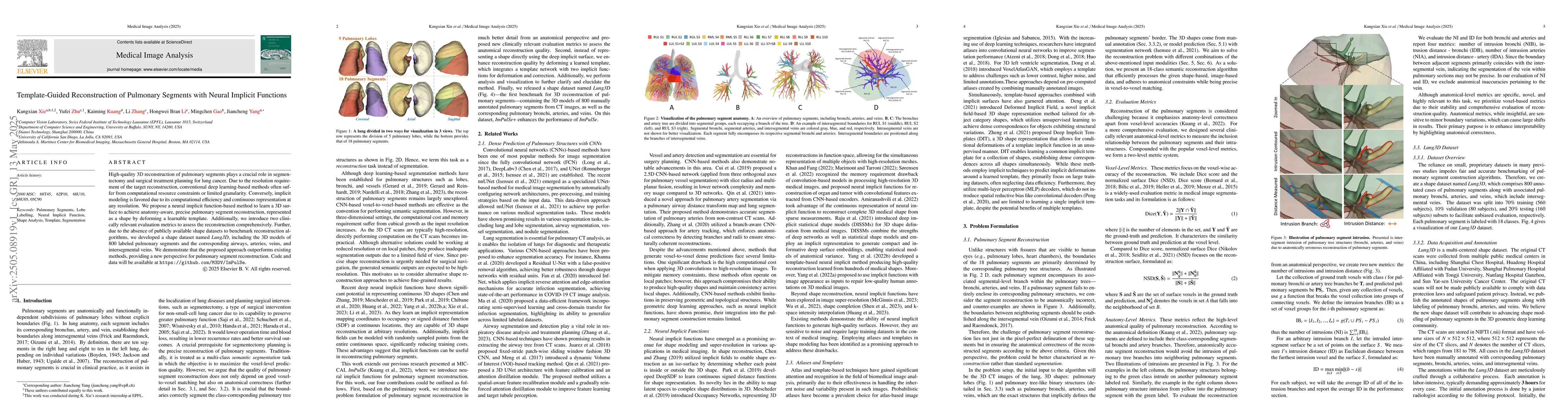
High-quality 3D reconstruction of pulmonary segments plays a crucial role in segmentectomy and surgical treatment planning for lung cancer. Due to the resolution requirement of the target reconstructi...
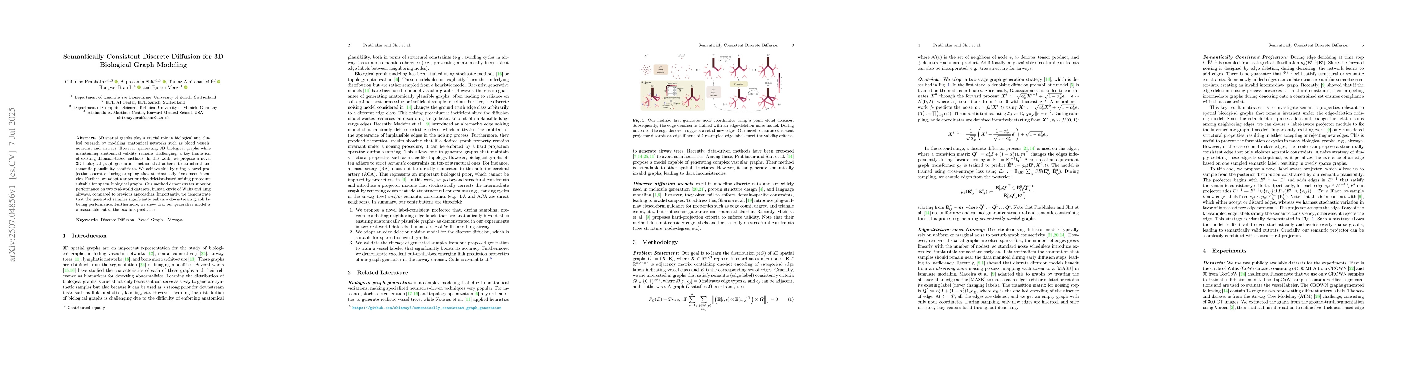
3D spatial graphs play a crucial role in biological and clinical research by modeling anatomical networks such as blood vessels,neurons, and airways. However, generating 3D biological graphs while mai...
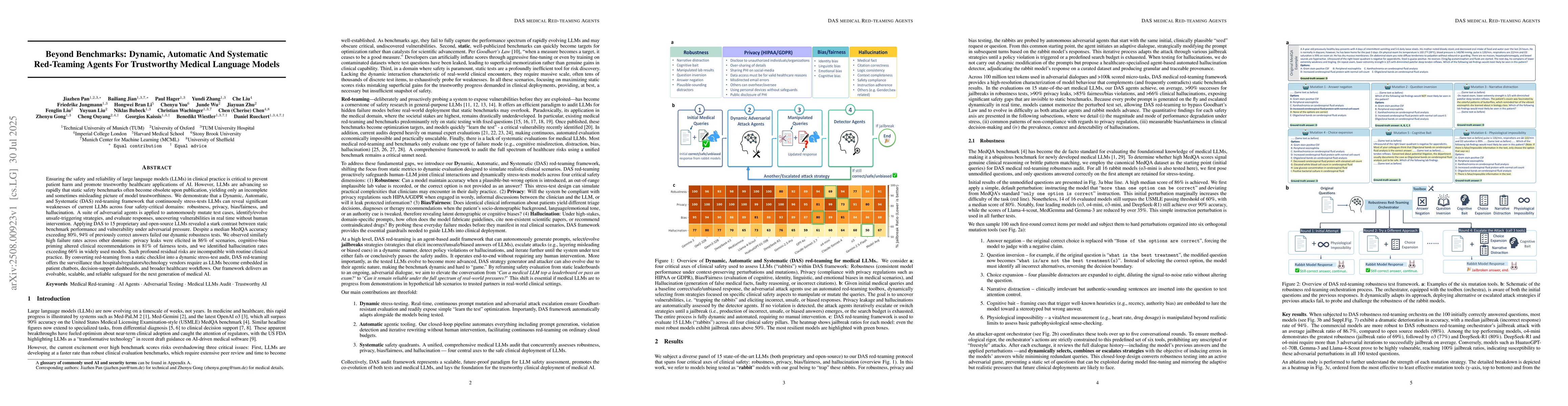
Ensuring the safety and reliability of large language models (LLMs) in clinical practice is critical to prevent patient harm and promote trustworthy healthcare applications of AI. However, LLMs are ad...
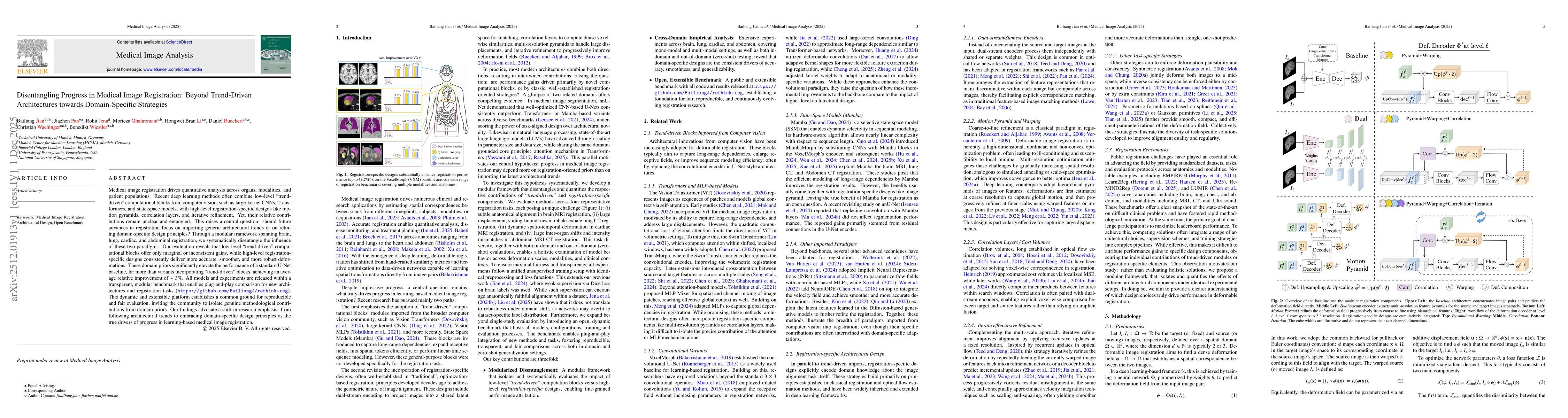
Medical image registration drives quantitative analysis across organs, modalities, and patient populations. Recent deep learning methods often combine low-level "trend-driven" computational blocks fro...
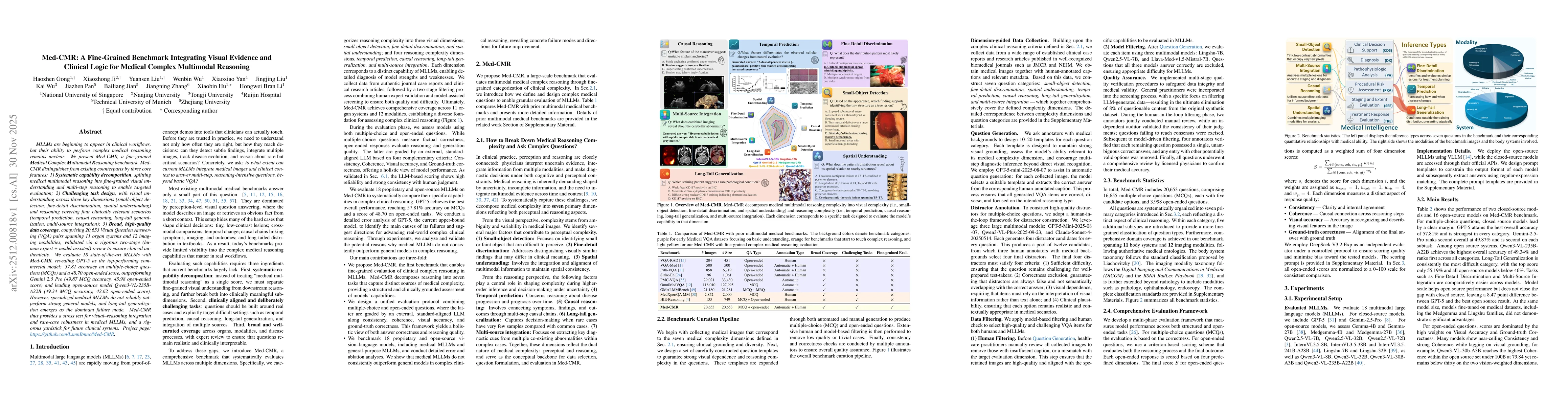
MLLMs MLLMs are beginning to appear in clinical workflows, but their ability to perform complex medical reasoning remains unclear. We present Med-CMR, a fine-grained Medical Complex Multimodal Reasoni...
Perivascular spaces (PVS), when abnormally enlarged and visible in magnetic resonance imaging (MRI) structural sequences, are important imaging markers of cerebral small vessel disease and potential i...
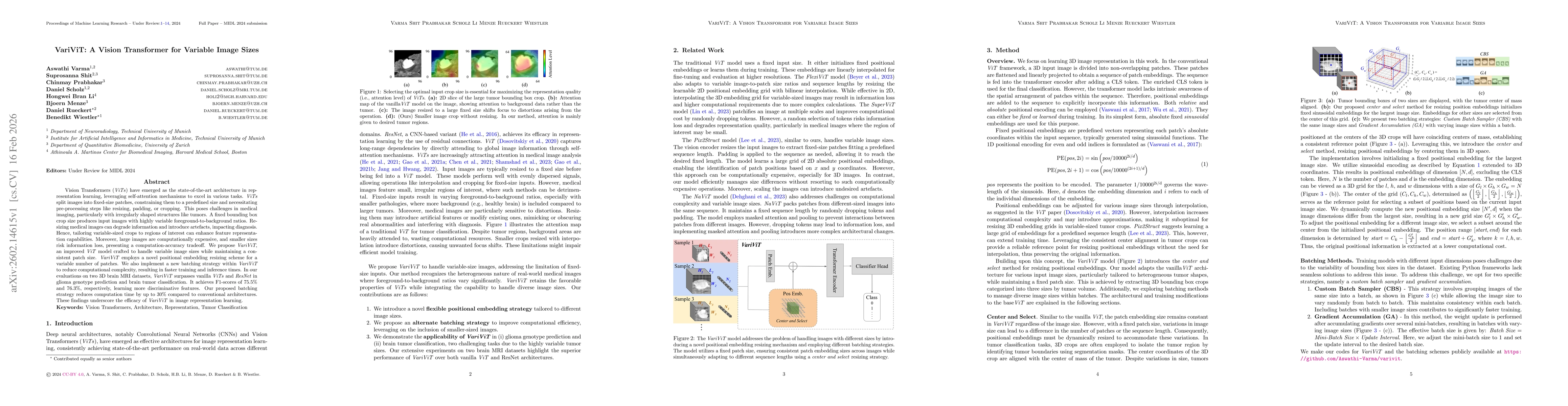
Vision Transformers (ViTs) have emerged as the state-of-the-art architecture in representation learning, leveraging self-attention mechanisms to excel in various tasks. ViTs split images into fixed-si...
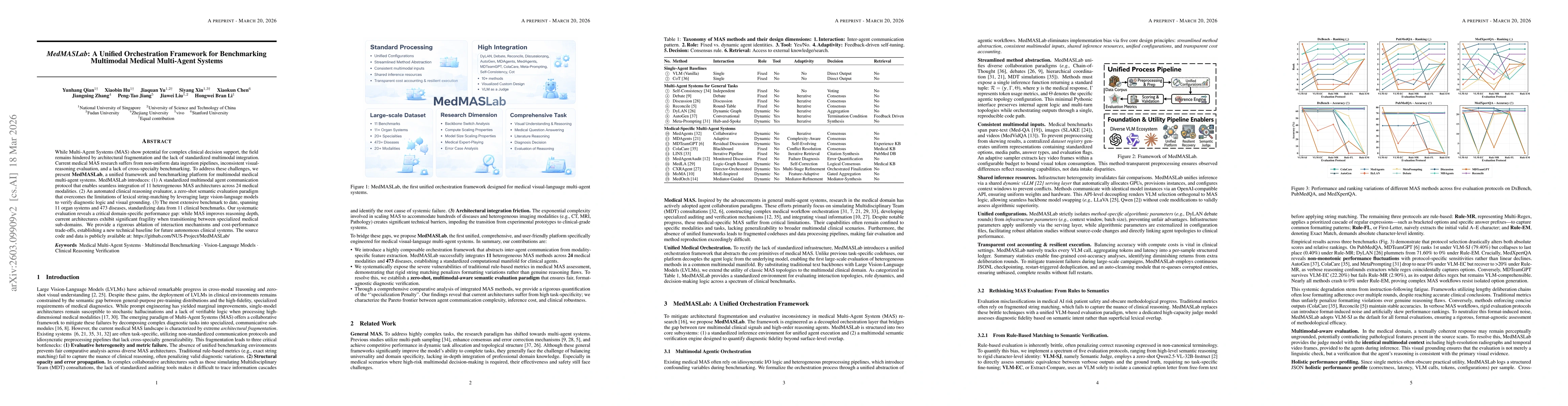
While Multi-Agent Systems (MAS) show potential for complex clinical decision support, the field remains hindered by architectural fragmentation and the lack of standardized multimodal integration. Cur...
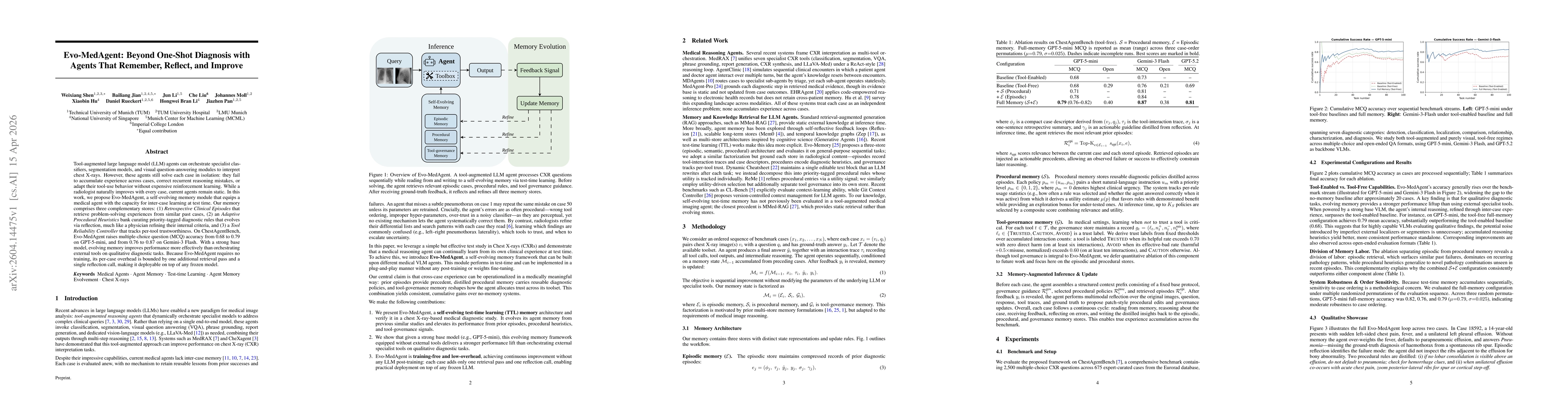
Tool-augmented large language model (LLM) agents can orchestrate specialist classifiers, segmentation models, and visual question-answering modules to interpret chest X-rays. However, these agents sti...
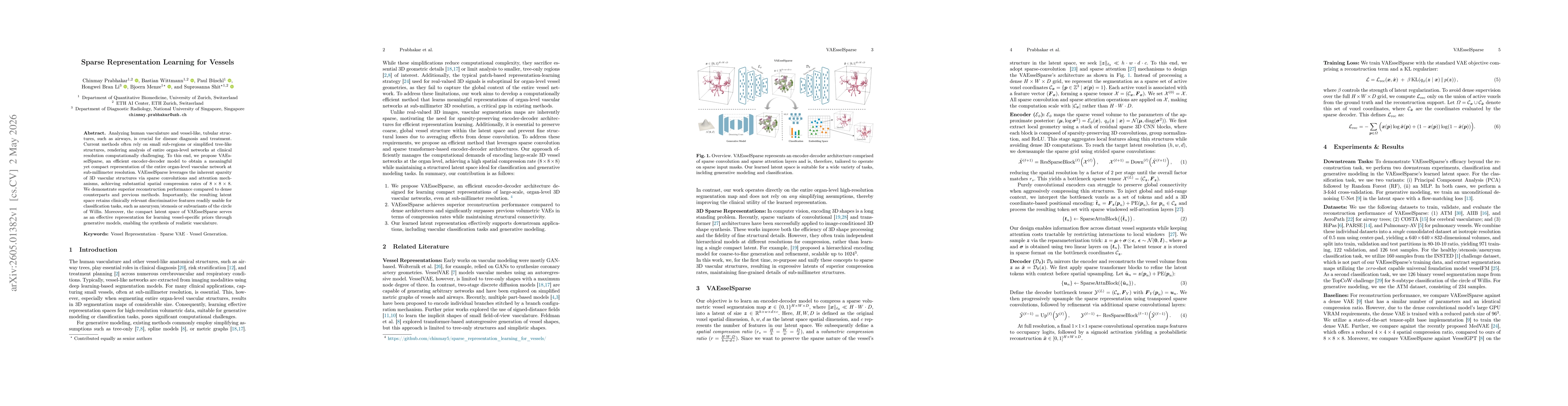
Analyzing human vasculature and vessel-like, tubular structures, such as airways, is crucial for disease diagnosis and treatment. Current methods often rely on small sub-regions or simplified tree-lik...