Academic Profile
Statistics
Similar Authors
Papers on arXiv
Placenta volume measurement from 3D ultrasound images is critical for predicting pregnancy outcomes, and manual annotation is the gold standard. However, such manual annotation is expensive and time-c...
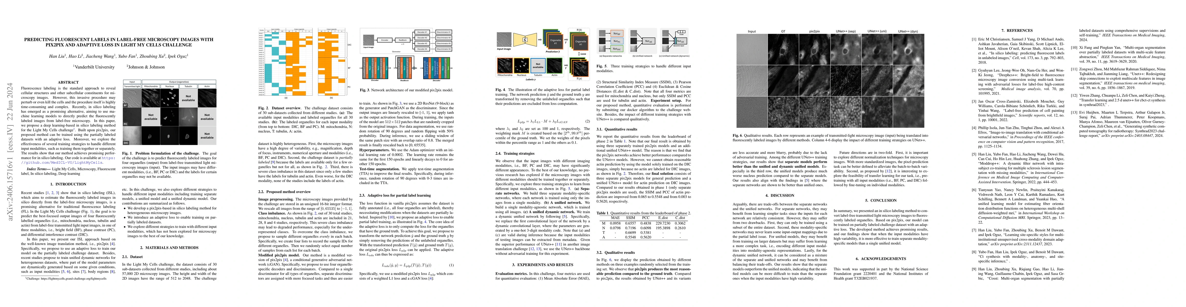
Fluorescence labeling is the standard approach to reveal cellular structures and other subcellular constituents for microscopy images. However, this invasive procedure may perturb or even kill the c...
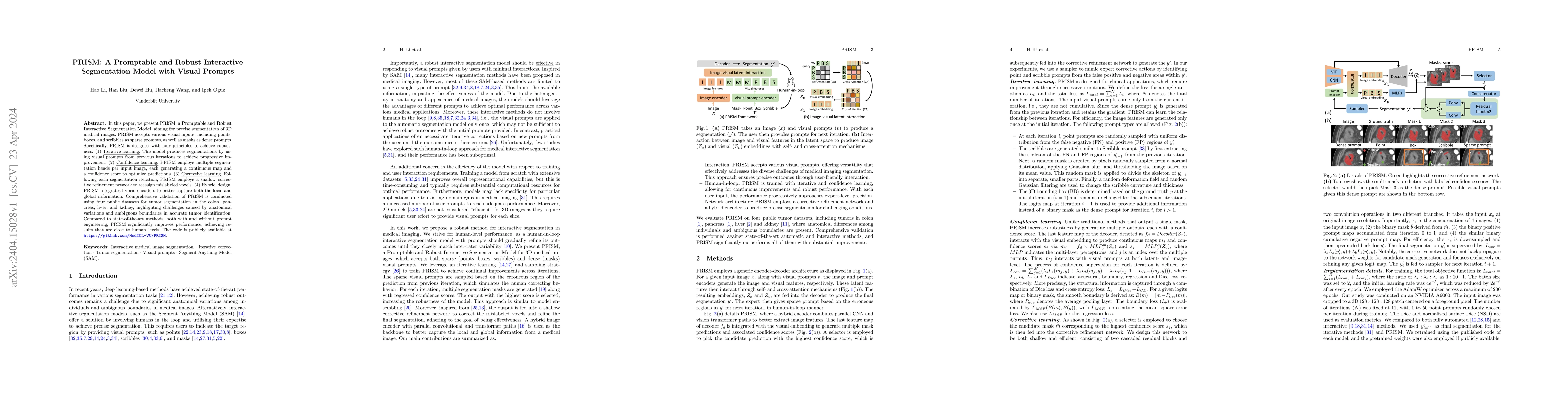
In this paper, we present PRISM, a Promptable and Robust Interactive Segmentation Model, aiming for precise segmentation of 3D medical images. PRISM accepts various visual inputs, including points, ...
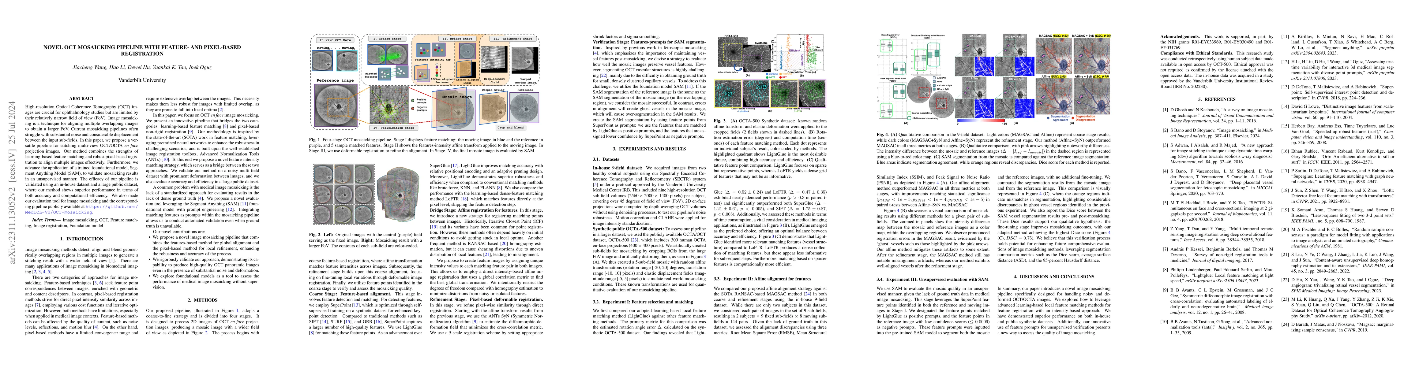
High-resolution Optical Coherence Tomography (OCT) images are crucial for ophthalmology studies but are limited by their relatively narrow field of view (FoV). Image mosaicking is a technique for al...
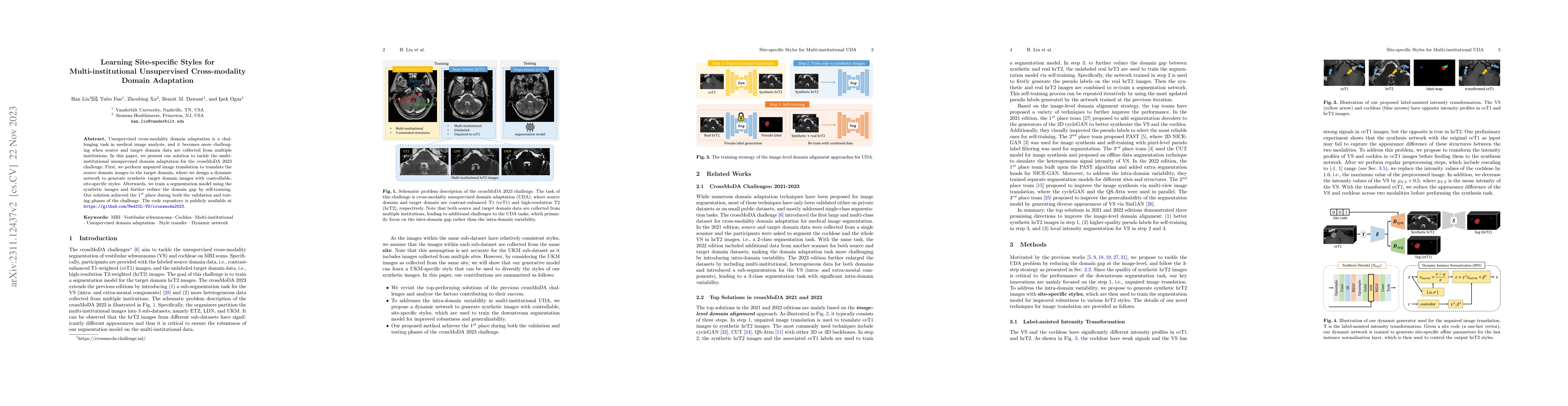
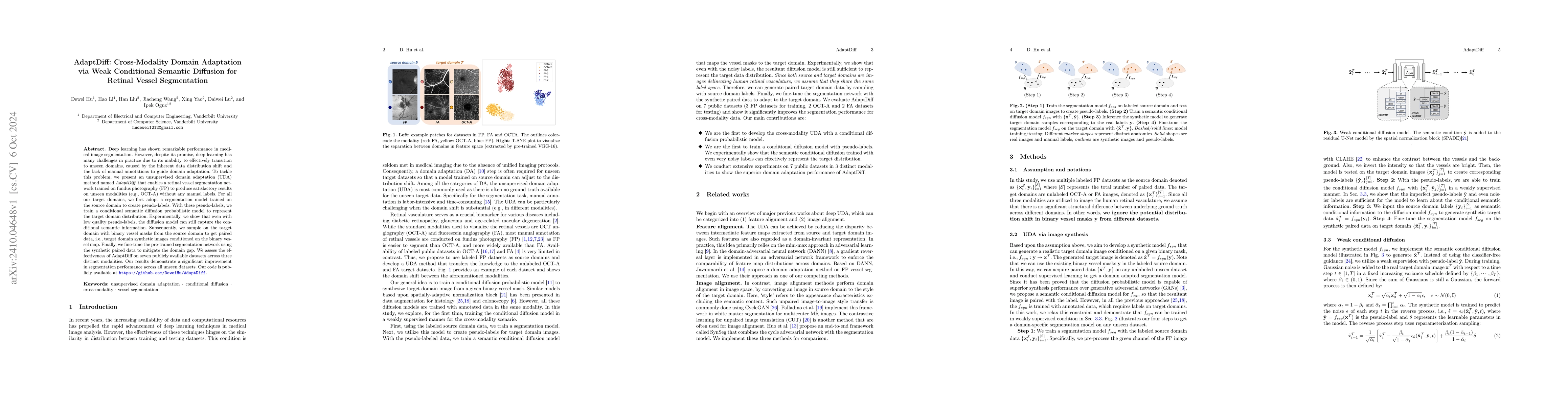
Unsupervised cross-modality domain adaptation is a challenging task in medical image analysis, and it becomes more challenging when source and target domain data are collected from multiple institut...
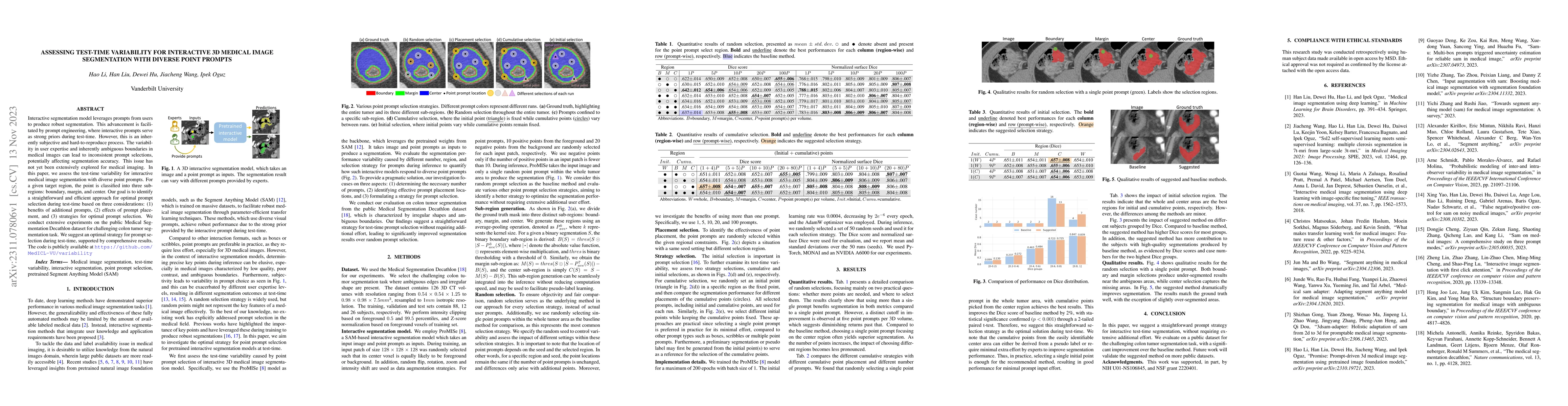
Interactive segmentation model leverages prompts from users to produce robust segmentation. This advancement is facilitated by prompt engineering, where interactive prompts serve as strong priors du...
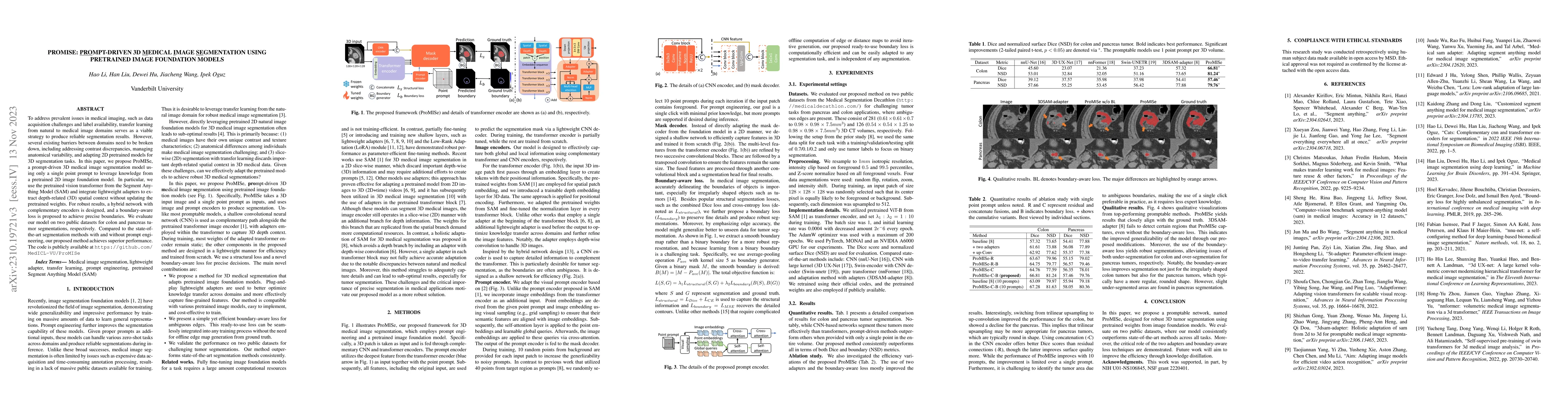
To address prevalent issues in medical imaging, such as data acquisition challenges and label availability, transfer learning from natural to medical image domains serves as a viable strategy to pro...
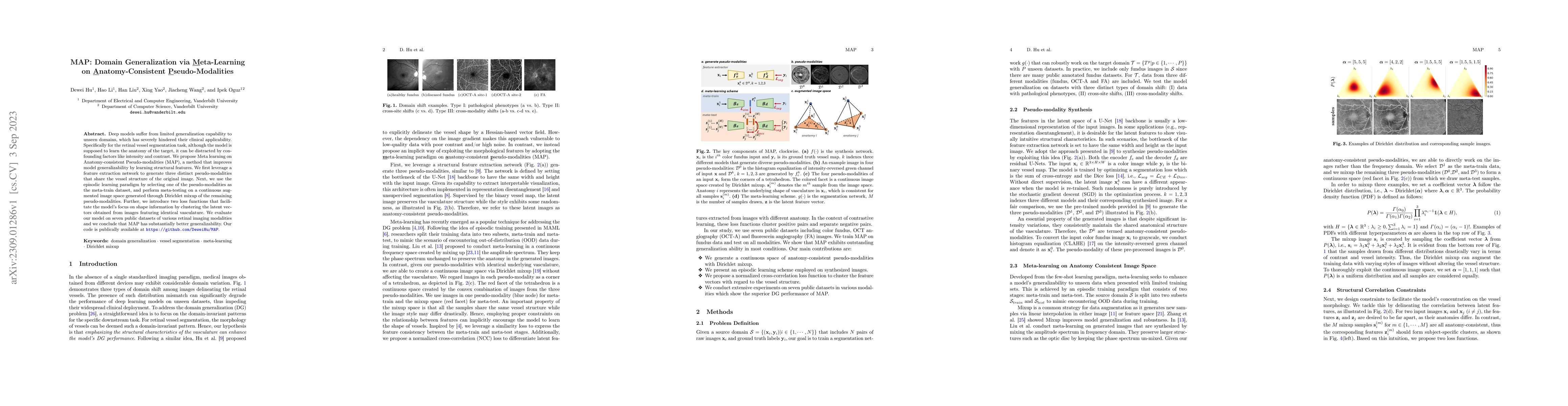
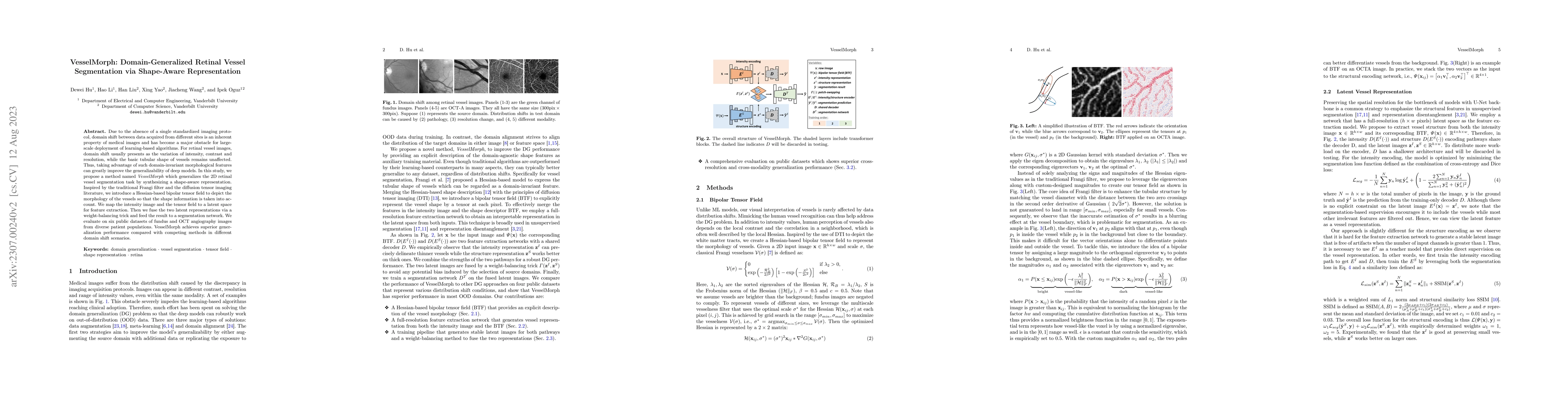
Deep models suffer from limited generalization capability to unseen domains, which has severely hindered their clinical applicability. Specifically for the retinal vessel segmentation task, although...
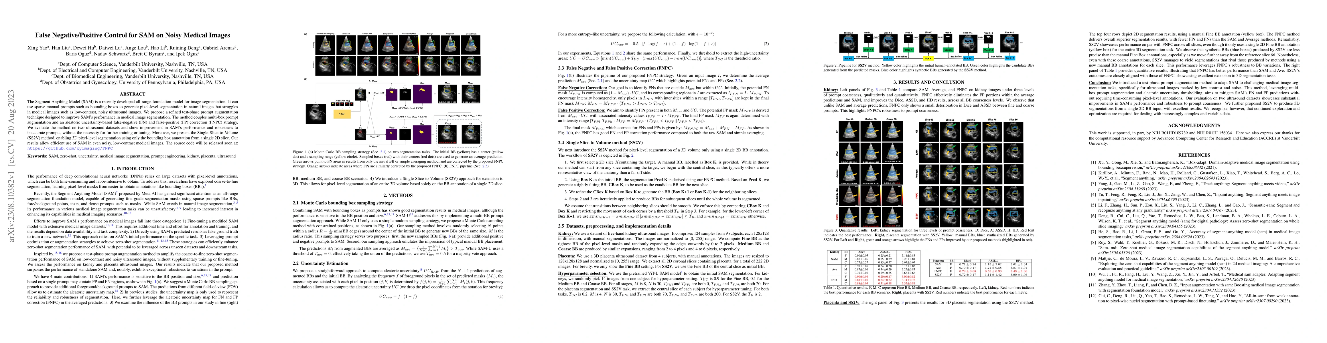
The Segment Anything Model (SAM) is a recently developed all-range foundation model for image segmentation. It can use sparse manual prompts such as bounding boxes to generate pixel-level segmentati...
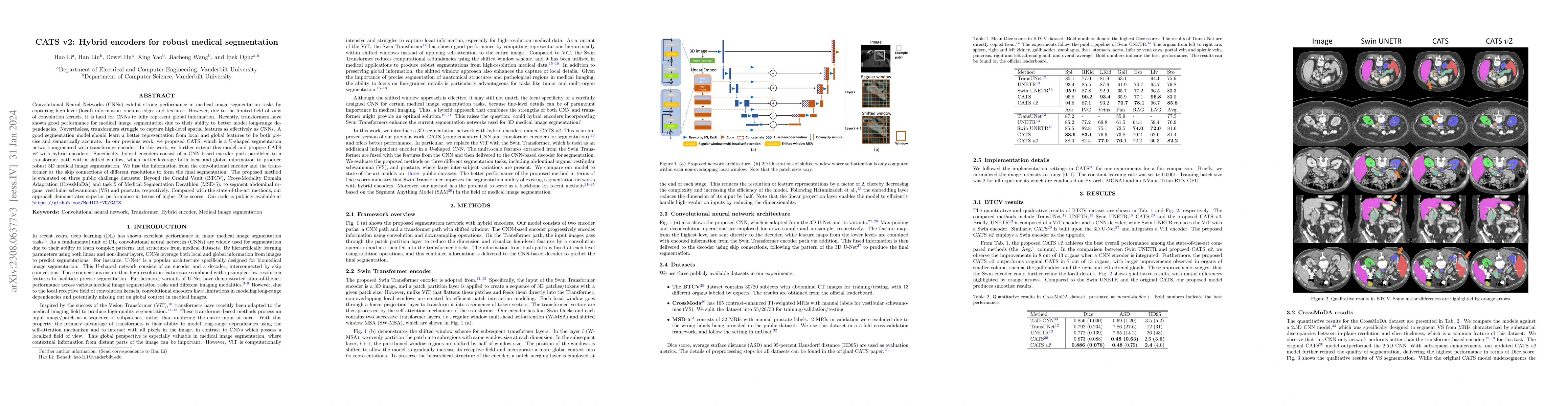
Convolutional Neural Networks (CNNs) have exhibited strong performance in medical image segmentation tasks by capturing high-level (local) information, such as edges and textures. However, due to th...
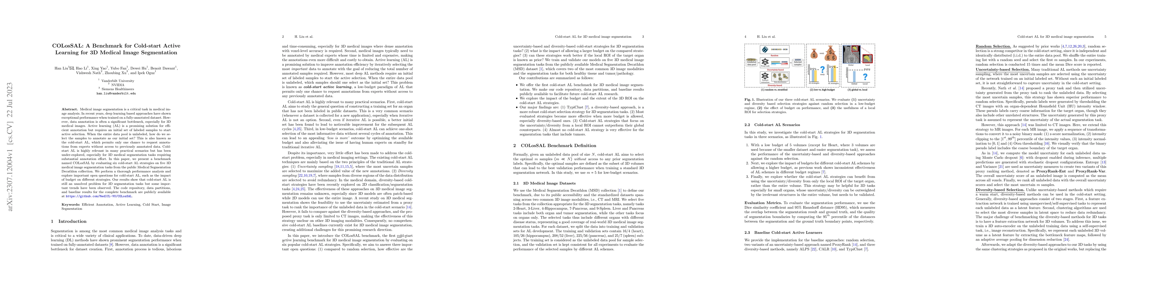
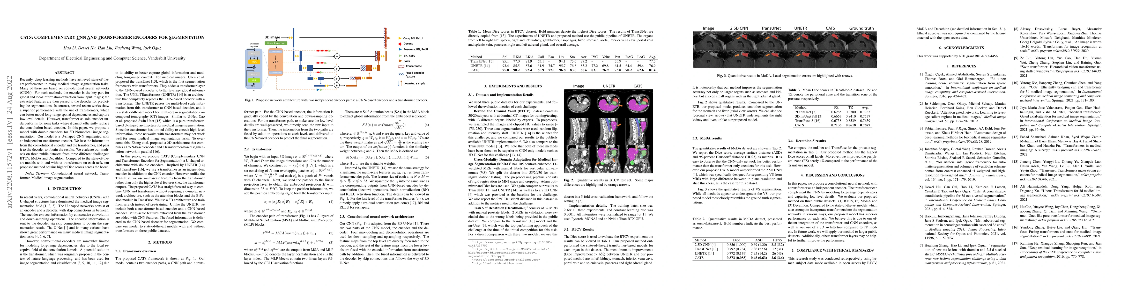
Medical image segmentation is a critical task in medical image analysis. In recent years, deep learning based approaches have shown exceptional performance when trained on a fully-annotated dataset....
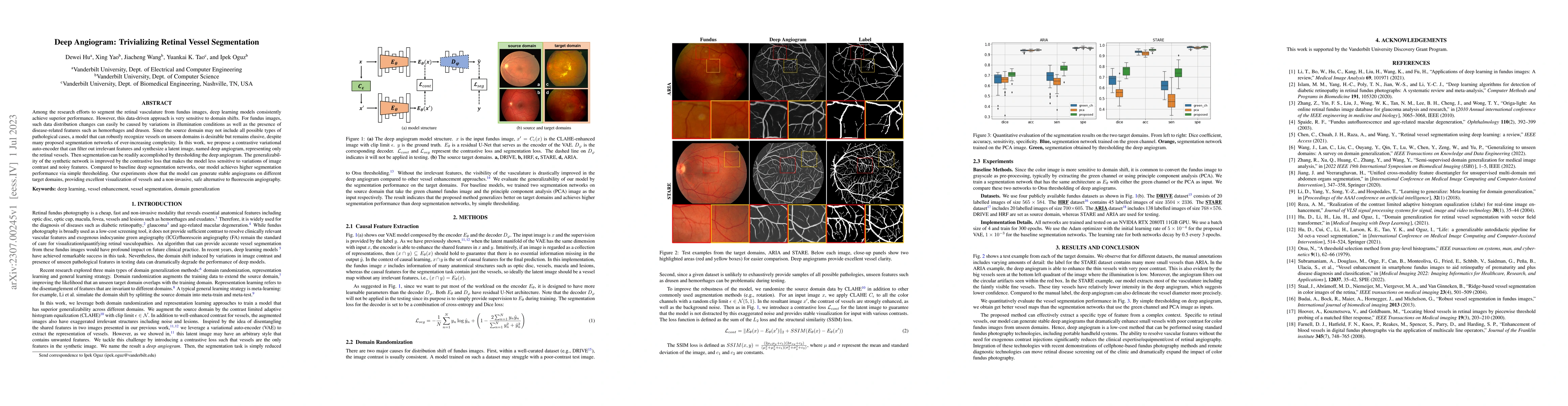
Among the research efforts to segment the retinal vasculature from fundus images, deep learning models consistently achieve superior performance. However, this data-driven approach is very sensitive...
Due to the absence of a single standardized imaging protocol, domain shift between data acquired from different sites is an inherent property of medical images and has become a major obstacle for la...
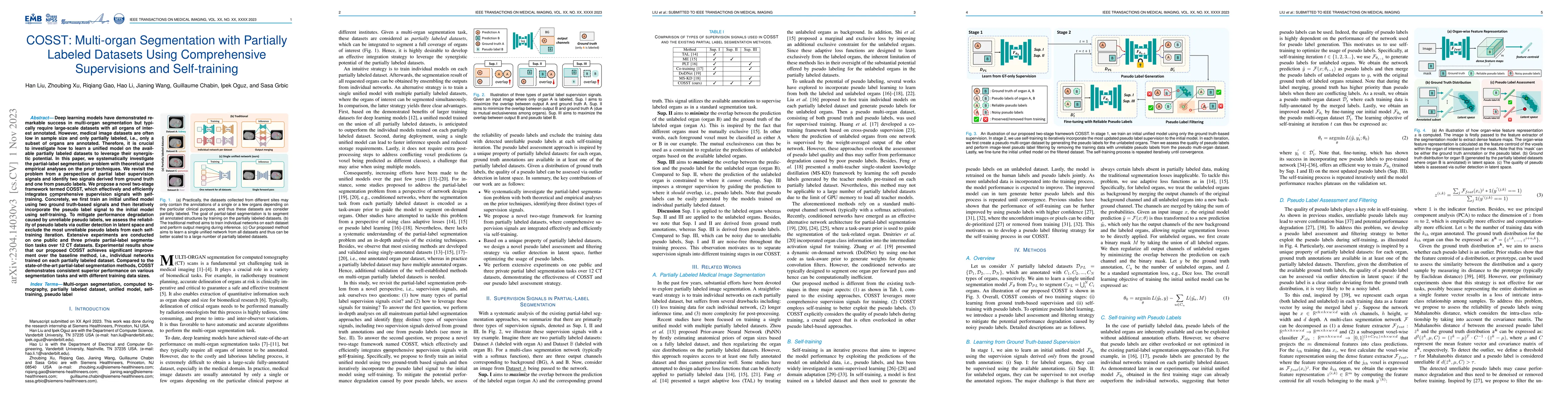
Deep learning models have demonstrated remarkable success in multi-organ segmentation but typically require large-scale datasets with all organs of interest annotated. However, medical image dataset...
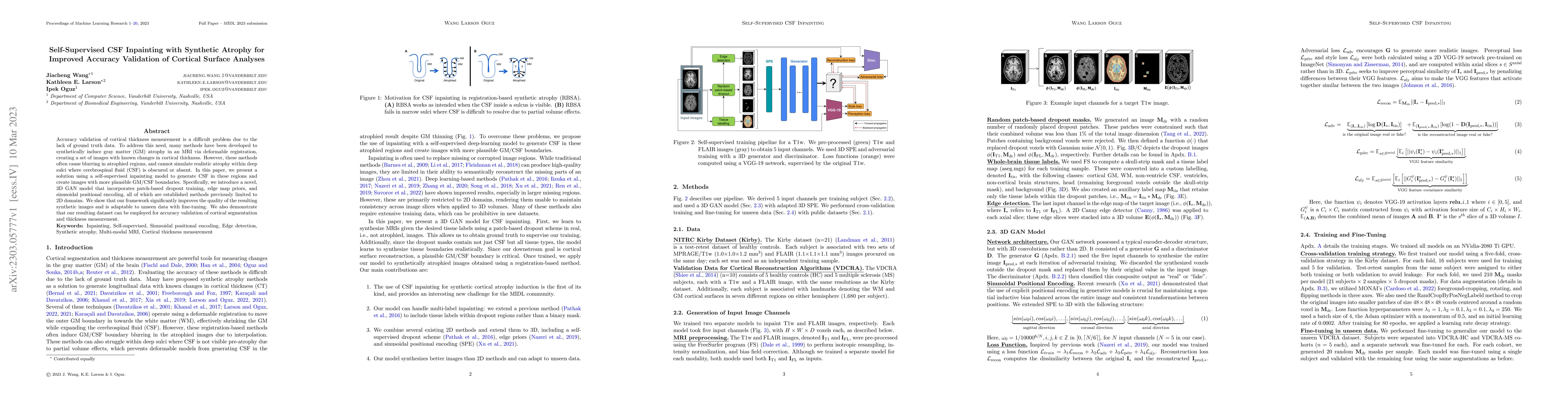
Accuracy validation of cortical thickness measurement is a difficult problem due to the lack of ground truth data. To address this need, many methods have been developed to synthetically induce gray...
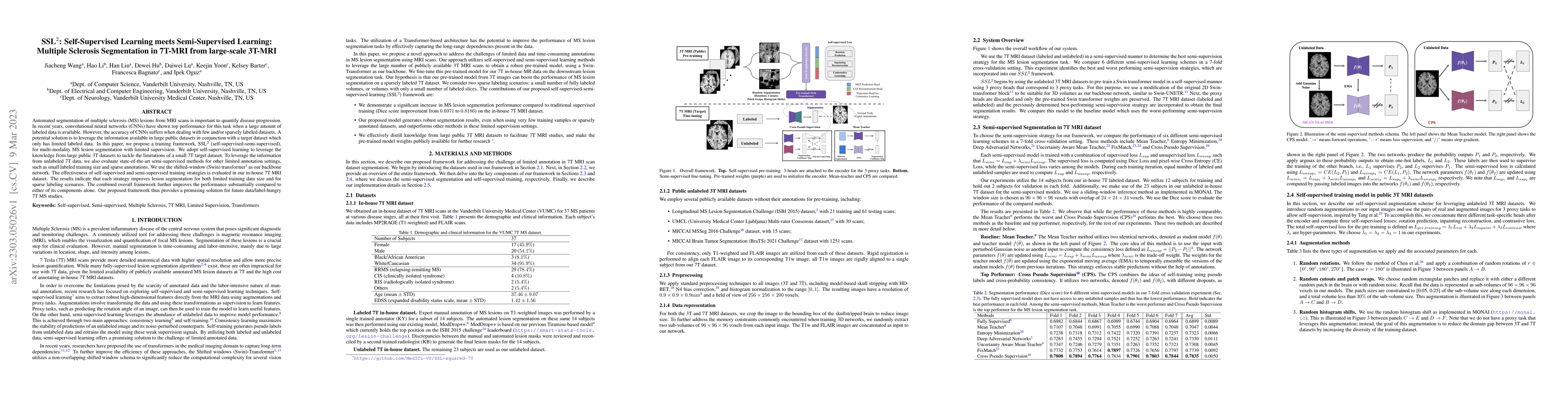
Automated segmentation of multiple sclerosis (MS) lesions from MRI scans is important to quantify disease progression. In recent years, convolutional neural networks (CNNs) have shown top performanc...
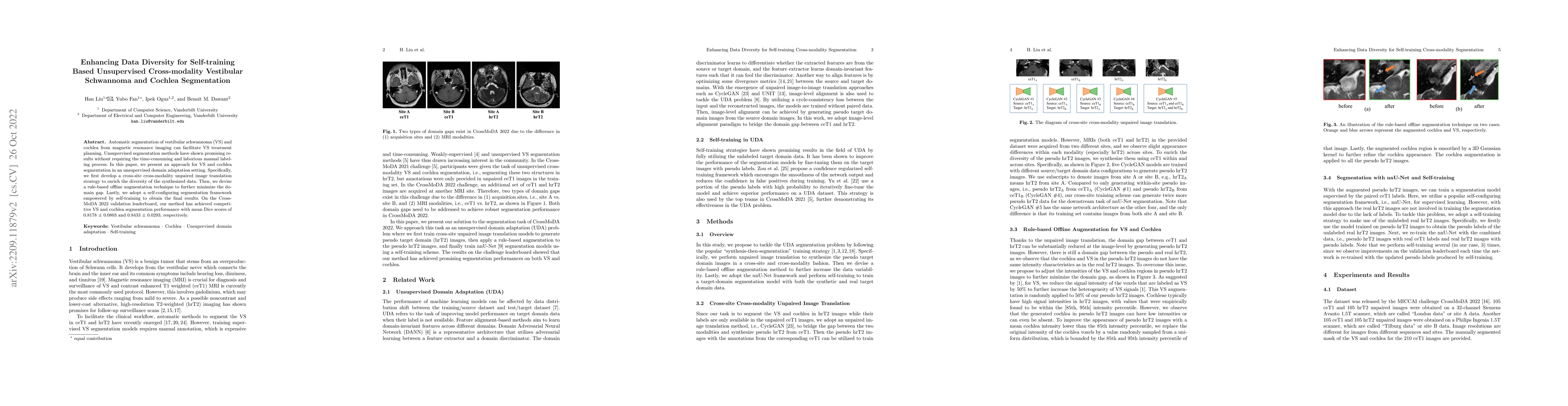
Automatic segmentation of vestibular schwannoma (VS) and cochlea from magnetic resonance imaging can facilitate VS treatment planning. Unsupervised segmentation methods have shown promising results ...
Recently, deep learning methods have achieved state-of-the-art performance in many medical image segmentation tasks. Many of these are based on convolutional neural networks (CNNs). For such methods...
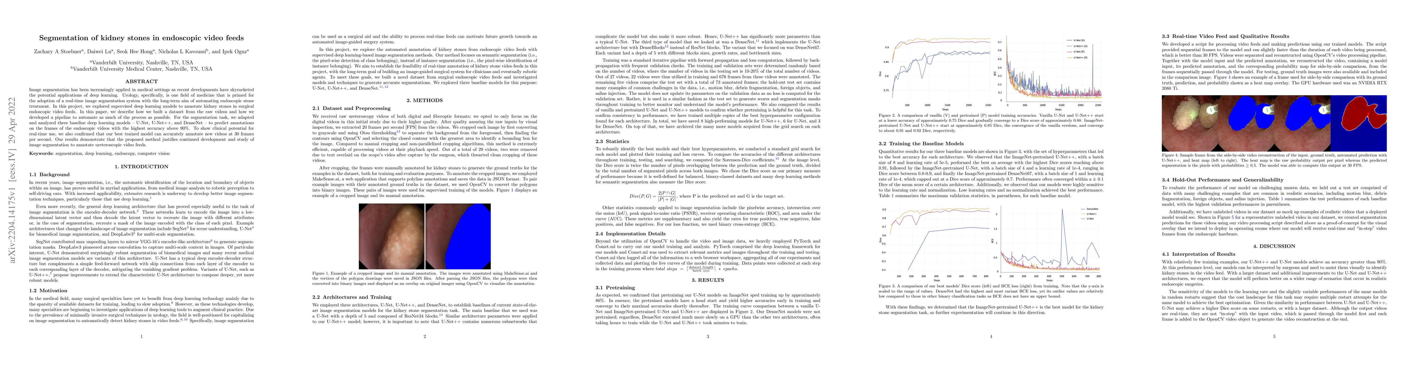
Image segmentation has been increasingly applied in medical settings as recent developments have skyrocketed the potential applications of deep learning. Urology, specifically, is one field of medic...
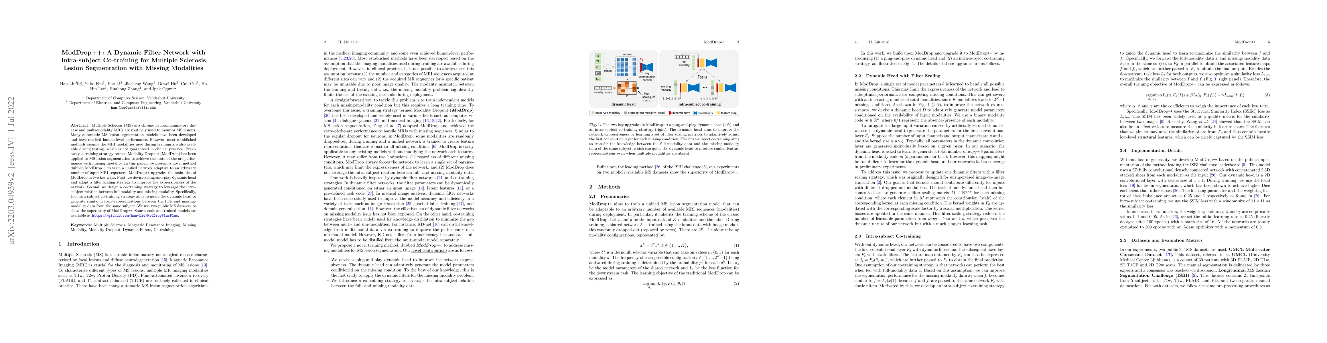
Multiple Sclerosis (MS) is a chronic neuroinflammatory disease and multi-modality MRIs are routinely used to monitor MS lesions. Many automatic MS lesion segmentation models have been developed and ...
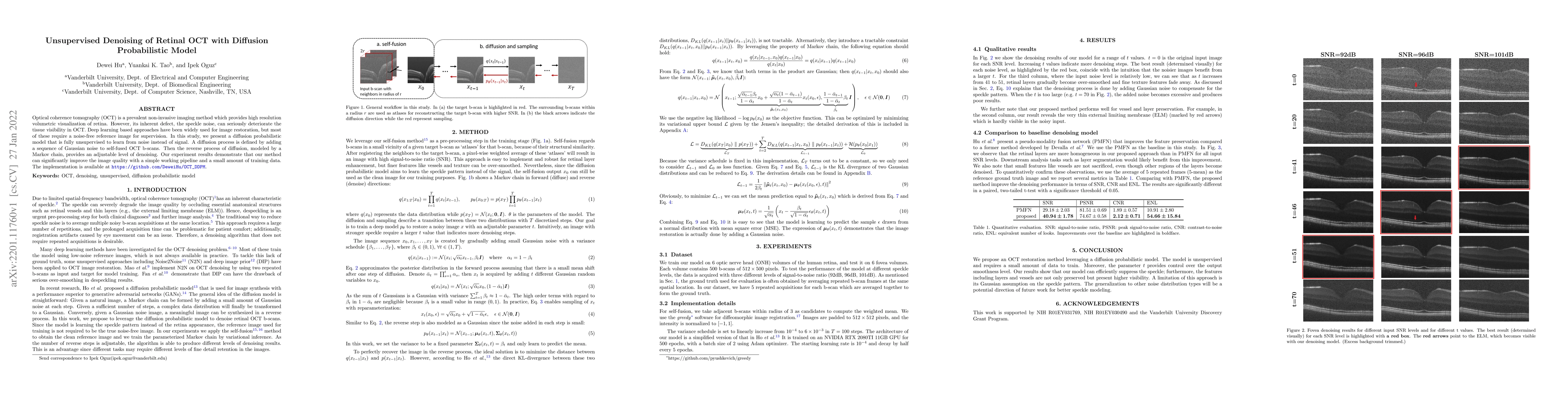
Optical coherence tomography (OCT) is a prevalent non-invasive imaging method which provides high resolution volumetric visualization of retina. However, its inherent defect, the speckle noise, can ...
Domain Adaptation (DA) has recently raised strong interests in the medical imaging community. While a large variety of DA techniques has been proposed for image segmentation, most of these technique...
Magnetic resonance images (MRIs) are widely used to quantify vestibular schwannoma and the cochlea. Recently, deep learning methods have shown state-of-the-art performance for segmenting these struc...
Optical coherence tomography (OCT) is a prevalent imaging technique for retina. However, it is affected by multiplicative speckle noise that can degrade the visibility of essential anatomical struct...
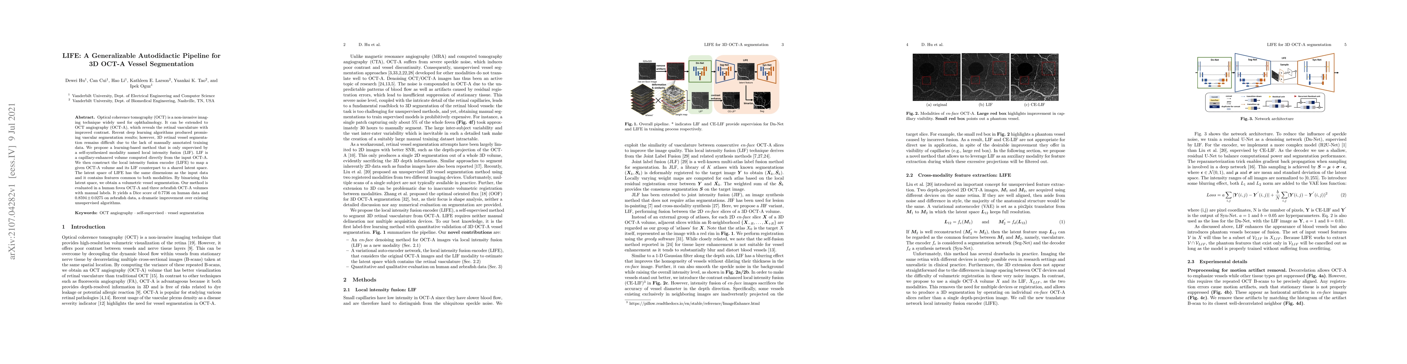
Optical coherence tomography (OCT) is a non-invasive imaging technique widely used for ophthalmology. It can be extended to OCT angiography (OCT-A), which reveals the retinal vasculature with improv...
The improvements in magnetic resonance imaging have led to the development of numerous techniques to better detect structural alterations caused by neurodegenerative diseases. Among these, the patch...
This compendium gathers all the accepted extended abstracts from the Second International Conference on Medical Imaging with Deep Learning (MIDL 2019), held in London, UK, 8-10 July 2019. Note that ...
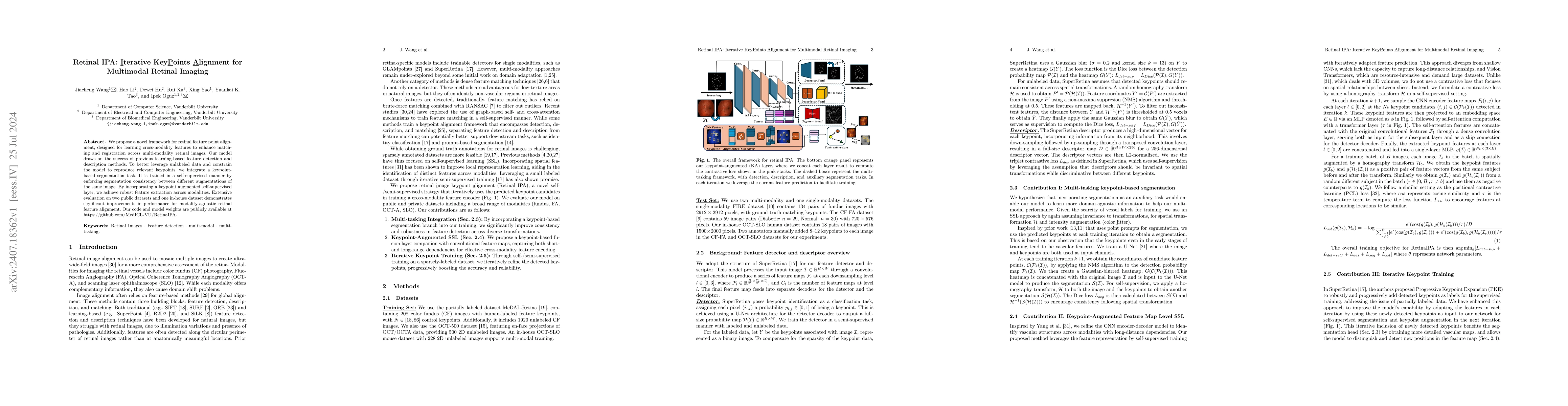
We propose a novel framework for retinal feature point alignment, designed for learning cross-modality features to enhance matching and registration across multi-modality retinal images. Our model dra...
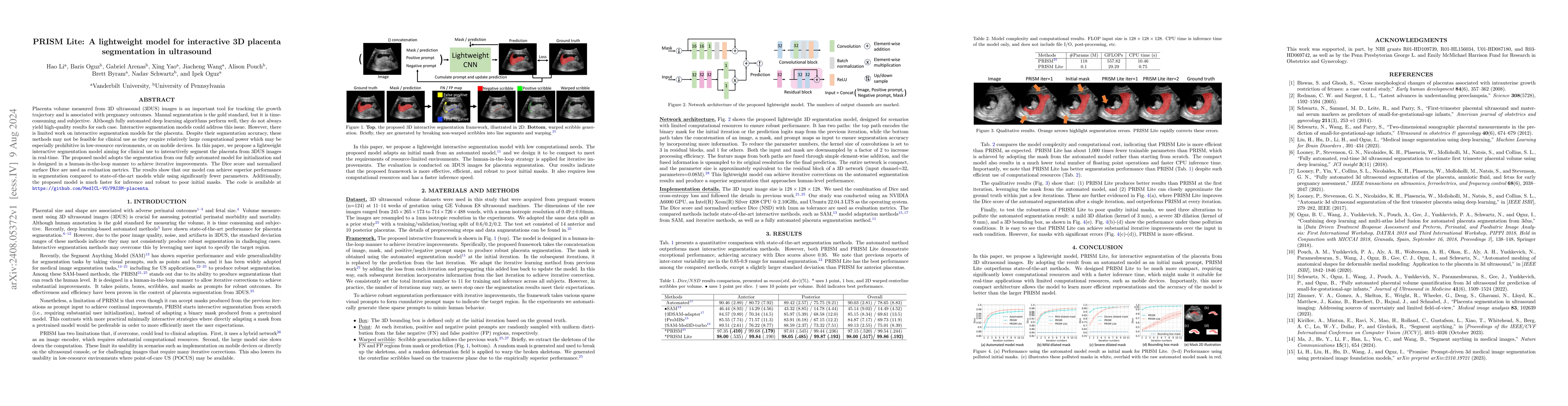
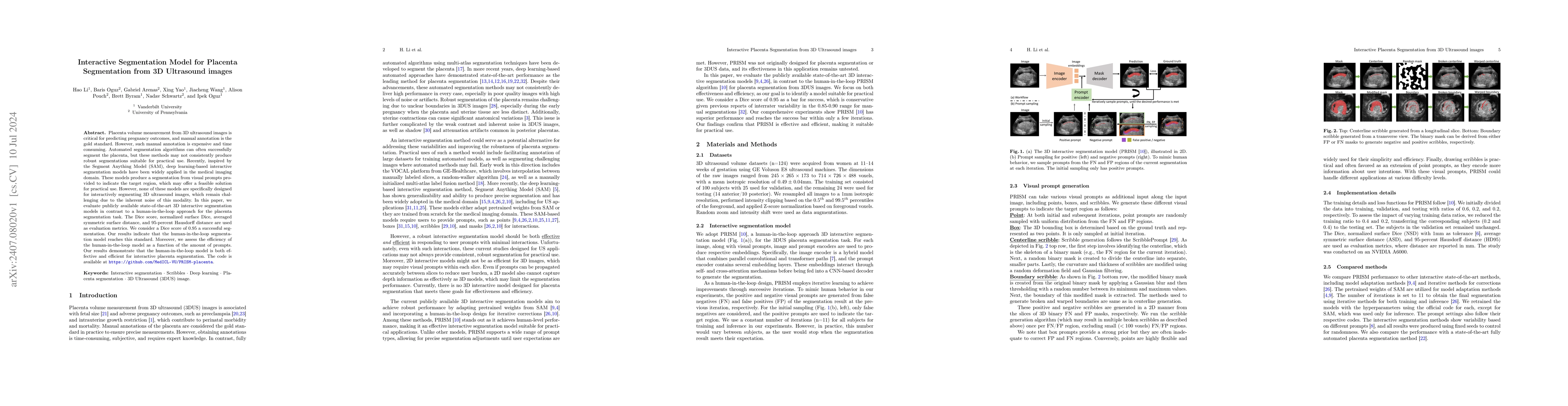
Placenta volume measured from 3D ultrasound (3DUS) images is an important tool for tracking the growth trajectory and is associated with pregnancy outcomes. Manual segmentation is the gold standard, b...
Our hypothesis is that UDA using diffusion-weighted images, generated with a unified model, offers a promising and reliable strategy for enhancing the performance of supervised learning models in mult...
Deep learning has shown remarkable performance in medical image segmentation. However, despite its promise, deep learning has many challenges in practice due to its inability to effectively transition...
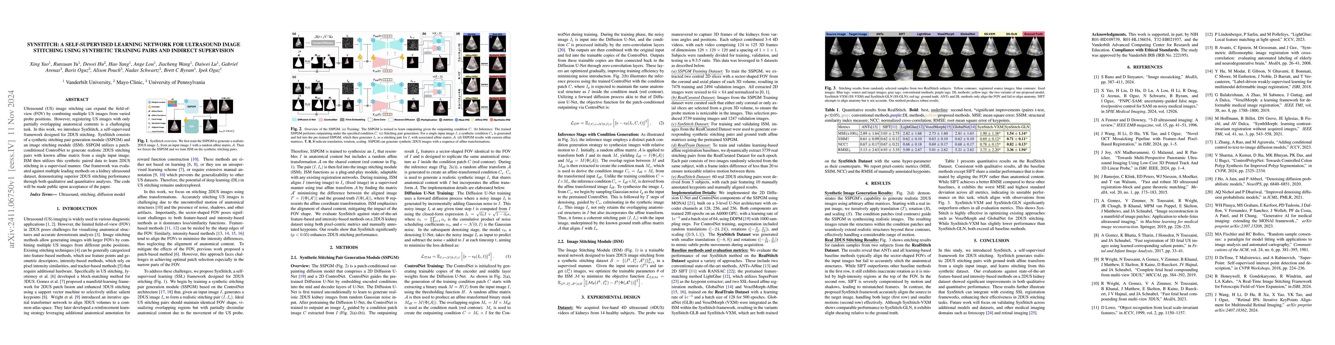
Ultrasound (US) image stitching can expand the field-of-view (FOV) by combining multiple US images from varied probe positions. However, registering US images with only partially overlapping anatomica...
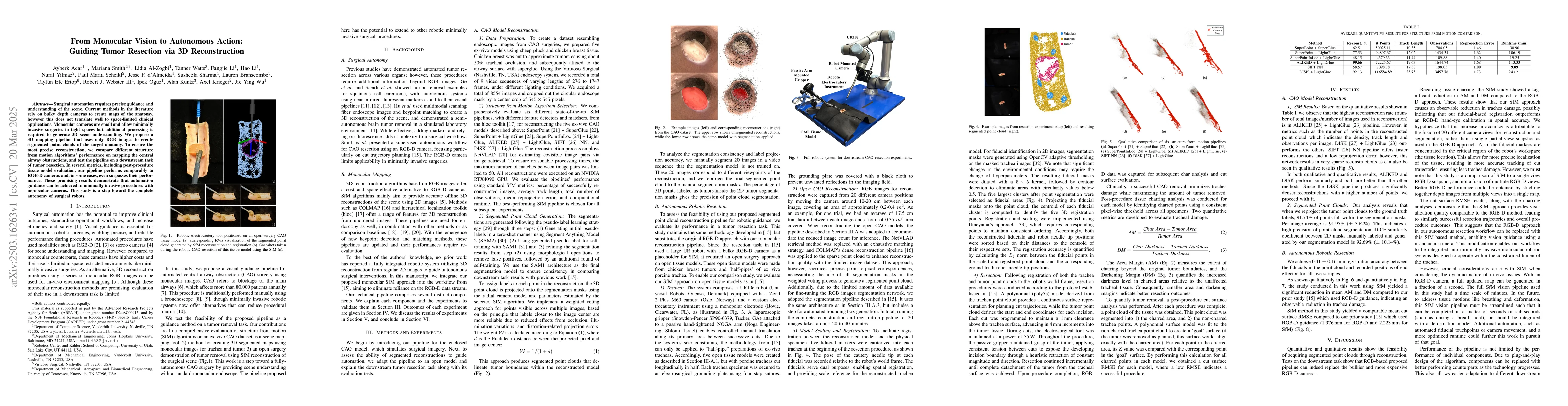
Surgical automation requires precise guidance and understanding of the scene. Current methods in the literature rely on bulky depth cameras to create maps of the anatomy, however this does not transla...
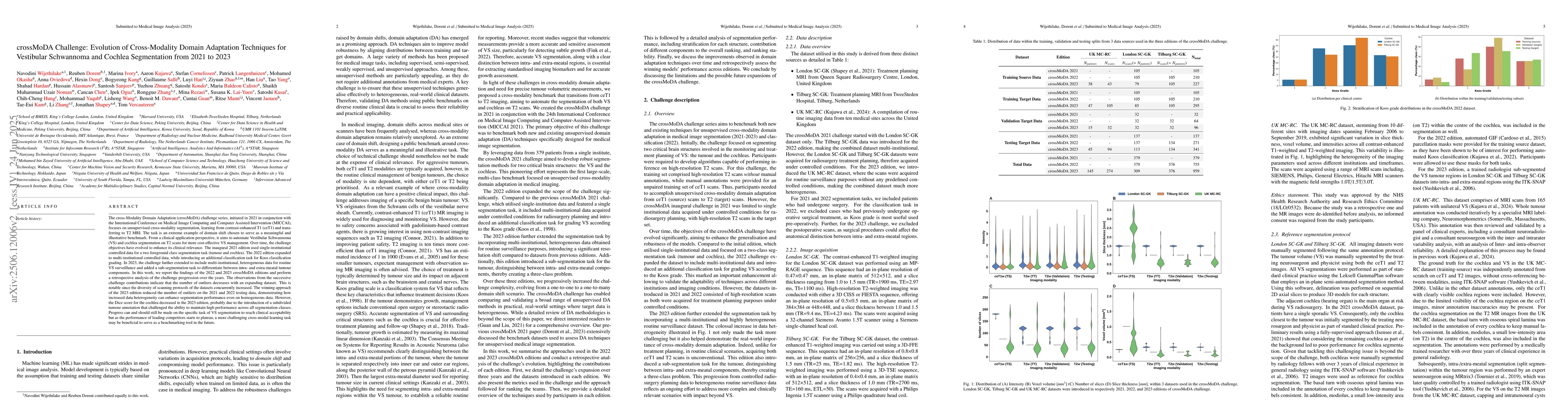
The cross-Modality Domain Adaptation (crossMoDA) challenge series, initiated in 2021 in conjunction with the International Conference on Medical Image Computing and Computer Assisted Intervention (MIC...
Semantic segmentation networks (SSNs) play a critical role in domains such as medical imaging, autonomous driving, and environmental monitoring, where safety hinges on reliable model behavior under un...
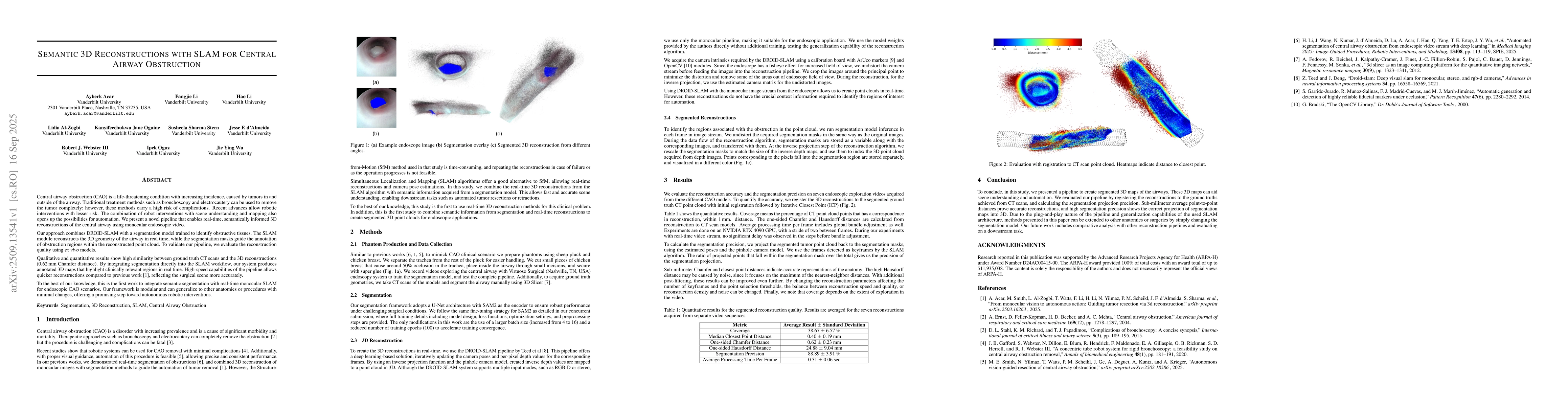
Central airway obstruction (CAO) is a life-threatening condition with increasing incidence, caused by tumors in and outside of the airway. Traditional treatment methods such as bronchoscopy and electr...
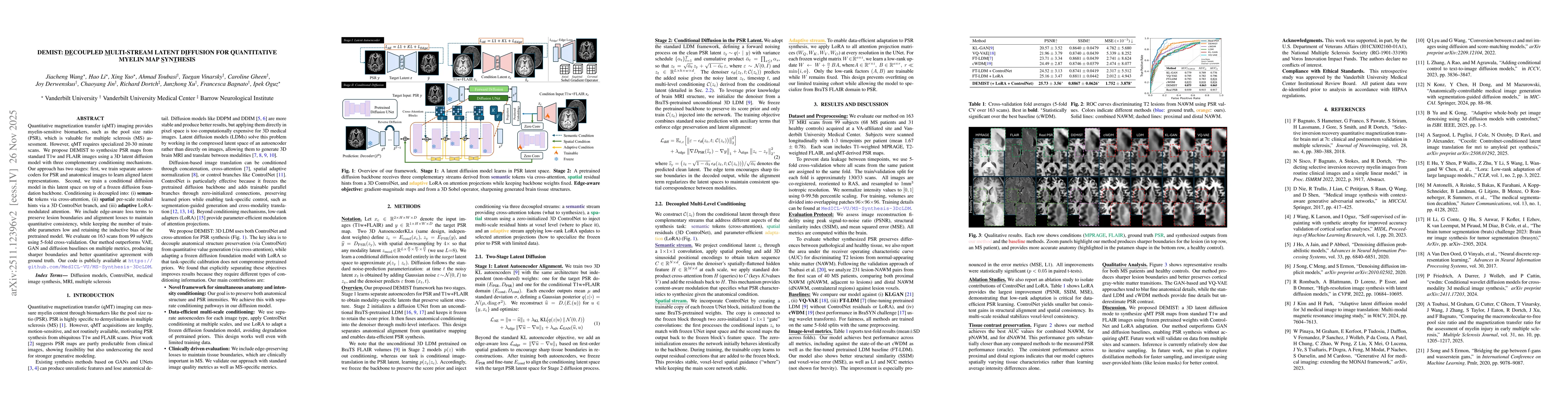
Quantitative magnetization transfer (qMT) imaging provides myelin-sensitive biomarkers, such as the pool size ratio (PSR), which is valuable for multiple sclerosis (MS) assessment. However, qMT requir...
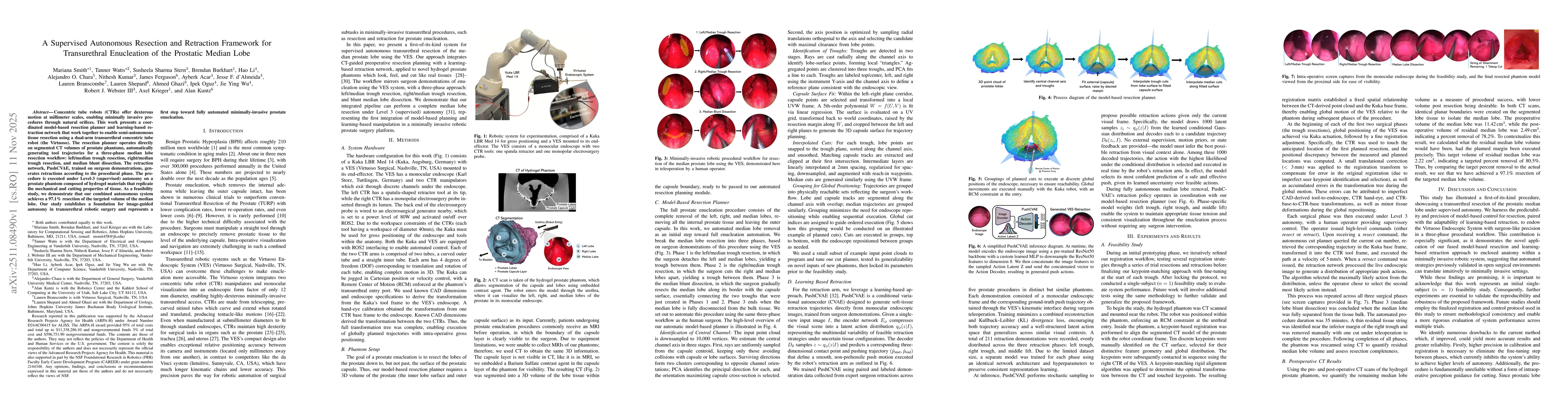
Concentric tube robots (CTRs) offer dexterous motion at millimeter scales, enabling minimally invasive procedures through natural orifices. This work presents a coordinated model-based resection plann...
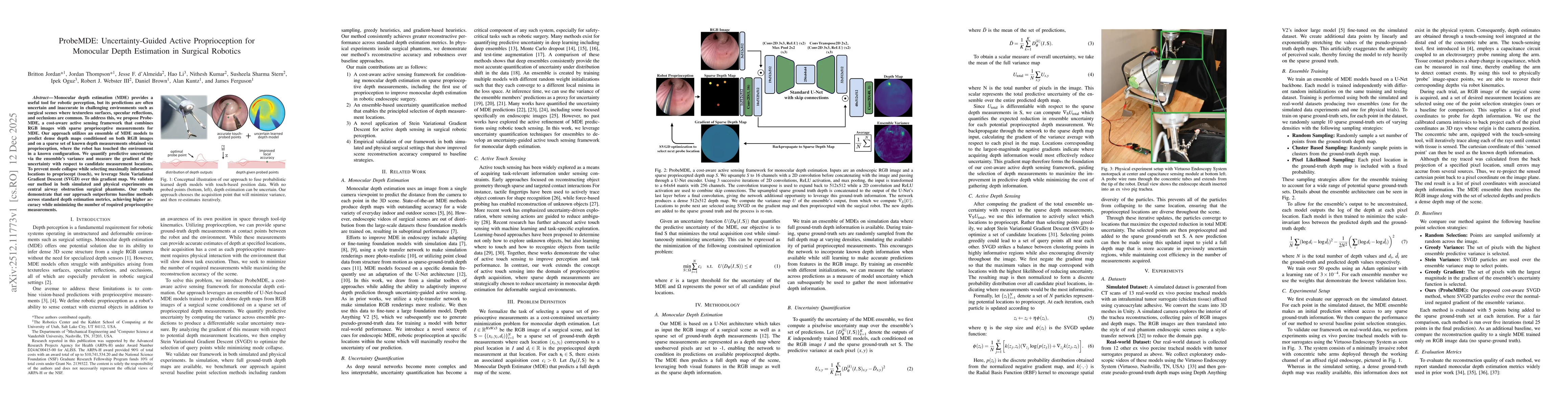
Monocular depth estimation (MDE) provides a useful tool for robotic perception, but its predictions are often uncertain and inaccurate in challenging environments such as surgical scenes where texture...
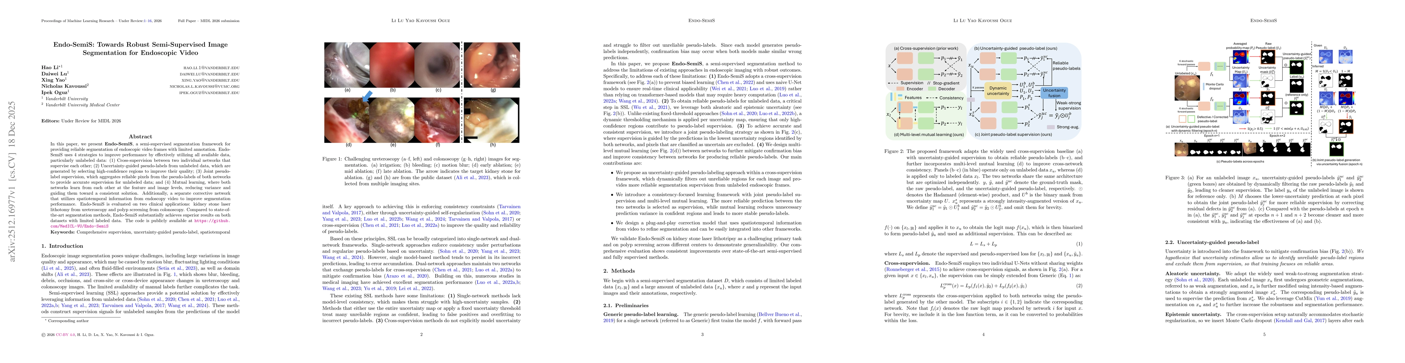
In this paper, we present Endo-SemiS, a semi-supervised segmentation framework for providing reliable segmentation of endoscopic video frames with limited annotation. EndoSemiS uses 4 strategies to im...

This work presents EndoStreamDepth, a monocular depth estimation framework for endoscopic video streams. It provides accurate depth maps with sharp anatomical boundaries for each frame, temporally con...
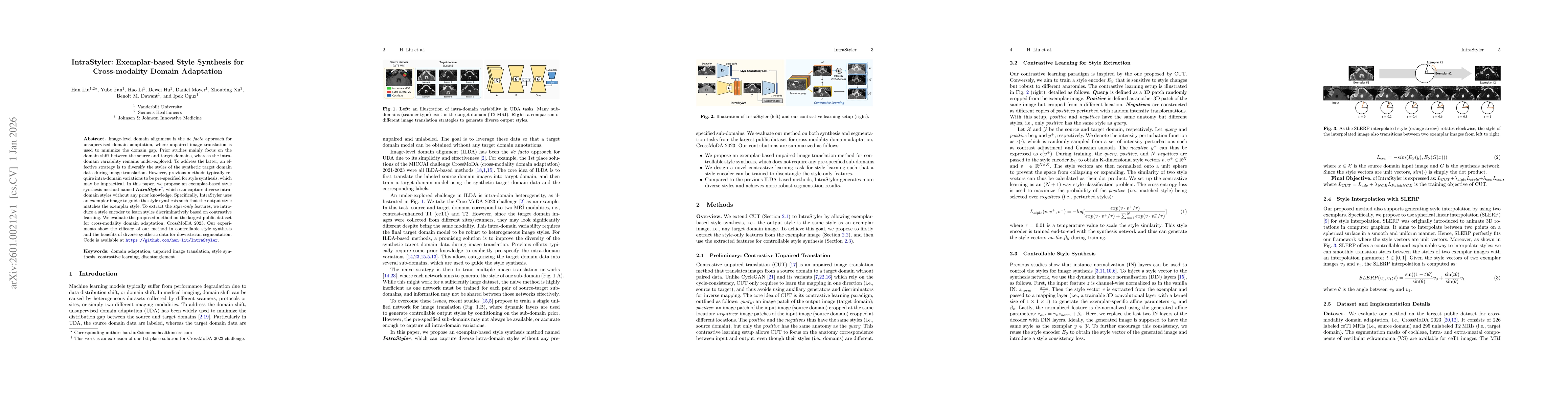
Image-level domain alignment is the de facto approach for unsupervised domain adaptation, where unpaired image translation is used to minimize the domain gap. Prior studies mainly focus on the domain ...
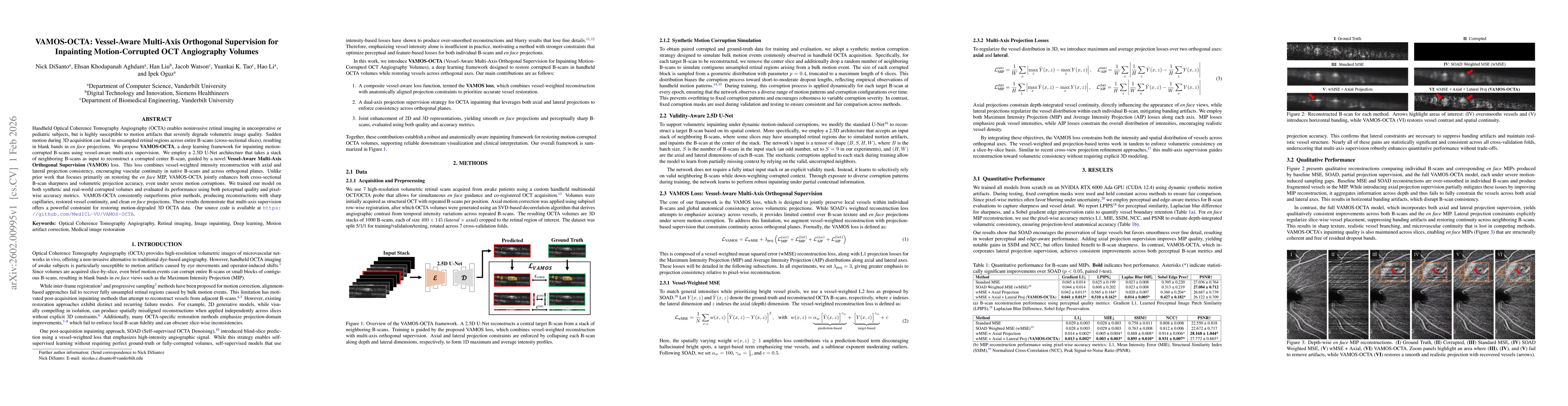
Handheld Optical Coherence Tomography Angiography (OCTA) enables noninvasive retinal imaging in uncooperative or pediatric subjects, but is highly susceptible to motion artifacts that severely degrade...
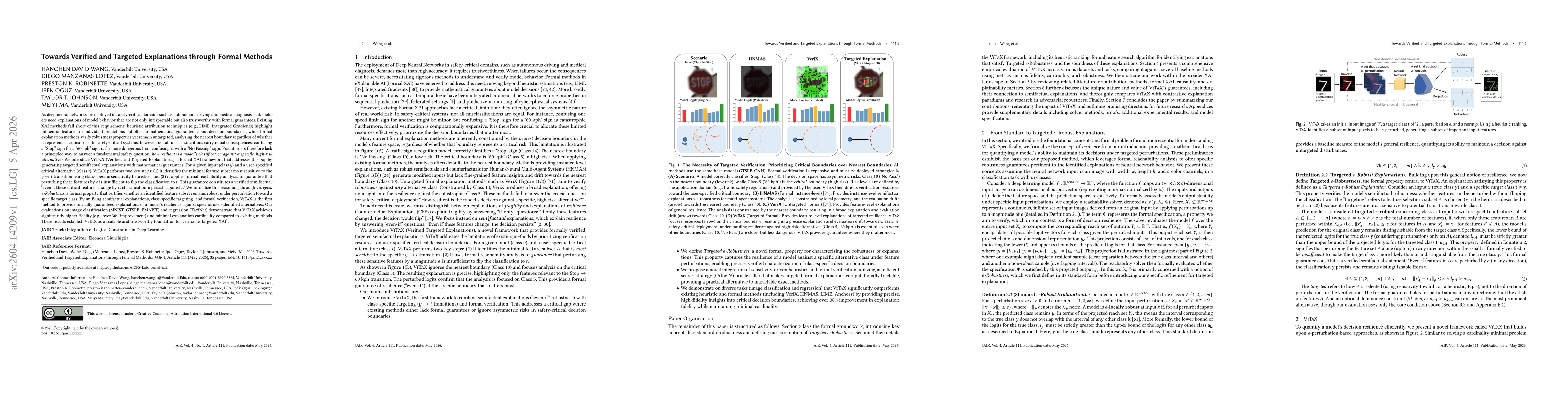
As deep neural networks are deployed in safety-critical domains such as autonomous driving and medical diagnosis, stakeholders need explanations that are interpretable but also trustworthy with formal...