Academic Profile
Statistics
Similar Authors
Papers on arXiv
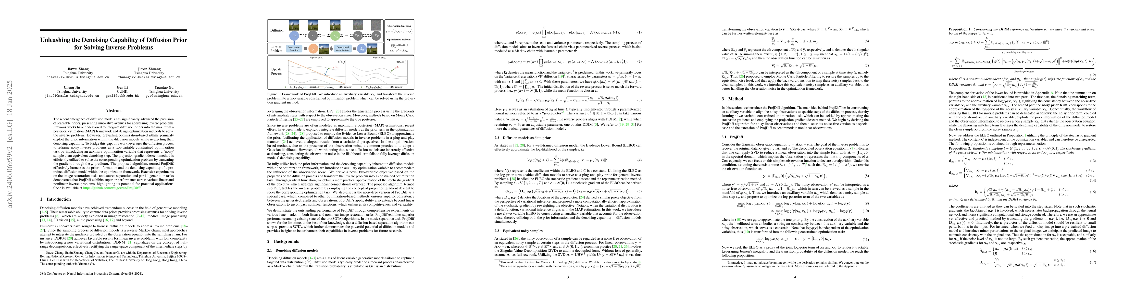
The recent emergence of diffusion models has significantly advanced the precision of learnable priors, presenting innovative avenues for addressing inverse problems. Since inverse problems inherentl...
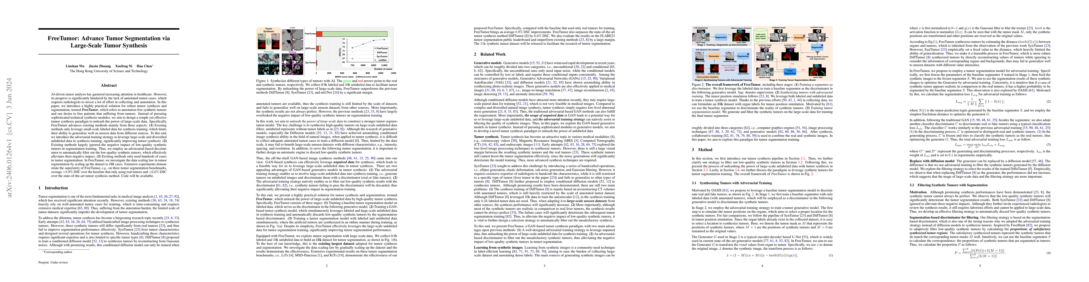
AI-driven tumor analysis has garnered increasing attention in healthcare. However, its progress is significantly hindered by the lack of annotated tumor cases, which requires radiologists to invest ...
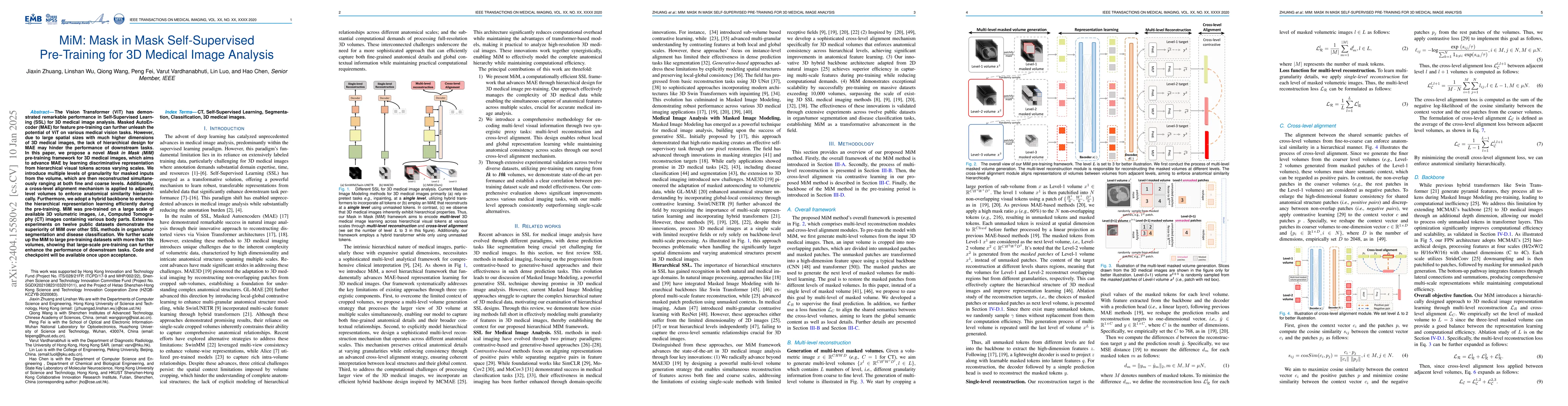
The Vision Transformer (ViT) has demonstrated remarkable performance in Self-Supervised Learning (SSL) for 3D medical image analysis. Mask AutoEncoder (MAE) for feature pre-training can further unle...
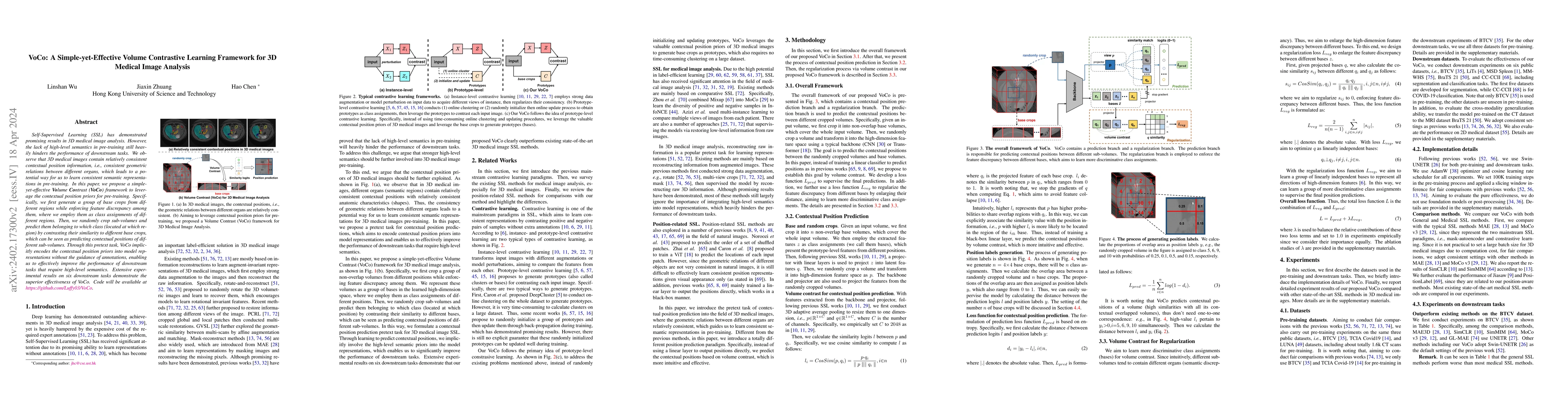
Self-Supervised Learning (SSL) has demonstrated promising results in 3D medical image analysis. However, the lack of high-level semantics in pre-training still heavily hinders the performance of dow...
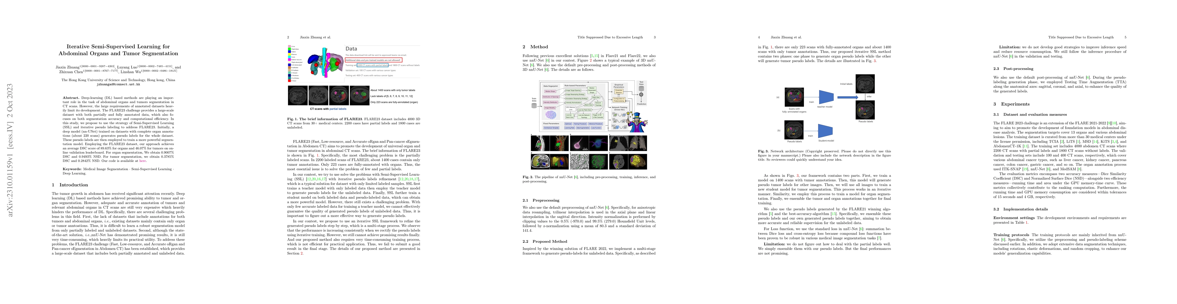
Deep-learning (DL) based methods are playing an important role in the task of abdominal organs and tumors segmentation in CT scans. However, the large requirements of annotated datasets heavily limi...
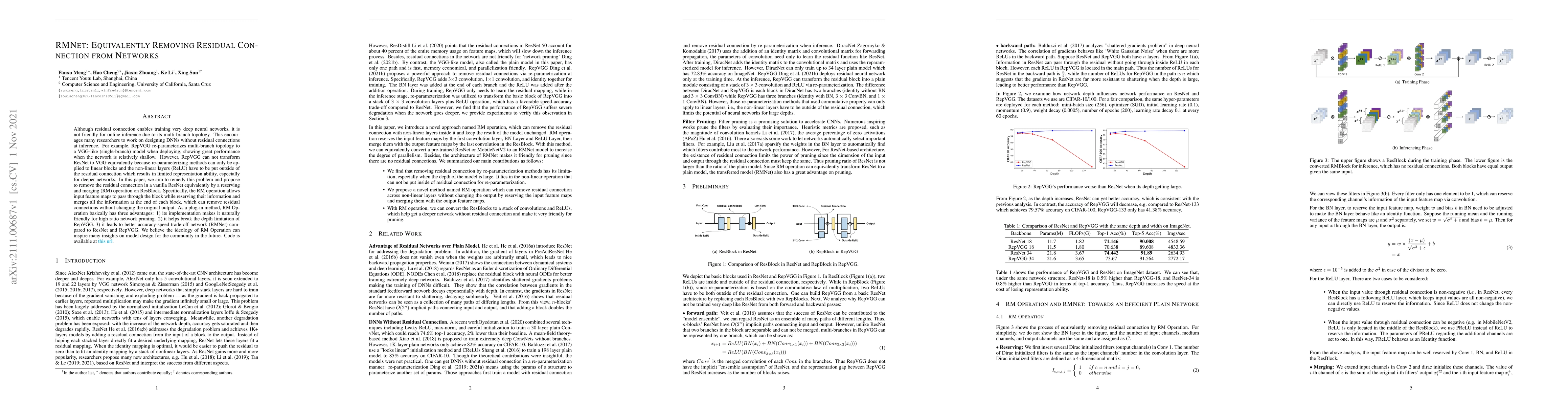
Although residual connection enables training very deep neural networks, it is not friendly for online inference due to its multi-branch topology. This encourages many researchers to work on designi...
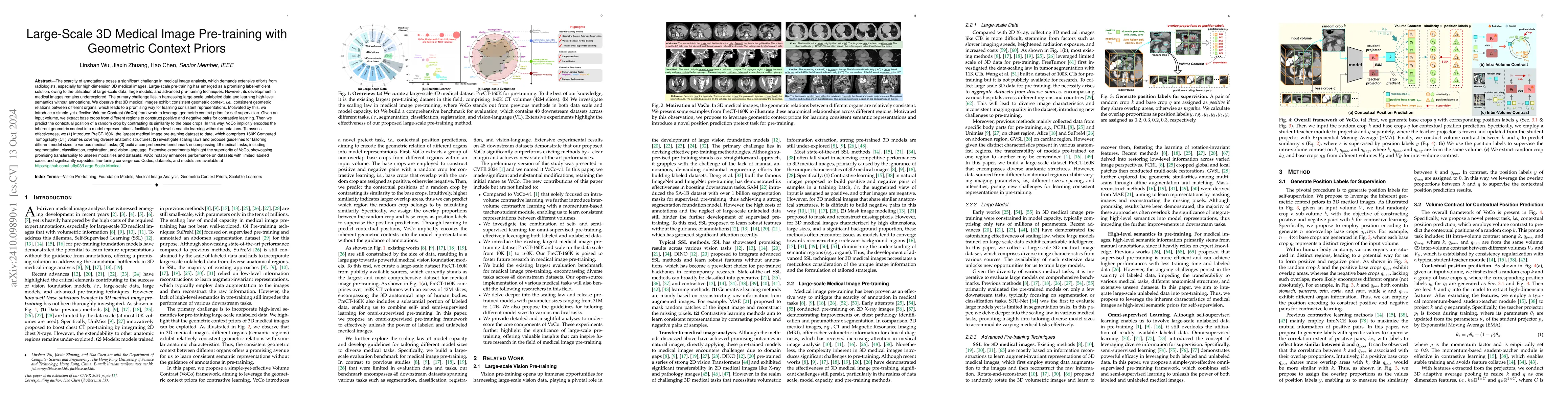
The scarcity of annotations poses a significant challenge in medical image analysis. Large-scale pre-training has emerged as a promising label-efficient solution, owing to the utilization of large-sca...
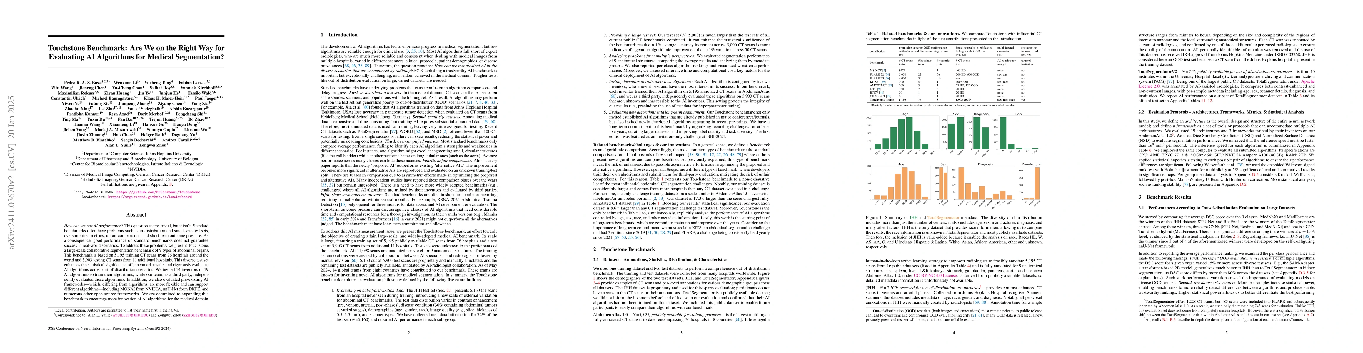
How can we test AI performance? This question seems trivial, but it isn't. Standard benchmarks often have problems such as in-distribution and small-size test sets, oversimplified metrics, unfair comp...
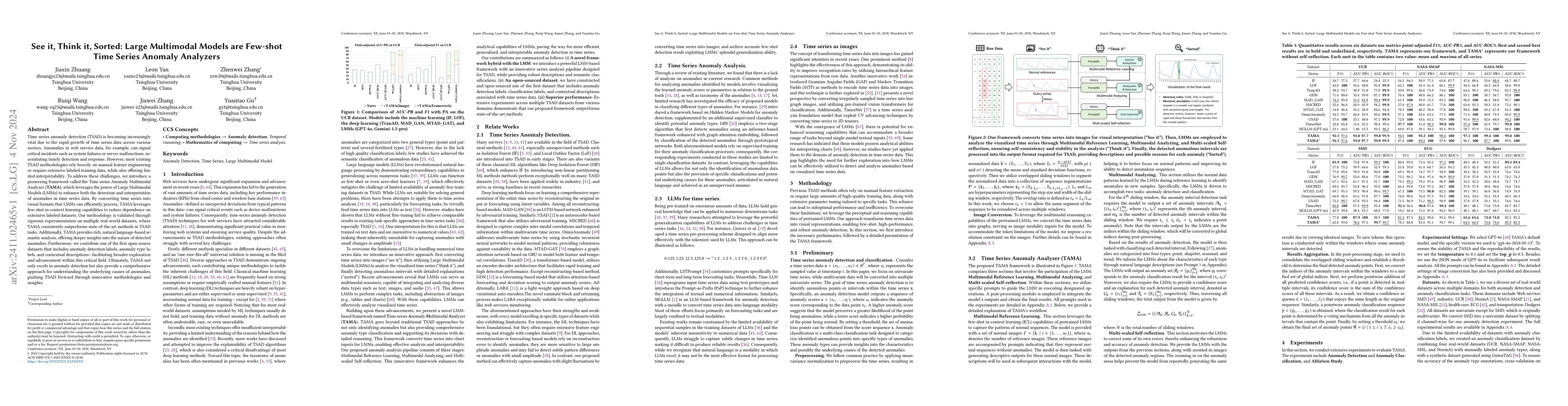
Time series anomaly detection (TSAD) is becoming increasingly vital due to the rapid growth of time series data across various sectors. Anomalies in web service data, for example, can signal critical ...
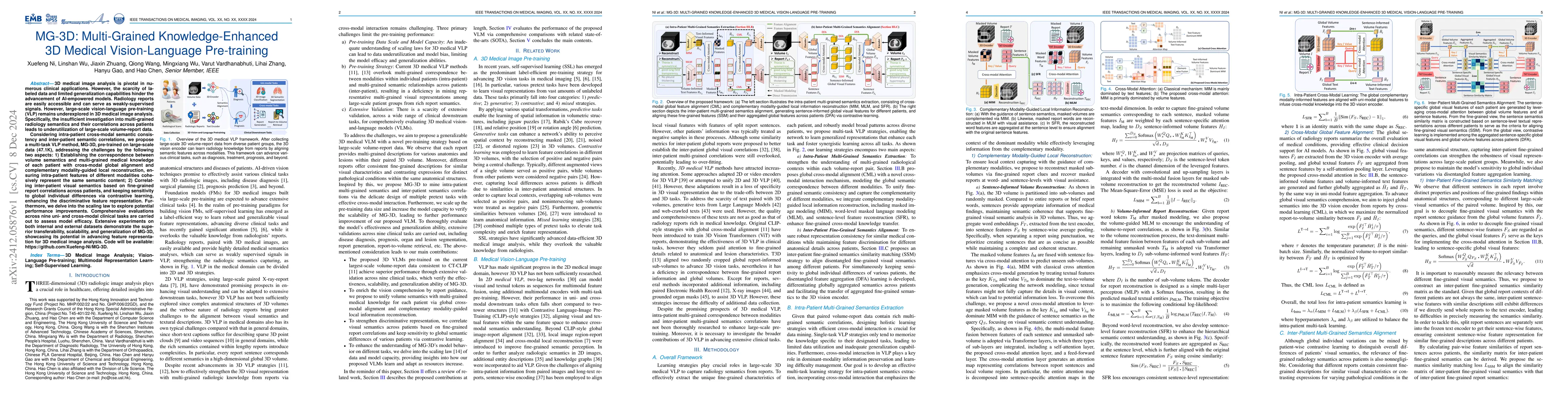
3D medical image analysis is pivotal in numerous clinical applications. However, the scarcity of labeled data and limited generalization capabilities hinder the advancement of AI-empowered models. Rad...
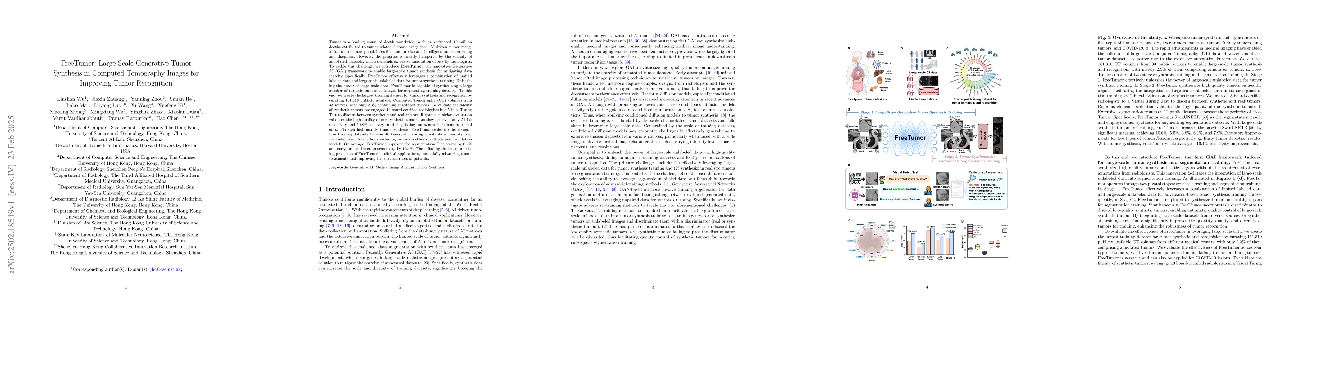
Tumor is a leading cause of death worldwide, with an estimated 10 million deaths attributed to tumor-related diseases every year. AI-driven tumor recognition unlocks new possibilities for more precise...
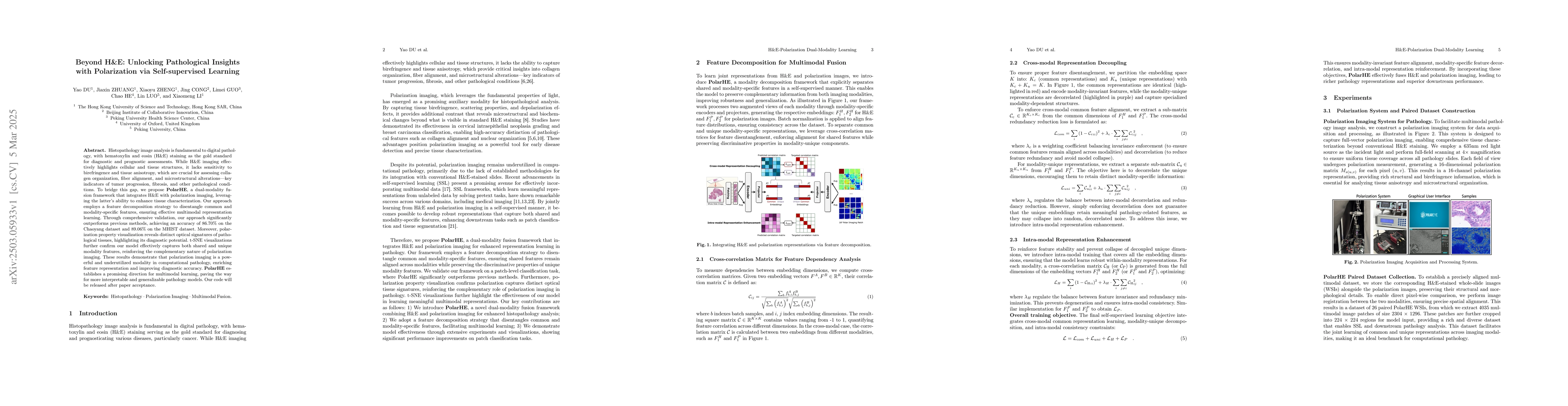
Histopathology image analysis is fundamental to digital pathology, with hematoxylin and eosin (H&E) staining as the gold standard for diagnostic and prognostic assessments. While H&E imaging effective...
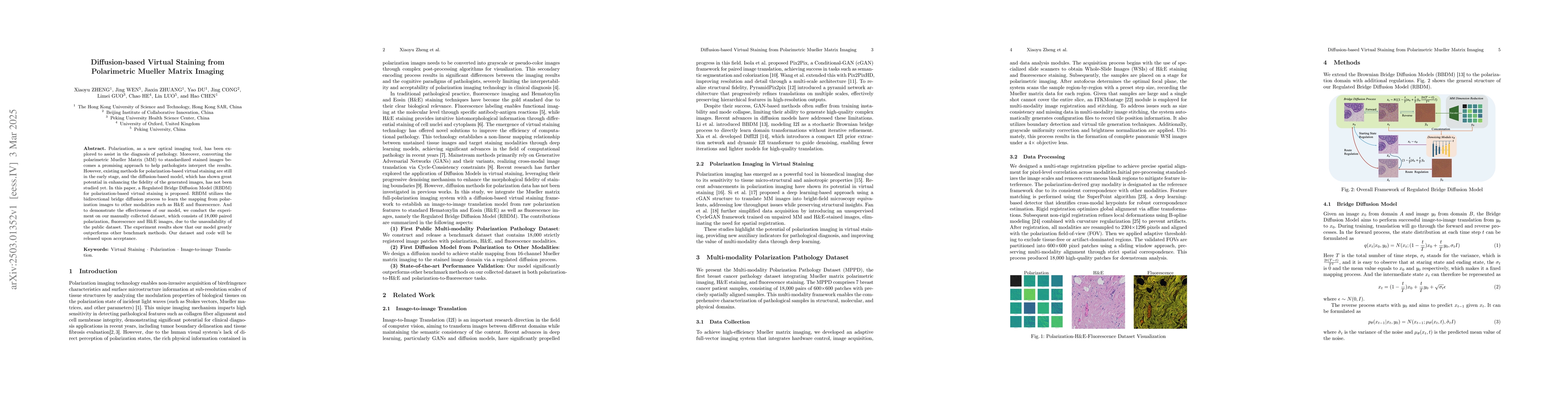
Polarization, as a new optical imaging tool, has been explored to assist in the diagnosis of pathology. Moreover, converting the polarimetric Mueller Matrix (MM) to standardized stained images becomes...
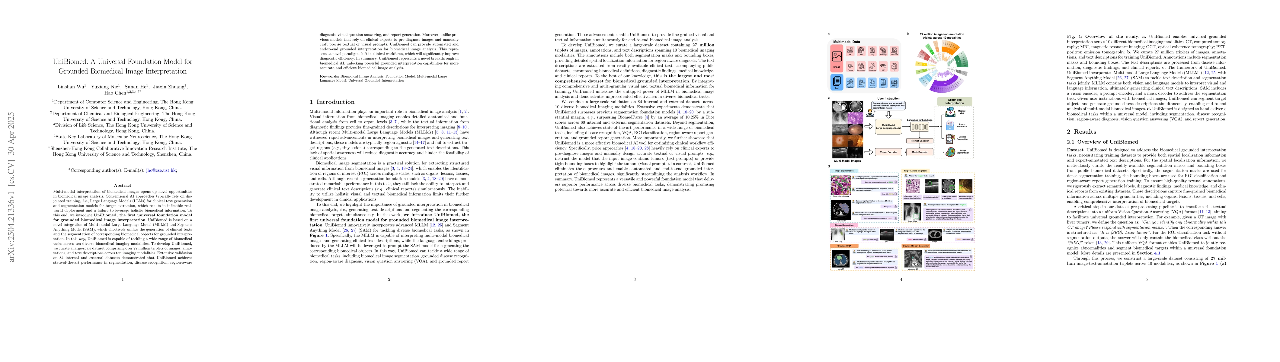
Multi-modal interpretation of biomedical images opens up novel opportunities in biomedical image analysis. Conventional AI approaches typically rely on disjointed training, i.e., Large Language Models...
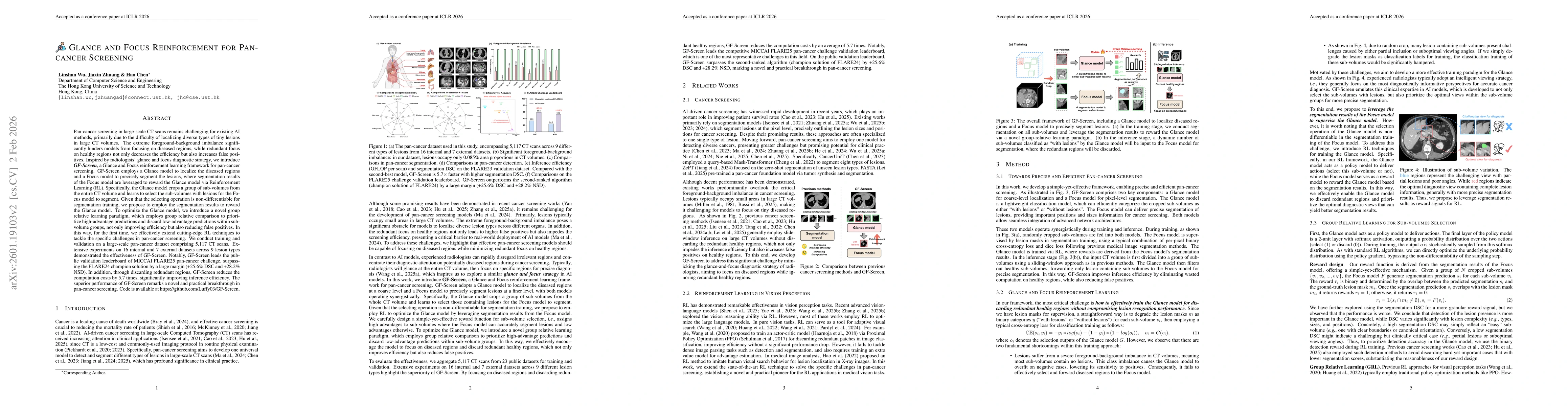
Pan-cancer screening in large-scale CT scans remains challenging for existing AI methods, primarily due to the difficulty of localizing diverse types of tiny lesions in large CT volumes. The extreme f...
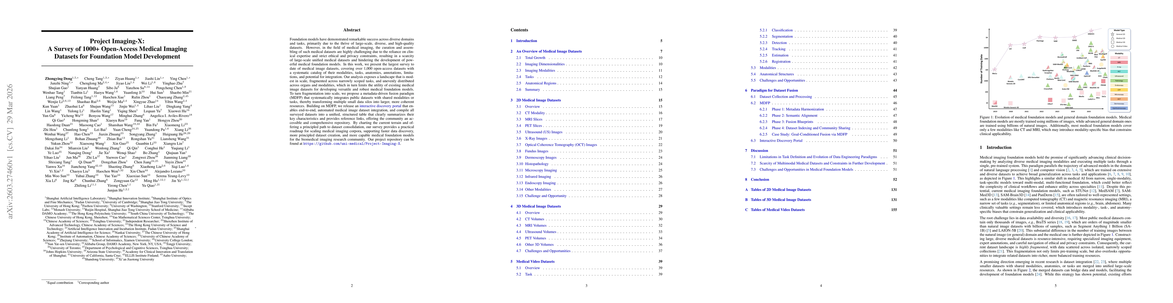
Foundation models have demonstrated remarkable success across diverse domains and tasks, primarily due to the thrive of large-scale, diverse, and high-quality datasets. However, in the field of medica...
Vision-language models (VLMs) such as CLIP exhibit strong Out-of-distribution (OOD) detection capabilities by aligning visual and textual representations. Recent CLIP-based test-time adaptation method...