Academic Profile
Statistics
Similar Authors
Papers on arXiv
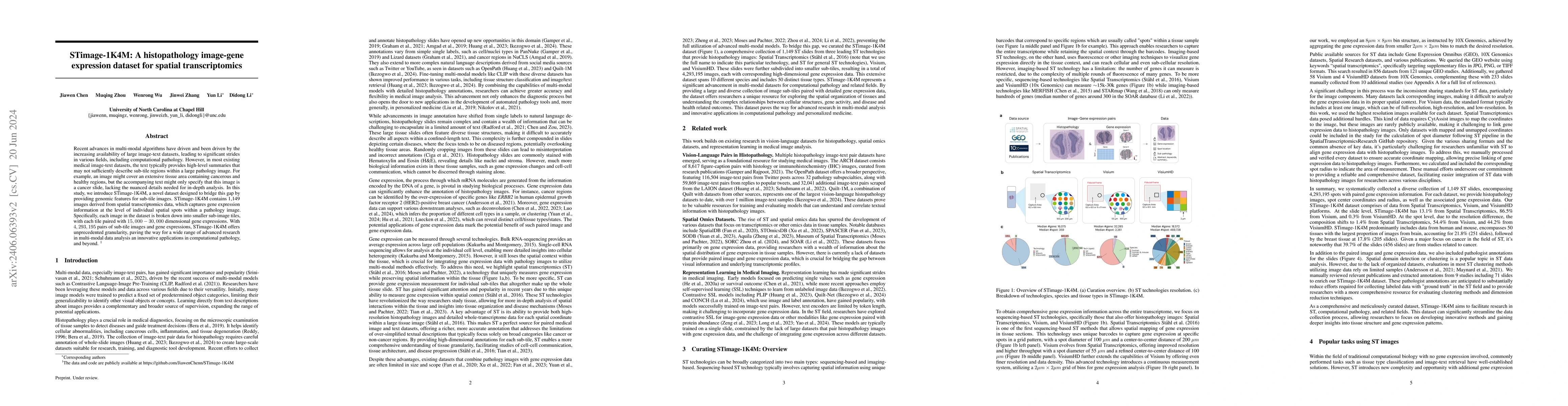
Recent advances in multi-modal algorithms have driven and been driven by the increasing availability of large image-text datasets, leading to significant strides in various fields, including computati...
Purpose: To develop a pipeline for motion artifact correction in mGRE and quantitative susceptibility mapping (QSM). Methods: Deep learning is integrated with autofocus to improve motion artifact su...
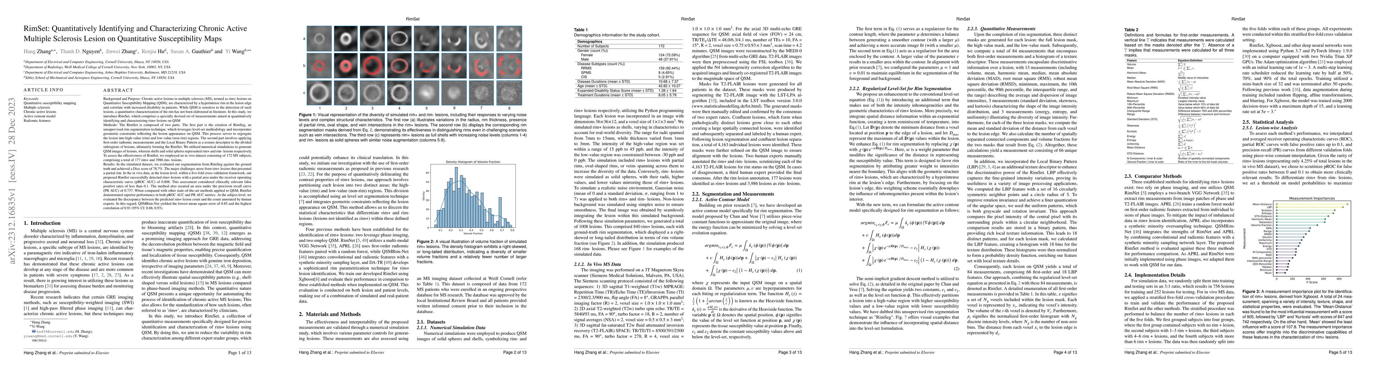
Background: Rim+ lesions in multiple sclerosis (MS), detectable via Quantitative Susceptibility Mapping (QSM), correlate with increased disability. Existing literature lacks quantitative analysis of...
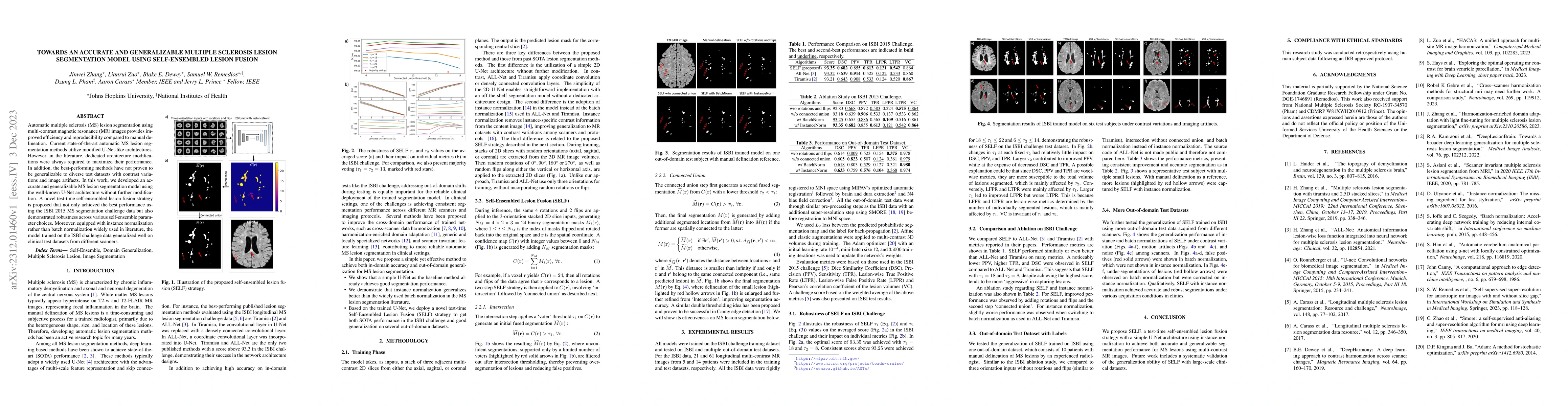
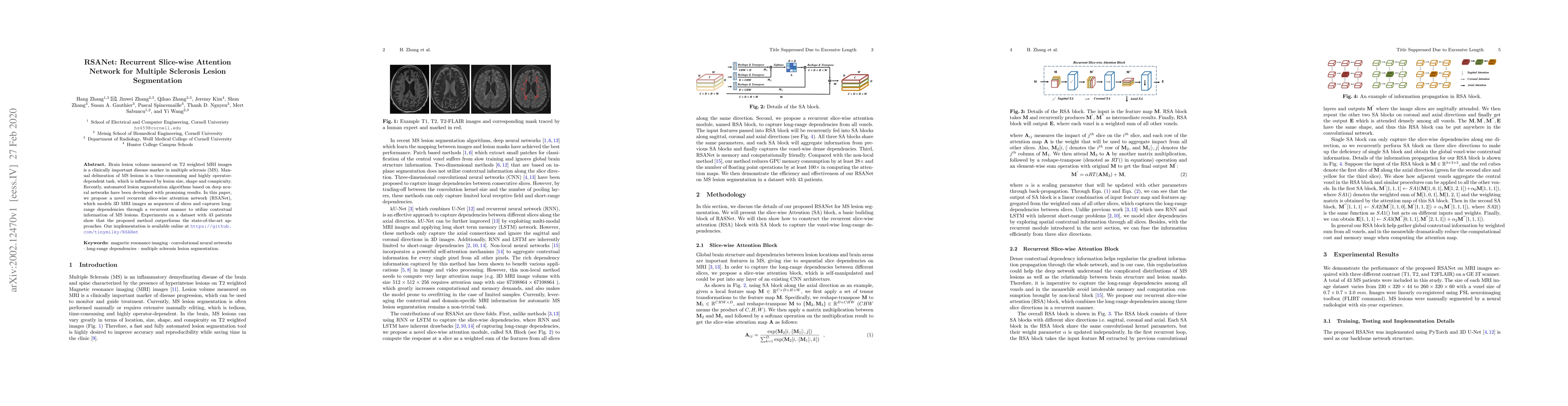
Automatic multiple sclerosis (MS) lesion segmentation using multi-contrast magnetic resonance (MR) images provides improved efficiency and reproducibility compared to manual delineation. Current sta...
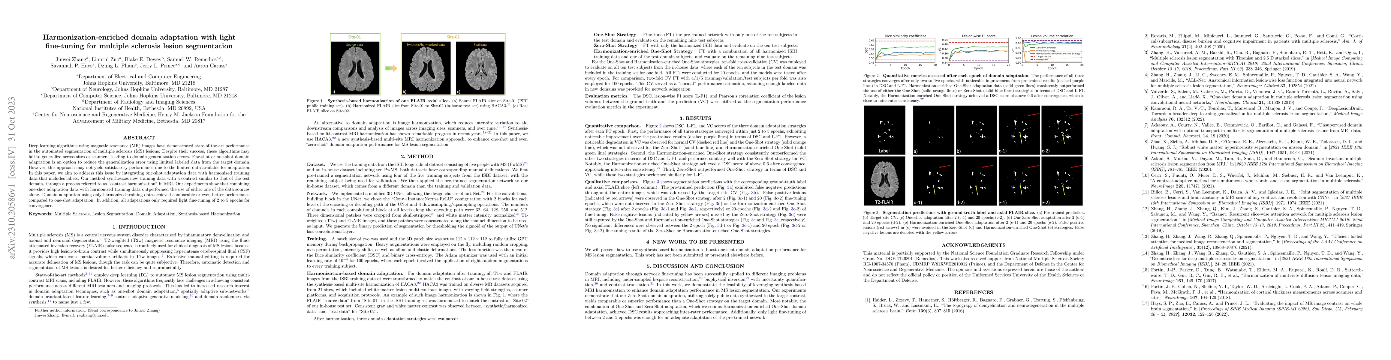
Deep learning algorithms utilizing magnetic resonance (MR) images have demonstrated cutting-edge proficiency in autonomously segmenting multiple sclerosis (MS) lesions. Despite their achievements, t...
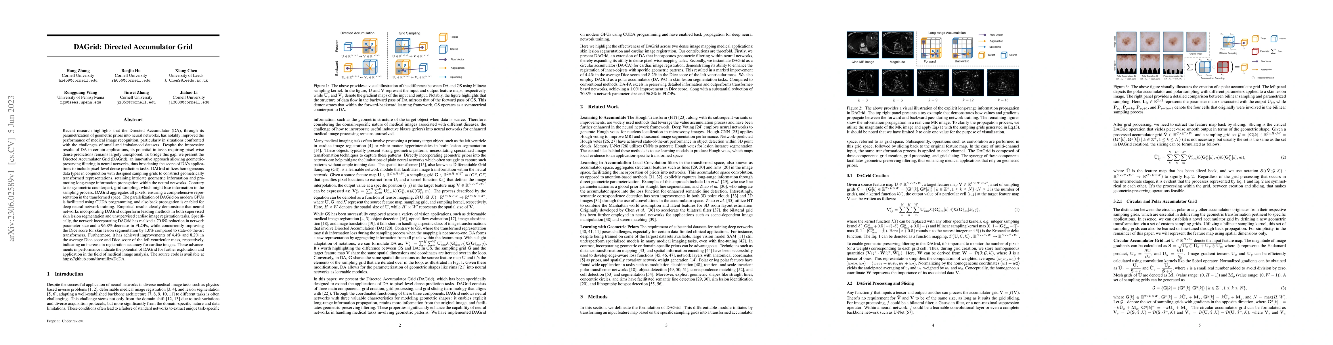
Recent research highlights that the Directed Accumulator (DA), through its parametrization of geometric priors into neural networks, has notably improved the performance of medical image recognition...
Purpose: To improve the generalization ability of convolutional neural network (CNN) based prediction of quantitative susceptibility mapping (QSM) from high-pass filtered phase (HPFP) image. Methods...
Purpose: To develop a method for rapid sub-millimeter T1, T2, T2* and QSM mapping in a single scan using multi-contrast Learned Acquisition and Reconstruction Optimization (mcLARO). Methods: A pul...
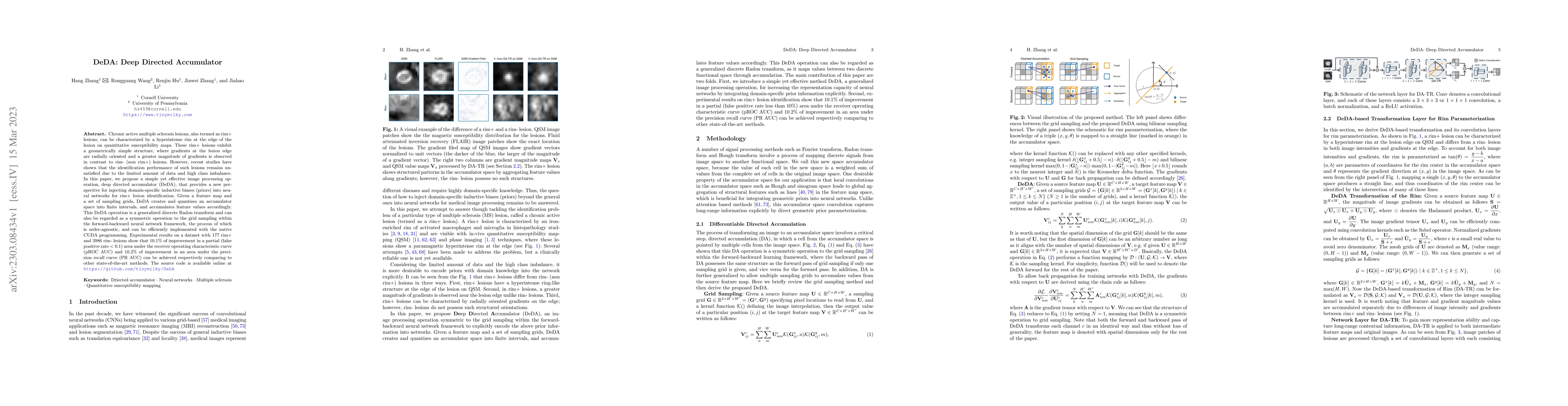
Chronic active multiple sclerosis lesions, also termed as rim+ lesions, can be characterized by a hyperintense rim at the edge of the lesion on quantitative susceptibility maps. These rim+ lesions e...
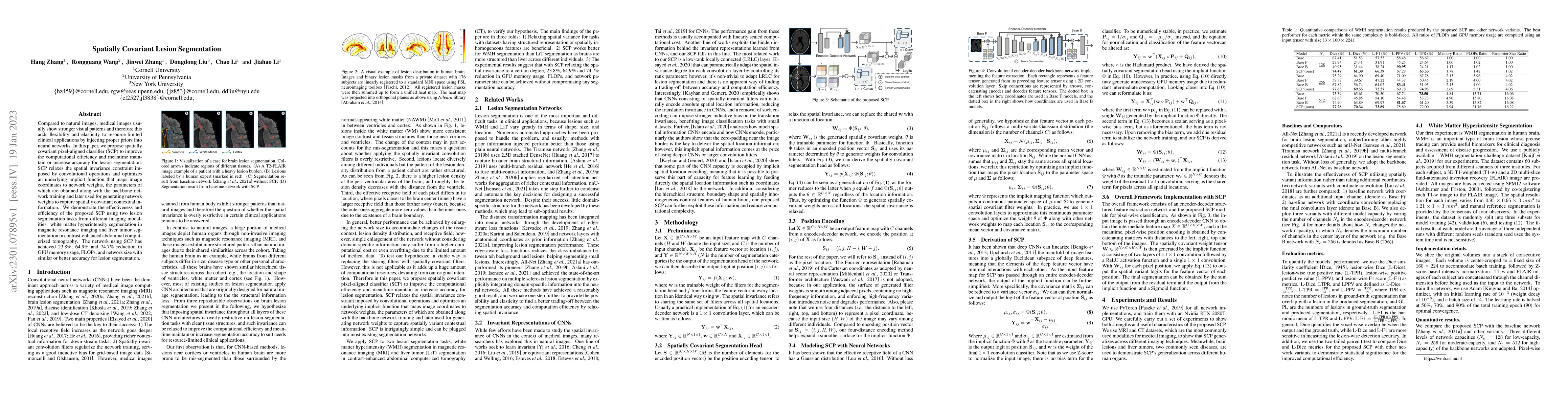
Compared to natural images, medical images usually show stronger visual patterns and therefore this adds flexibility and elasticity to resource-limited clinical applications by injecting proper prio...
Quantitative susceptibility mapping (QSM) involves acquisition and reconstruction of a series of images at multi-echo time points to estimate tissue field, which prolongs scan time and requires spec...
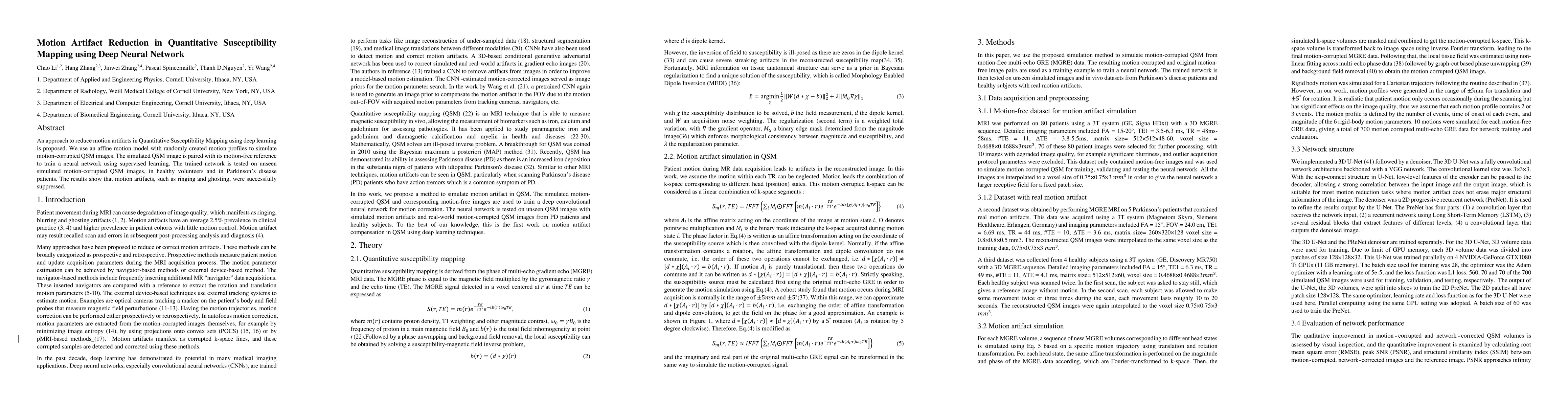
An approach to reduce motion artifacts in Quantitative Susceptibility Mapping using deep learning is proposed. We use an affine motion model with randomly created motion profiles to simulate motion-...
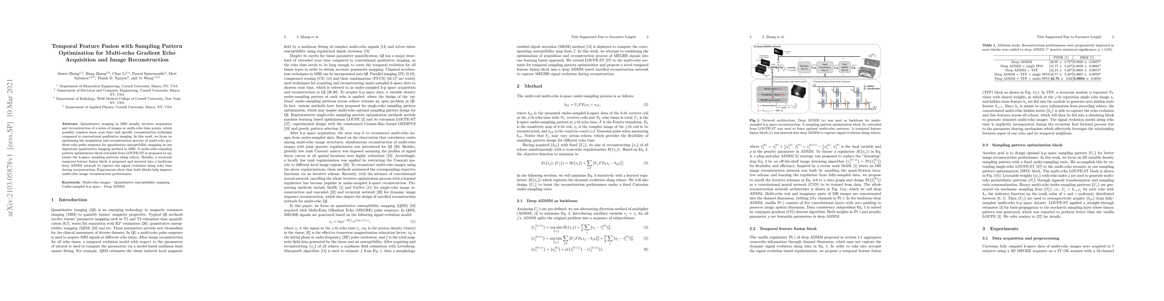
Quantitative imaging in MRI usually involves acquisition and reconstruction of a series of images at multi-echo time points, which possibly requires more scan time and specific reconstruction techni...
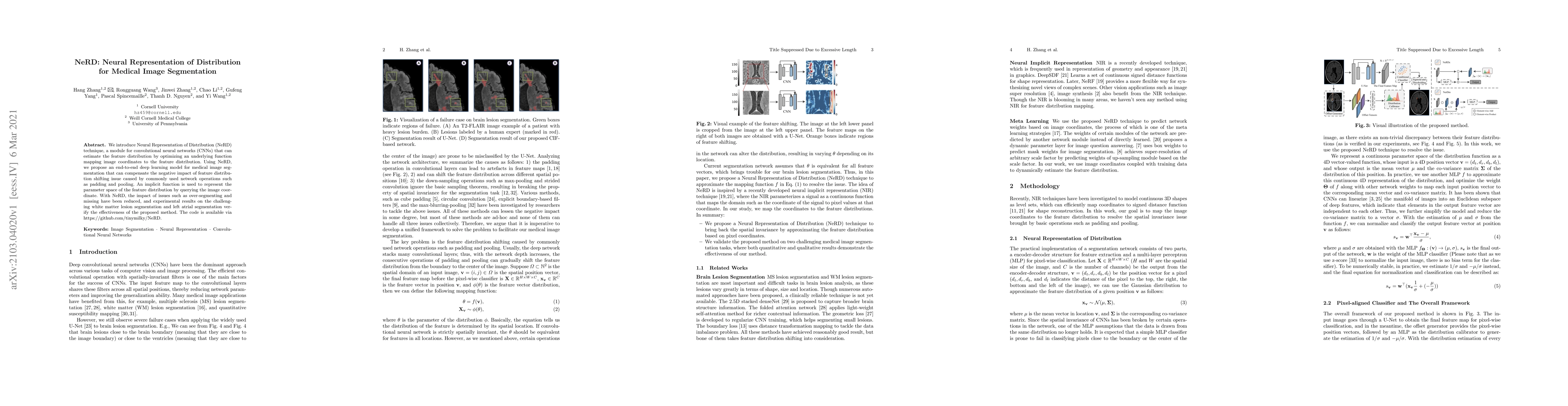
We introduce Neural Representation of Distribution (NeRD) technique, a module for convolutional neural networks (CNNs) that can estimate the feature distribution by optimizing an underlying function...
Multiple sclerosis (MS) lesions occupy a small fraction of the brain volume, and are heterogeneous with regards to shape, size and locations, which poses a great challenge for training deep learning...
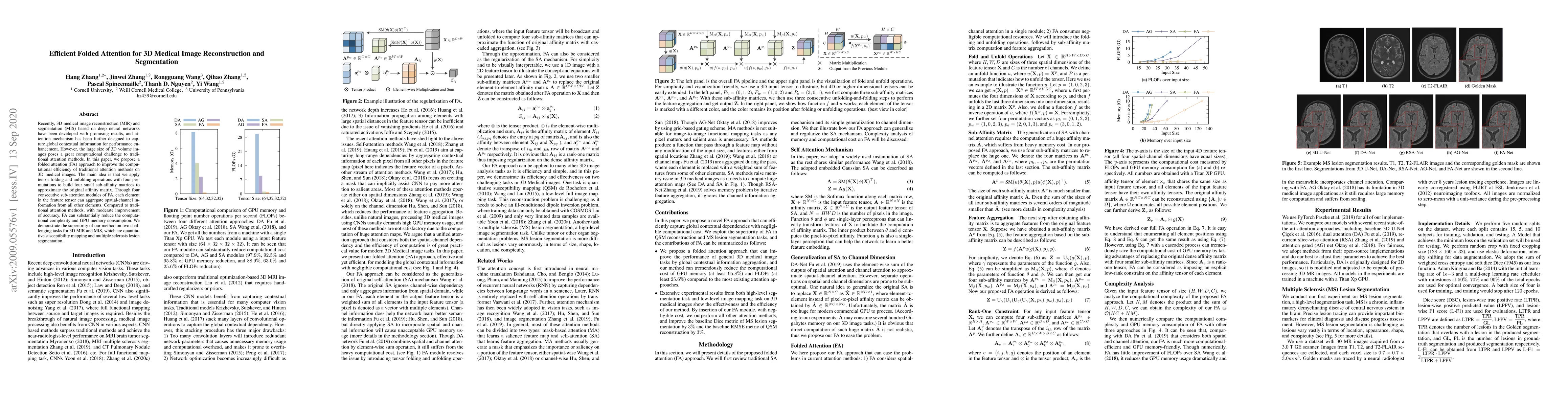
Recently, 3D medical image reconstruction (MIR) and segmentation (MIS) based on deep neural networks have been developed with promising results, and attention mechanism has been further designed to ...
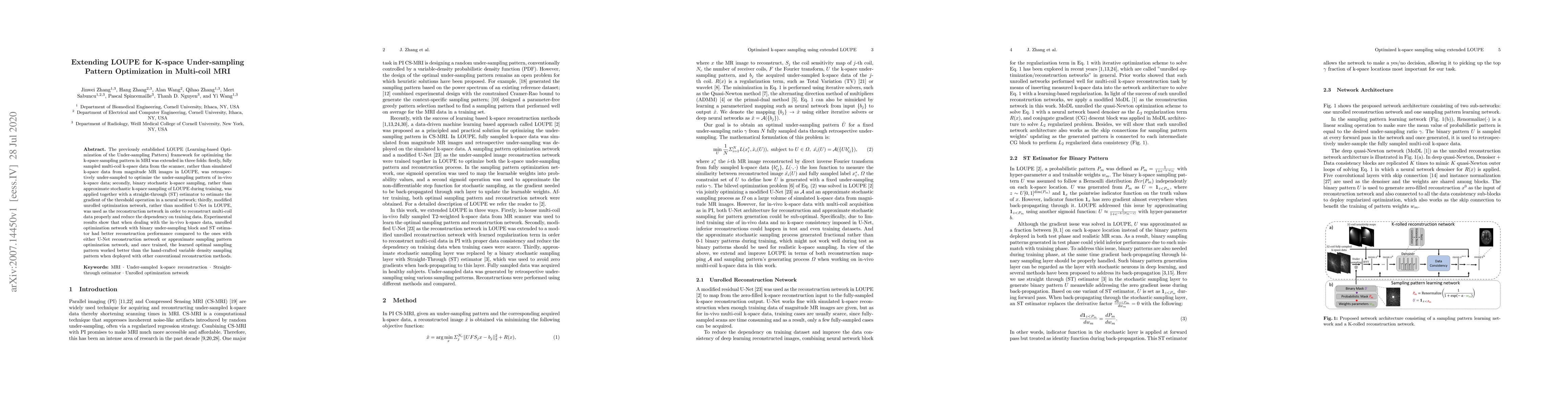
The previously established LOUPE (Learning-based Optimization of the Under-sampling Pattern) framework for optimizing the k-space sampling pattern in MRI was extended in three folds: firstly, fully ...
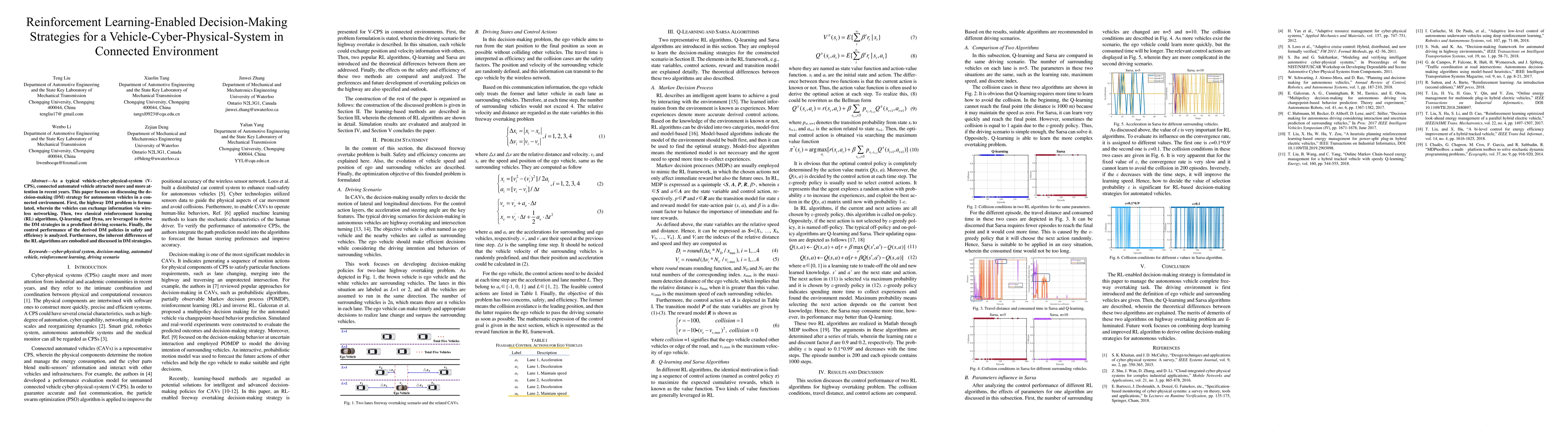
As a typical vehicle-cyber-physical-system (V-CPS), connected automated vehicles attracted more and more attention in recent years. This paper focuses on discussing the decision-making (DM) strategy...
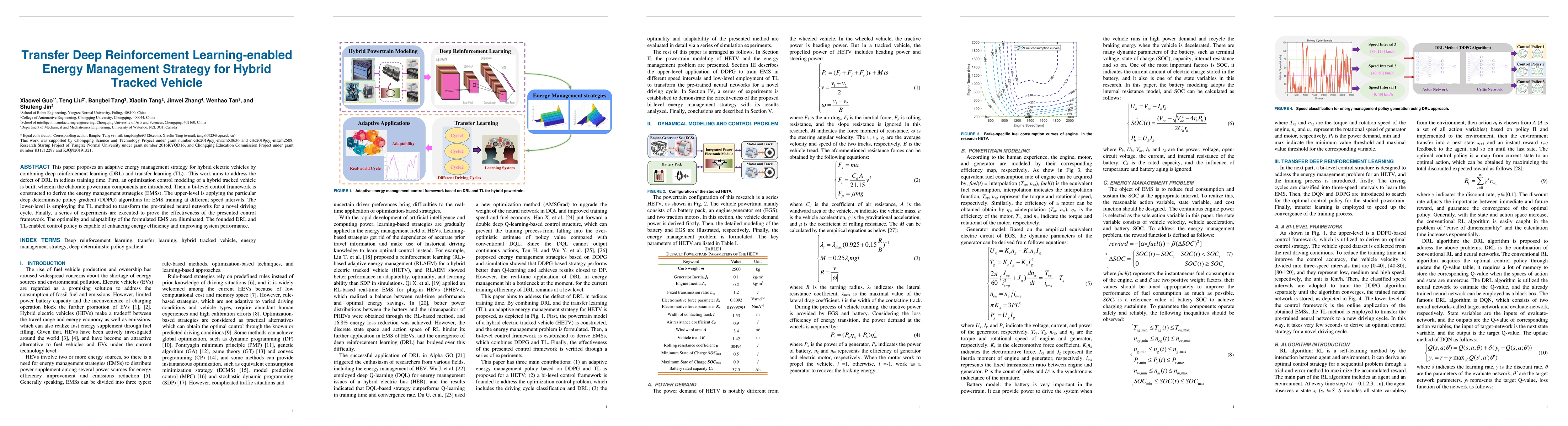
This paper proposes an adaptive energy management strategy for hybrid electric vehicles by combining deep reinforcement learning (DRL) and transfer learning (TL). This work aims to address the defec...
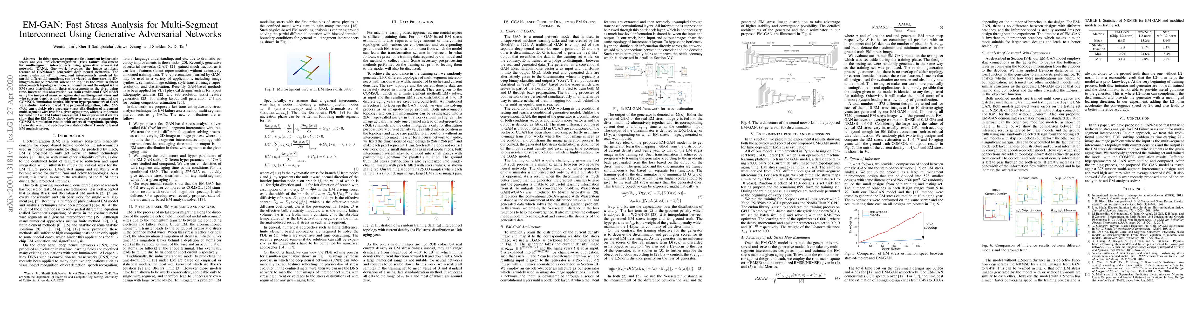
In this paper, we propose a fast transient hydrostatic stress analysis for electromigration (EM) failure assessment for multi-segment interconnects using generative adversarial networks (GANs). Our ...
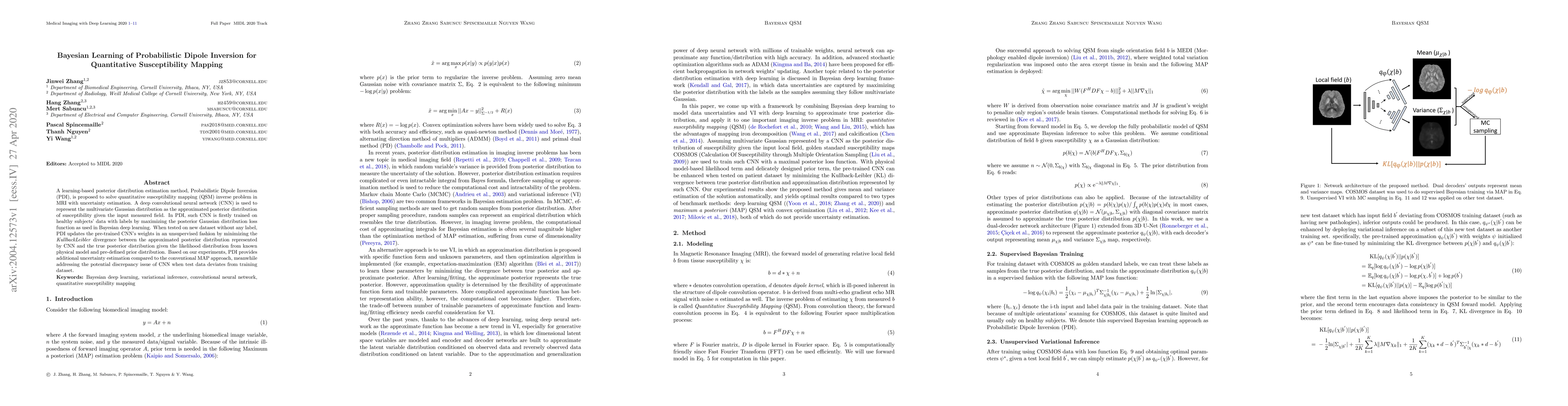
A learning-based posterior distribution estimation method, Probabilistic Dipole Inversion (PDI), is proposed to solve quantitative susceptibility mapping (QSM) inverse problem in MRI with uncertaint...
Brain lesion volume measured on T2 weighted MRI images is a clinically important disease marker in multiple sclerosis (MS). Manual delineation of MS lesions is a time-consuming and highly operator-d...
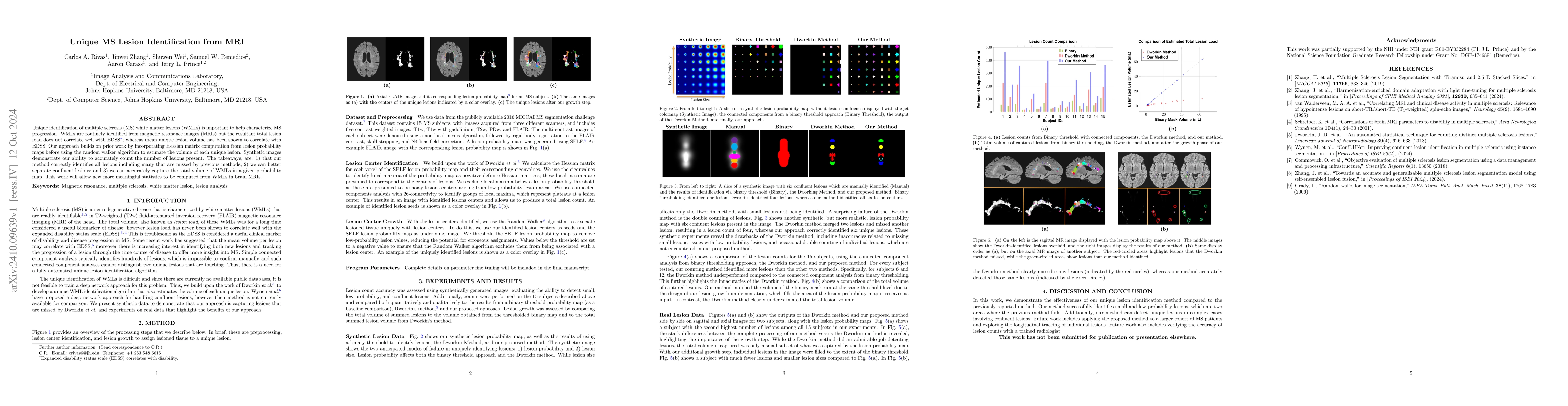
Unique identification of multiple sclerosis (MS) white matter lesions (WMLs) is important to help characterize MS progression. WMLs are routinely identified from magnetic resonance images (MRIs) but t...
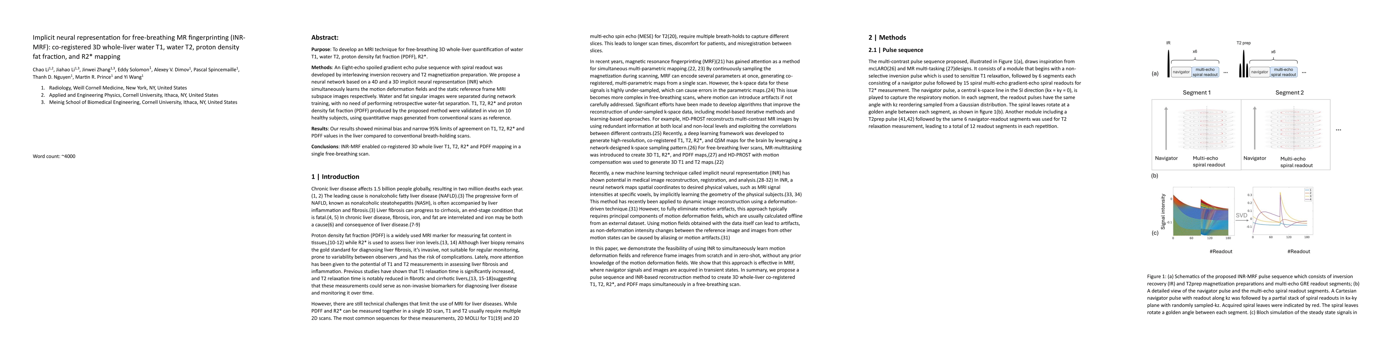
Purpose: To develop an MRI technique for free-breathing 3D whole-liver quantification of water T1, water T2, proton density fat fraction (PDFF), R2*. Methods: An Eight-echo spoiled gradient echo pulse...
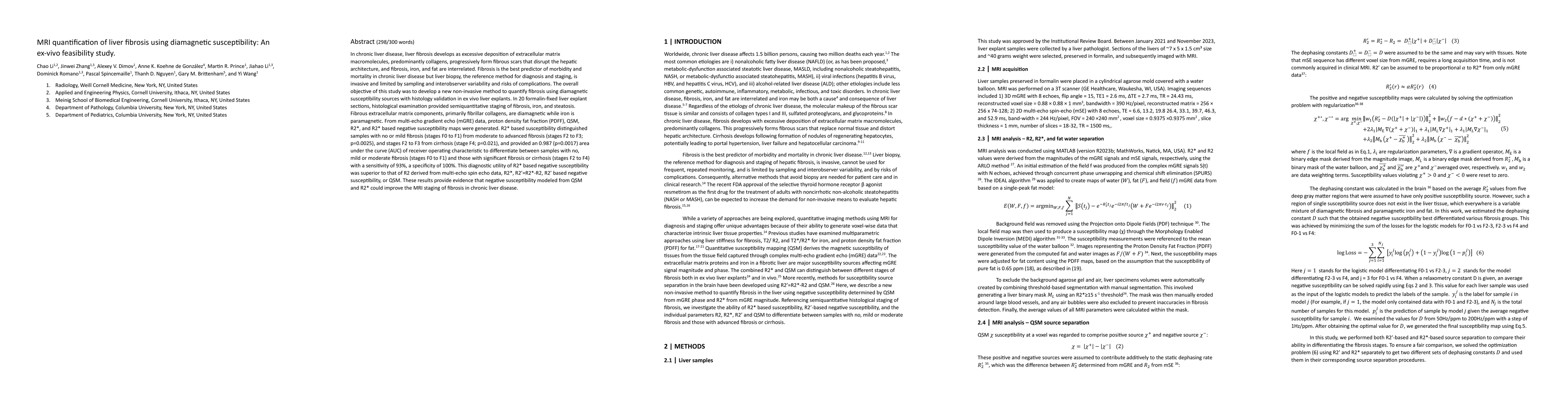
In chronic liver disease, liver fibrosis develops as excessive deposition of extracellular matrix macromolecules, predominantly collagens, progressively form fibrous scars that disrupt the hepatic arc...
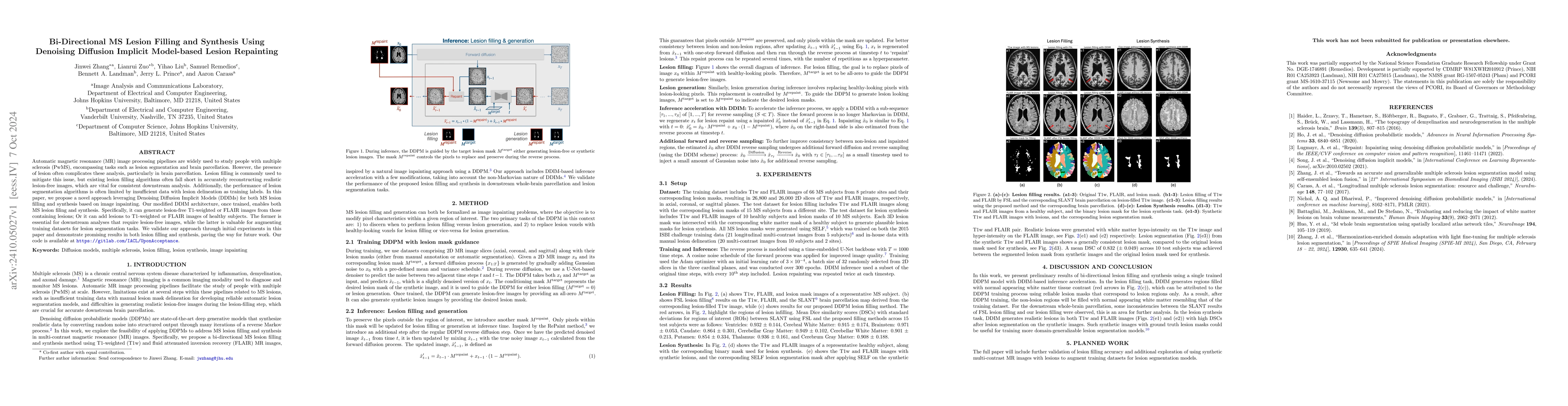
Automatic magnetic resonance (MR) image processing pipelines are widely used to study people with multiple sclerosis (PwMS), encompassing tasks such as lesion segmentation and brain parcellation. Howe...
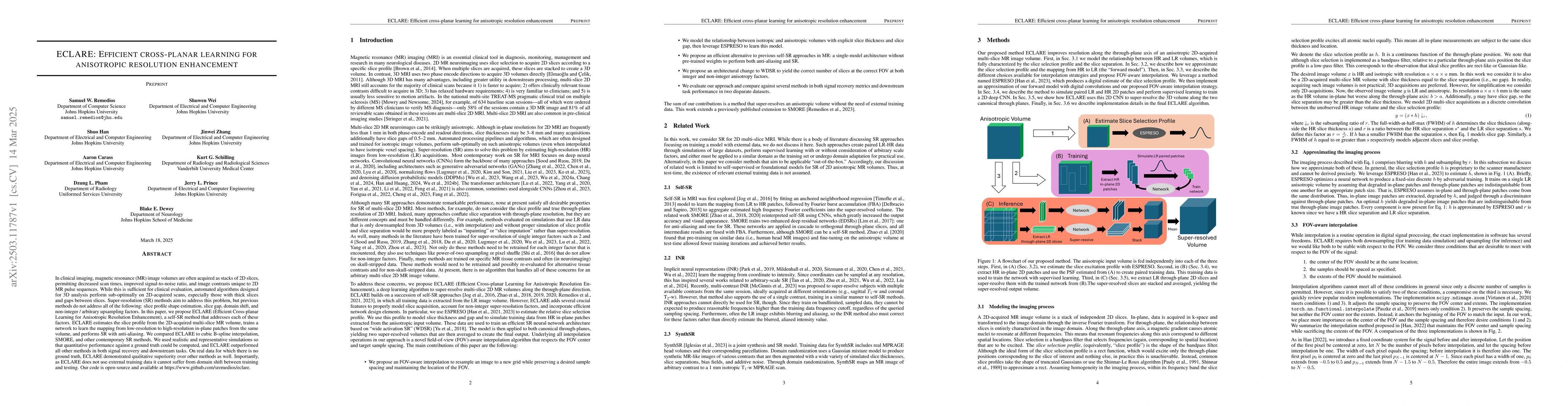
In clinical imaging, magnetic resonance (MR) image volumes are often acquired as stacks of 2D slices, permitting decreased scan times, improved signal-to-noise ratio, and image contrasts unique to 2D ...
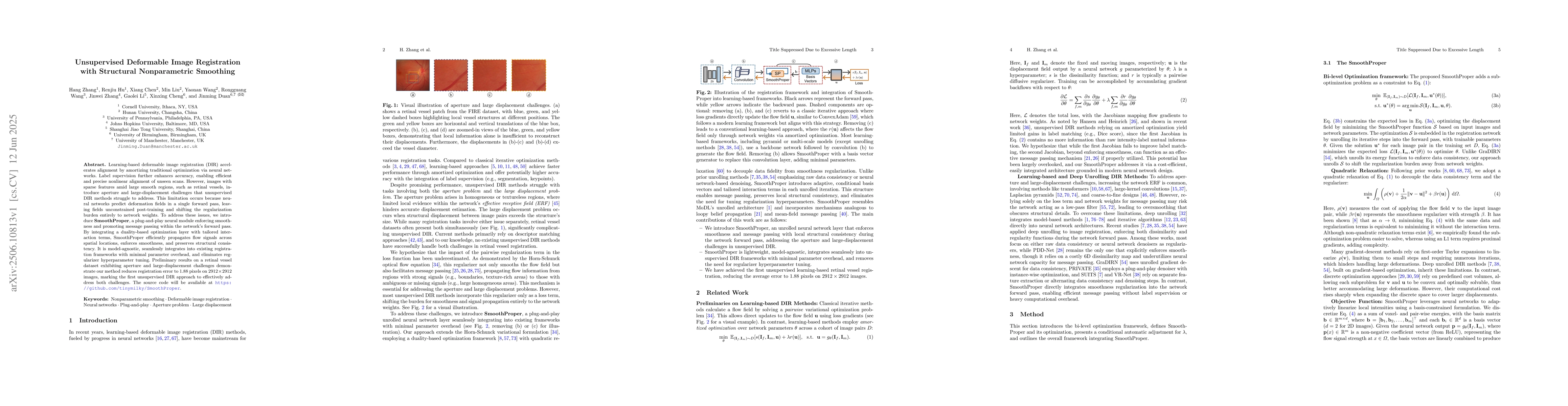
Learning-based deformable image registration (DIR) accelerates alignment by amortizing traditional optimization via neural networks. Label supervision further enhances accuracy, enabling efficient and...
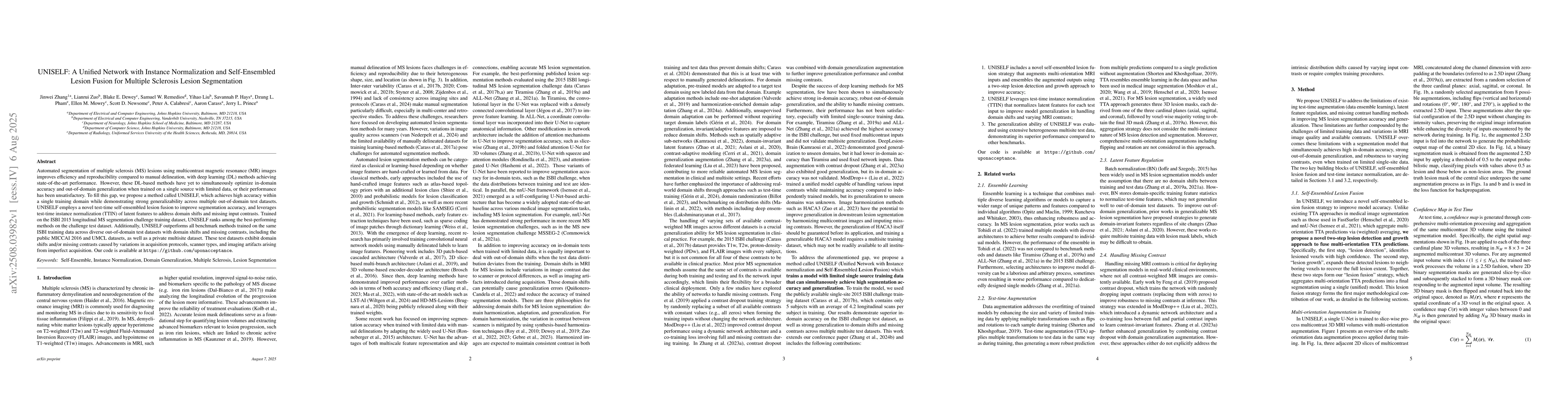
Automated segmentation of multiple sclerosis (MS) lesions using multicontrast magnetic resonance (MR) images improves efficiency and reproducibility compared to manual delineation, with deep learning ...
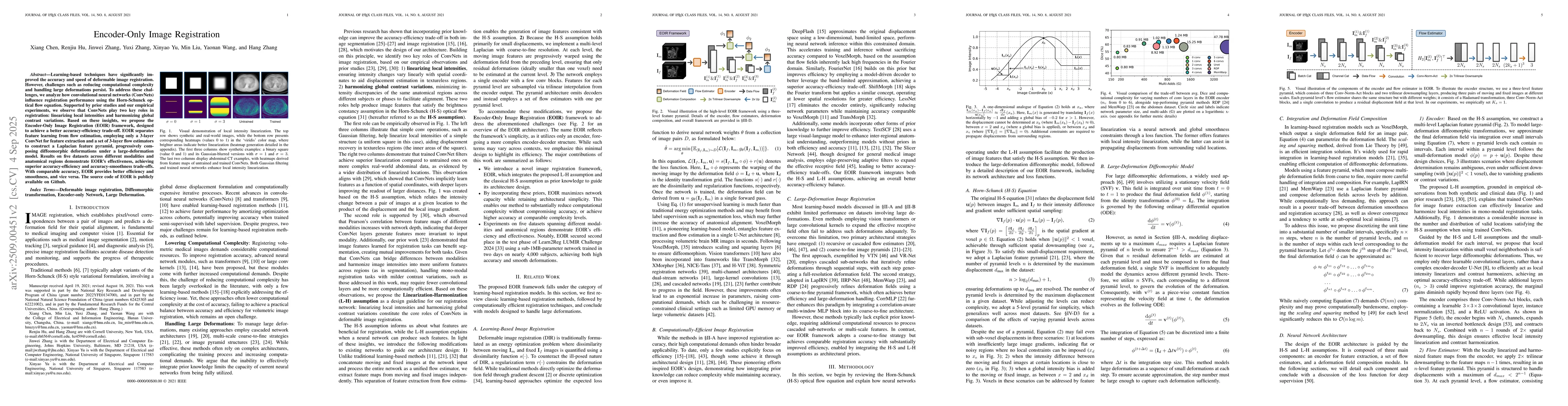
Learning-based techniques have significantly improved the accuracy and speed of deformable image registration. However, challenges such as reducing computational complexity and handling large deformat...
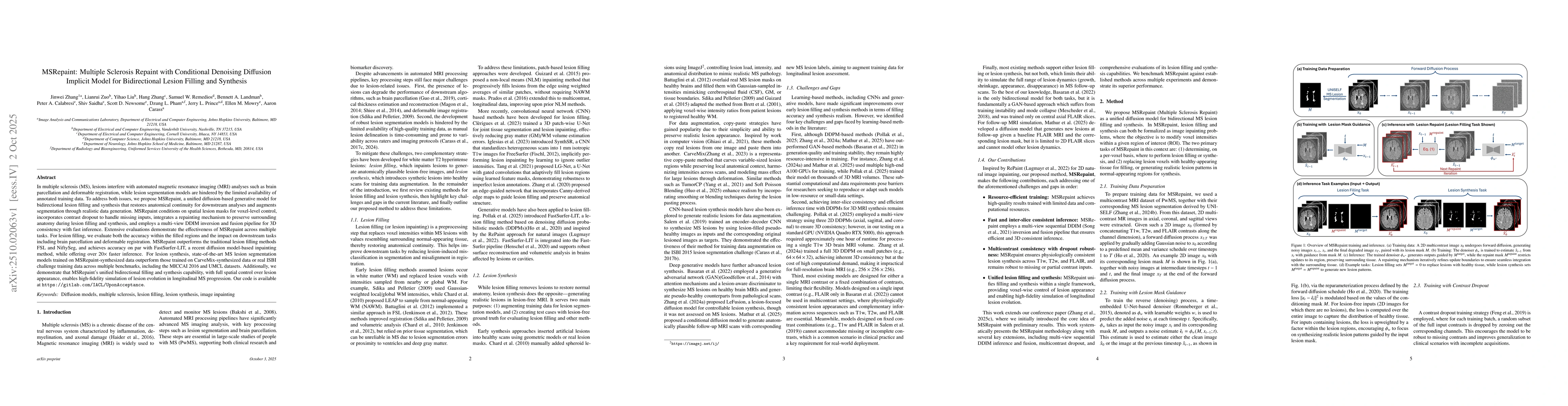
In multiple sclerosis, lesions interfere with automated magnetic resonance imaging analyses such as brain parcellation and deformable registration, while lesion segmentation models are hindered by the...
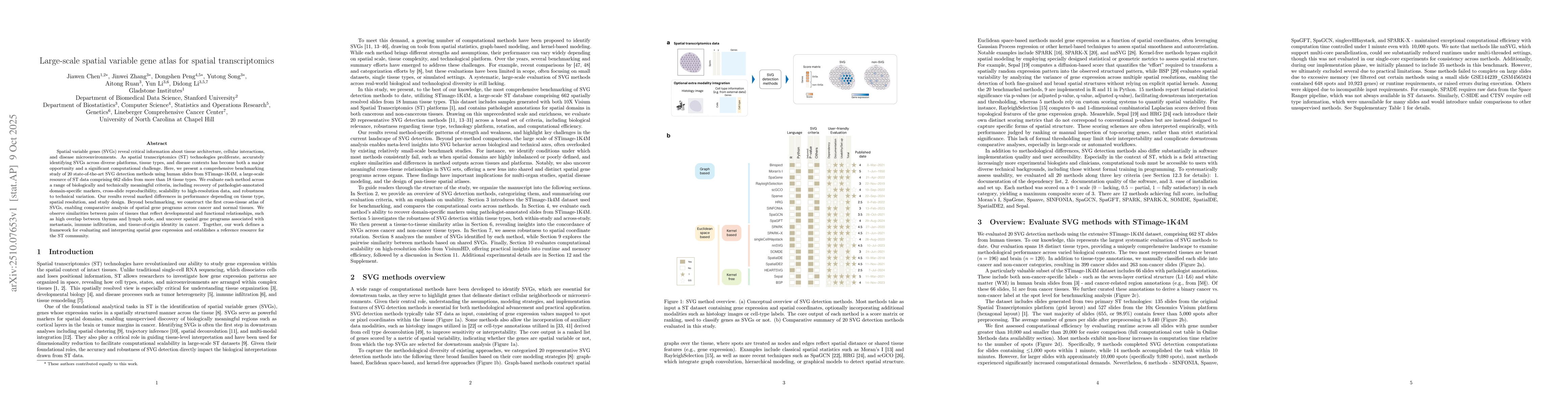
Spatial variable genes (SVGs) reveal critical information about tissue architecture, cellular interactions, and disease microenvironments. As spatial transcriptomics (ST) technologies proliferate, acc...
Reliable harmonization of heterogeneous magnetic resonance~(MR) image datasets, especially those acquired in pragmatic clinical trials, is critical to advance multi-center neuroimaging studies and tra...

We propose a novel spectral vision transformer architecture for efficient tokenization in limited data, with an emphasis on medical imaging. We outline convenient theoretical properties arising from t...