Academic Profile
Statistics
Similar Authors
Papers on arXiv
Deep learning-based semantic segmentation in neuroimaging currently requires high-resolution scans and extensive annotated datasets, posing significant barriers to clinical applicability. We present...
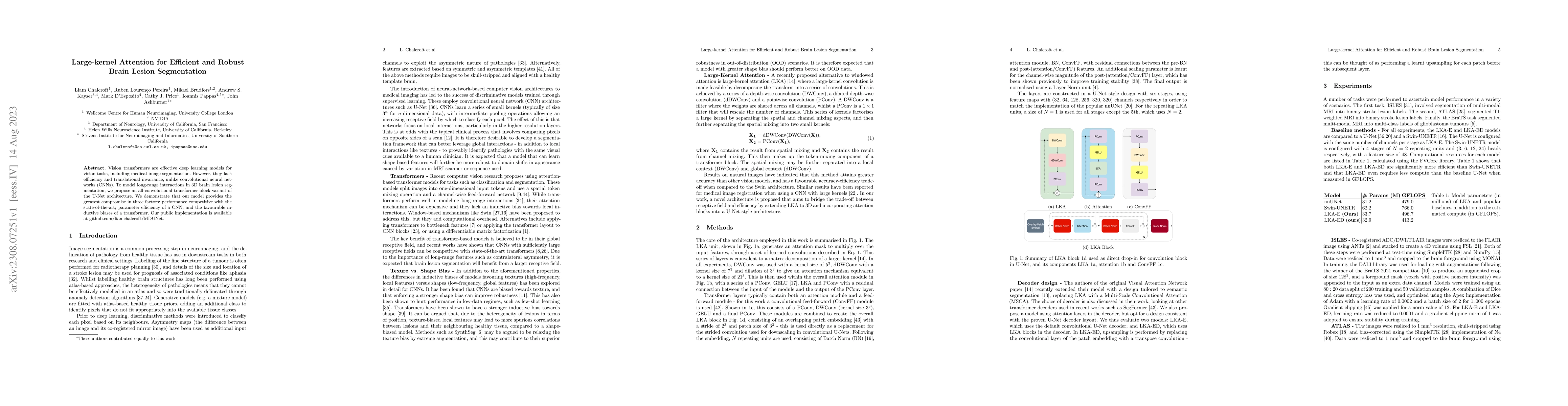
Vision transformers are effective deep learning models for vision tasks, including medical image segmentation. However, they lack efficiency and translational invariance, unlike convolutional neural...
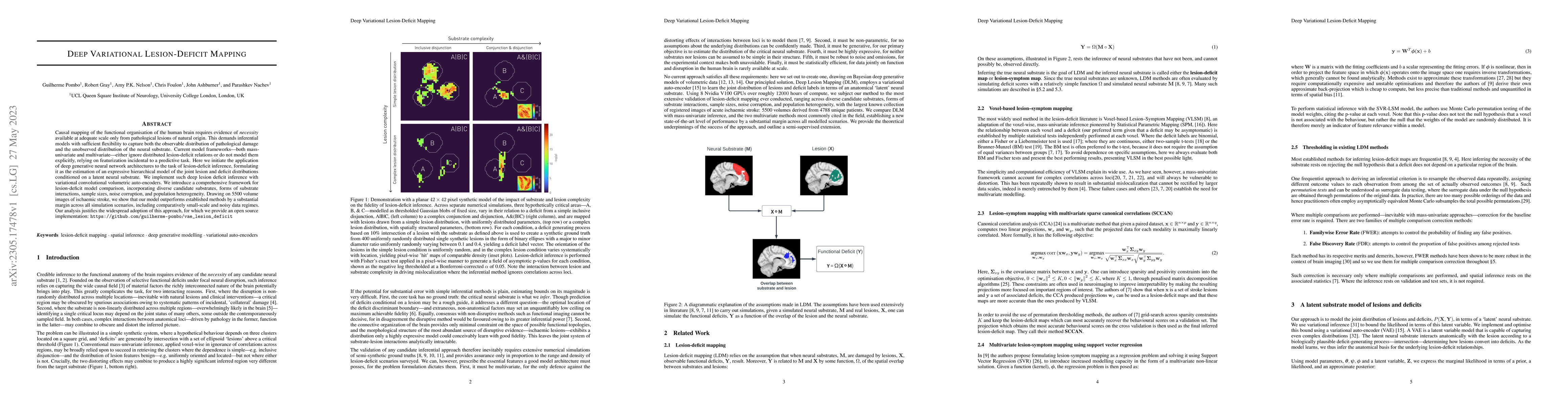
Causal mapping of the functional organisation of the human brain requires evidence of \textit{necessity} available at adequate scale only from pathological lesions of natural origin. This demands in...
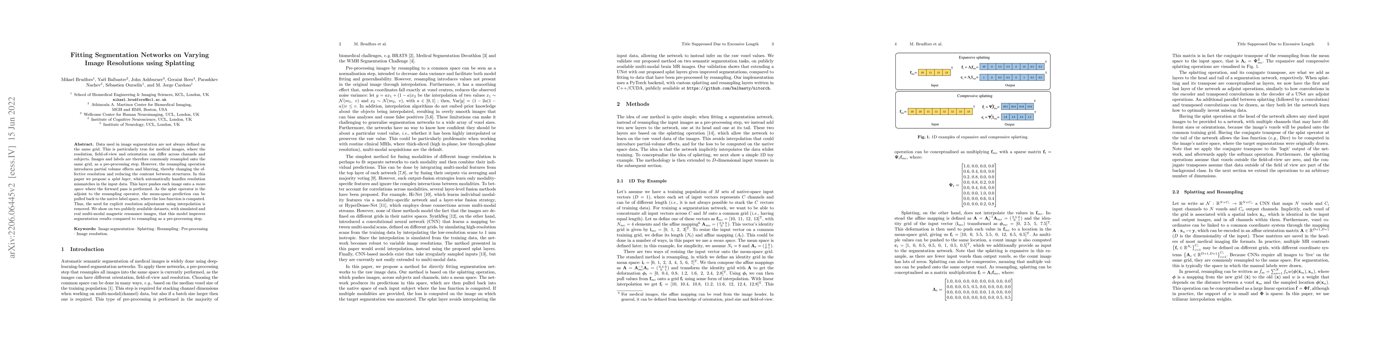
Data used in image segmentation are not always defined on the same grid. This is particularly true for medical images, where the resolution, field-of-view and orientation can differ across channels ...
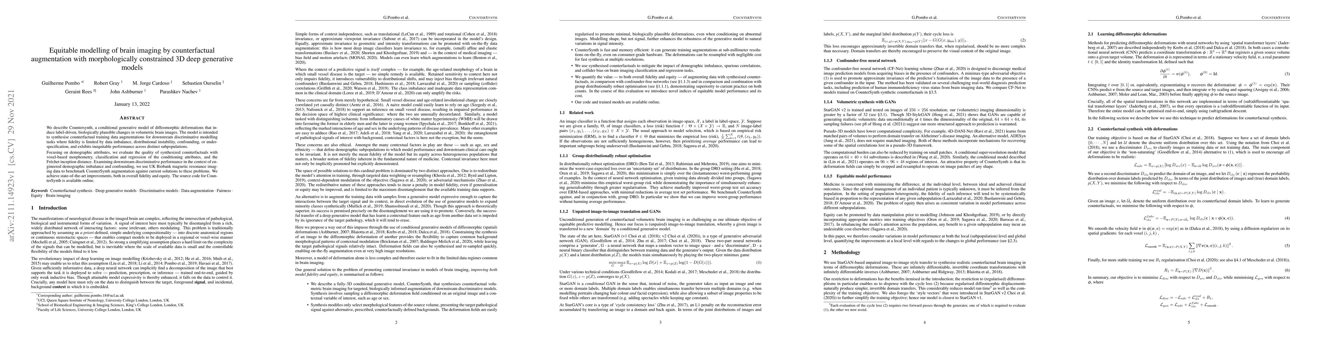
We describe Countersynth, a conditional generative model of diffeomorphic deformations that induce label-driven, biologically plausible changes in volumetric brain images. The model is intended to s...
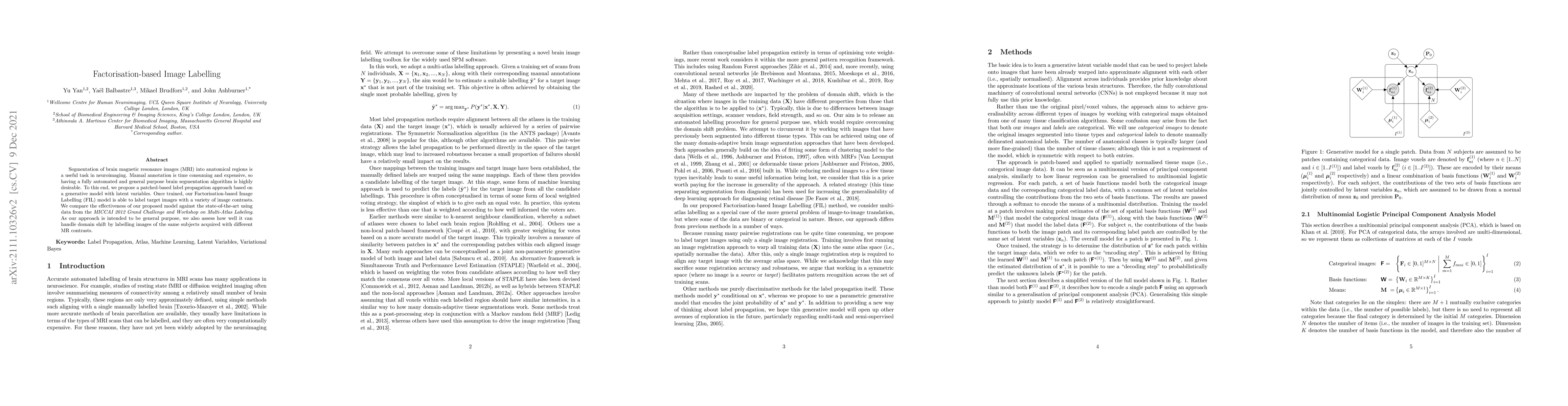
Segmentation of brain magnetic resonance images (MRI) into anatomical regions is a useful task in neuroimaging. Manual annotation is time consuming and expensive, so having a fully automated and gen...
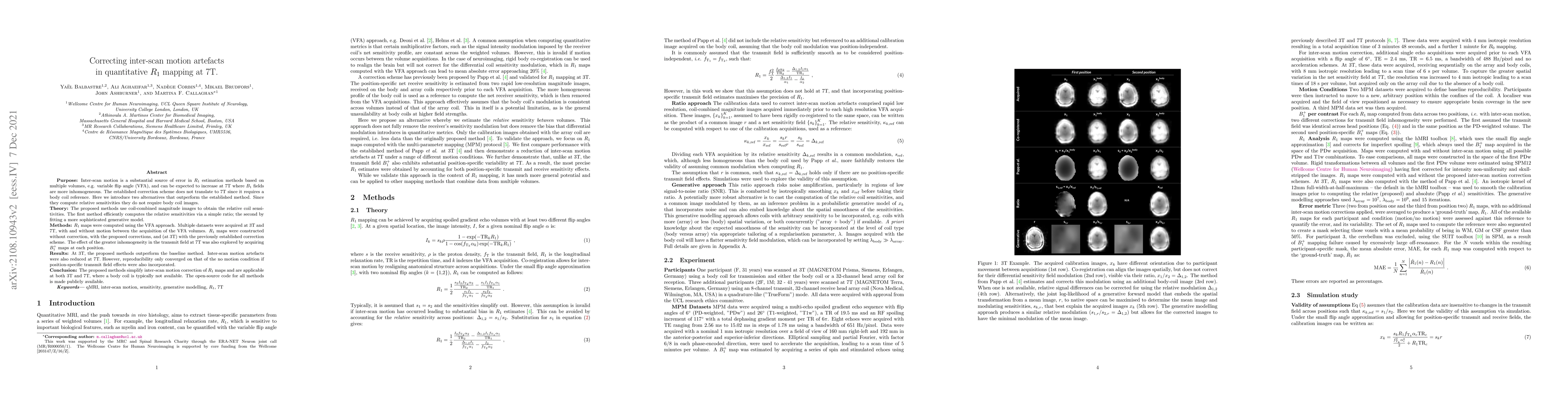
Purpose: Inter-scan motion is a substantial source of error in $R_1$ estimation, and can be expected to increase at 7T where $B_1$ fields are more inhomogeneous. The established correction scheme do...
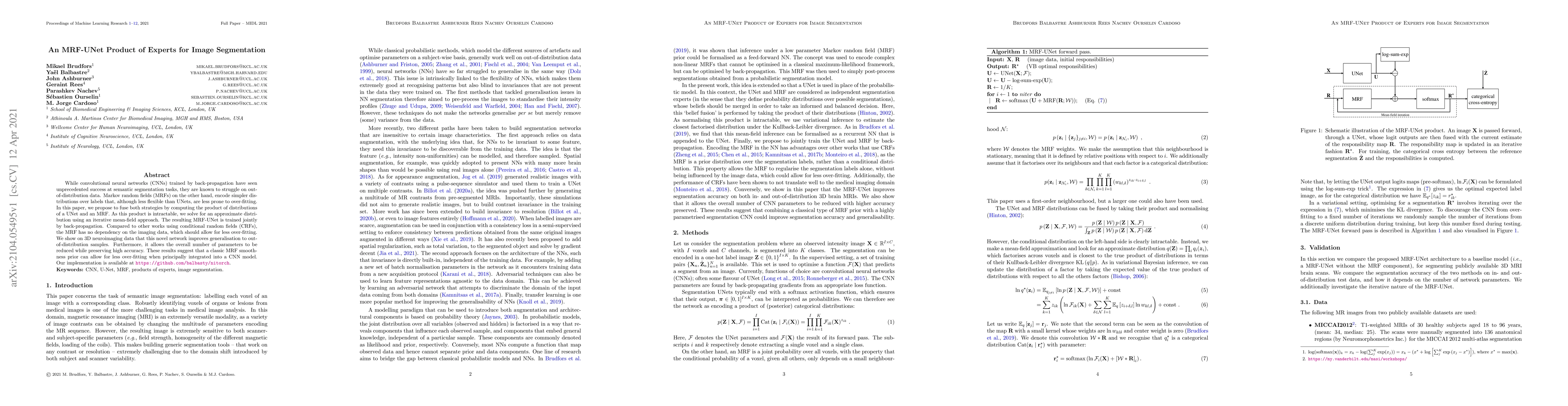
While convolutional neural networks (CNNs) trained by back-propagation have seen unprecedented success at semantic segmentation tasks, they are known to struggle on out-of-distribution data. Markov ...
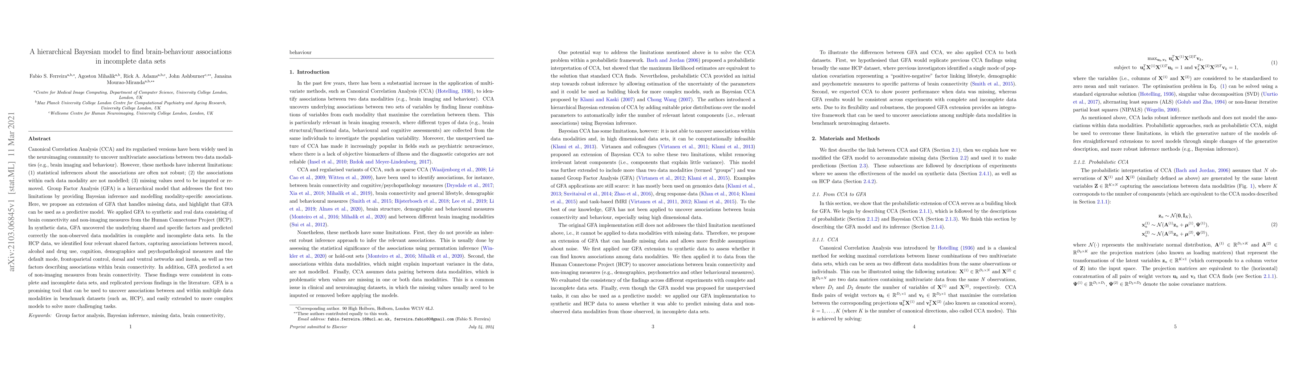
Canonical Correlation Analysis (CCA) and its regularised versions have been widely used in the neuroimaging community to uncover multivariate associations between two data modalities (e.g., brain im...
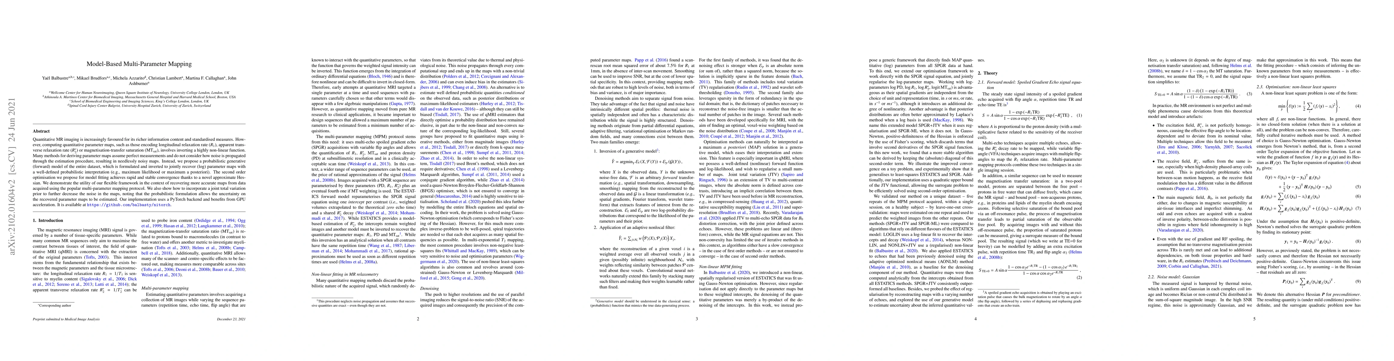
Quantitative MR imaging is increasingly favoured for its richer information content and standardised measures. However, computing quantitative parameter maps, such as those encoding longitudinal rel...
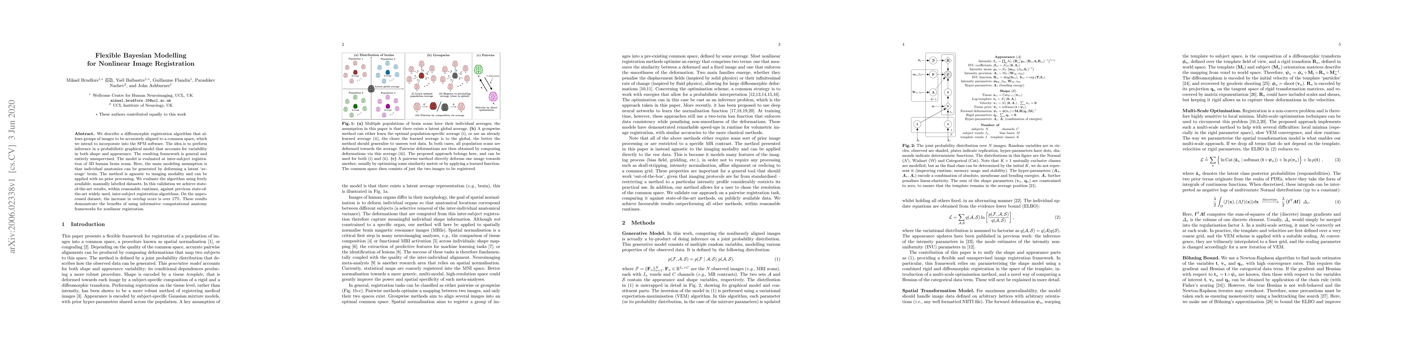
We describe a diffeomorphic registration algorithm that allows groups of images to be accurately aligned to a common space, which we intend to incorporate into the SPM software. The idea is to perfo...
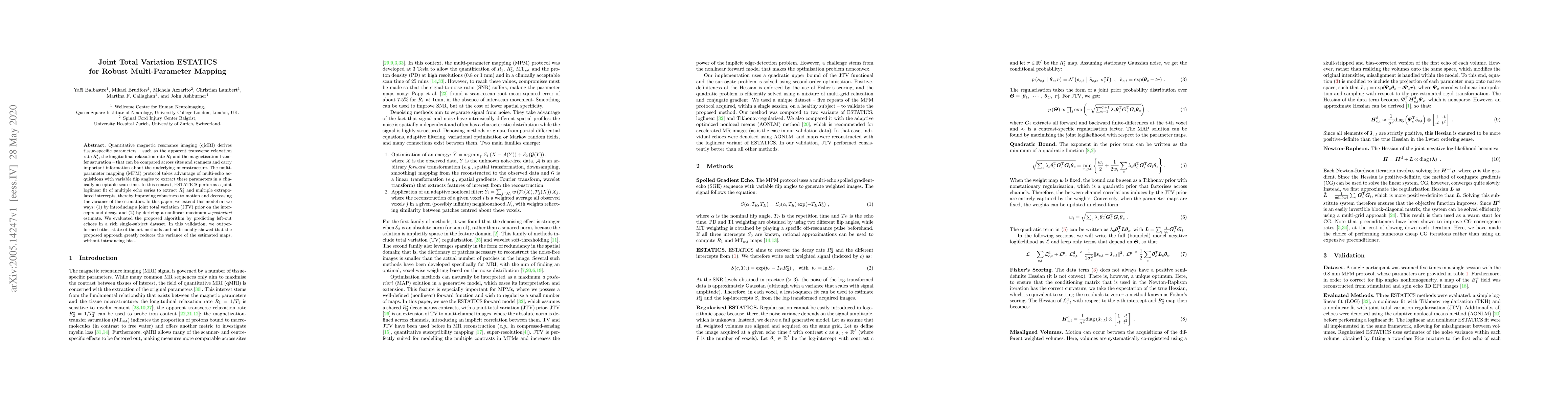
Quantitative magnetic resonance imaging (qMRI) derives tissue-specific parameters -- such as the apparent transverse relaxation rate R2*, the longitudinal relaxation rate R1 and the magnetisation tr...
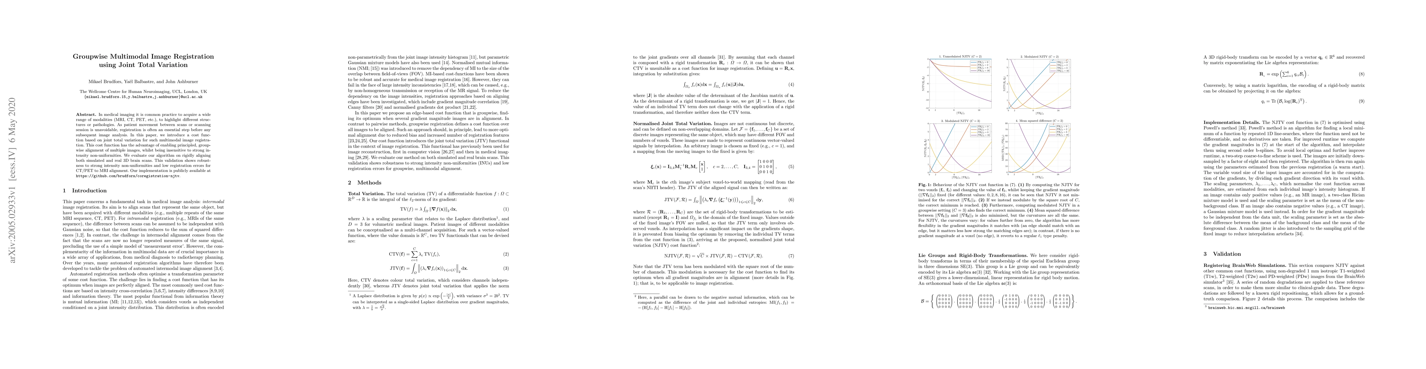
In medical imaging it is common practice to acquire a wide range of modalities (MRI, CT, PET, etc.), to highlight different structures or pathologies. As patient movement between scans or scanning s...
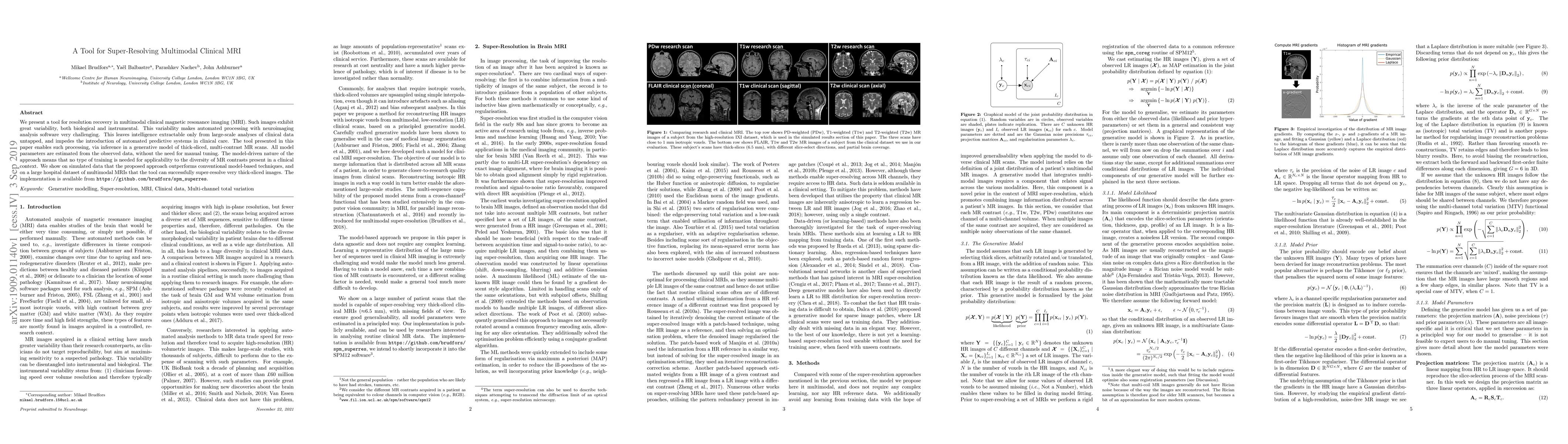
We present a tool for resolution recovery in multimodal clinical magnetic resonance imaging (MRI). Such images exhibit great variability, both biological and instrumental. This variability makes aut...
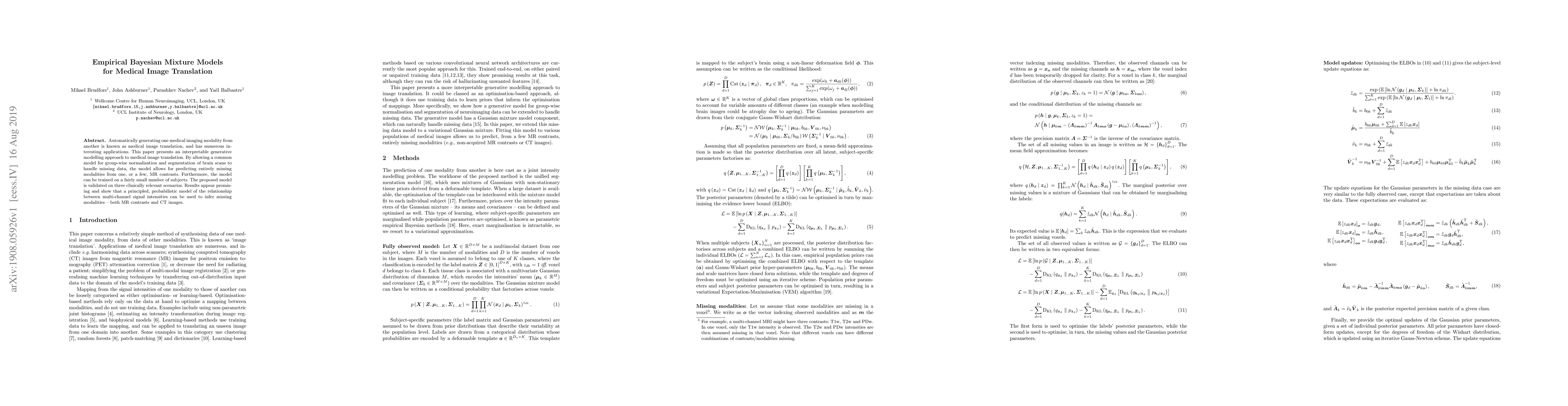
Automatically generating one medical imaging modality from another is known as medical image translation, and has numerous interesting applications. This paper presents an interpretable generative m...
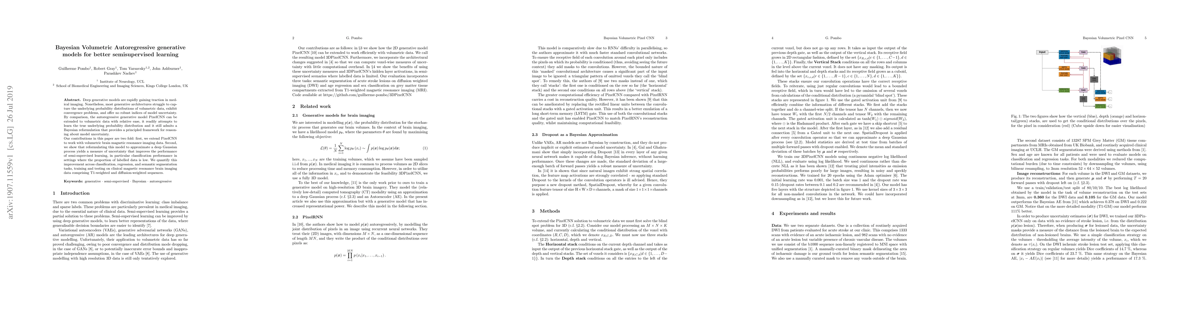
Deep generative models are rapidly gaining traction in medical imaging. Nonetheless, most generative architectures struggle to capture the underlying probability distributions of volumetric data, ex...
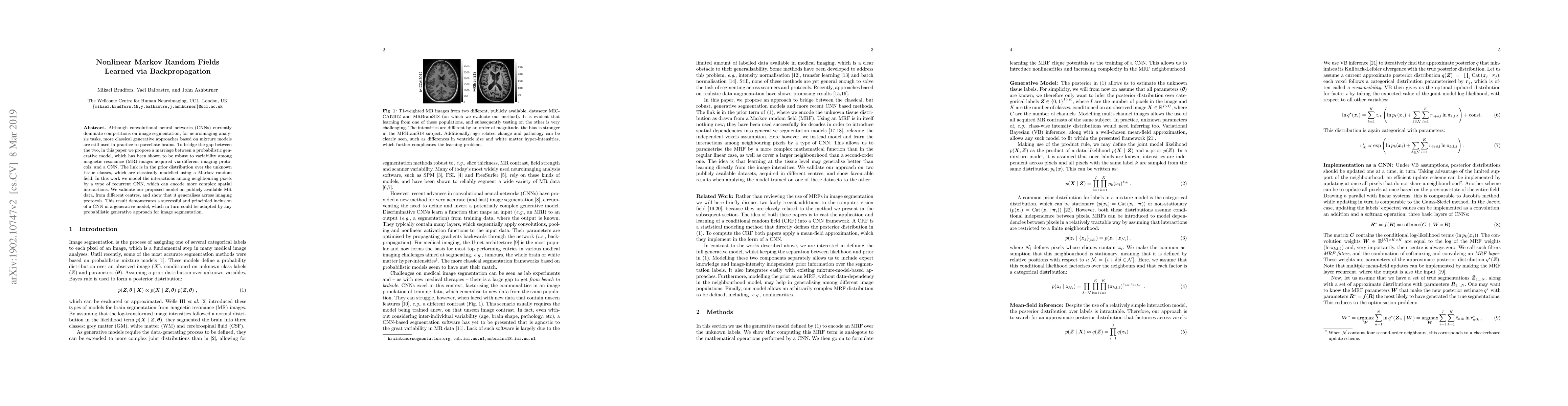
Although convolutional neural networks (CNNs) currently dominate competitions on image segmentation, for neuroimaging analysis tasks, more classical generative approaches based on mixture models are...
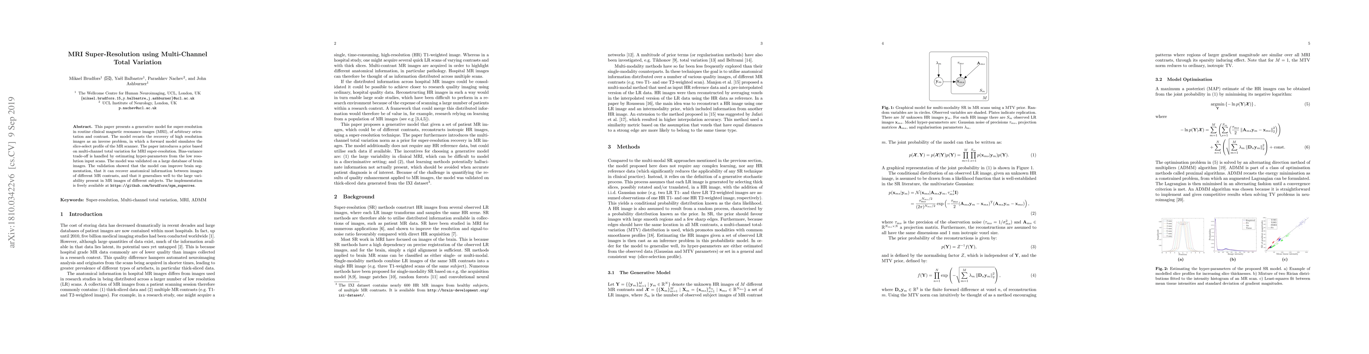
This paper presents a generative model for super-resolution in routine clinical magnetic resonance images (MRI), of arbitrary orientation and contrast. The model recasts the recovery of high resolut...
This paper presents a framework for automatically learning shape and appearance models for medical (and certain other) images. It is based on the idea that having a more accurate shape and appearanc...
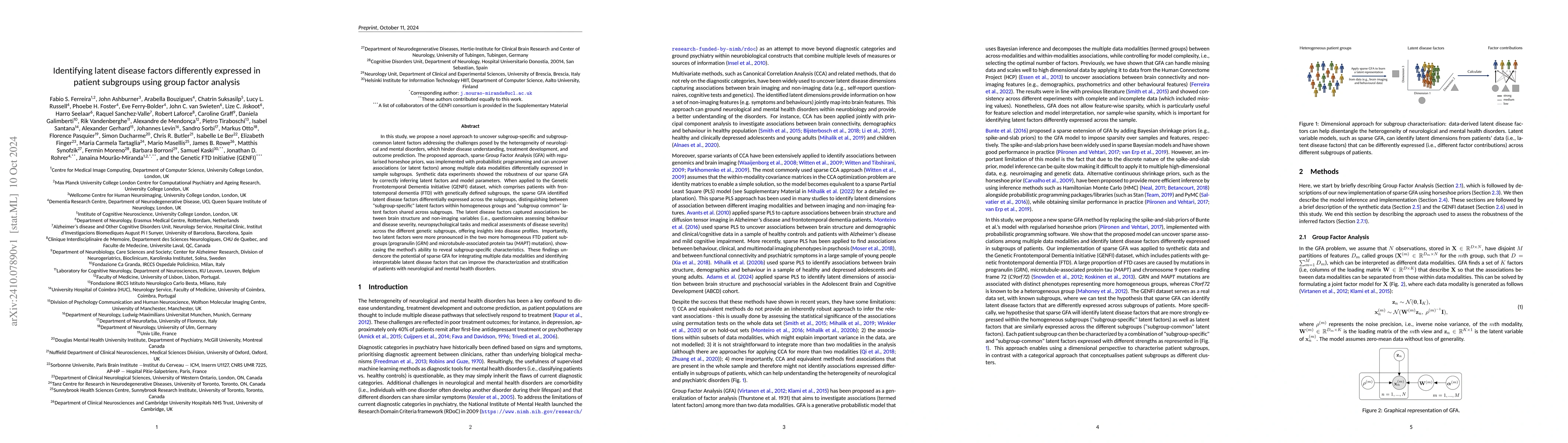
In this study, we propose a novel approach to uncover subgroup-specific and subgroup-common latent factors addressing the challenges posed by the heterogeneity of neurological and mental disorders, wh...
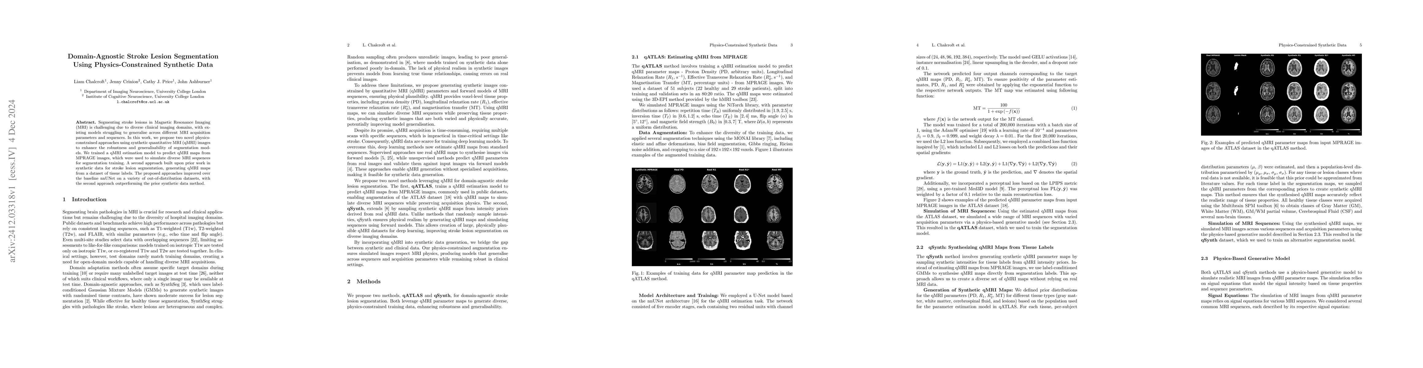
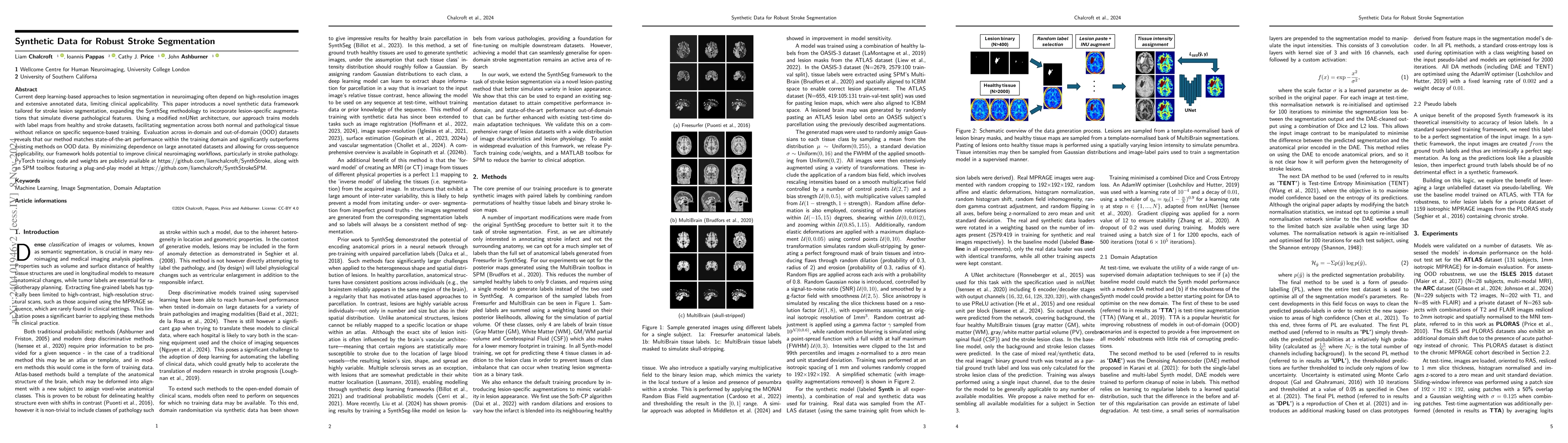
Segmenting stroke lesions in Magnetic Resonance Imaging (MRI) is challenging due to diverse clinical imaging domains, with existing models struggling to generalise across different MRI acquisition par...
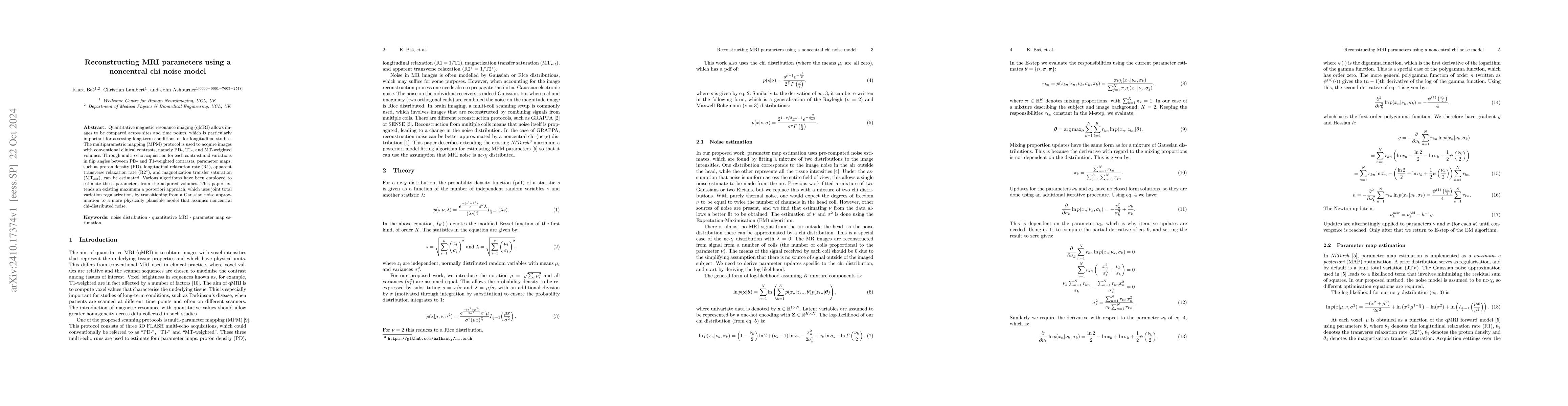
Quantitative magnetic resonance imaging (qMRI) allows images to be compared across sites and time points, which is particularly important for assessing long-term conditions or for longitudinal studies...
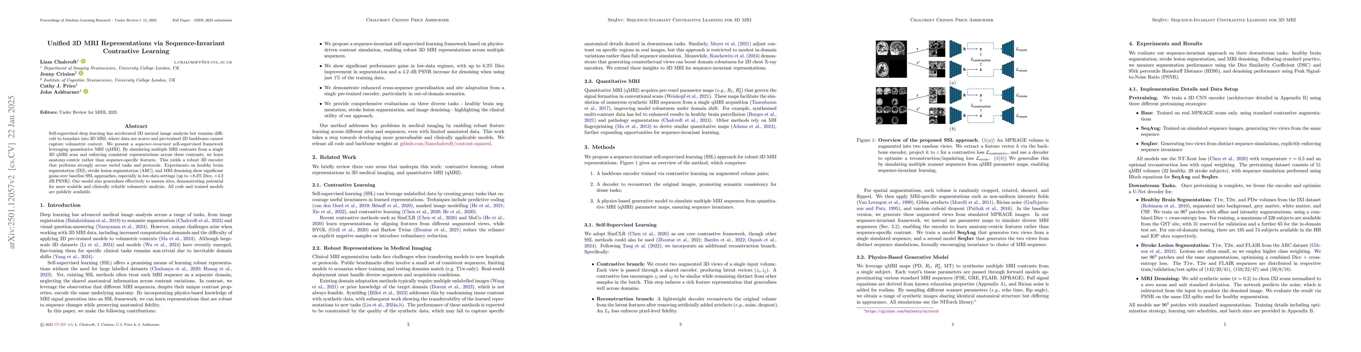
Self-supervised deep learning has accelerated 2D natural image analysis but remains difficult to translate into 3D MRI, where data are scarce and pre-trained 2D backbones cannot capture volumetric con...