ArxivLens
J
Researcher

Jonathan Rafael-Patino

4 papers on arXiv

4 arXiv Papers
21 Total Publications
Profile

Academic Profile

Name Jonathan Rafael-Patiño
Total Papers 21
Scholar Profile View on Semantic Scholar
Metrics

Statistics

4 arXiv Papers
21 Total Publications
Publications

Papers on arXiv

Thumbnail for CACTUS: A Computational Framework for Generating Realistic White Matter
  Microstructure Substrates
arXiv
CACTUS: A Computational Framework for Generating Realistic White Matter Microstructure Substrates

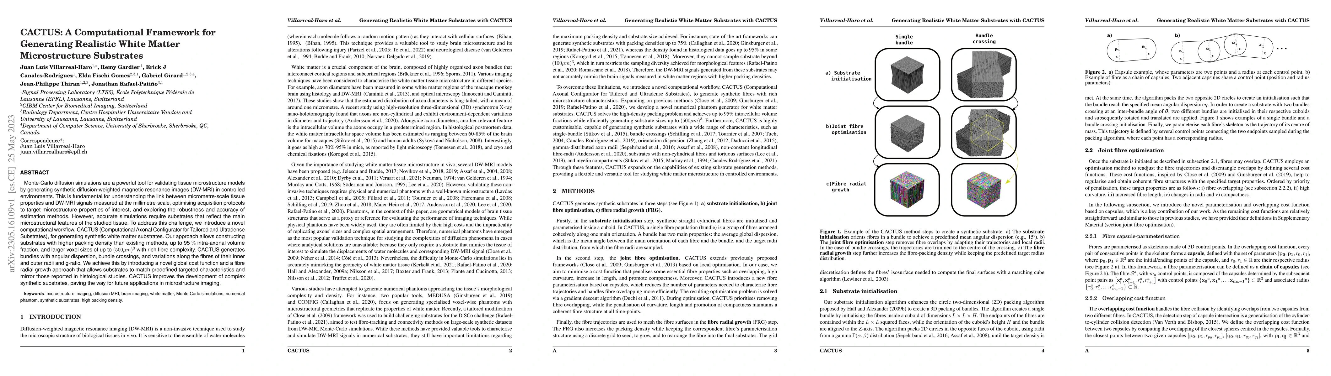
Monte-Carlo diffusion simulations are a powerful tool for validating tissue microstructure models by generating synthetic diffusion-weighted magnetic resonance images (DW-MRI) in controlled environm...

Thumbnail for Cellular EXchange Imaging (CEXI): Evaluation of a diffusion model
  including water exchange in cells using numerical phantoms of permeable
  spheres
arXiv
Cellular EXchange Imaging (CEXI): Evaluation of a diffusion model including water exchange in cells using numerical phantoms of permeable spheres

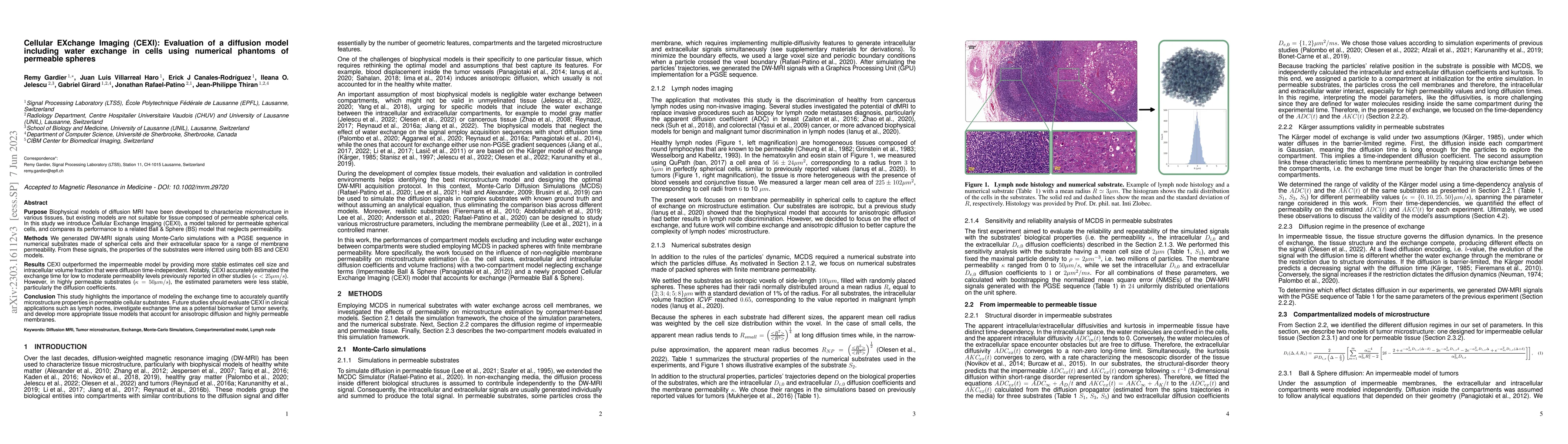
Purpose: Biophysical models of diffusion MRI have been developed to characterize microstructure in various tissues, but existing models are not suitable for tissue composed of permeable spherical ce...

Thumbnail for Microstructure estimation from diffusion-MRI: Compartmentalized models
  in permeable cellular tissue
arXiv
Microstructure estimation from diffusion-MRI: Compartmentalized models in permeable cellular tissue

Diffusion-weighted magnetic resonance imaging (DW-MRI) is used to characterize brain tissue microstructure employing tissue-specific biophysical models. A current limitation, however, is that most o...

Thumbnail for Multi-compartment diffusion MRI, T2 relaxometry and myelin water imaging
  as neuroimaging descriptors for anomalous tissue detection
arXiv
Multi-compartment diffusion MRI, T2 relaxometry and myelin water imaging as neuroimaging descriptors for anomalous tissue detection

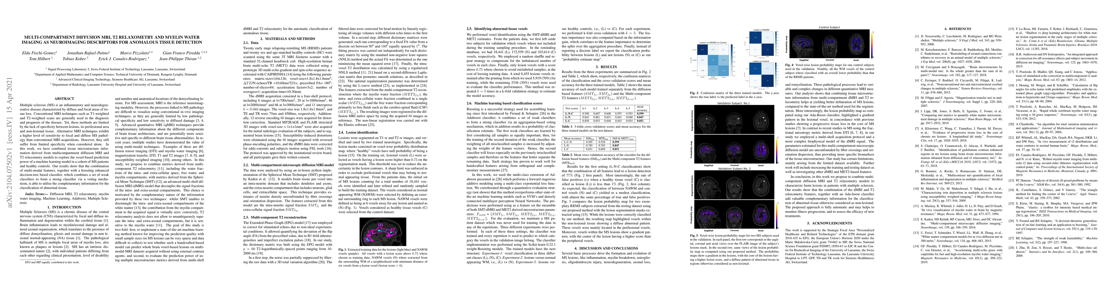
Multiple sclerosis (MS) is an inflammatory and neurodegenerative disease characterized by diffuse and focal areas of tissue loss. Conventional MRI techniques such as T1-weighted and T2-weighted scan...

This website uses cookies to ensure you get the best experience. Learn more