Academic Profile
Statistics
Similar Authors
Papers on arXiv

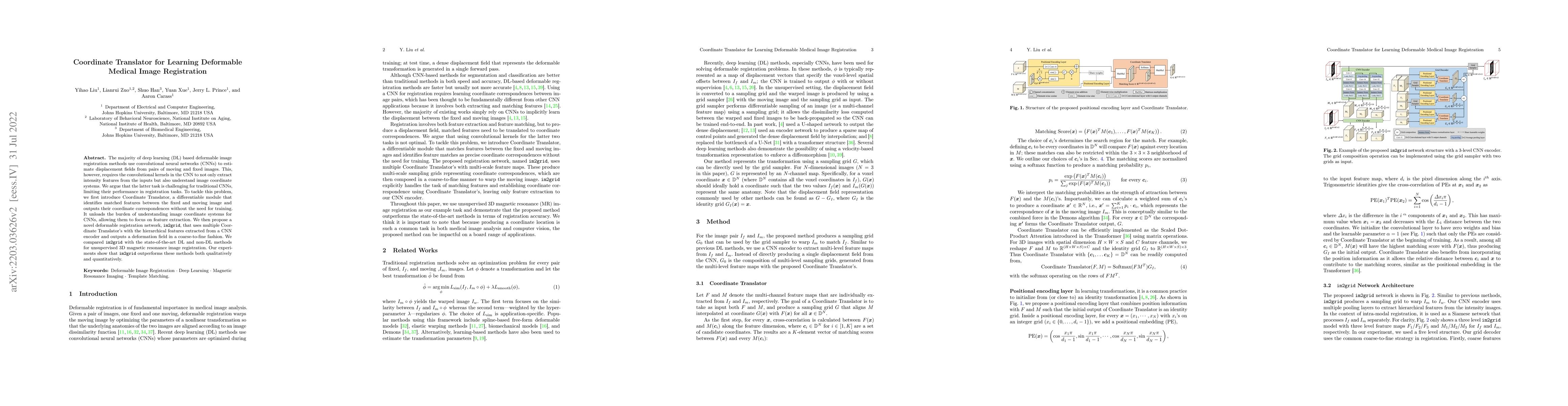
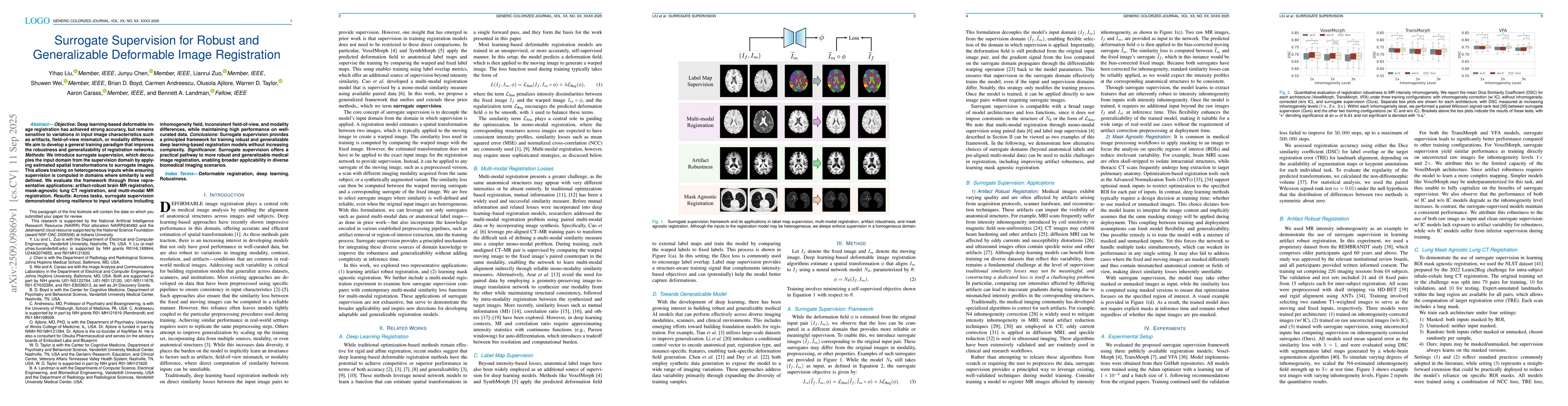
Deformable image registration establishes non-linear spatial correspondences between fixed and moving images. Deep learning-based deformable registration methods have been widely studied in recent yea...
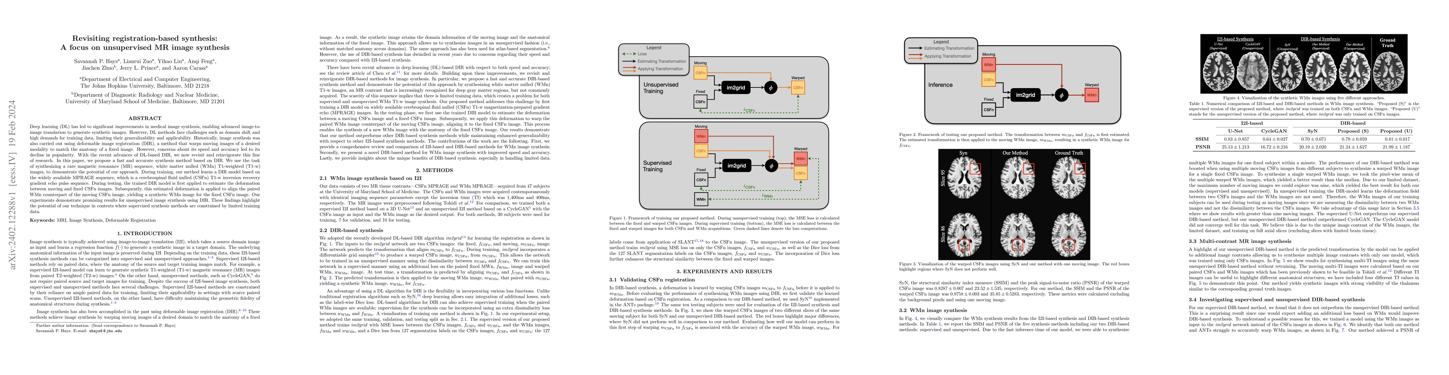
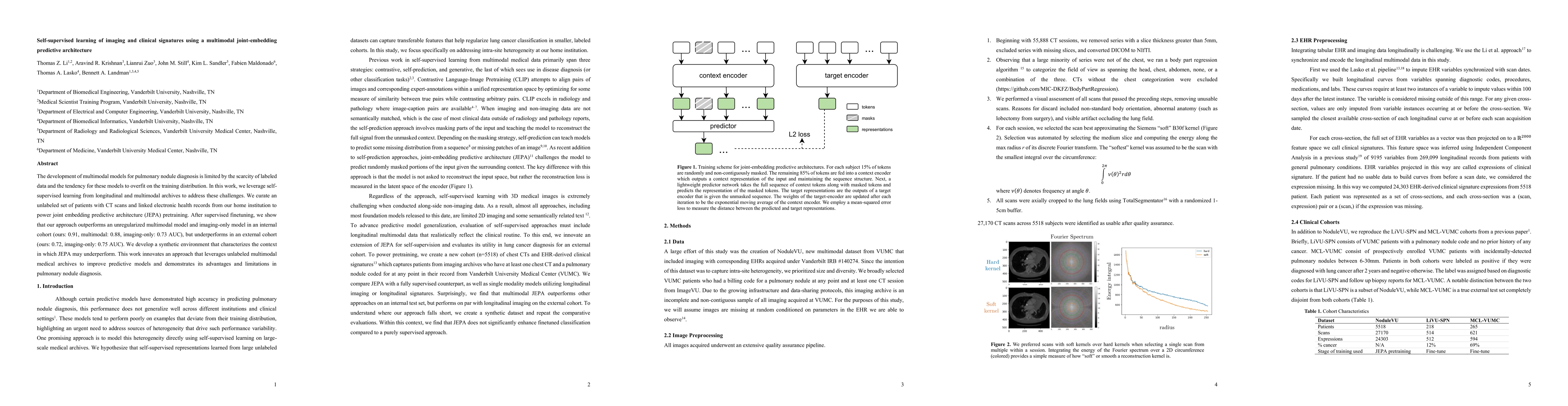
Deep learning (DL) has led to significant improvements in medical image synthesis, enabling advanced image-to-image translation to generate synthetic images. However, DL methods face challenges such...
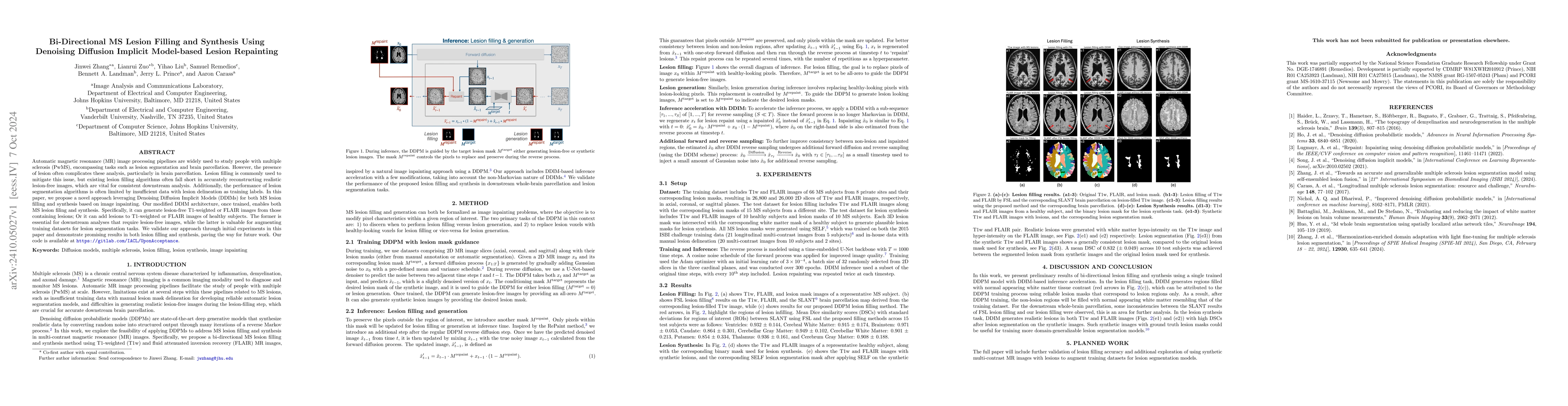
Automatic multiple sclerosis (MS) lesion segmentation using multi-contrast magnetic resonance (MR) images provides improved efficiency and reproducibility compared to manual delineation. Current sta...
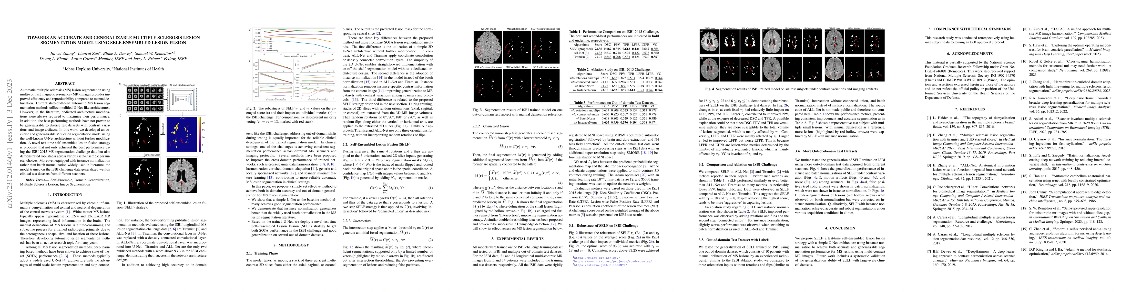
Deep learning algorithms utilizing magnetic resonance (MR) images have demonstrated cutting-edge proficiency in autonomously segmenting multiple sclerosis (MS) lesions. Despite their achievements, t...
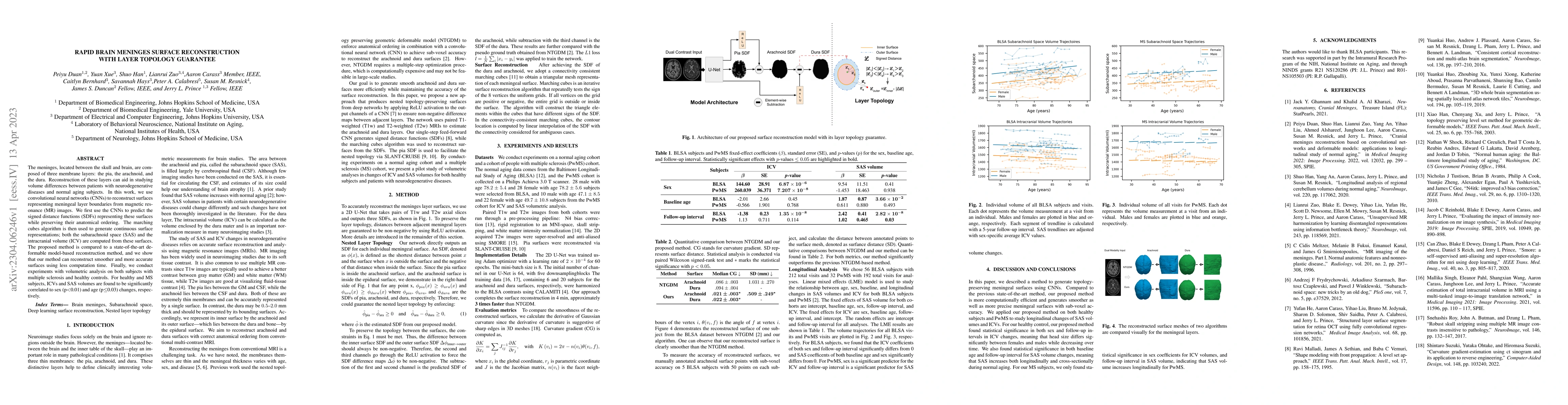
The meninges, located between the skull and brain, are composed of three membrane layers: the pia, the arachnoid, and the dura. Reconstruction of these layers can aid in studying volume differences ...
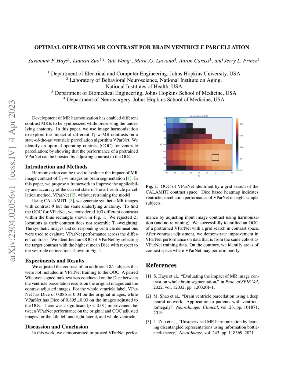
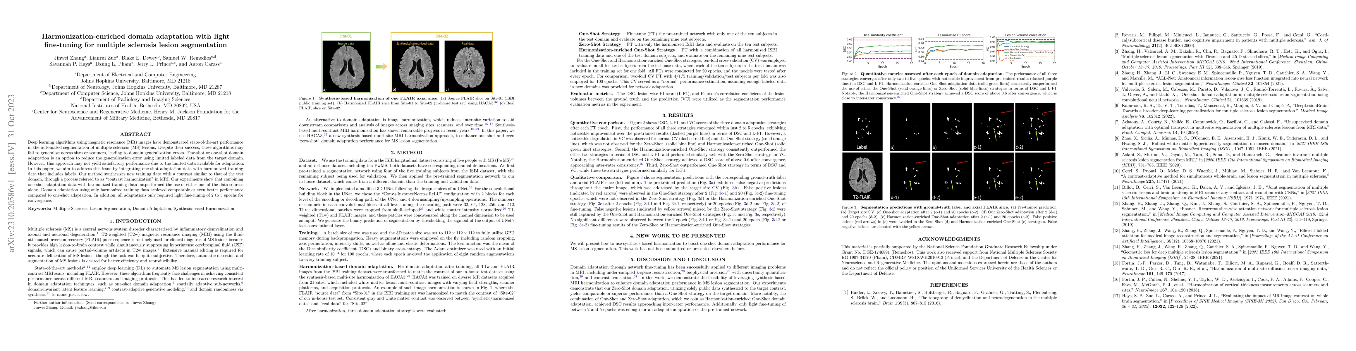
Development of MR harmonization has enabled different contrast MRIs to be synthesized while preserving the underlying anatomy. In this paper, we use image harmonization to explore the impact of diff...
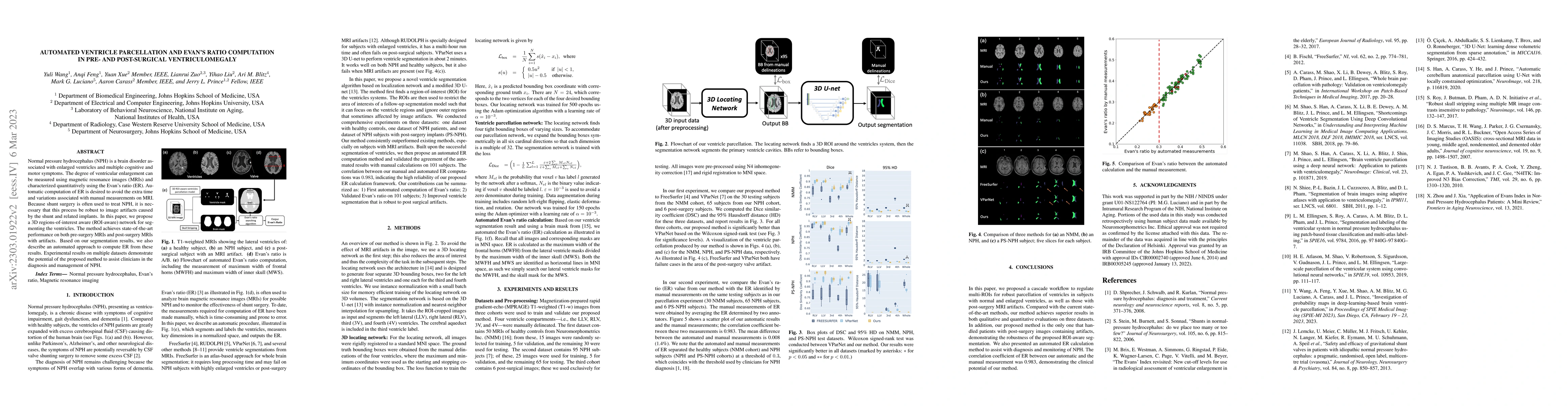
Normal pressure hydrocephalus~(NPH) is a brain disorder associated with enlarged ventricles and multiple cognitive and motor symptoms. The degree of ventricular enlargement can be measured using mag...
Image quality control (IQC) can be used in automated magnetic resonance (MR) image analysis to exclude erroneous results caused by poorly acquired or artifact-laden images. Existing IQC methods for ...
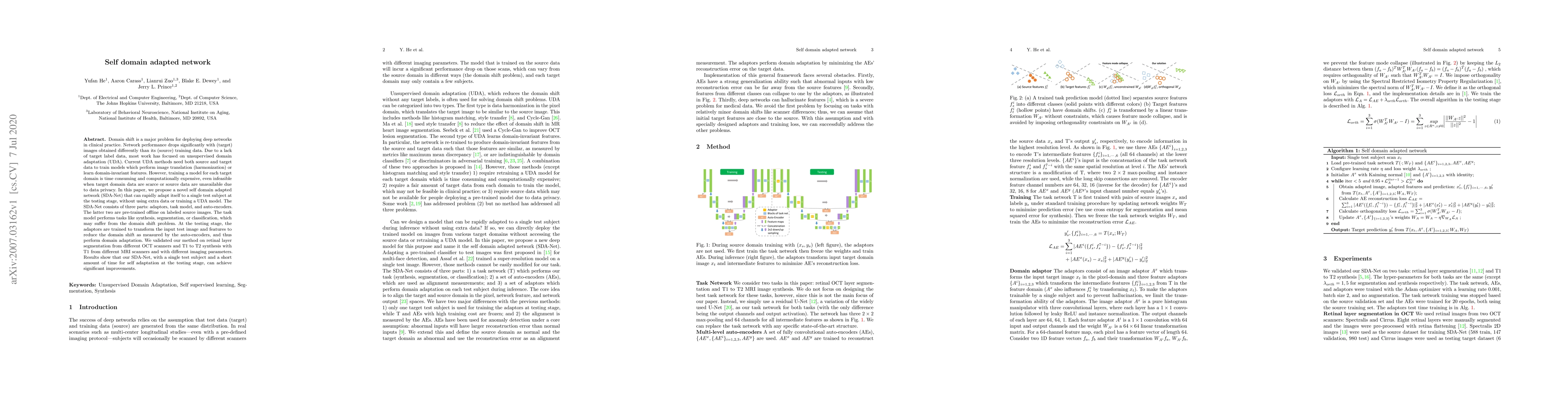
The lack of standardization is a prominent issue in magnetic resonance (MR) imaging. This often causes undesired contrast variations in the acquired images due to differences in hardware and acquisi...
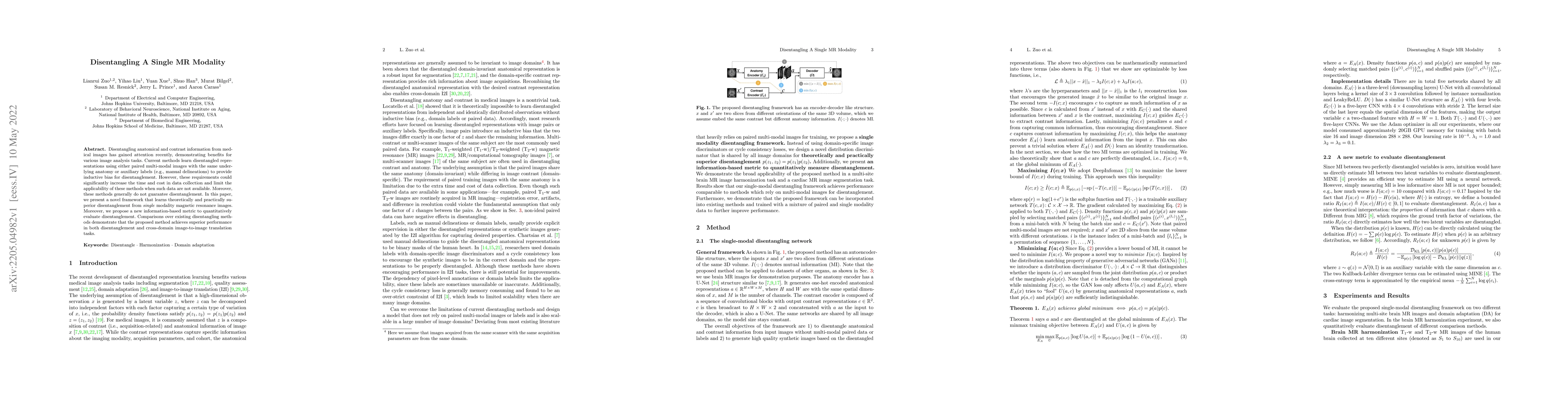
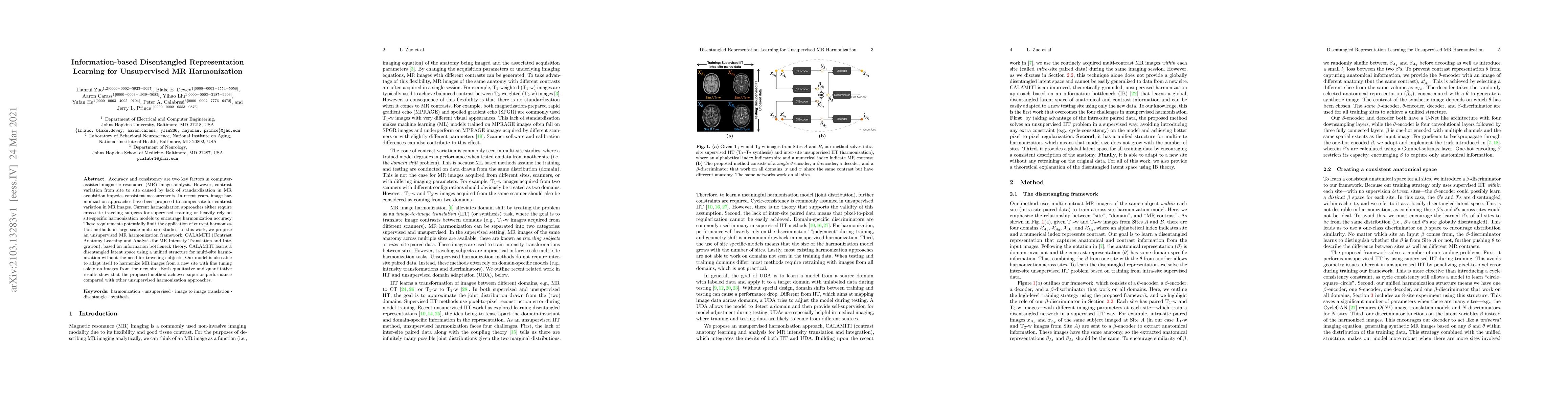
Disentangling anatomical and contrast information from medical images has gained attention recently, demonstrating benefits for various image analysis tasks. Current methods learn disentangled repre...
The majority of deep learning (DL) based deformable image registration methods use convolutional neural networks (CNNs) to estimate displacement fields from pairs of moving and fixed images. This, h...
Accuracy and consistency are two key factors in computer-assisted magnetic resonance (MR) image analysis. However, contrast variation from site to site caused by lack of standardization in MR acquis...
Domain shift is a major problem for deploying deep networks in clinical practice. Network performance drops significantly with (target) images obtained differently than its (source) training data. D...
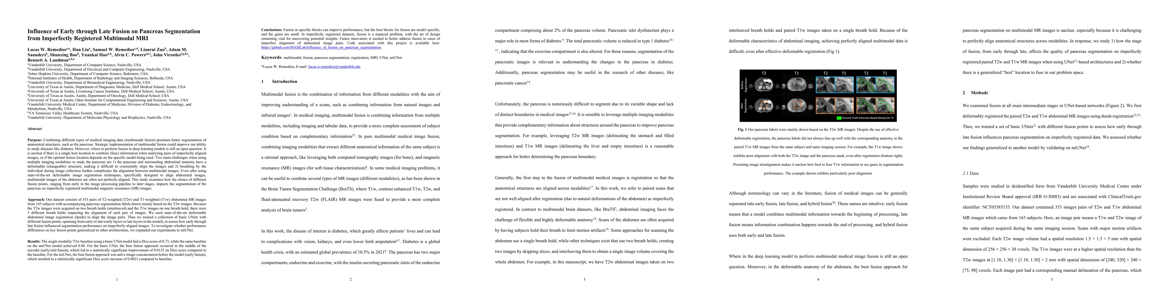
Multimodal fusion promises better pancreas segmentation. However, where to perform fusion in models is still an open question. It is unclear if there is a best location to fuse information when analyz...
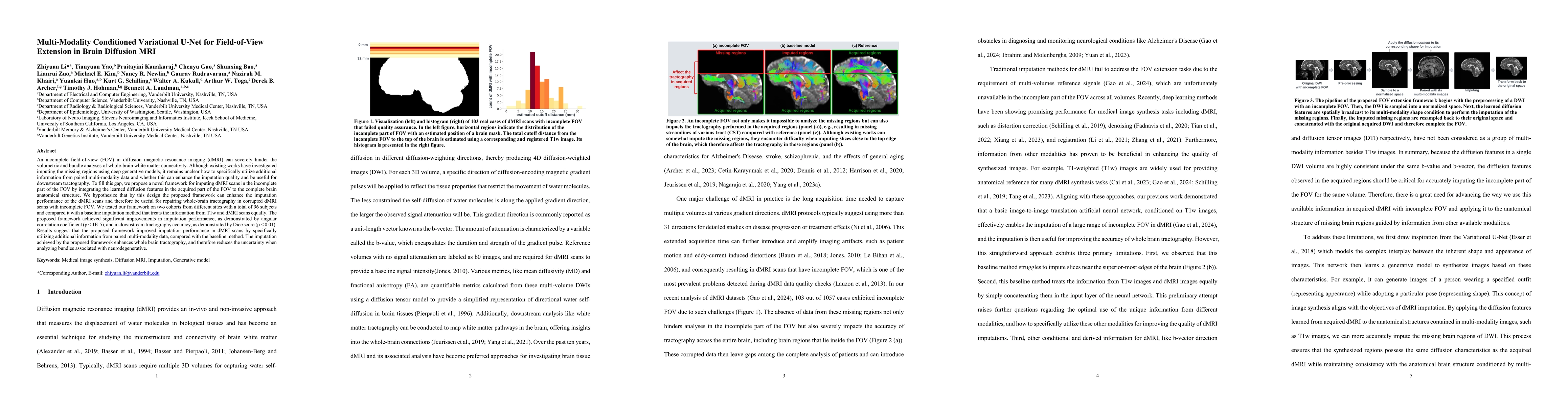
An incomplete field-of-view (FOV) in diffusion magnetic resonance imaging (dMRI) can severely hinder the volumetric and bundle analyses of whole-brain white matter connectivity. Although existing work...
Estimated brain age from magnetic resonance image (MRI) and its deviation from chronological age can provide early insights into potential neurodegenerative diseases, supporting early detection and im...
Automatic magnetic resonance (MR) image processing pipelines are widely used to study people with multiple sclerosis (PwMS), encompassing tasks such as lesion segmentation and brain parcellation. Howe...
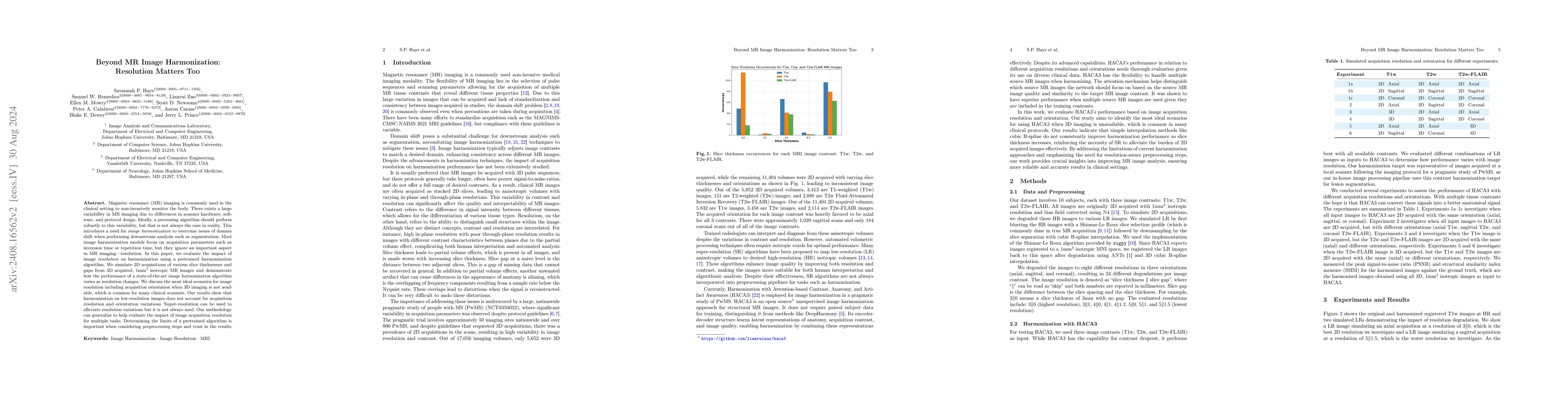
Magnetic resonance (MR) imaging is commonly used in the clinical setting to non-invasively monitor the body. There exists a large variability in MR imaging due to differences in scanner hardware, soft...
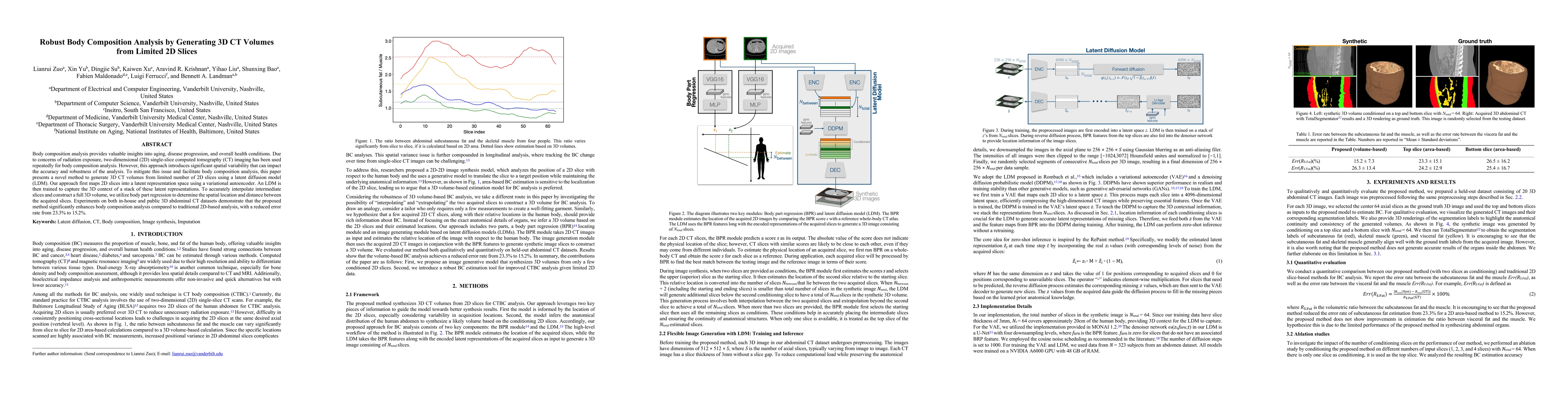
Body composition analysis provides valuable insights into aging, disease progression, and overall health conditions. Due to concerns of radiation exposure, two-dimensional (2D) single-slice computed t...
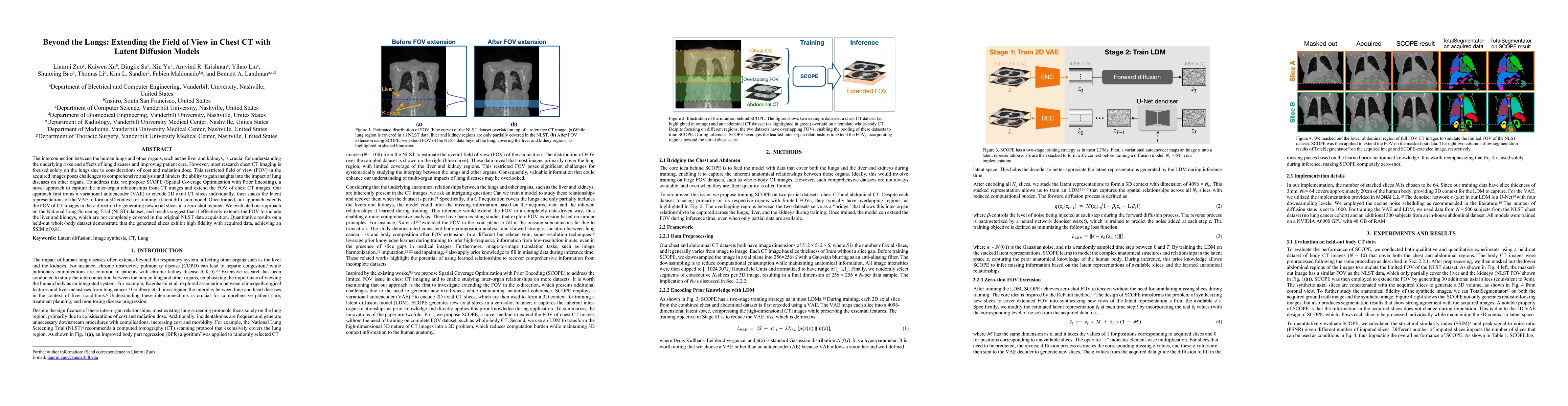
The interconnection between the human lungs and other organs, such as the liver and kidneys, is crucial for understanding the underlying risks and effects of lung diseases and improving patient care. ...
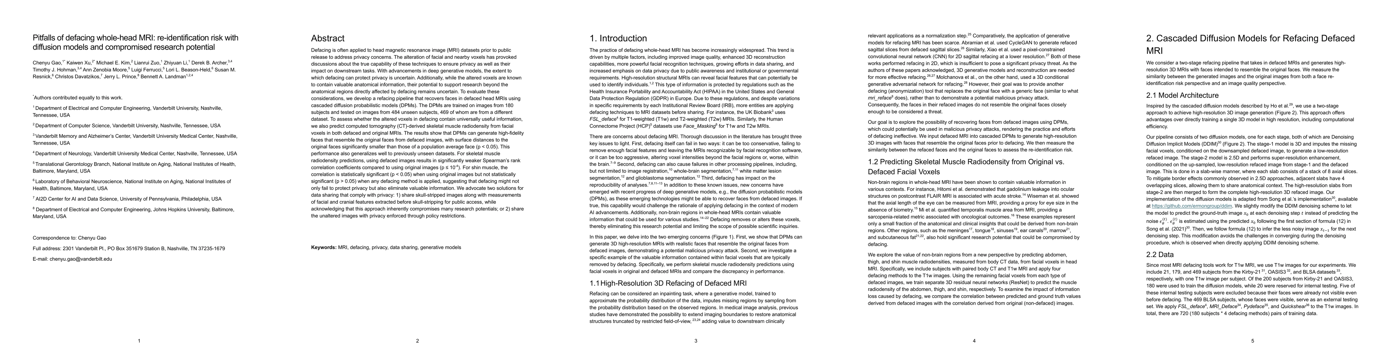
Defacing is often applied to head magnetic resonance image (MRI) datasets prior to public release to address privacy concerns. The alteration of facial and nearby voxels has provoked discussions about...
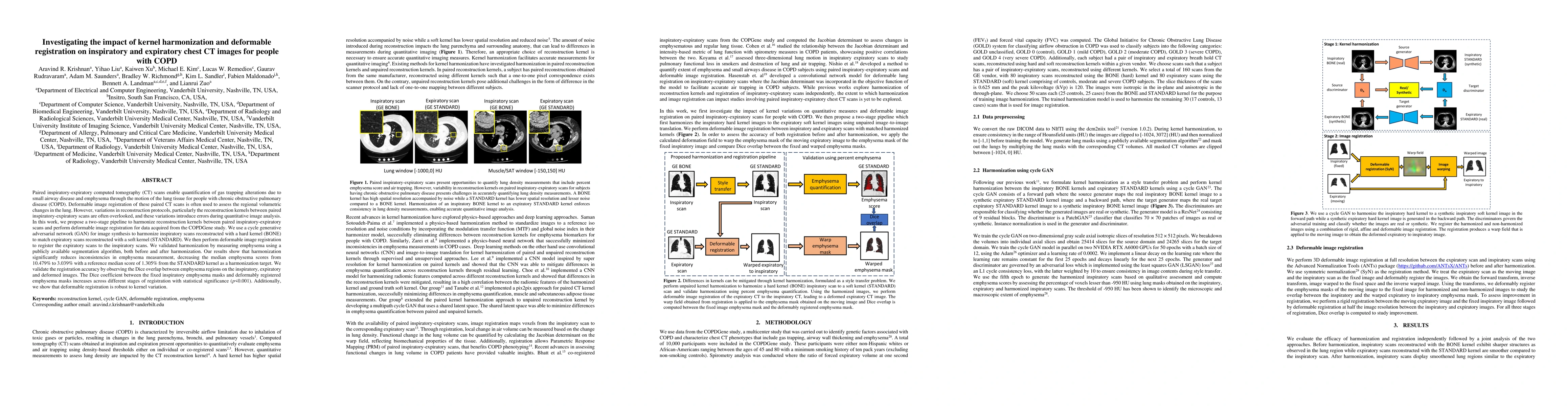
Paired inspiratory-expiratory CT scans enable the quantification of gas trapping due to small airway disease and emphysema by analyzing lung tissue motion in COPD patients. Deformable image registrati...
Reconstruction kernels in computed tomography (CT) affect spatial resolution and noise characteristics, introducing systematic variability in quantitative imaging measurements such as emphysema quanti...
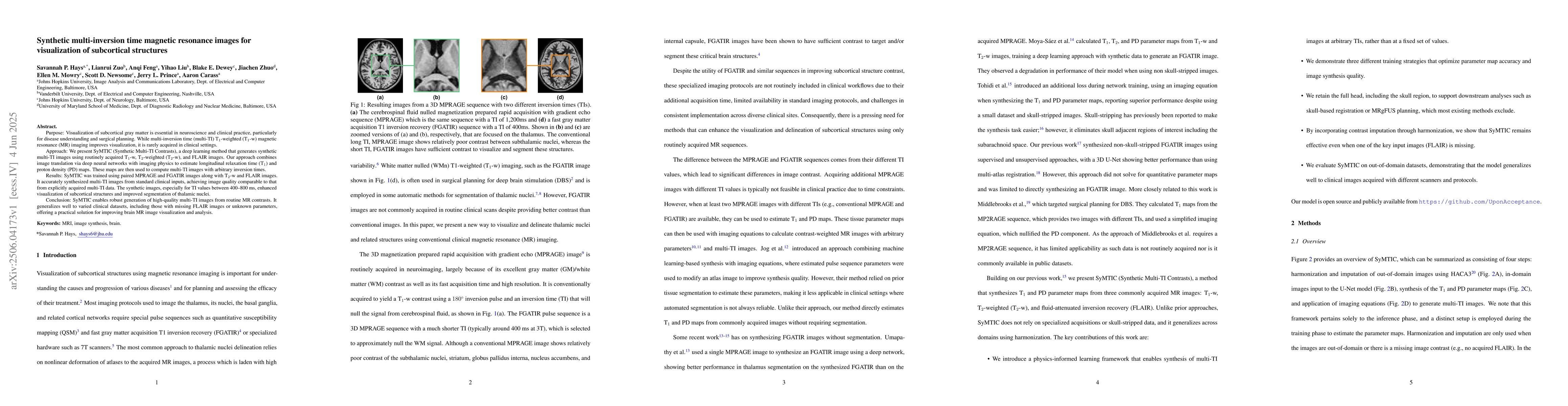
Purpose: Visualization of subcortical gray matter is essential in neuroscience and clinical practice, particularly for disease understanding and surgical planning.While multi-inversion time (multi-TI)...
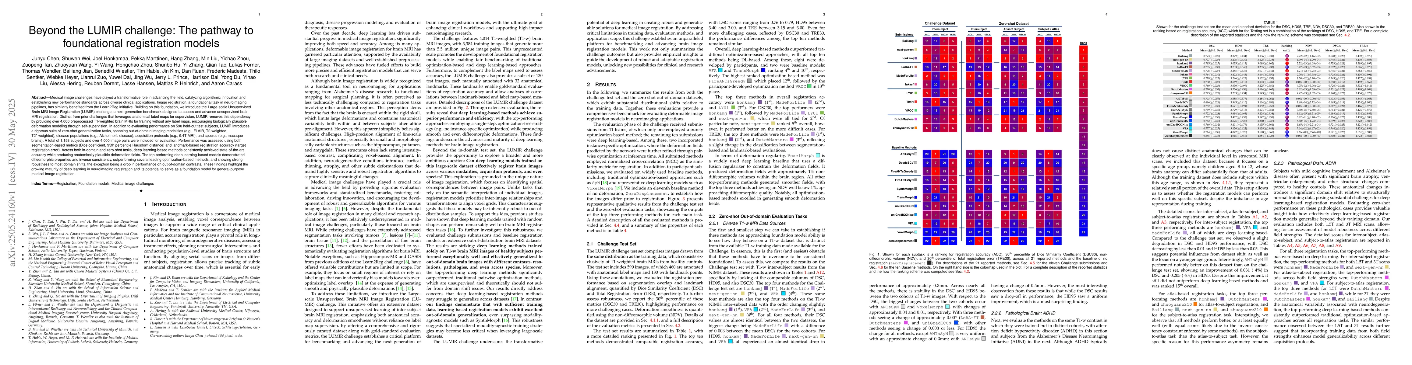
Medical image challenges have played a transformative role in advancing the field, catalyzing algorithmic innovation and establishing new performance standards across diverse clinical applications. Im...
Automated segmentation of multiple sclerosis (MS) lesions using multicontrast magnetic resonance (MR) images improves efficiency and reproducibility compared to manual delineation, with deep learning ...
Objective: Deep learning-based deformable image registration has achieved strong accuracy, but remains sensitive to variations in input image characteristics such as artifacts, field-of-view mismatch,...
The development of multimodal models for pulmonary nodule diagnosis is limited by the scarcity of labeled data and the tendency for these models to overfit on the training distribution. In this work, ...
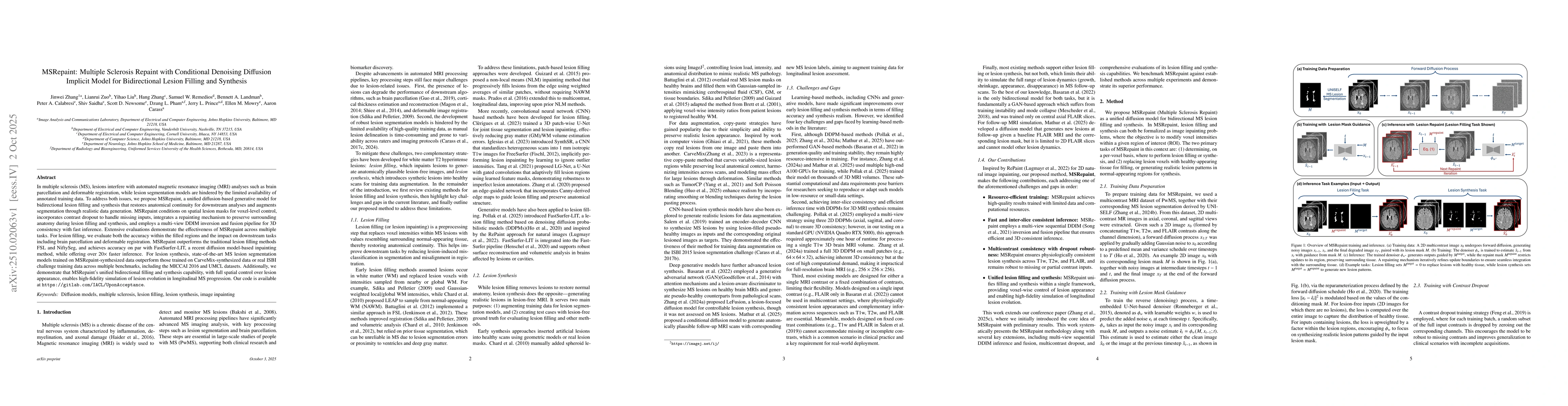
In multiple sclerosis, lesions interfere with automated magnetic resonance imaging analyses such as brain parcellation and deformable registration, while lesion segmentation models are hindered by the...
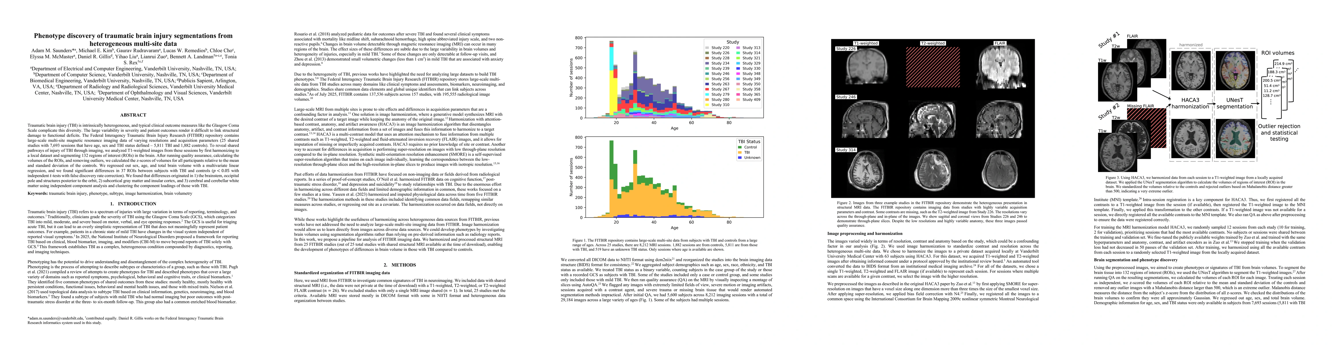
Traumatic brain injury (TBI) is intrinsically heterogeneous, and typical clinical outcome measures like the Glasgow Coma Scale complicate this diversity. The large variability in severity and patient ...
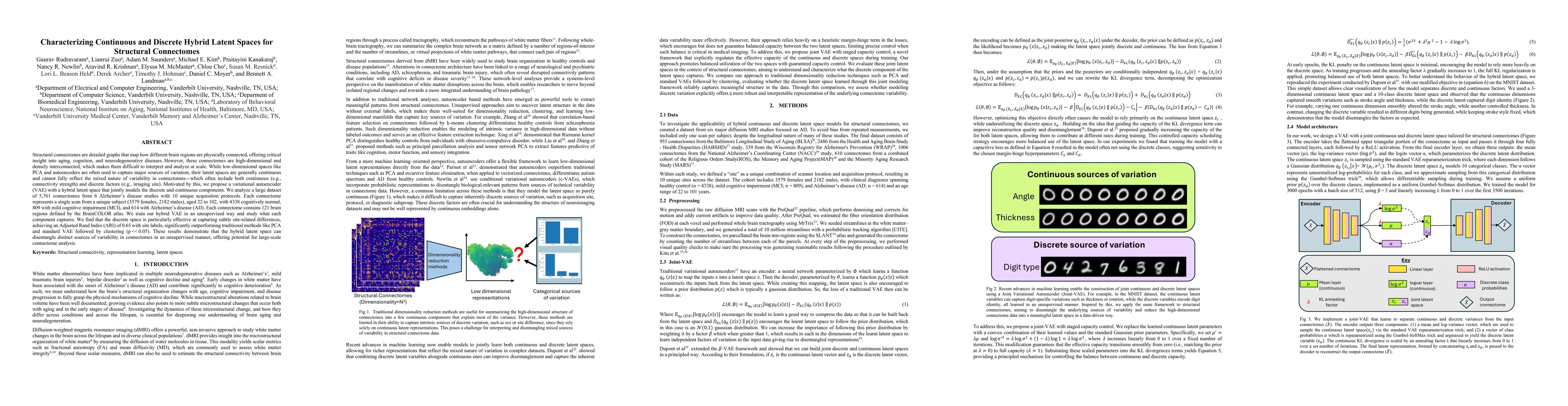
Structural connectomes are detailed graphs that map how different brain regions are physically connected, offering critical insight into aging, cognition, and neurodegenerative diseases. However, thes...
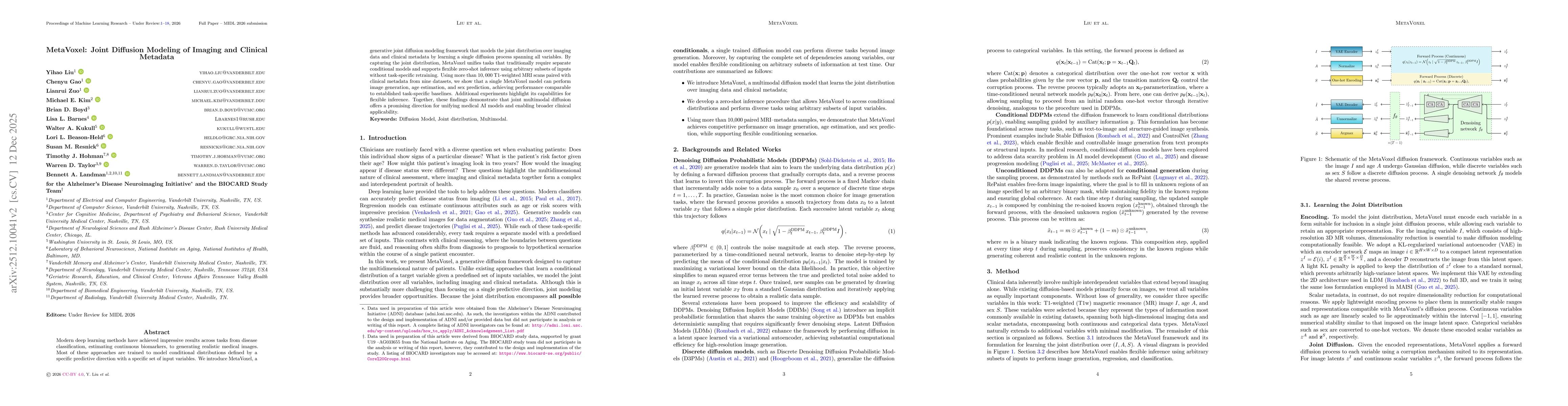
Modern deep learning methods have achieved impressive results across tasks from disease classification, estimating continuous biomarkers, to generating realistic medical images. Most of these approach...
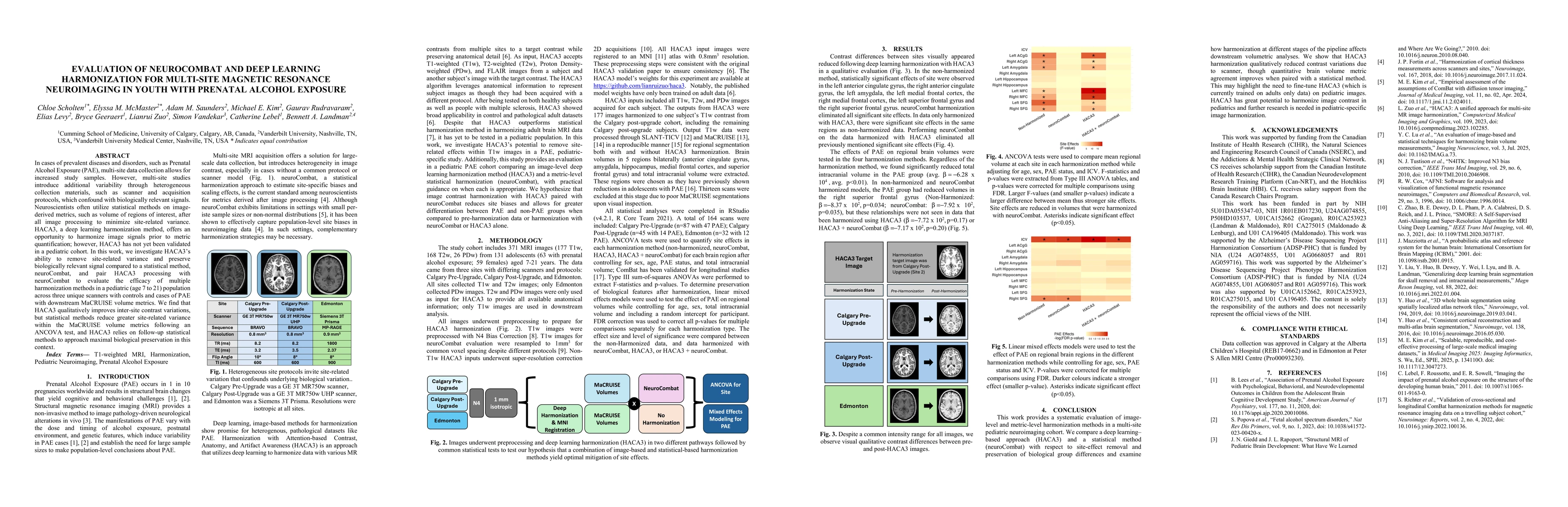
In cases of prevalent diseases and disorders, such as Prenatal Alcohol Exposure (PAE), multi-site data collection allows for increased study samples. However, multi-site studies introduce additional v...
Reliable harmonization of heterogeneous magnetic resonance~(MR) image datasets, especially those acquired in pragmatic clinical trials, is critical to advance multi-center neuroimaging studies and tra...
Acquisition differences across sites, scanners, and protocols in dMRI introduce variability that complicates structural connectome analysis. This motivates deep learning models that can represent high...