Academic Profile
Statistics
Similar Authors
Papers on arXiv
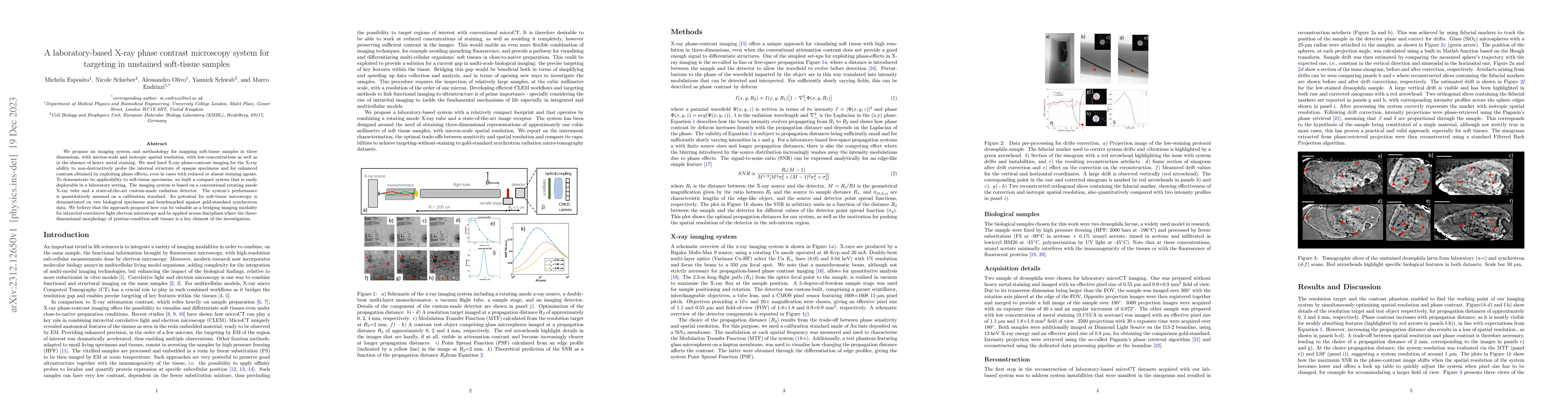
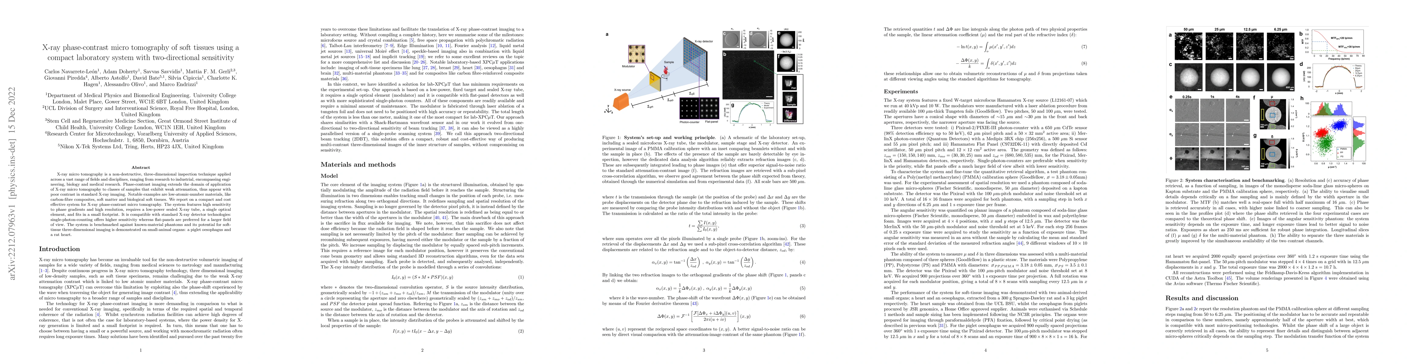
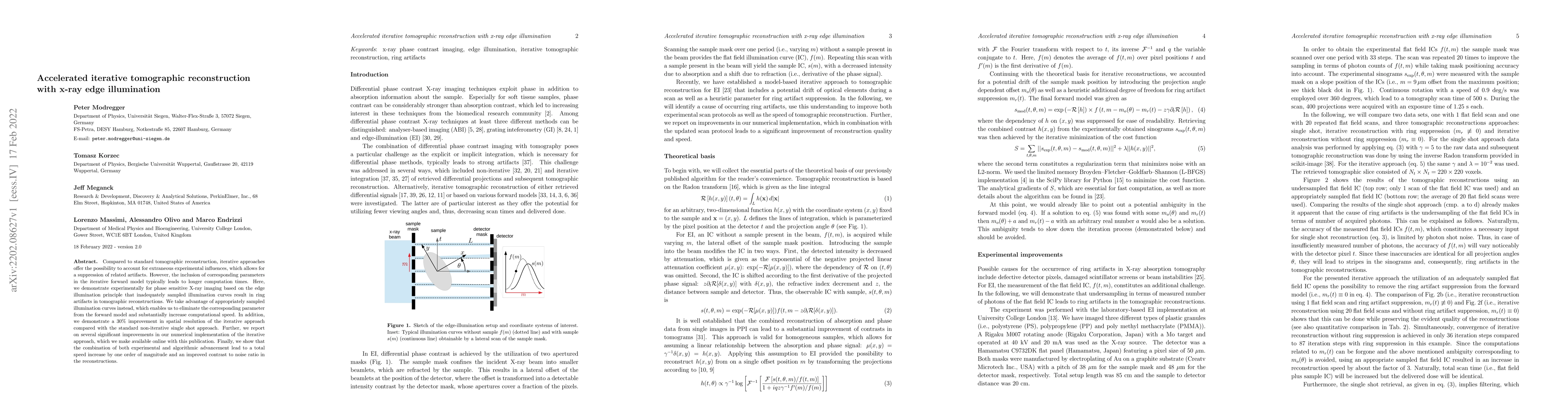
We propose an imaging system and methodology for mapping soft-tissue samples in three dimensions, with micron-scale and isotropic spatial resolution, with low-concentrations as well as in the absenc...
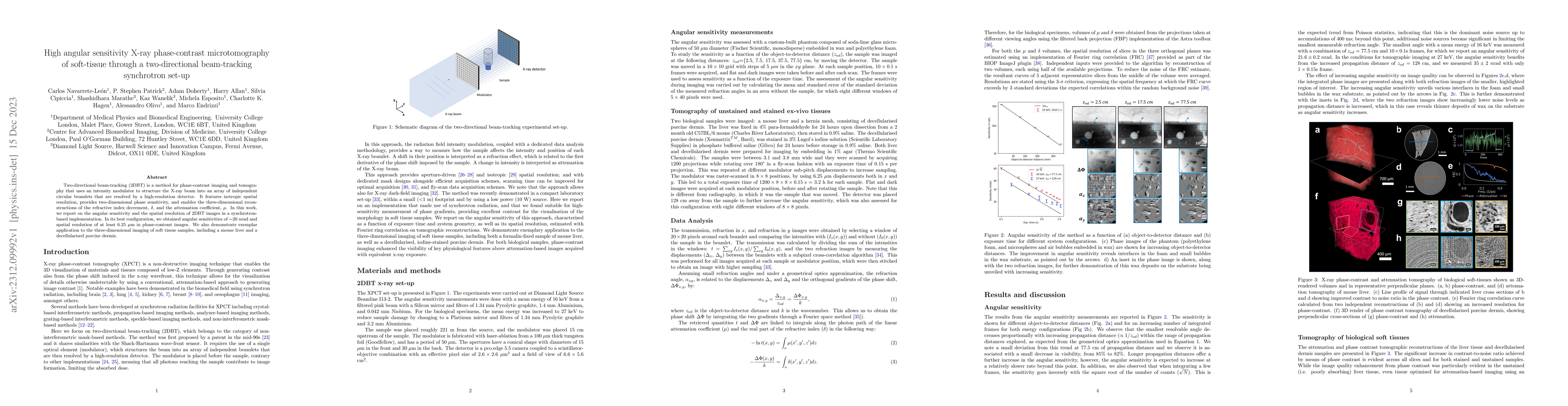
Two-directional beam-tracking (2DBT) is a method for phase-contrast imaging and tomography that uses an intensity modulator to structure the X-ray beam into an array of independent circular beamlets...
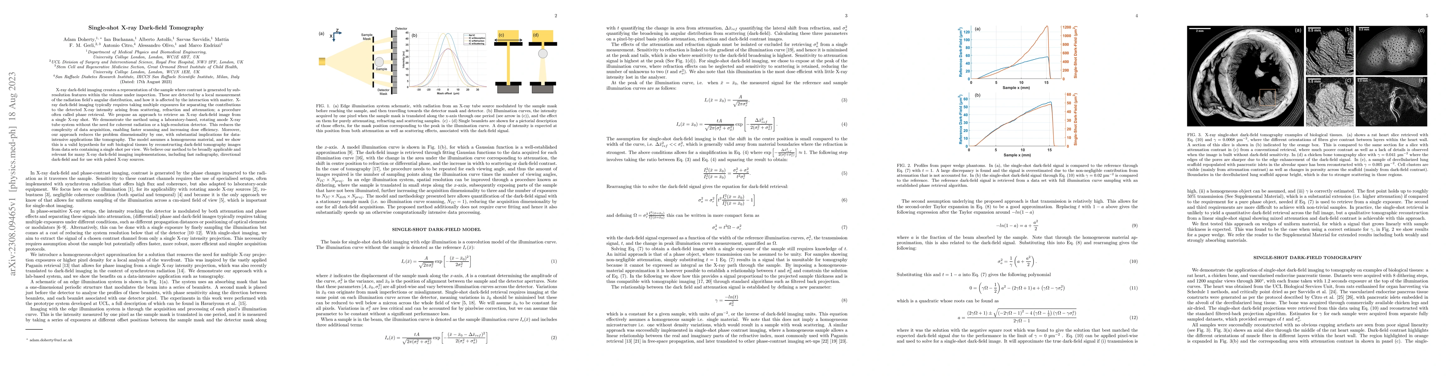
X-ray dark-field imaging creates a representation of the sample where contrast is generated by subresolution features within the volume under inspection. These are detected by a local measurement of...
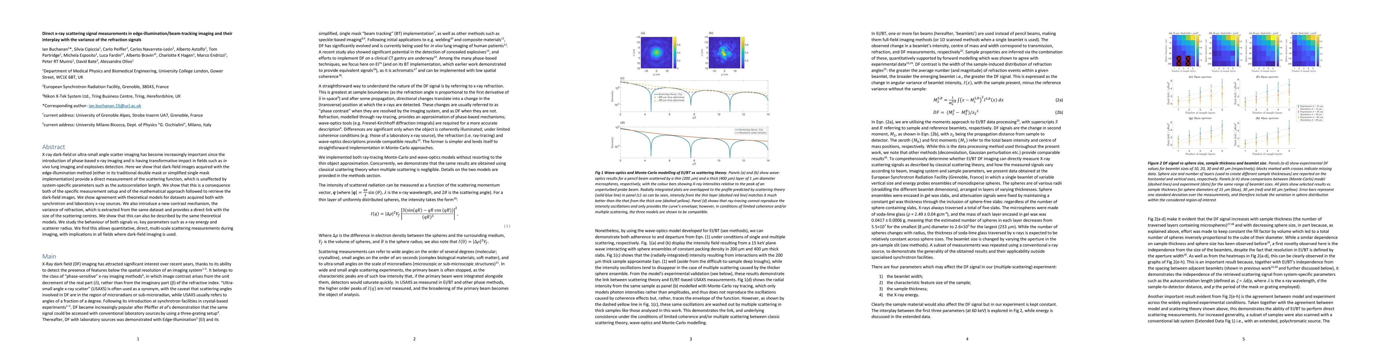
X-ray dark-field or ultra-small angle scatter imaging has become increasingly important since the introduction of phase-based x-ray imaging and is having transformative impact in fields such as in v...
X-ray micro tomography is a non-destructive, three-dimensional inspection technique applied across a vast range of fields and disciplines, ranging from research to industrial, encompassing engineeri...
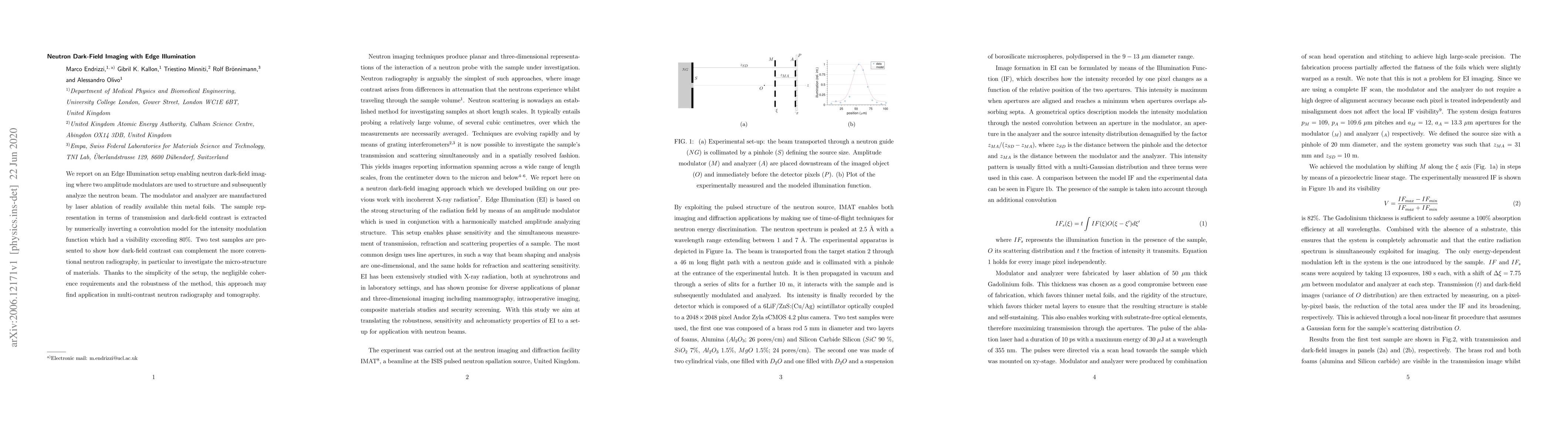
Compared to standard tomographic reconstruction, iterative approaches offer the possibility to account for extraneous experimental influences, which allows for a suppression of related artifacts. Ho...
We report on an Edge Illumination setup enabling neutron dark-field imaging where two amplitude modulators are used to structure and subsequently analyze the neutron beam. The modulator and analyzer...
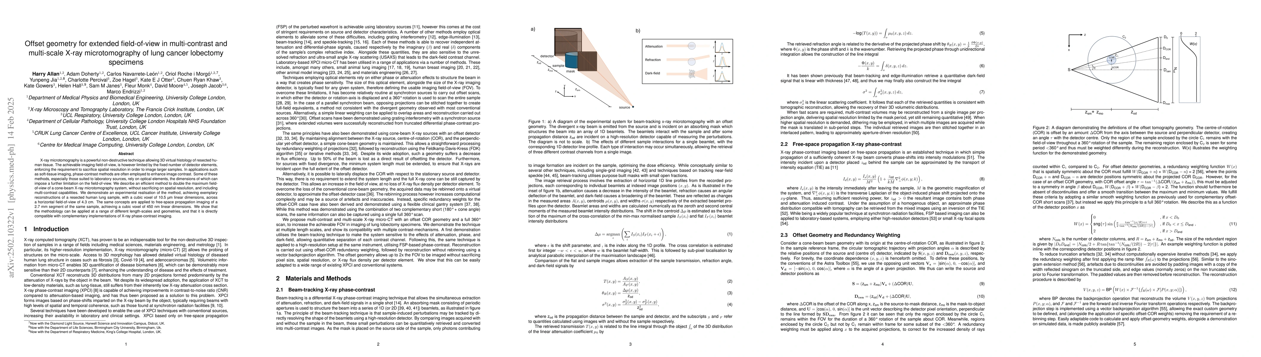
X-ray microtomography is a powerful non-destructive technique allowing 3D virtual histology of resected human tissue. The achievable imaging field-of-view, is however limited by the fixed number of de...
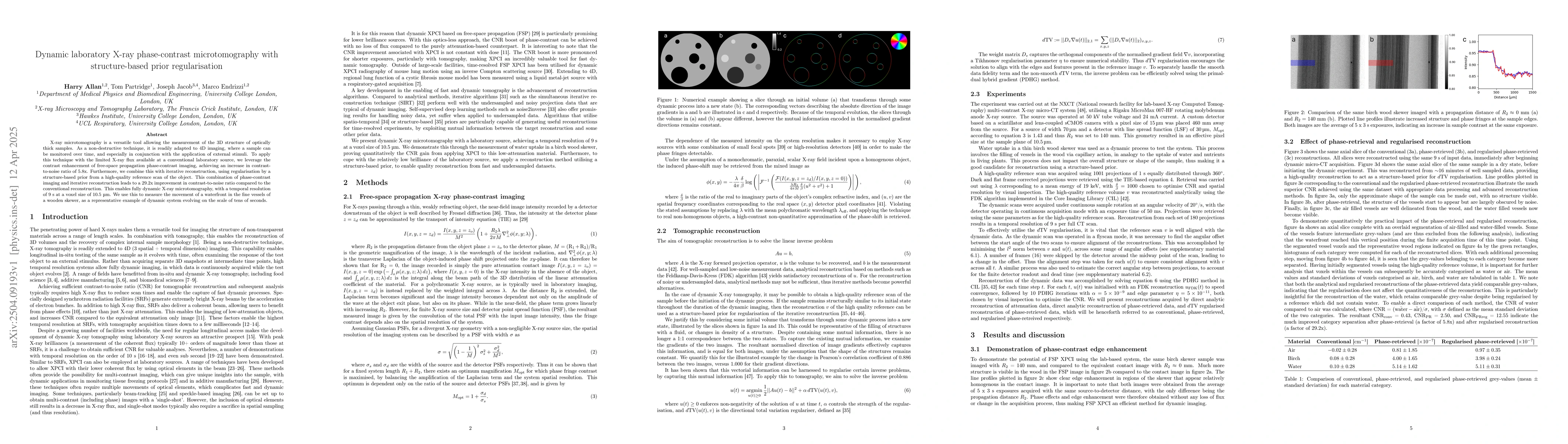
X-ray microtomography is a versatile tool allowing the measurement of the 3D structure of optically thick samples. As a non-destructive technique, it is readily adapted to 4D imaging, where a sample c...
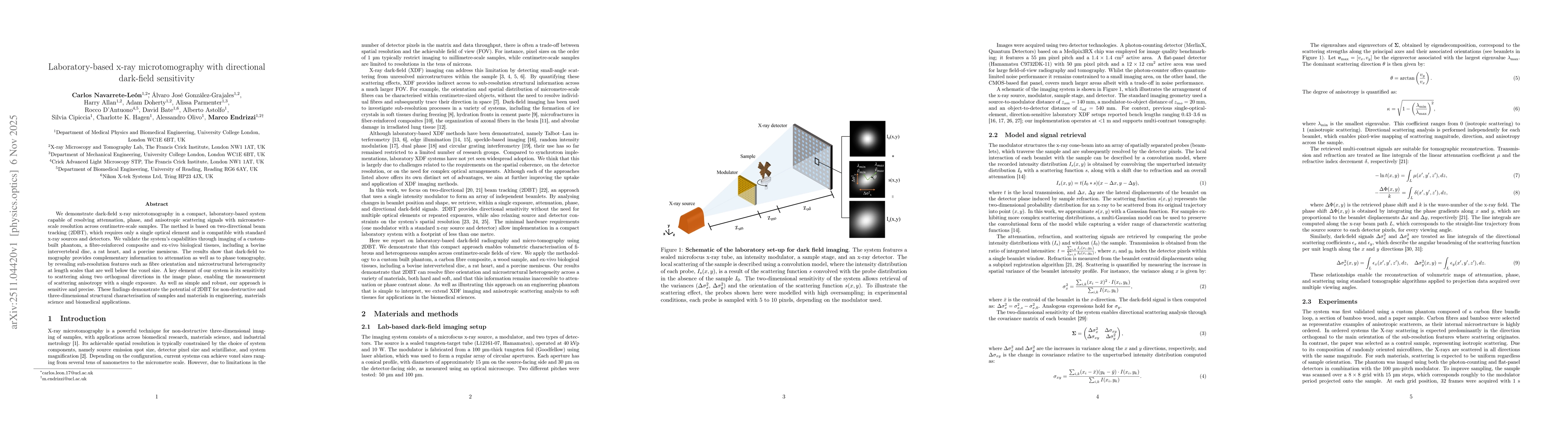
We demonstrate dark-field x-ray microtomography in a compact, laboratory-based system capable of resolving attenuation, phase, and anisotropic scattering signals with micrometer-scale resolution acros...
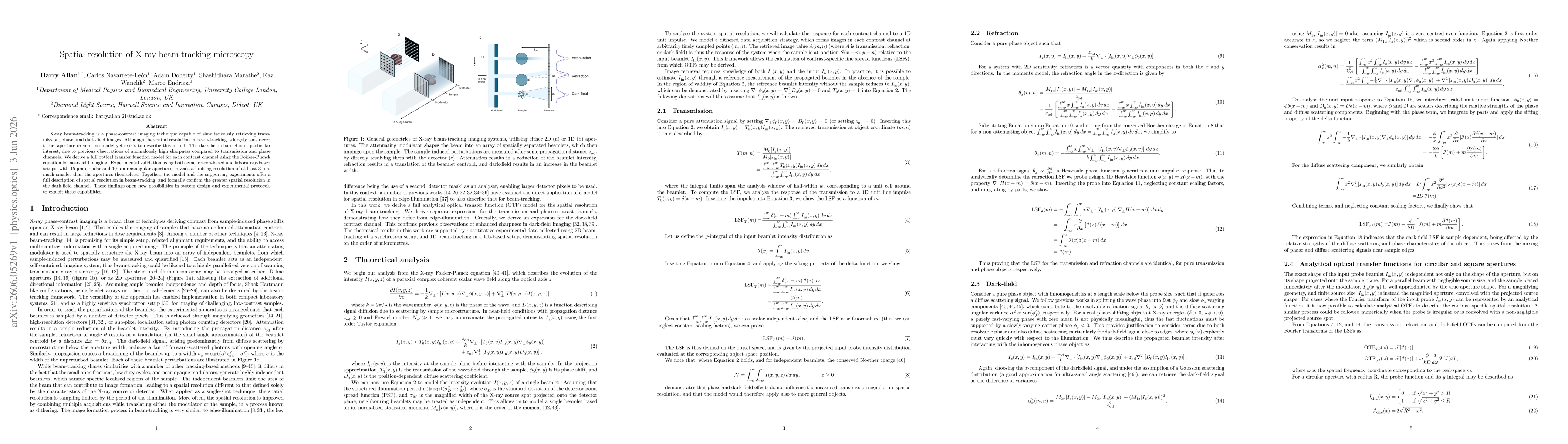
X-ray beam-tracking is a phase-contrast imaging technique capable of simultaneously retrieving transmission, phase, and dark-field images. Although the spatial resolution in beam-tracking is largely c...
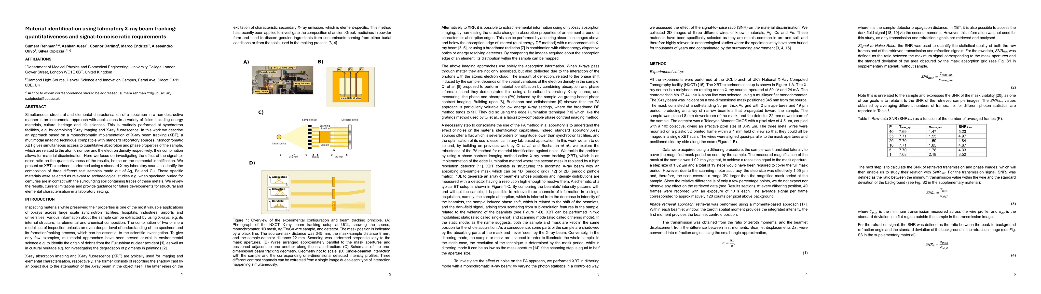
Simultaneous structural and elemental characterisation of a specimen in a non-destructive manner is an instrumental approach with applications in a variety of fields including energy materials, cultur...