ArxivLens
M
Researcher

Megumi Nakao

10 papers on arXiv

10 arXiv Papers
10 Total Publications
Profile

Academic Profile

Name M. Nakao
Total Papers 10
Scholar Profile View on Semantic Scholar
Metrics

Statistics

10 arXiv Papers
10 Total Publications
Publications

Papers on arXiv

Thumbnail for Shape Reconstruction from Thoracoscopic Images using Self-supervised
  Virtual Learning
arXiv
Shape Reconstruction from Thoracoscopic Images using Self-supervised Virtual Learning

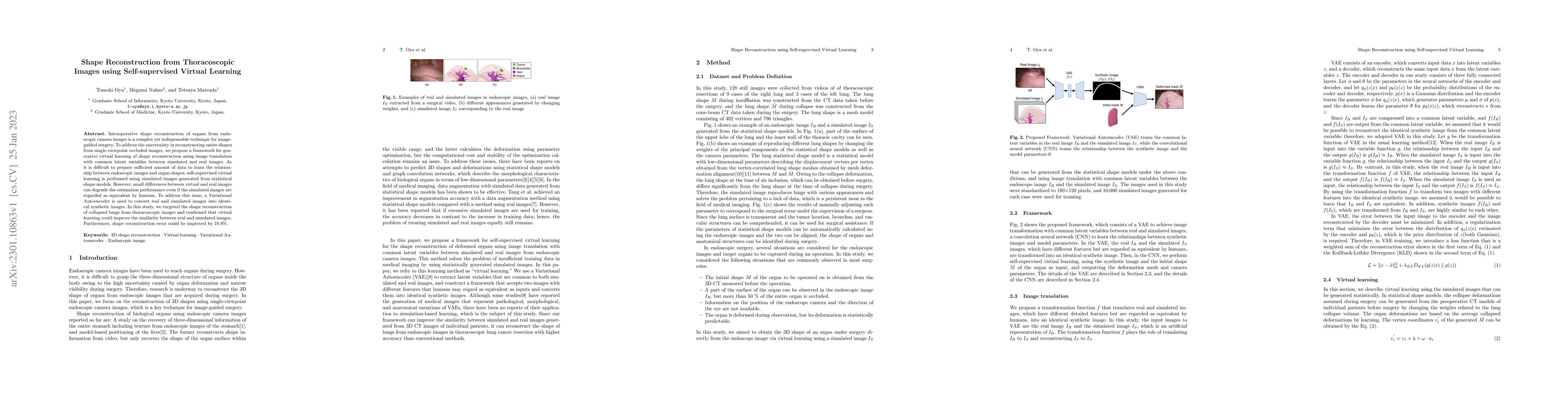
Intraoperative shape reconstruction of organs from endoscopic camera images is a complex yet indispensable technique for image-guided surgery. To address the uncertainty in reconstructing entire sha...

Thumbnail for 2D/3D Deep Image Registration by Learning 3D Displacement Fields for
  Abdominal Organs
arXiv
2D/3D Deep Image Registration by Learning 3D Displacement Fields for Abdominal Organs

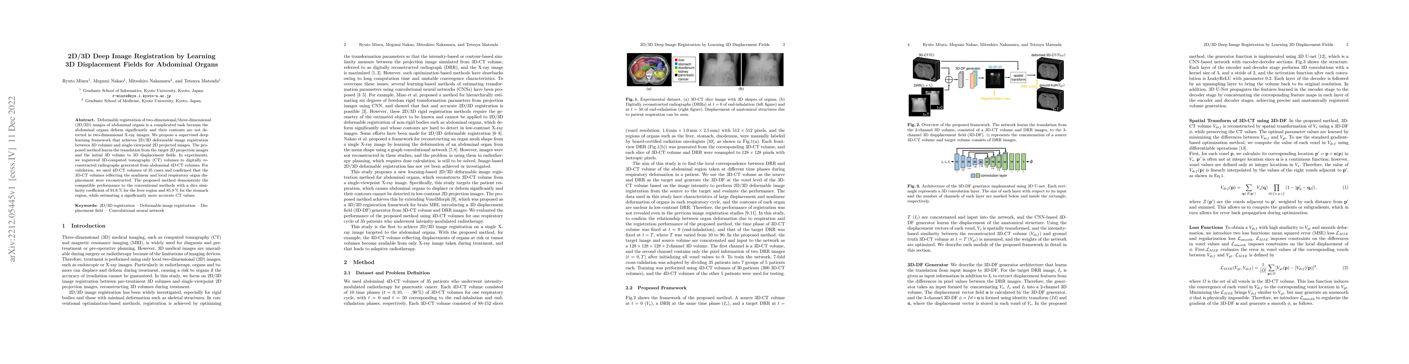
Deformable registration of two-dimensional/three-dimensional (2D/3D) images of abdominal organs is a complicated task because the abdominal organs deform significantly and their contours are not det...

Thumbnail for Feedback Assisted Adversarial Learning to Improve the Quality of
  Cone-beam CT Images
arXiv
Feedback Assisted Adversarial Learning to Improve the Quality of Cone-beam CT Images

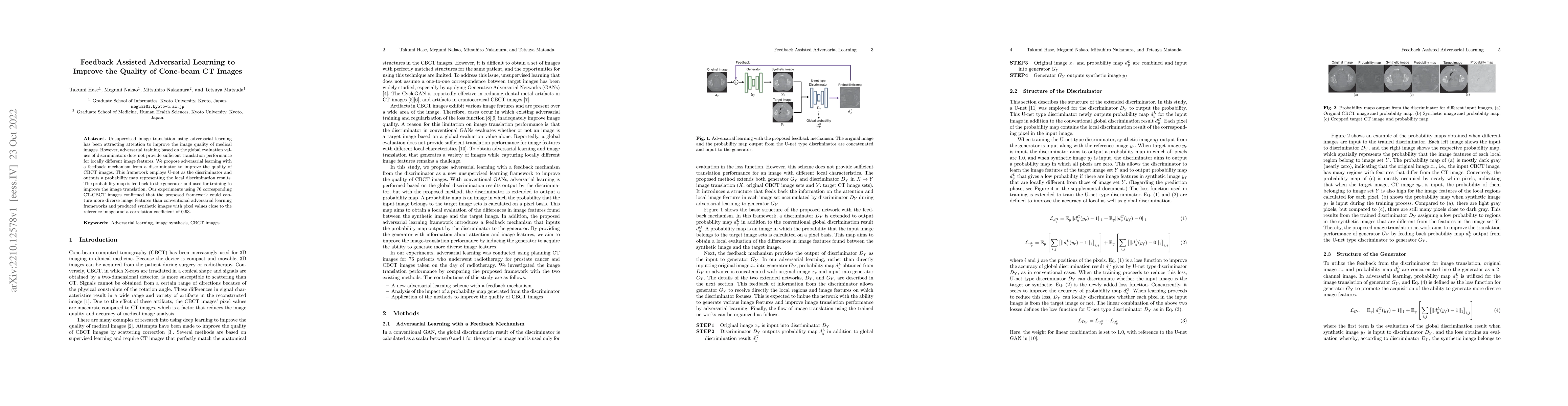
Unsupervised image translation using adversarial learning has been attracting attention to improve the image quality of medical images. However, adversarial training based on the global evaluation v...

Thumbnail for IGCN: Image-to-graph Convolutional Network for 2D/3D Deformable
  Registration
arXiv
IGCN: Image-to-graph Convolutional Network for 2D/3D Deformable Registration

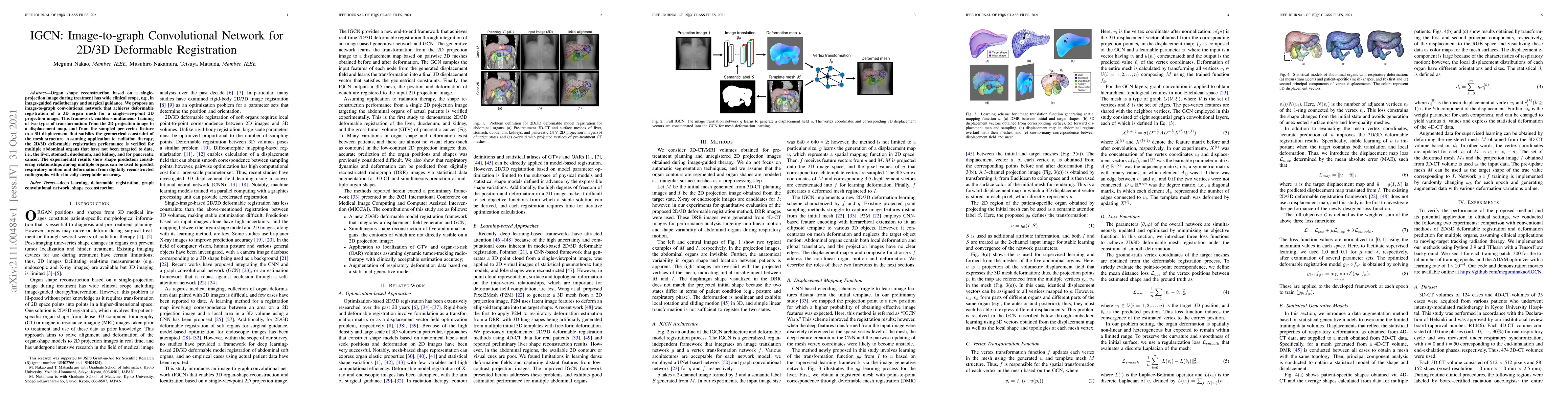
Organ shape reconstruction based on a single-projection image during treatment has wide clinical scope, e.g., in image-guided radiotherapy and surgical guidance. We propose an image-to-graph convolu...

Thumbnail for Statistical modeling of pneumothorax deformation by mapping CT and
  cone-beam CT images
arXiv
Statistical modeling of pneumothorax deformation by mapping CT and cone-beam CT images

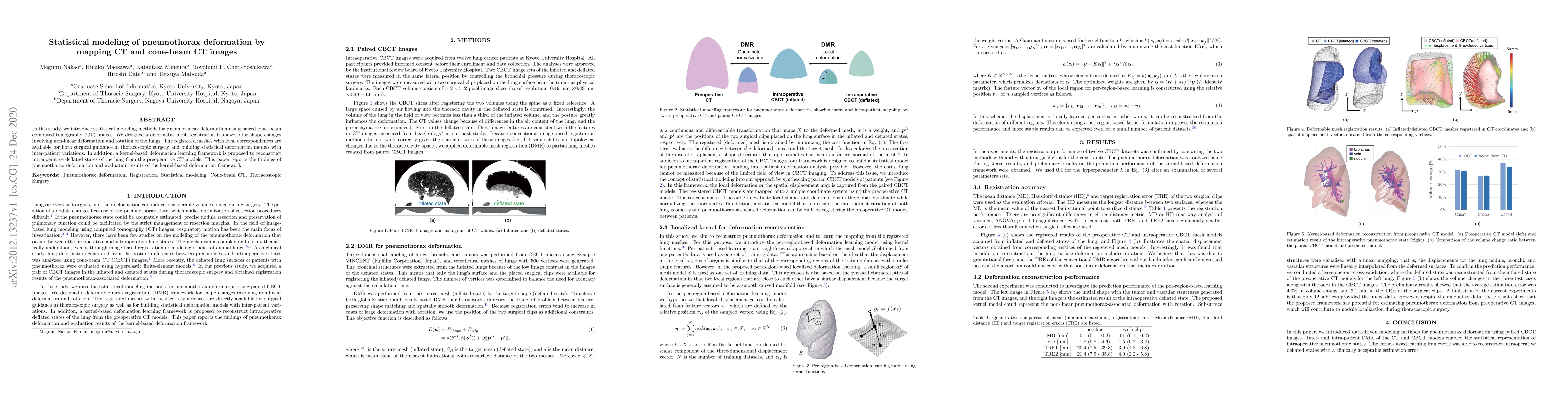
In this study, we introduce statistical modeling methods for pneumothorax deformation using paired cone-beam computed tomography (CT) images. We designed a deformable mesh registration framework for...

Thumbnail for Analysis of Heterogeneity of Pneumothorax-associated Deformation using
  Model-based Registration
arXiv
Analysis of Heterogeneity of Pneumothorax-associated Deformation using Model-based Registration

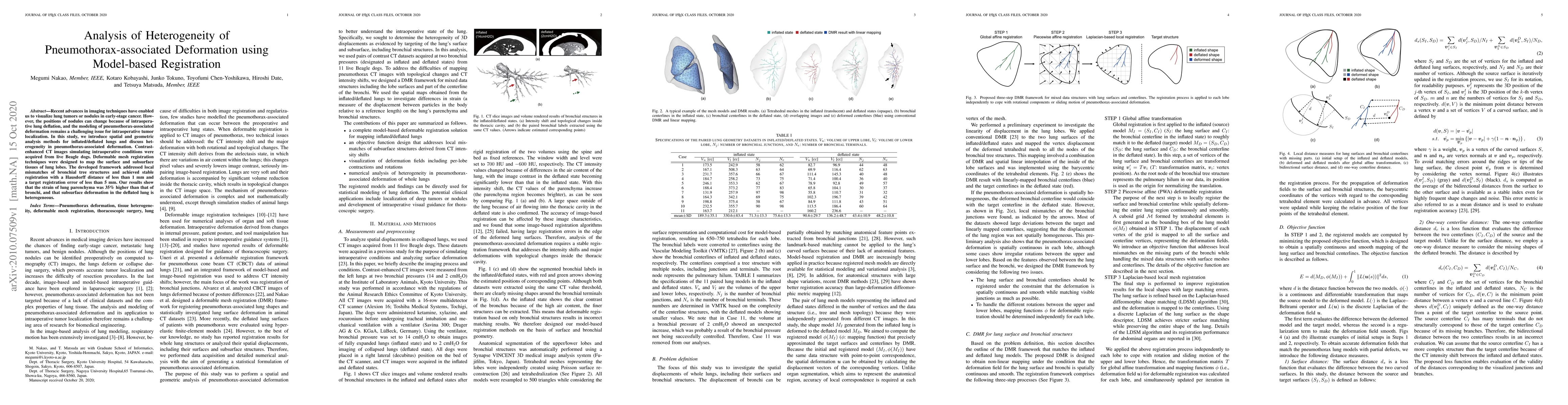
Recent advances in imaging techniques have enabled us to visualize lung tumors or nodules in early-stage cancer. However, the positions of nodules can change because of intraoperative lung deflation...

Thumbnail for Statistical Deformation Reconstruction Using Multi-organ Shape Features
  for Pancreatic Cancer Localization
arXiv
Statistical Deformation Reconstruction Using Multi-organ Shape Features for Pancreatic Cancer Localization

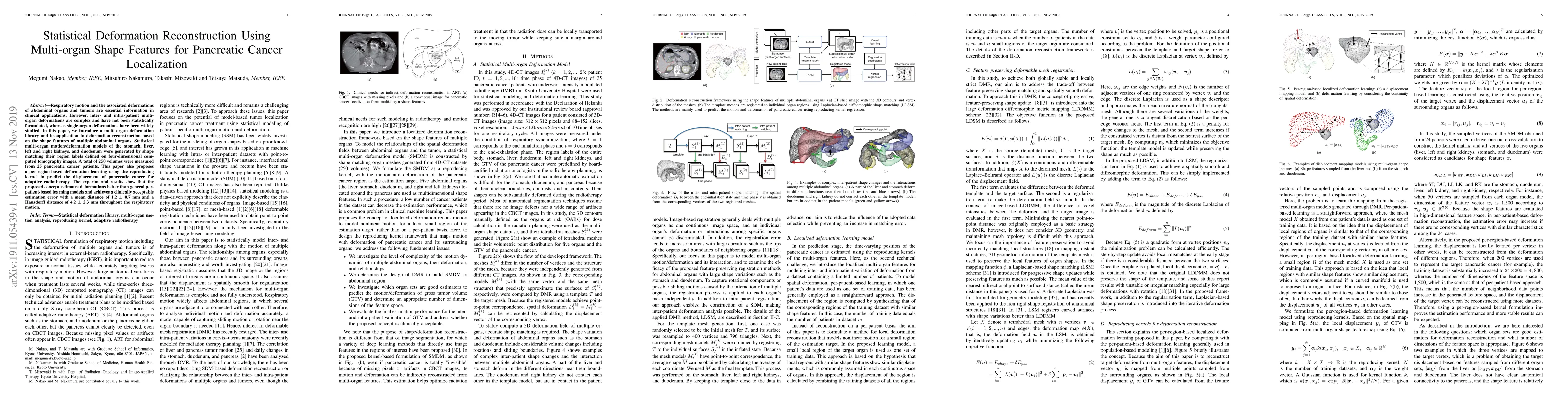
Respiratory motion and the associated deformations of abdominal organs and tumors are essential information in clinical applications. However, inter- and intra-patient multi-organ deformations are c...

Thumbnail for Construction of an Organ Shape Atlas Using a Hierarchical Mesh
  Variational Autoencoder
arXiv
Construction of an Organ Shape Atlas Using a Hierarchical Mesh Variational Autoencoder

An organ shape atlas, which represents the shape and position of the organs and skeleton of a living body using a small number of parameters, is expected to have a wide range of clinical applications,...

Thumbnail for Limited-Angle CT Reconstruction Using Multi-Volume Latent Consistency Model
arXiv
Limited-Angle CT Reconstruction Using Multi-Volume Latent Consistency Model

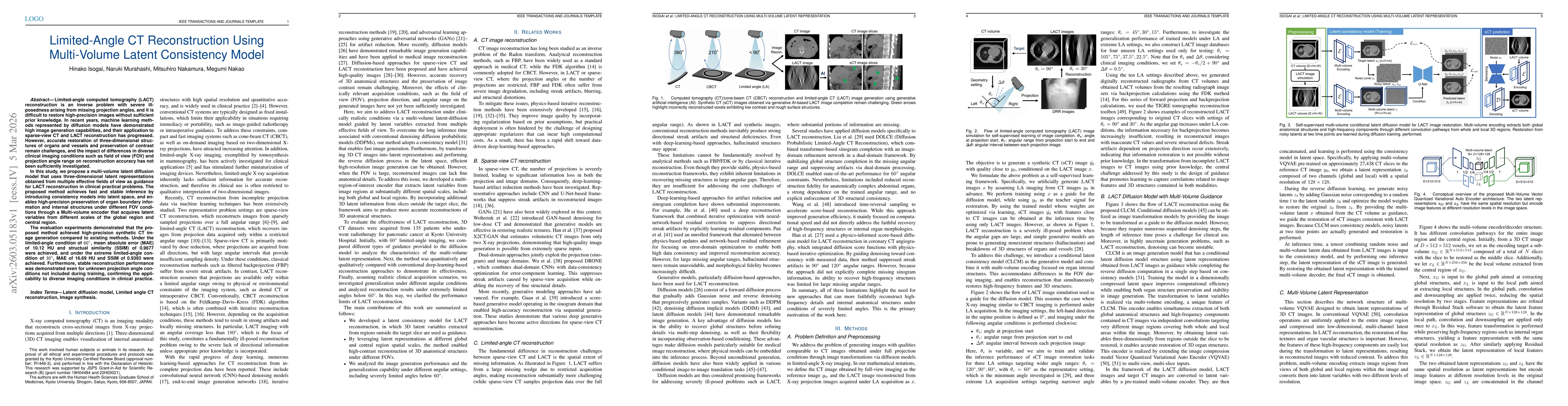
Limited-angle computed tomography (LACT) reconstruction is an inverse problem with severe ill-posedness arising from missing projection angles, and it is difficult to restore high-precision images wit...

Thumbnail for Cone-Beam CT Image Quality Enhancement Using A Latent Diffusion Model Trained with Simulated CBCT Artifacts
arXiv
Cone-Beam CT Image Quality Enhancement Using A Latent Diffusion Model Trained with Simulated CBCT Artifacts

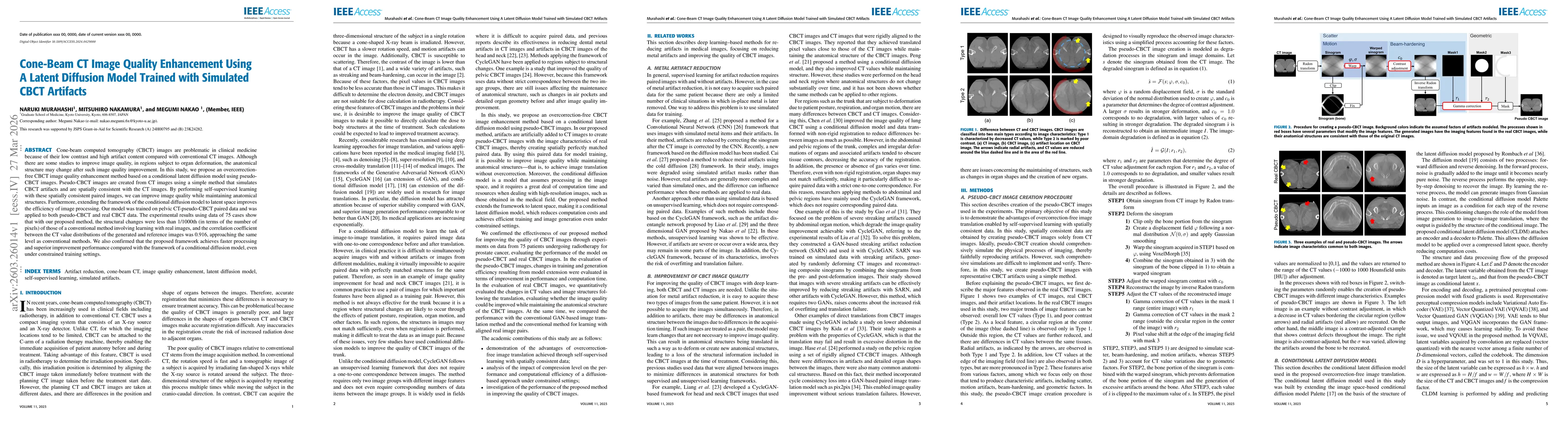
Cone-beam computed tomography (CBCT) images are problematic in clinical medicine because of their low contrast and high artifact content compared with conventional CT images. Although there are some s...

This website uses cookies to ensure you get the best experience. Learn more