Academic Profile
Statistics
Similar Authors
Papers on arXiv
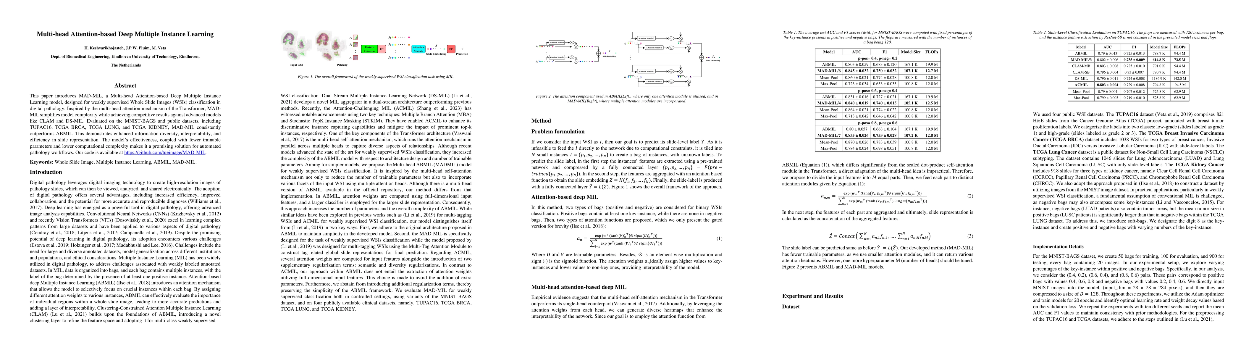
This paper introduces MAD-MIL, a Multi-head Attention-based Deep Multiple Instance Learning model, designed for weakly supervised Whole Slide Images (WSIs) classification in digital pathology. Inspi...
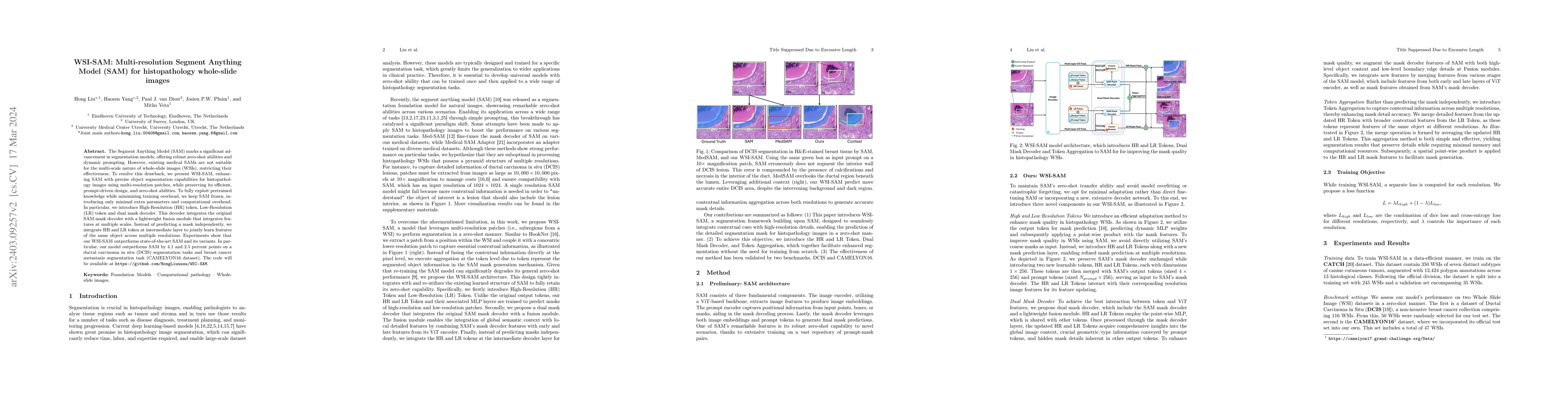
The Segment Anything Model (SAM) marks a significant advancement in segmentation models, offering robust zero-shot abilities and dynamic prompting. However, existing medical SAMs are not suitable fo...
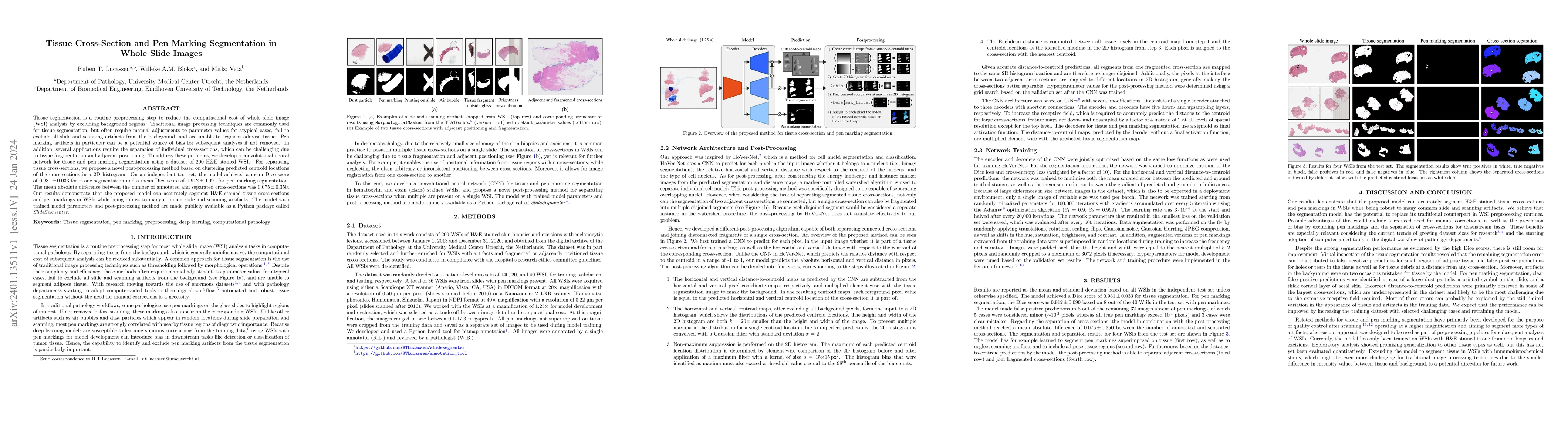
Tissue segmentation is a routine preprocessing step to reduce the computational cost of whole slide image (WSI) analysis by excluding background regions. Traditional image processing techniques are ...
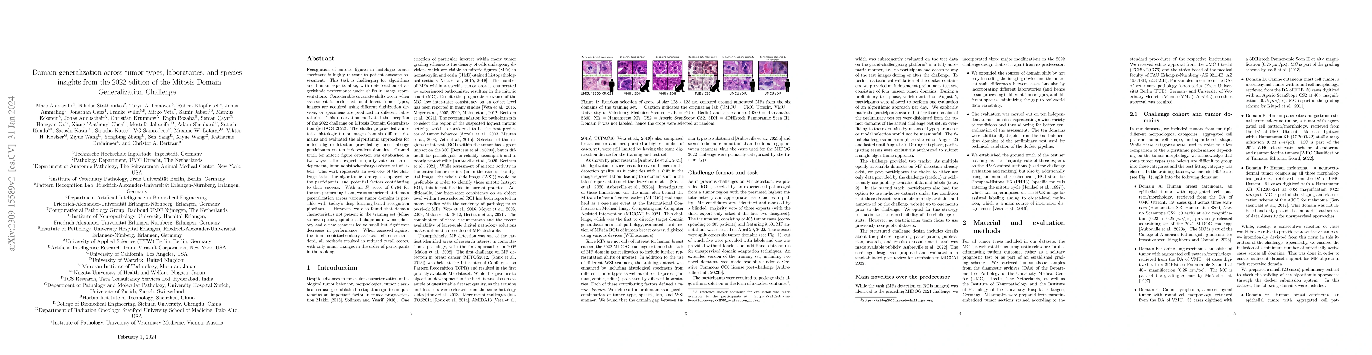
Recognition of mitotic figures in histologic tumor specimens is highly relevant to patient outcome assessment. This task is challenging for algorithms and human experts alike, with deterioration of ...
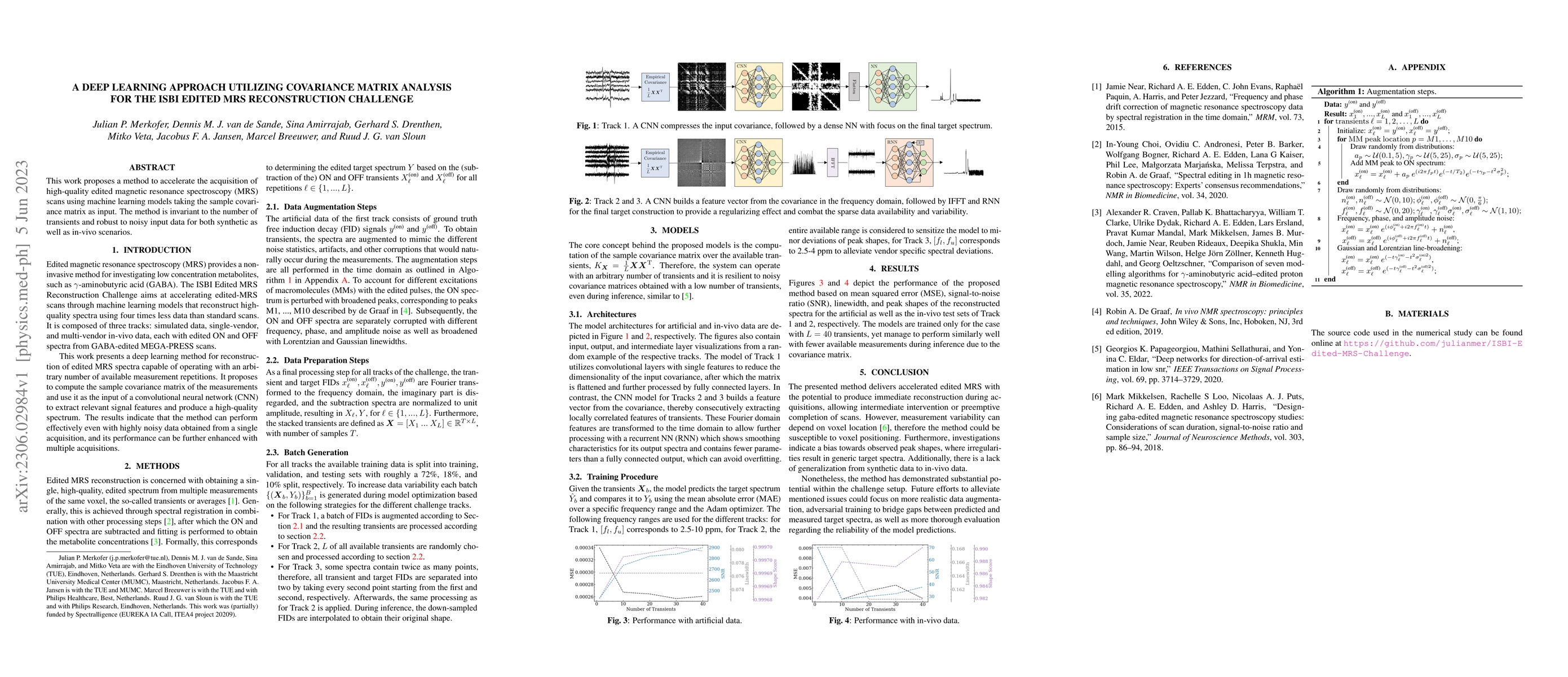
This work proposes a method to accelerate the acquisition of high-quality edited magnetic resonance spectroscopy (MRS) scans using machine learning models taking the sample covariance matrix as inpu...
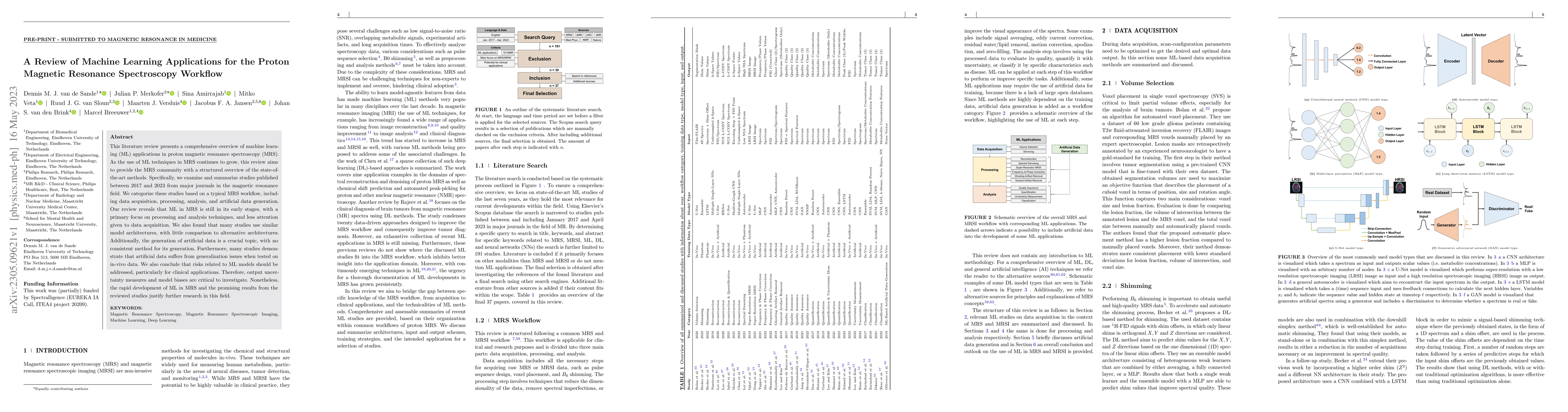
This literature review presents a comprehensive overview of machine learning (ML) applications in proton magnetic resonance spectroscopy (MRS). As the use of ML techniques in MRS continues to grow, ...
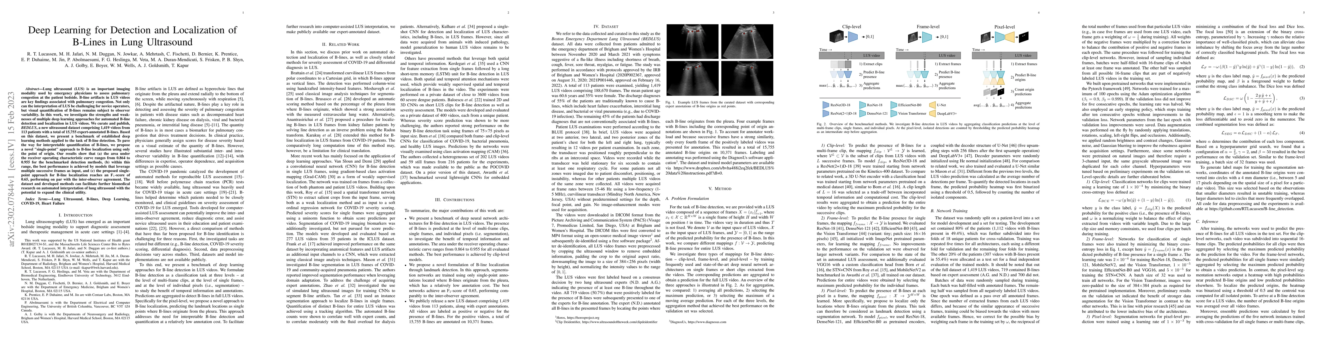
Lung ultrasound (LUS) is an important imaging modality used by emergency physicians to assess pulmonary congestion at the patient bedside. B-line artifacts in LUS videos are key findings associated ...
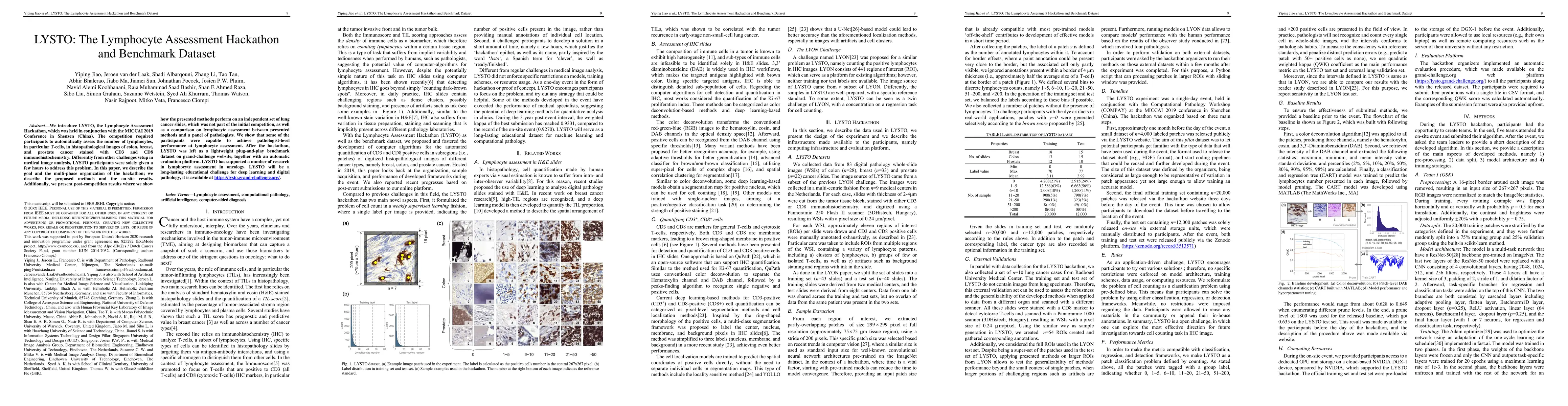
We introduce LYSTO, the Lymphocyte Assessment Hackathon, which was held in conjunction with the MICCAI 2019 Conference in Shenzen (China). The competition required participants to automatically asse...
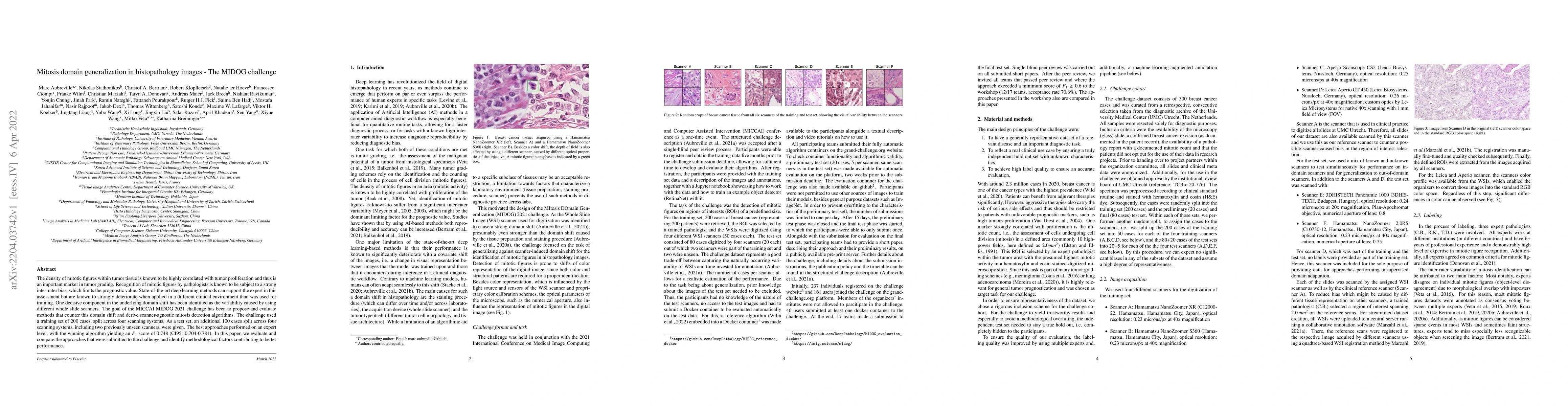
The density of mitotic figures within tumor tissue is known to be highly correlated with tumor proliferation and thus is an important marker in tumor grading. Recognition of mitotic figures by patho...
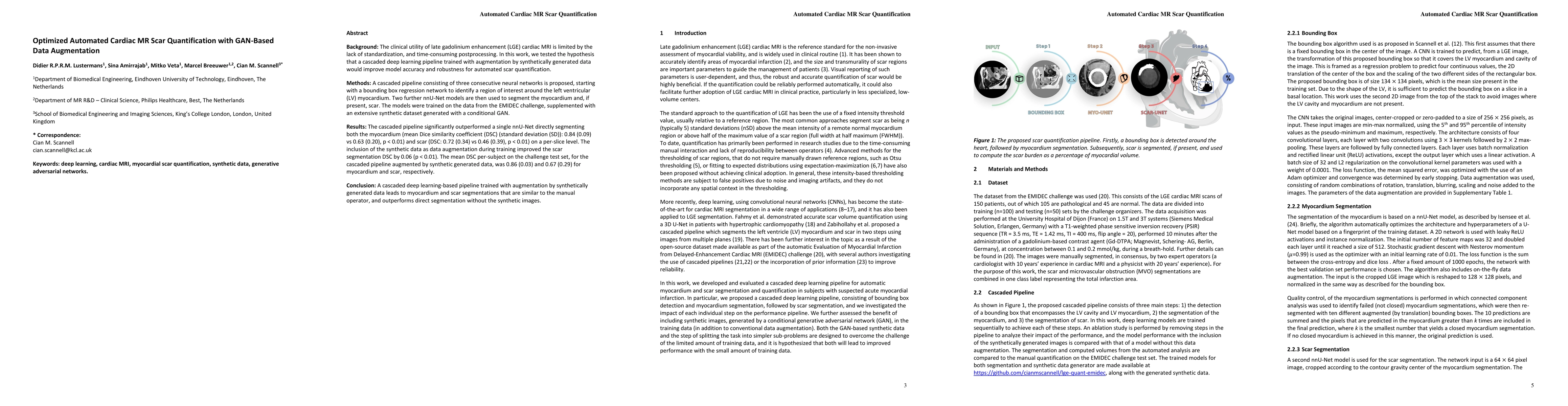
Background: The clinical utility of late gadolinium enhancement (LGE) cardiac MRI is limited by the lack of standardization, and time-consuming postprocessing. In this work, we tested the hypothesis...
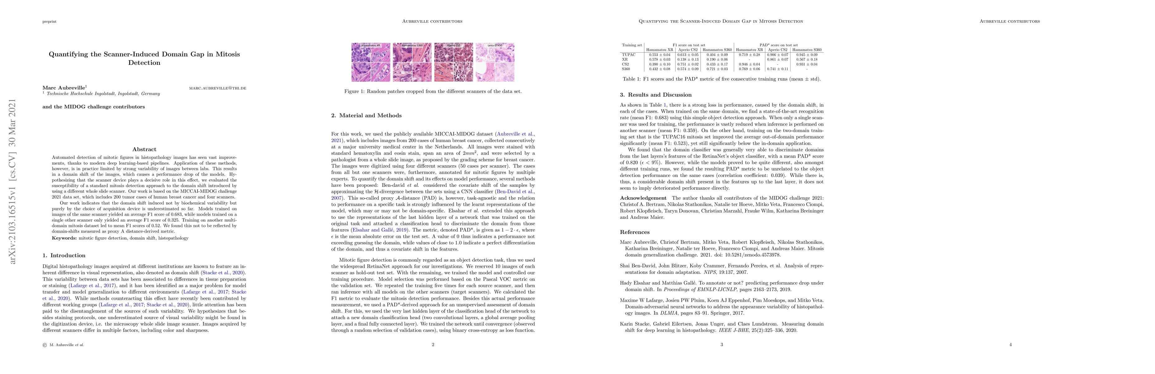
Automated detection of mitotic figures in histopathology images has seen vast improvements, thanks to modern deep learning-based pipelines. Application of these methods, however, is in practice limi...
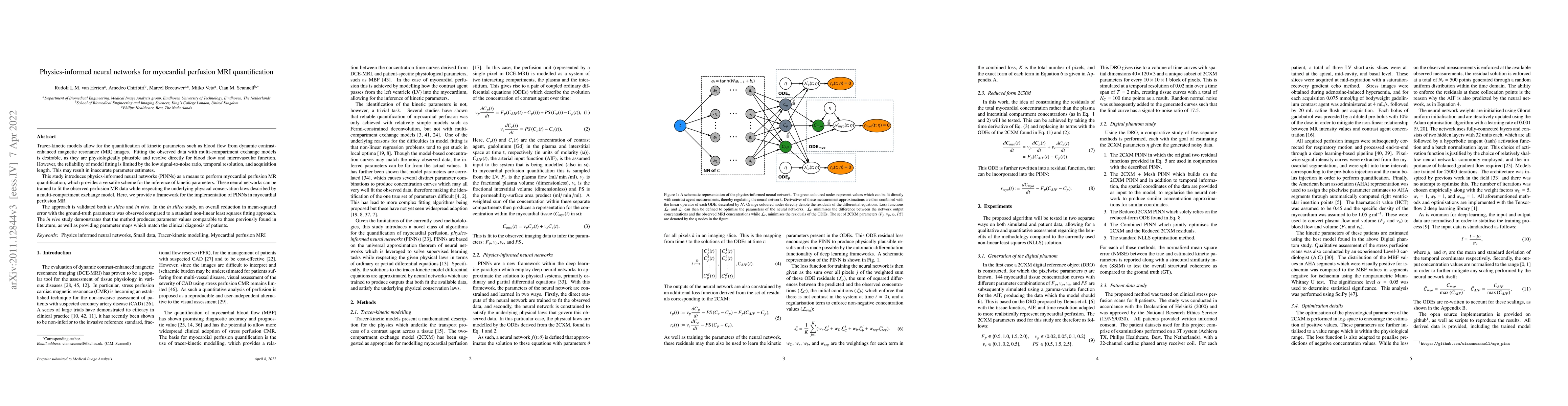
Tracer-kinetic models allow for the quantification of kinetic parameters such as blood flow from dynamic contrast-enhanced magnetic resonance (MR) images. Fitting the observed data with multi-compar...
Ductal carcinoma in situ (DCIS) is a non-invasive breast cancer that can progress into invasive ductal carcinoma (IDC). Studies suggest DCIS is often overtreated since a considerable part of DCIS le...
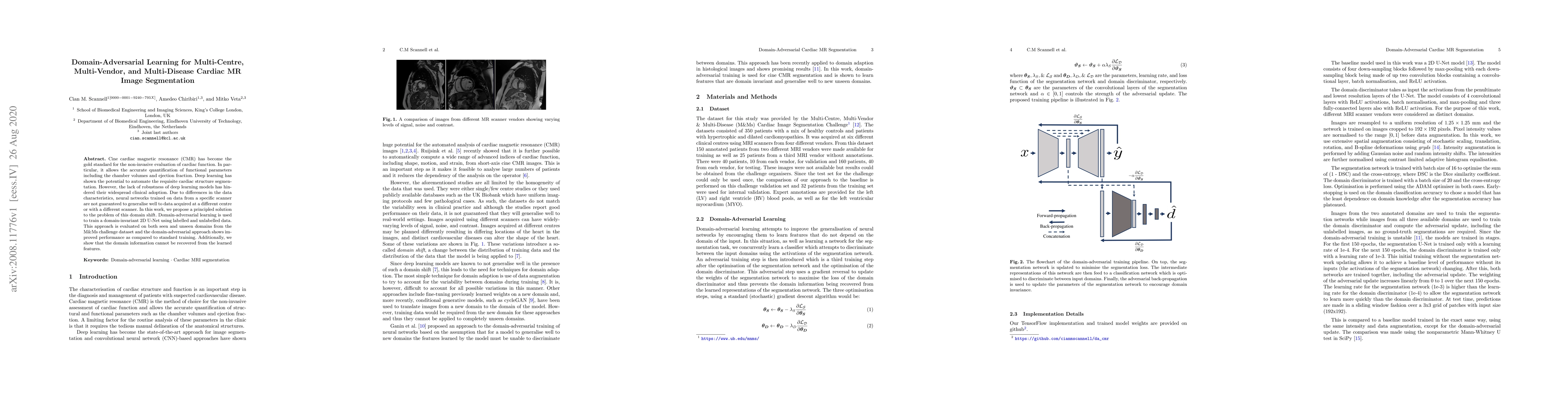
Cine cardiac magnetic resonance (CMR) has become the gold standard for the non-invasive evaluation of cardiac function. In particular, it allows the accurate quantification of functional parameters ...
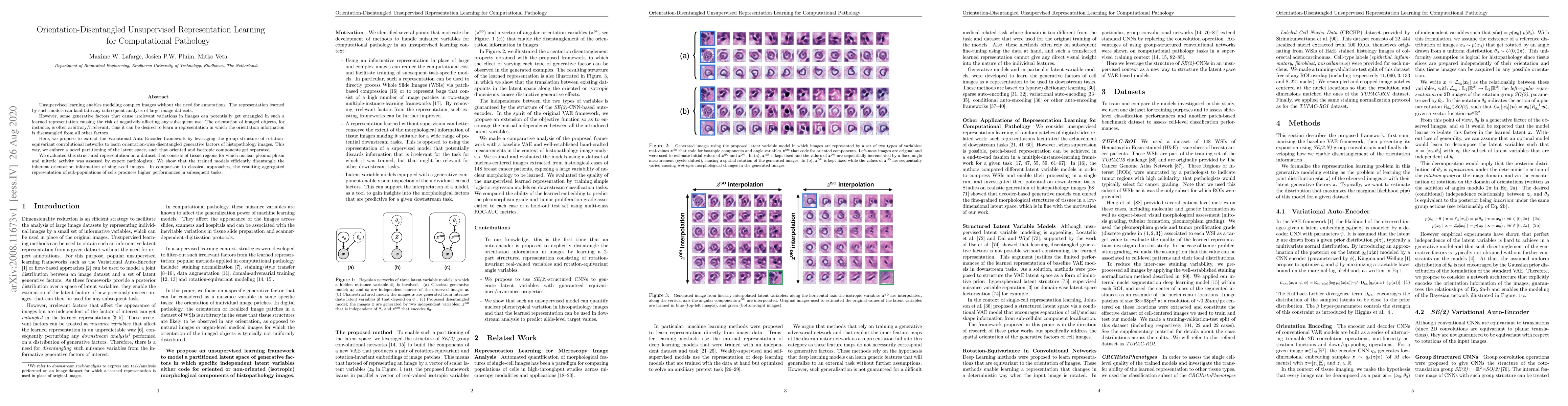
Unsupervised learning enables modeling complex images without the need for annotations. The representation learned by such models can facilitate any subsequent analysis of large image datasets. Ho...
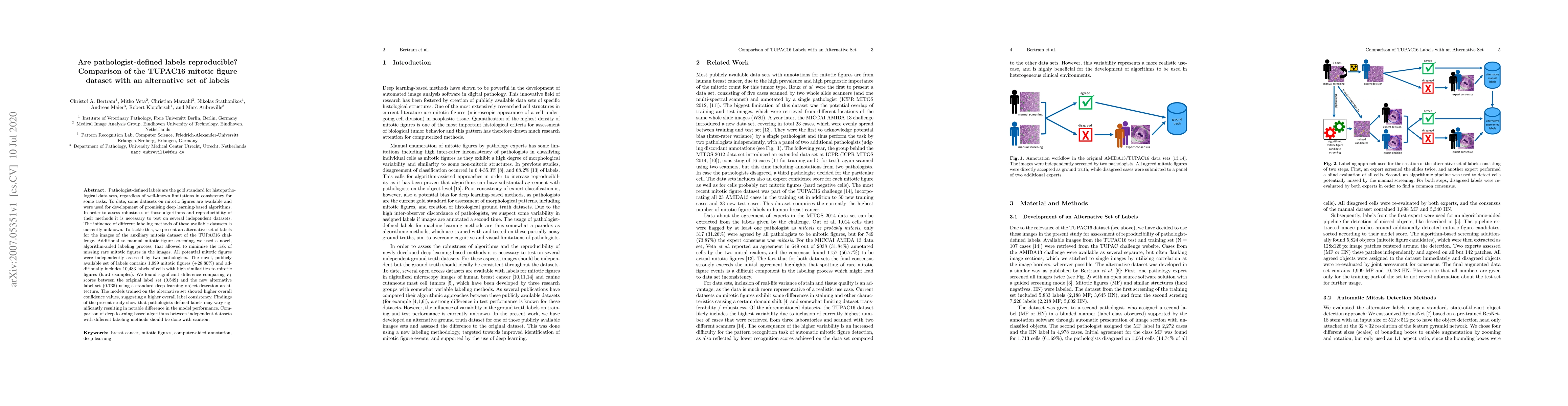
Pathologist-defined labels are the gold standard for histopathological data sets, regardless of well-known limitations in consistency for some tasks. To date, some datasets on mitotic figures are av...
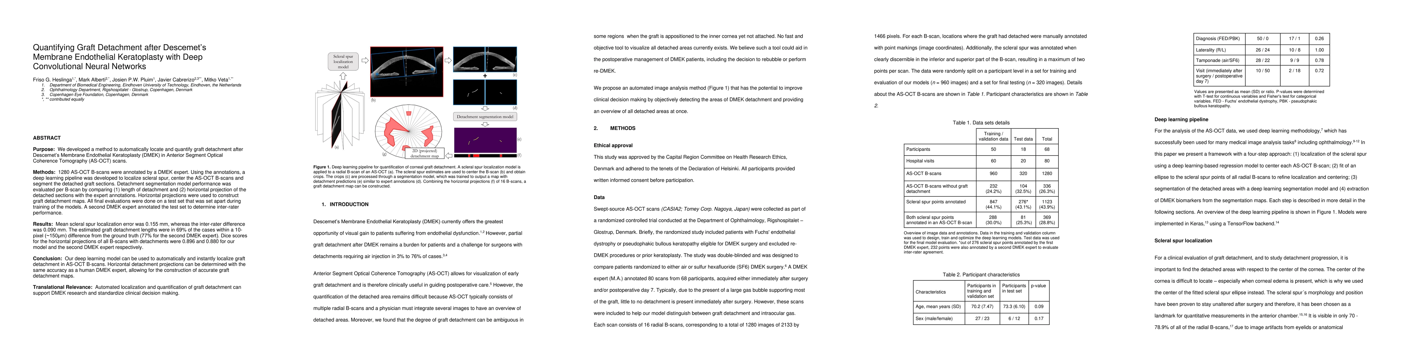
Purpose: We developed a method to automatically locate and quantify graft detachment after Descemet's Membrane Endothelial Keratoplasty (DMEK) in Anterior Segment Optical Coherence Tomography (AS-OC...
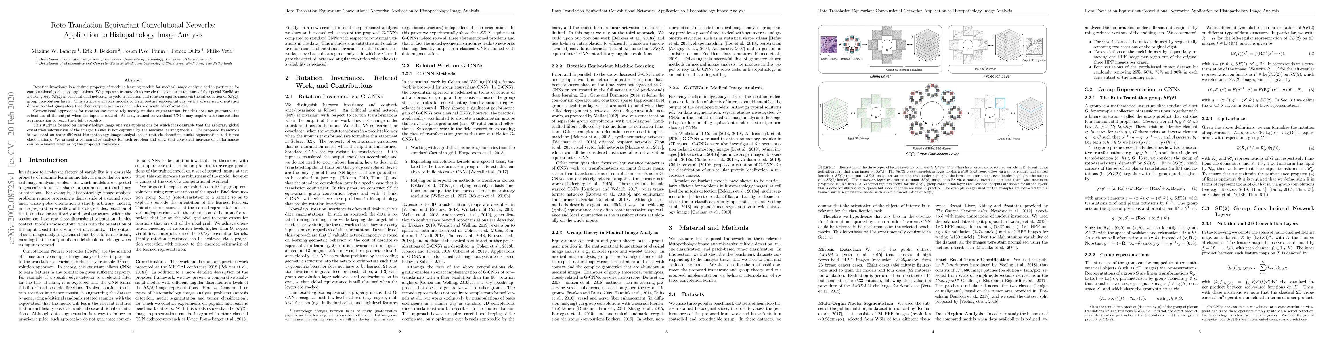
Rotation-invariance is a desired property of machine-learning models for medical image analysis and in particular for computational pathology applications. We propose a framework to encode the geome...
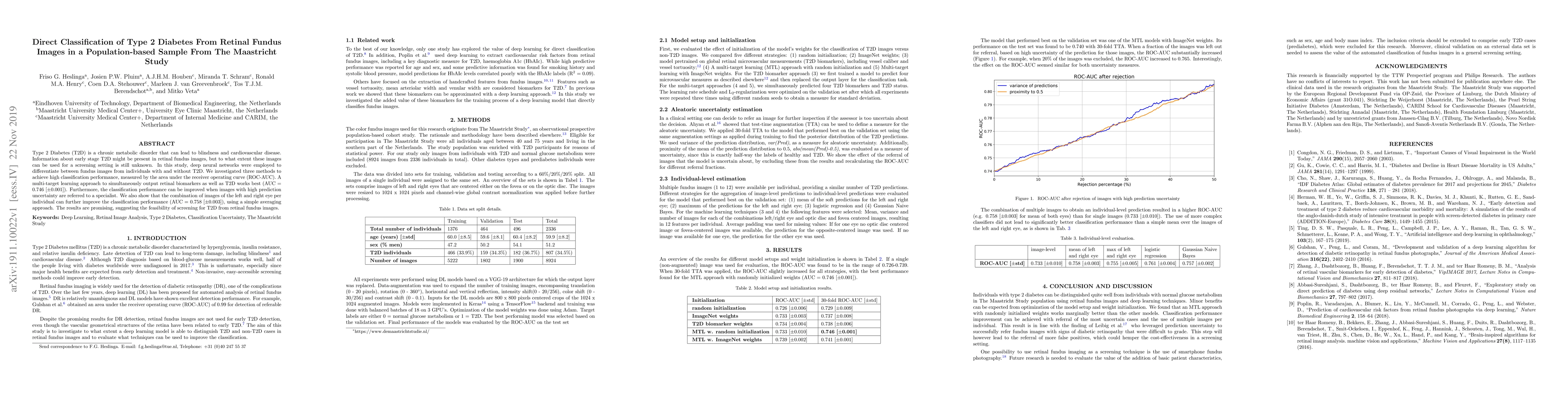
Type 2 Diabetes (T2D) is a chronic metabolic disorder that can lead to blindness and cardiovascular disease. Information about early stage T2D might be present in retinal fundus images, but to what ...
Terminal ductal lobular unit (TDLU) involution is the regression of milk-producing structures in the breast. Women with less TDLU involution are more likely to develop breast cancer. A major bottlen...
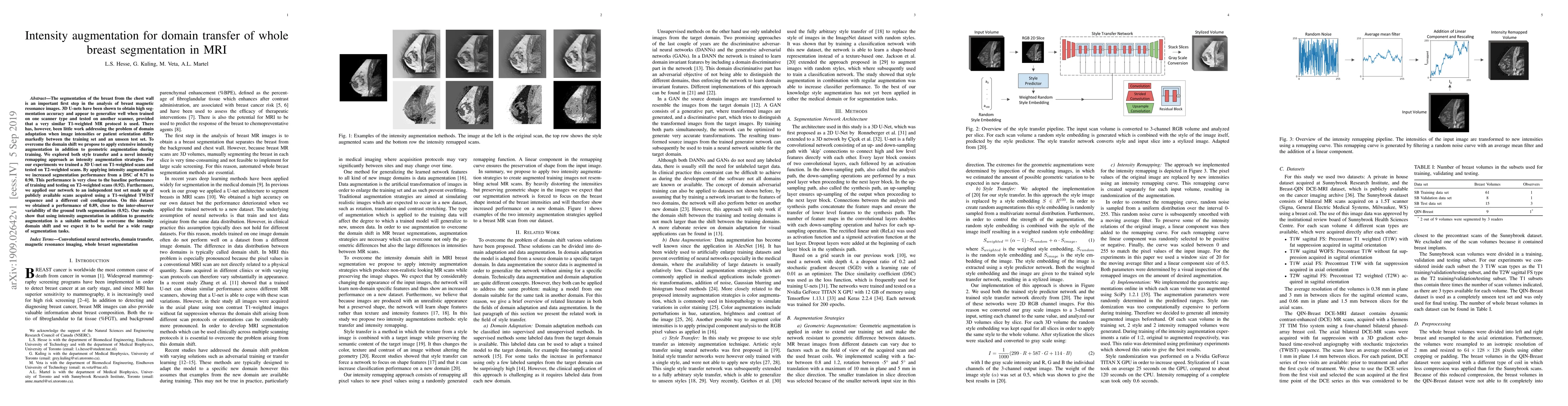
The segmentation of the breast from the chest wall is an important first step in the analysis of breast magnetic resonance images. 3D U-nets have been shown to obtain high segmentation accuracy and ...
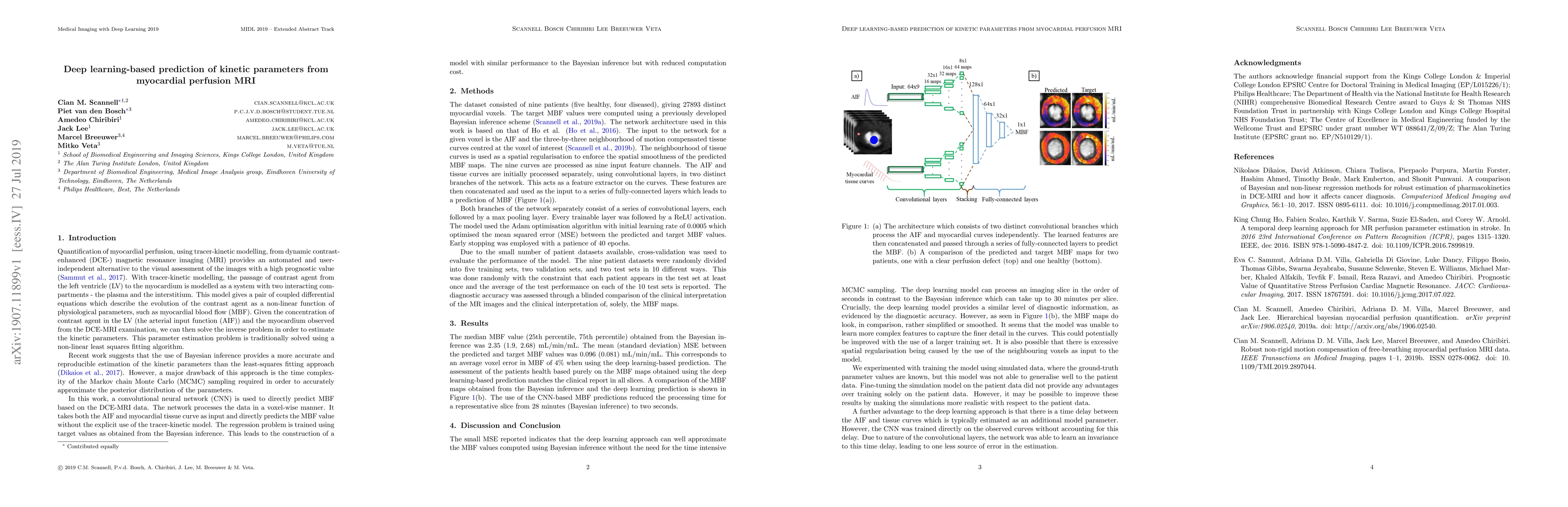
The quantification of myocardial perfusion MRI has the potential to provide a fast, automated and user-independent assessment of myocardial ischaemia. However, due to the relatively high noise level...
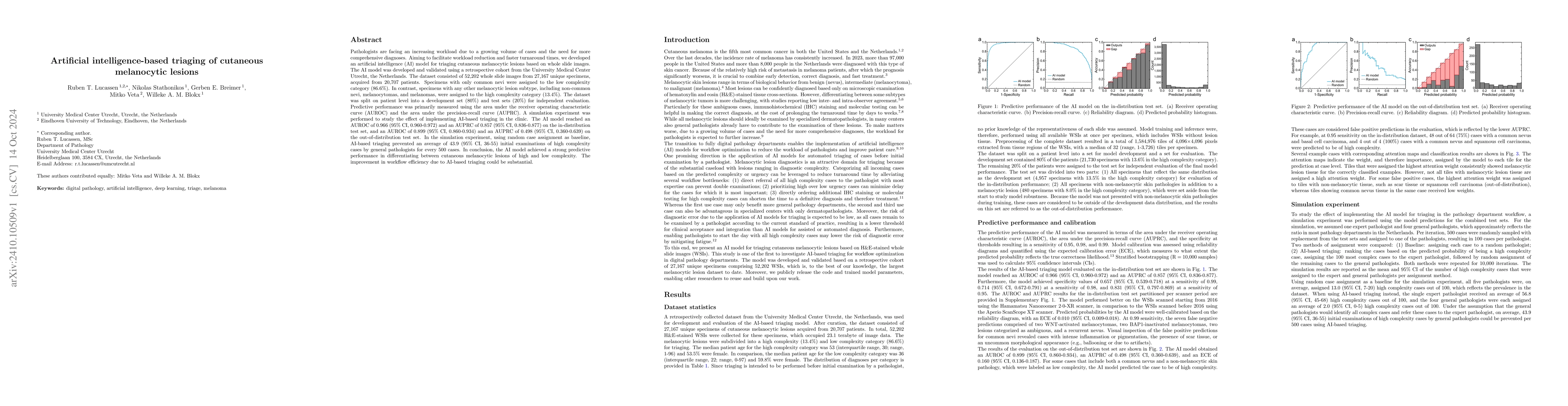
Pathologists are facing an increasing workload due to a growing volume of cases and the need for more comprehensive diagnoses. Aiming to facilitate workload reduction and faster turnaround times, we d...
Millions of melanocytic skin lesions are examined by pathologists each year, the majority of which concern common nevi (i.e., ordinary moles). While most of these lesions can be diagnosed in seconds, ...
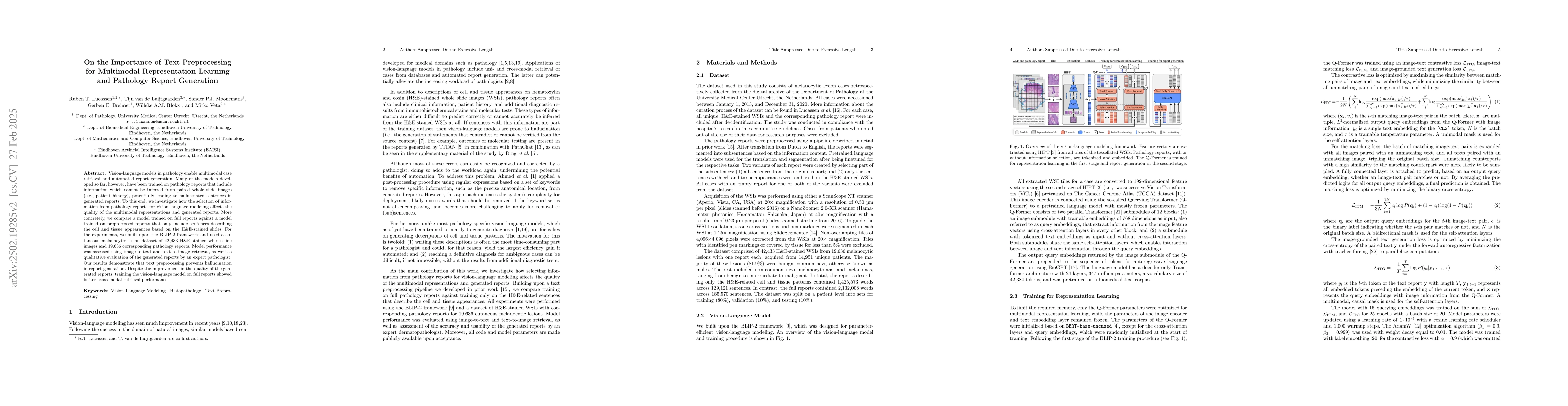
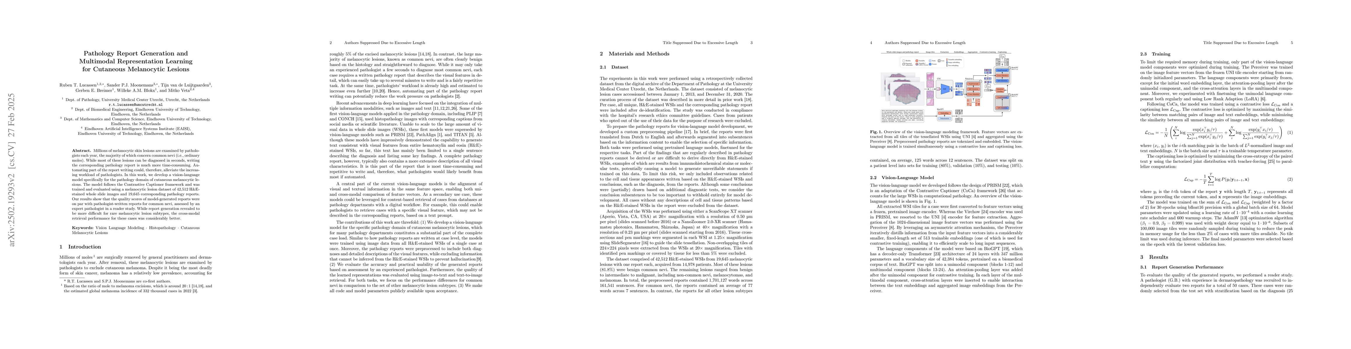
Vision-language models in pathology enable multimodal case retrieval and automated report generation. Many of the models developed so far, however, have been trained on pathology reports that include ...
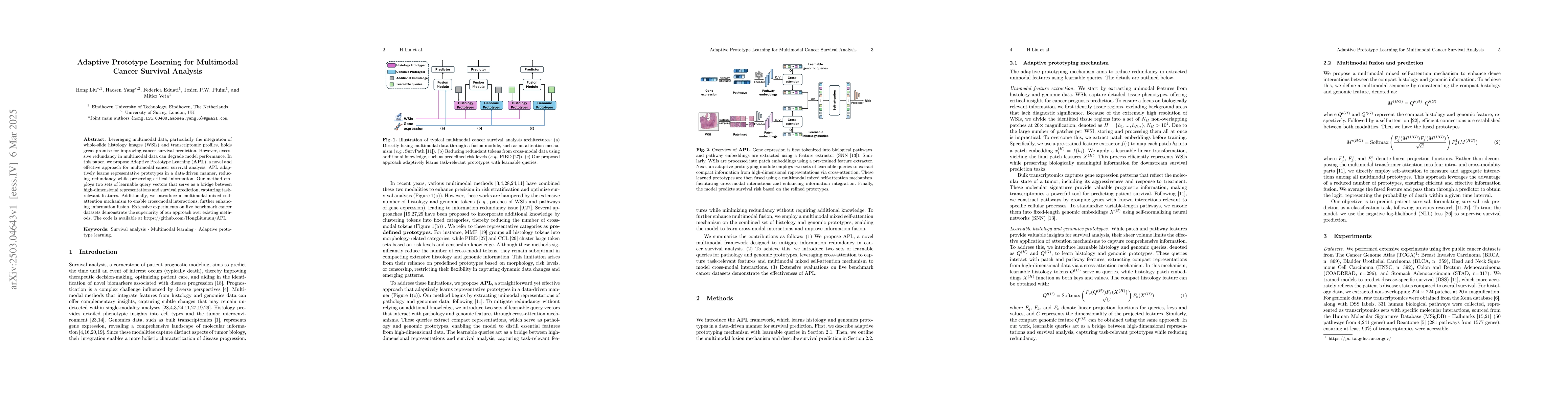
Leveraging multimodal data, particularly the integration of whole-slide histology images (WSIs) and transcriptomic profiles, holds great promise for improving cancer survival prediction. However, exce...
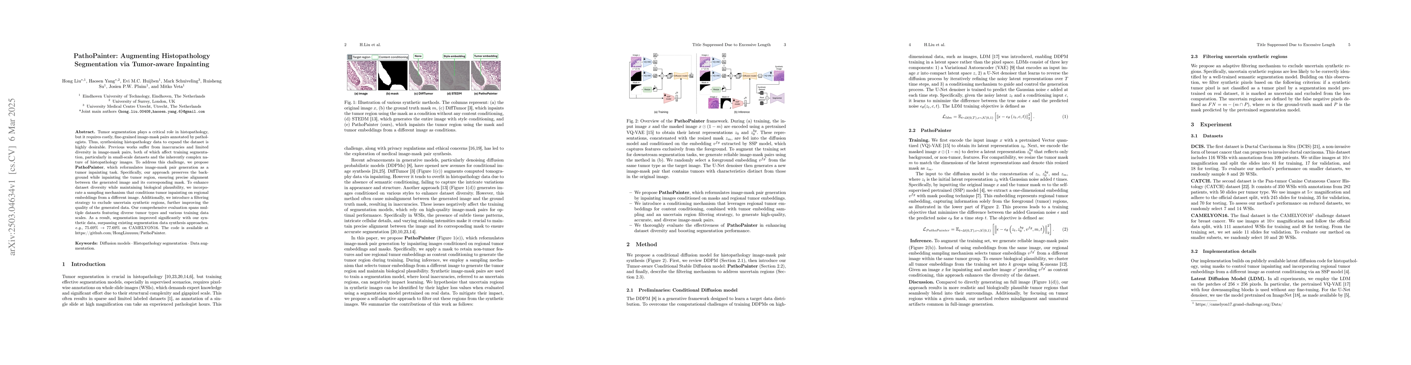
Tumor segmentation plays a critical role in histopathology, but it requires costly, fine-grained image-mask pairs annotated by pathologists. Thus, synthesizing histopathology data to expand the datase...
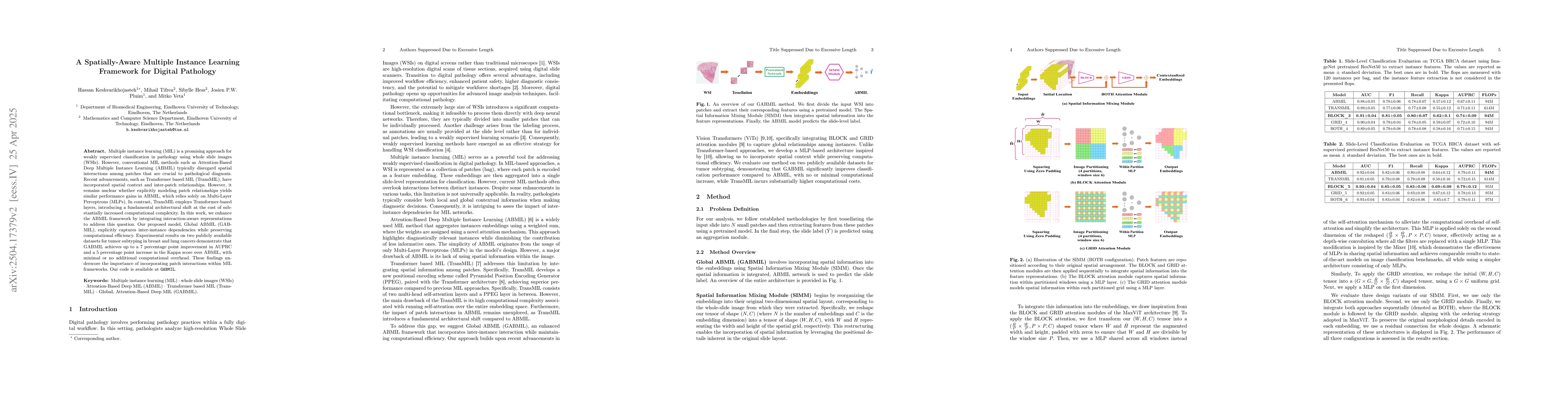
Multiple instance learning (MIL) is a promising approach for weakly supervised classification in pathology using whole slide images (WSIs). However, conventional MIL methods such as Attention-Based De...
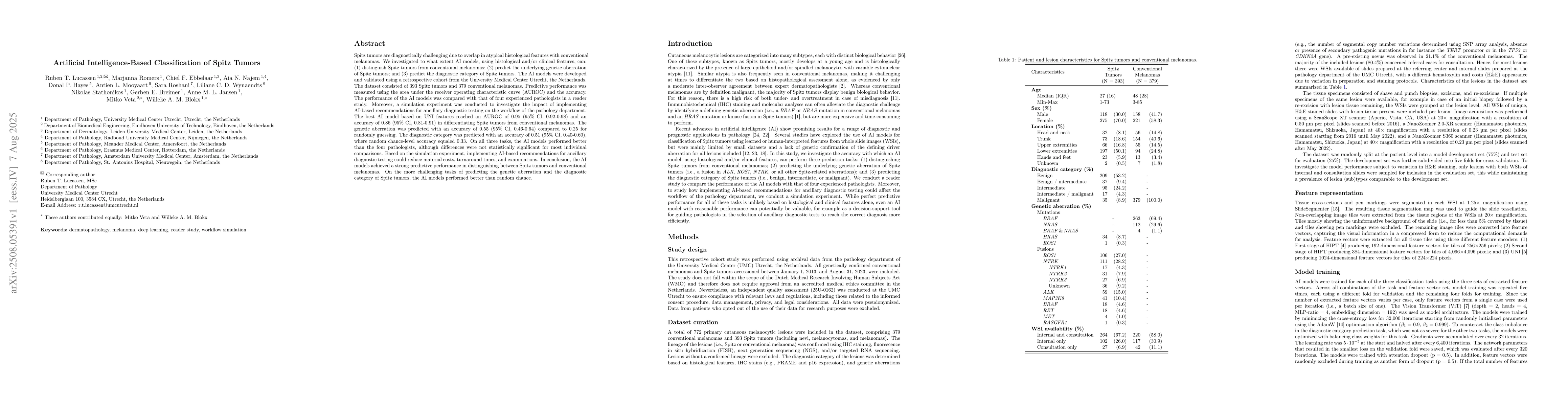
Spitz tumors are diagnostically challenging due to overlap in atypical histological features with conventional melanomas. We investigated to what extent AI models, using histological and/or clinical f...
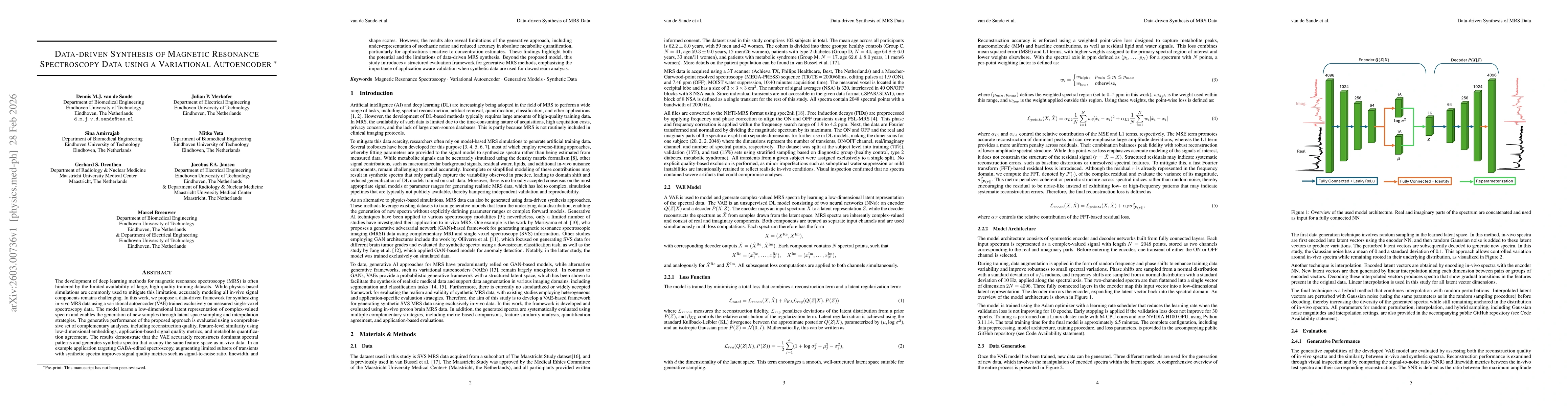
The development of deep learning methods for magnetic resonance spectroscopy (MRS) is often hindered by limited availability of large, high-quality training datasets. While physics-based simulations a...
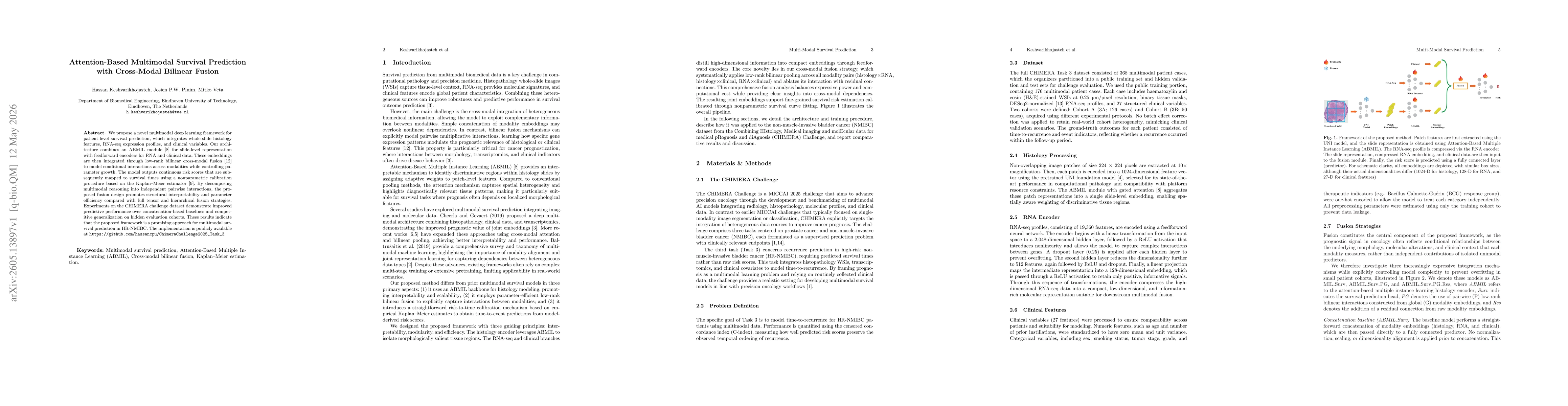
We propose a novel multimodal deep learning framework for patient-level survival prediction, which integrates whole-slide histology features, RNA-seq expression profiles, and clinical variables. Our a...
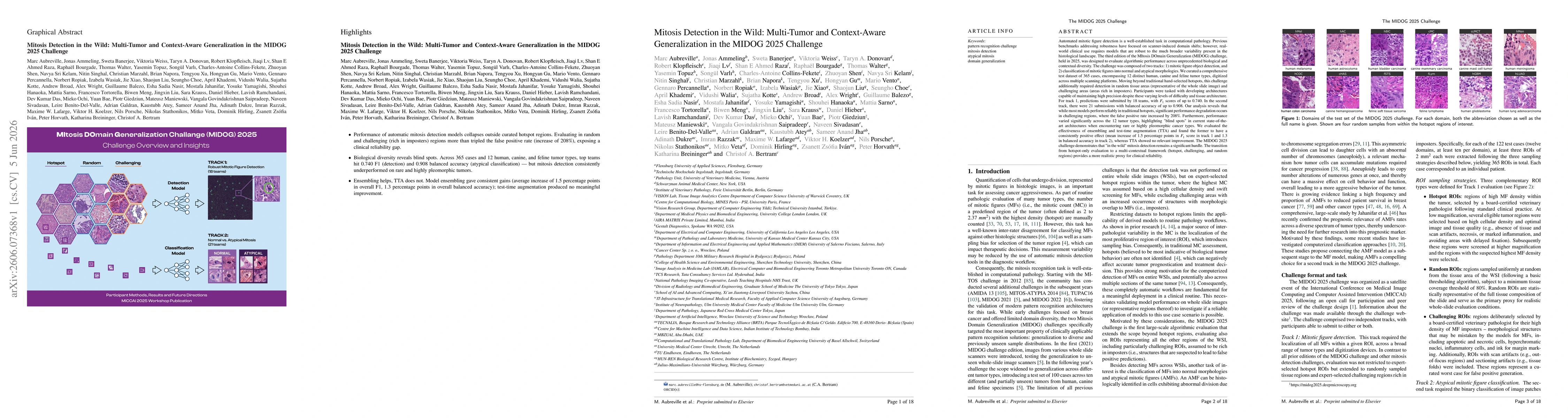
Automated mitosis detection is a well-established task in computational pathology. While previous benchmarks focused on scanner-induced domain shift, clinical "real-world" application requires models ...