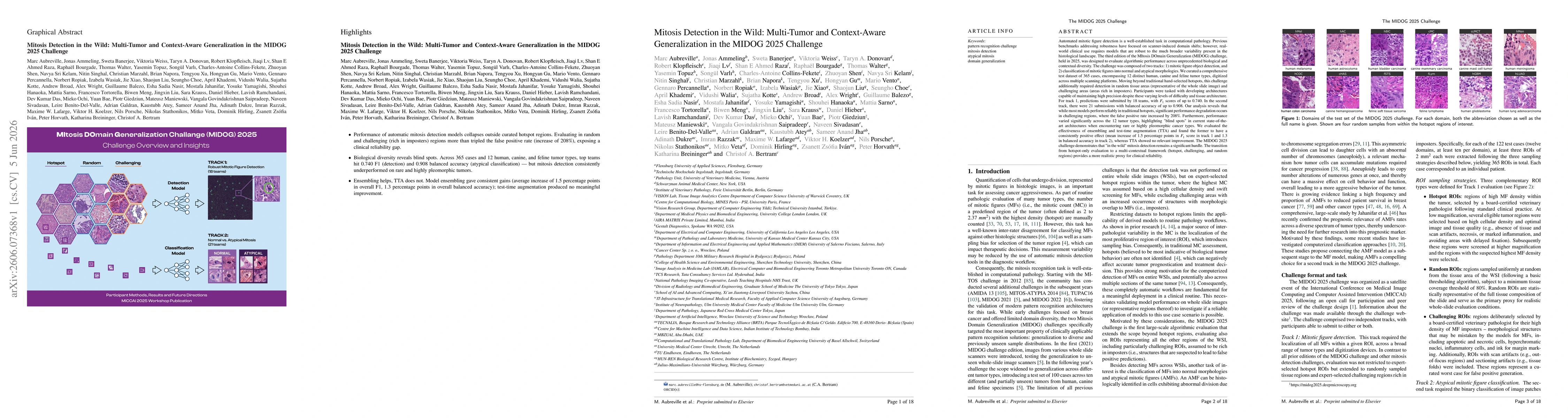
Academic Profile
Statistics
Similar Authors
Papers on arXiv
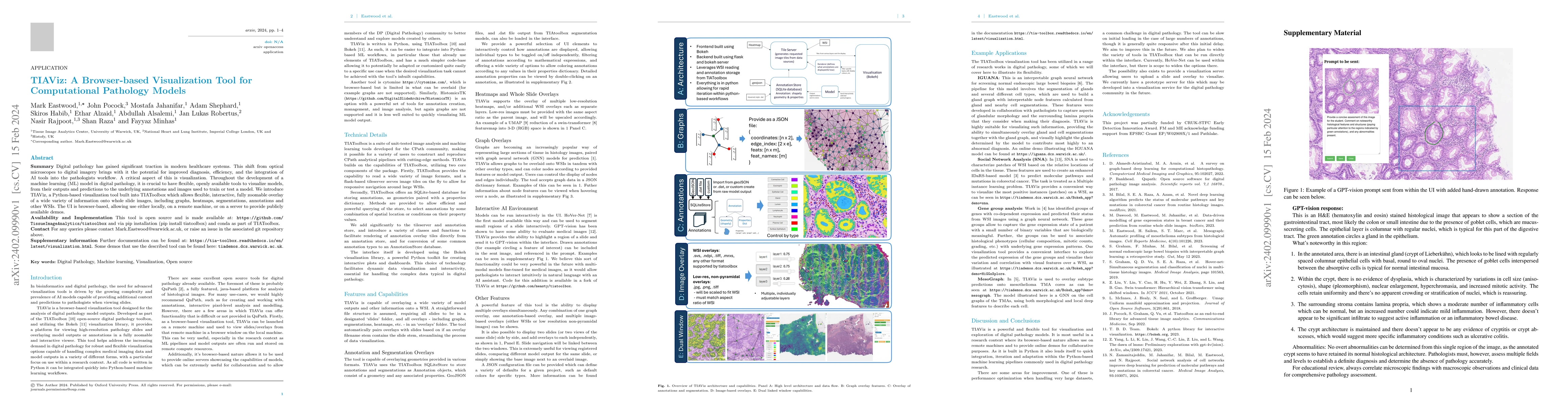
Digital pathology has gained significant traction in modern healthcare systems. This shift from optical microscopes to digital imagery brings with it the potential for improved diagnosis, efficiency...
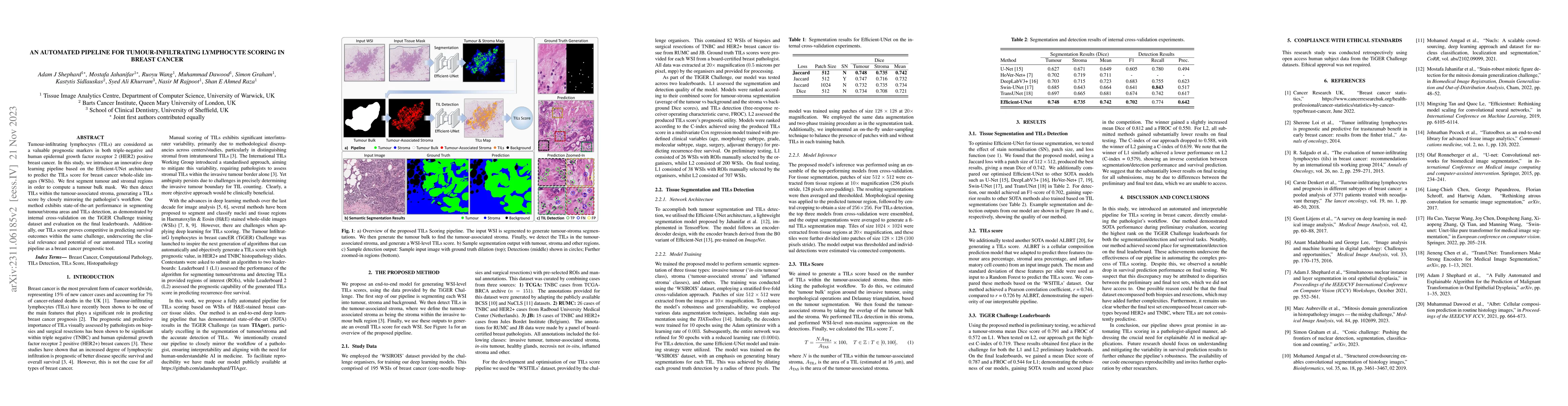
Tumour-infiltrating lymphocytes (TILs) are considered as a valuable prognostic markers in both triple-negative and human epidermal growth factor receptor 2 (HER2) positive breast cancer. In this stu...

Deep learning models have exhibited exceptional effectiveness in Computational Pathology (CPath) by tackling intricate tasks across an array of histology image analysis applications. Nevertheless, t...
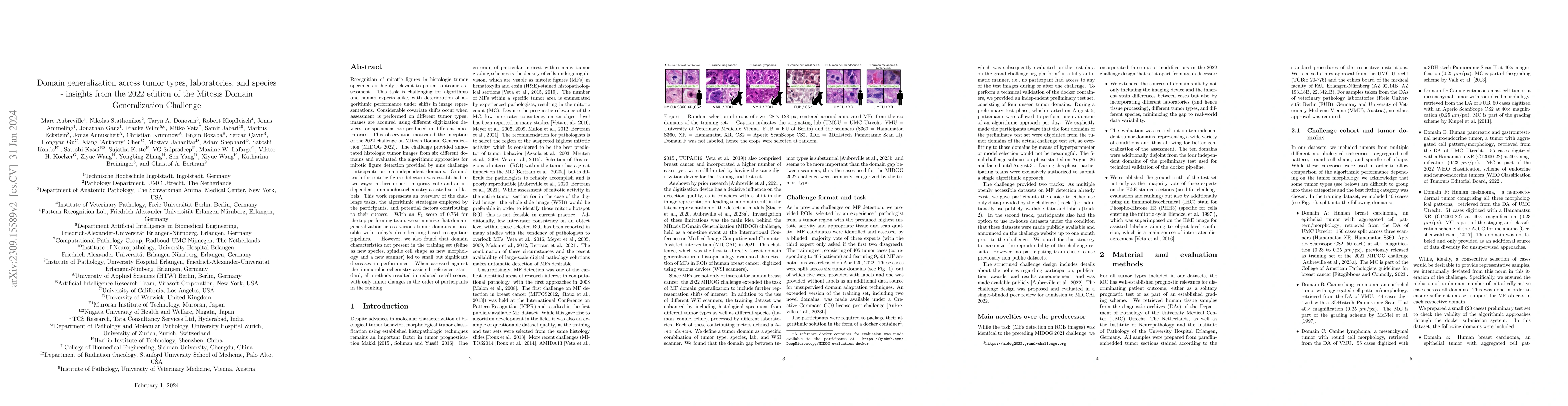
Recognition of mitotic figures in histologic tumor specimens is highly relevant to patient outcome assessment. This task is challenging for algorithms and human experts alike, with deterioration of ...
Oral epithelial dysplasia (OED) is a premalignant histopathological diagnosis given to lesions of the oral cavity. Its grading suffers from significant inter-/intra- observer variability, and does n...
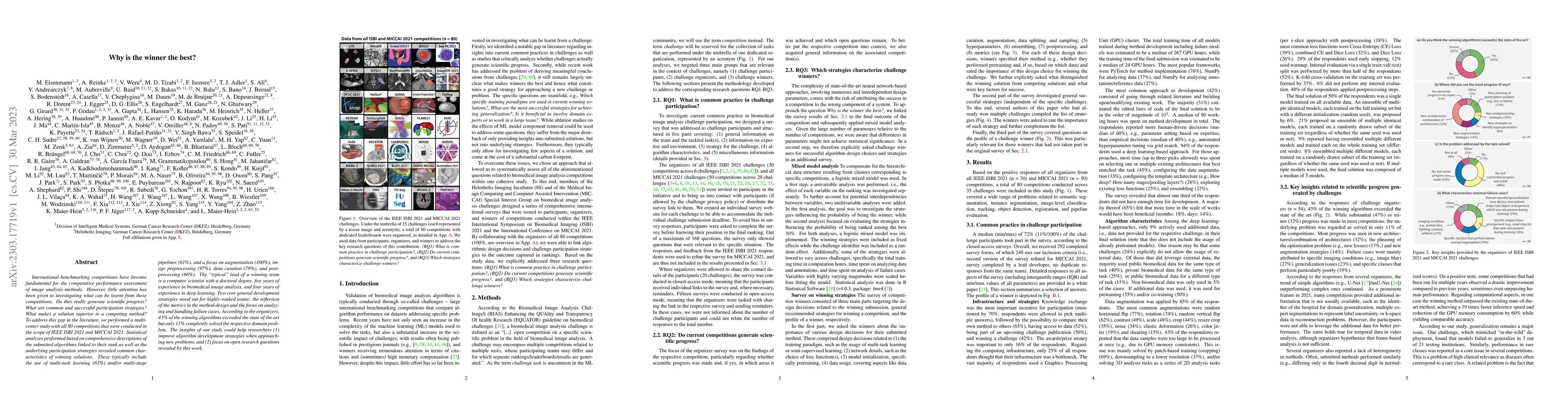
International benchmarking competitions have become fundamental for the comparative performance assessment of image analysis methods. However, little attention has been given to investigating what c...
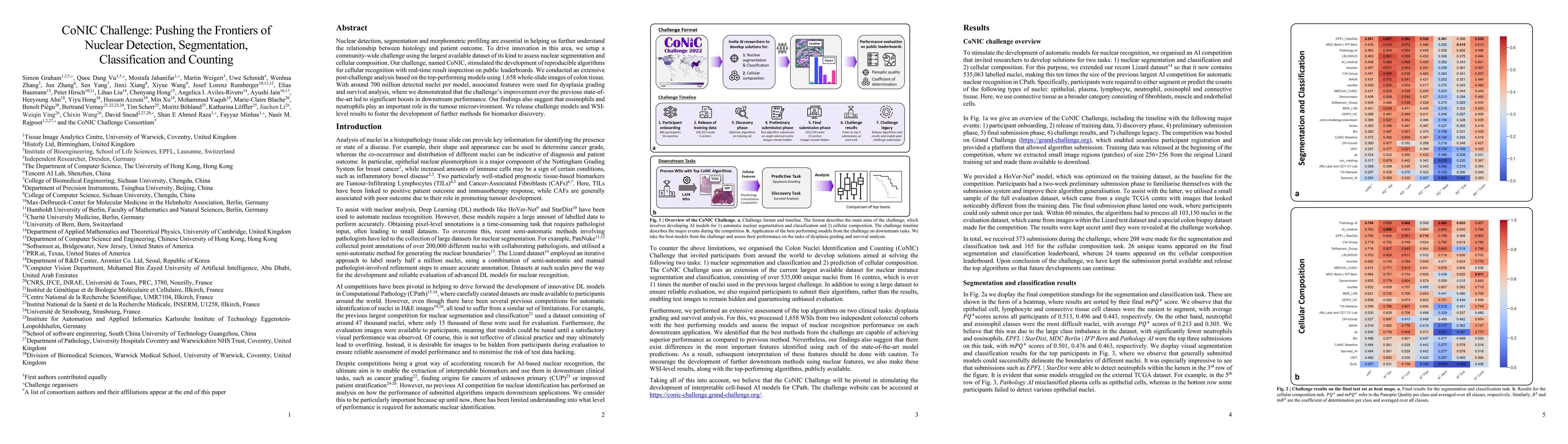
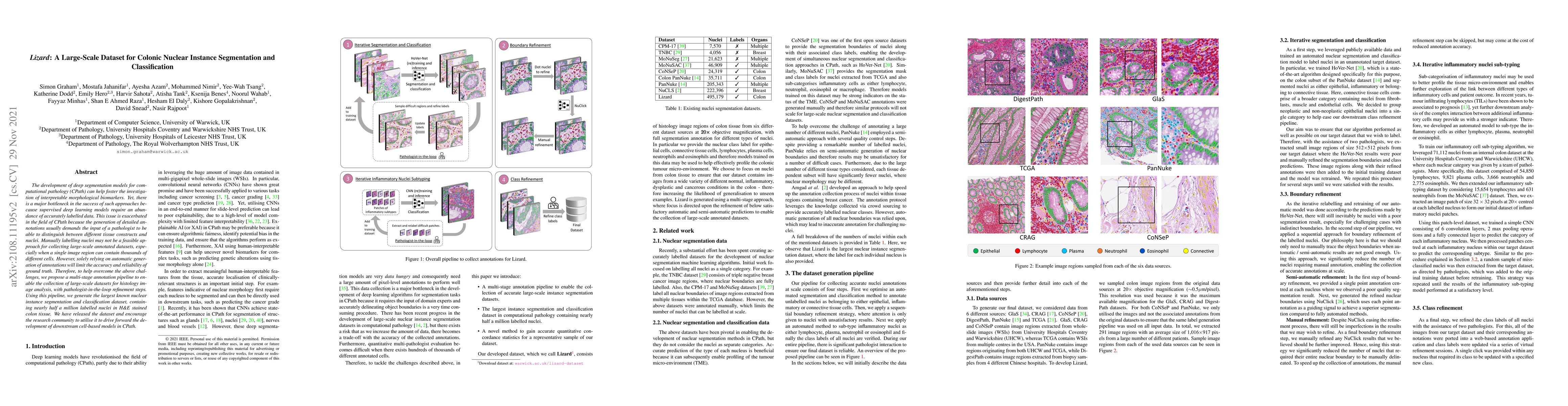
Nuclear detection, segmentation and morphometric profiling are essential in helping us further understand the relationship between histology and patient outcome. To drive innovation in this area, we...
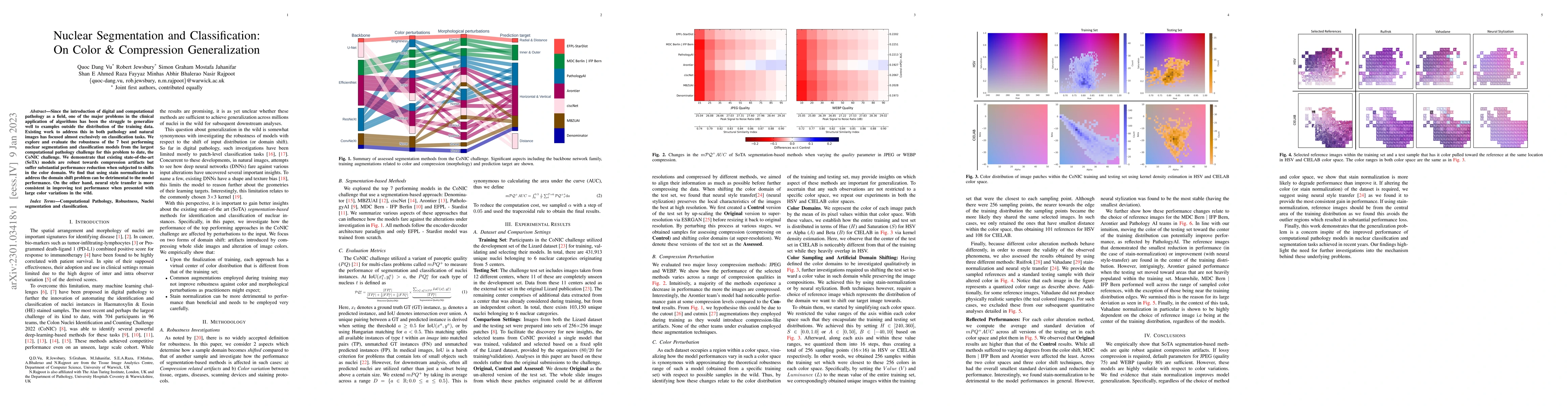
Since the introduction of digital and computational pathology as a field, one of the major problems in the clinical application of algorithms has been the struggle to generalize well to examples out...
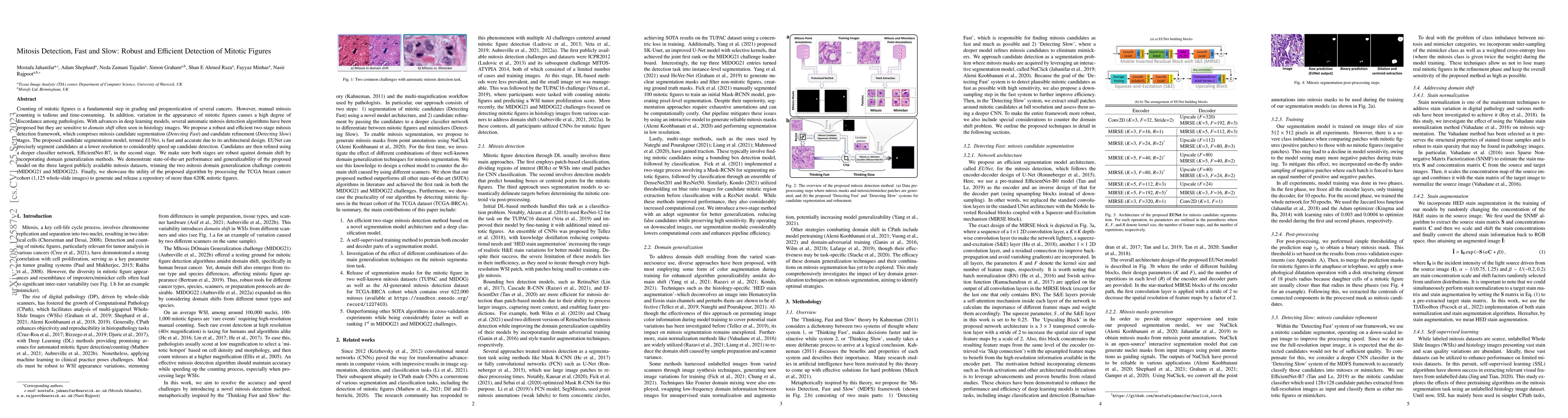
Counting of mitotic figures is a fundamental step in grading and prognostication of several cancers. However, manual mitosis counting is tedious and time-consuming. In addition, variation in the app...
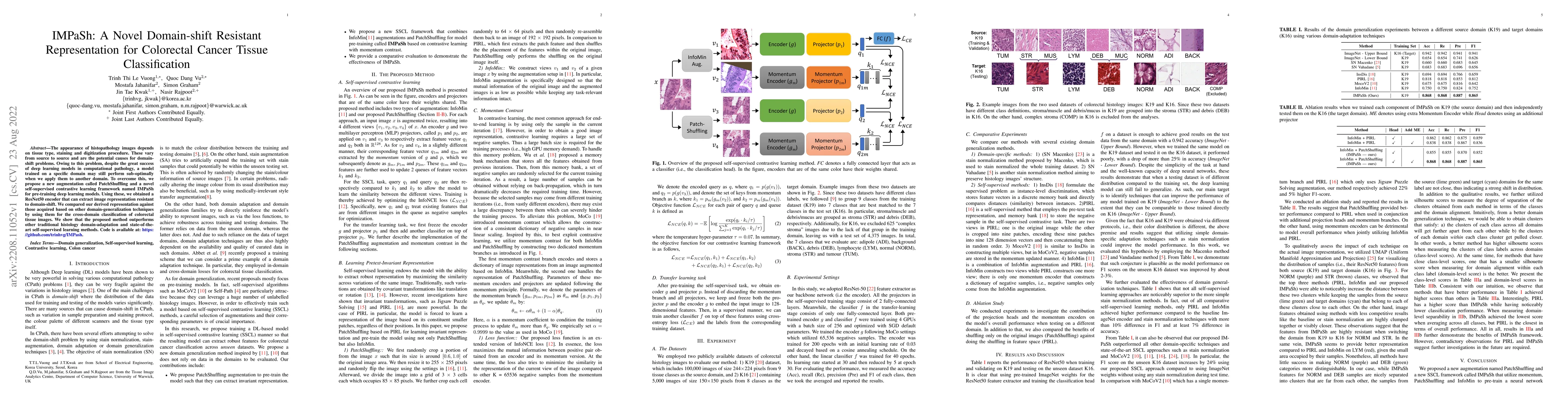
The appearance of histopathology images depends on tissue type, staining and digitization procedure. These vary from source to source and are the potential causes for domain-shift problems. Owing to...
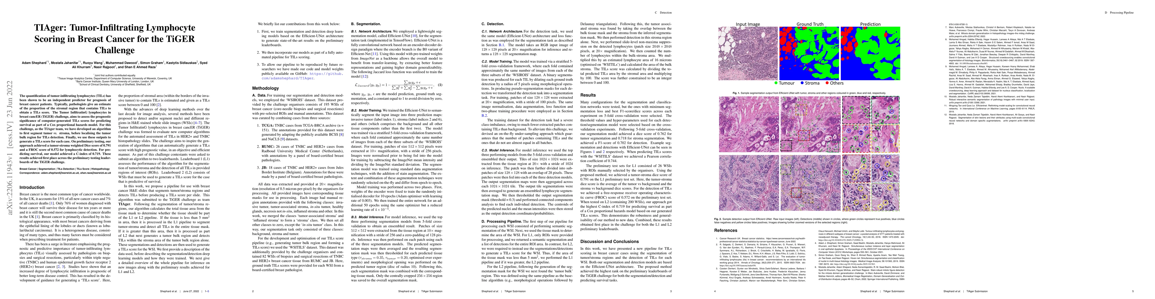
The quantification of tumor-infiltrating lymphocytes (TILs) has been shown to be an independent predictor for prognosis of breast cancer patients. Typically, pathologists give an estimate of the pro...
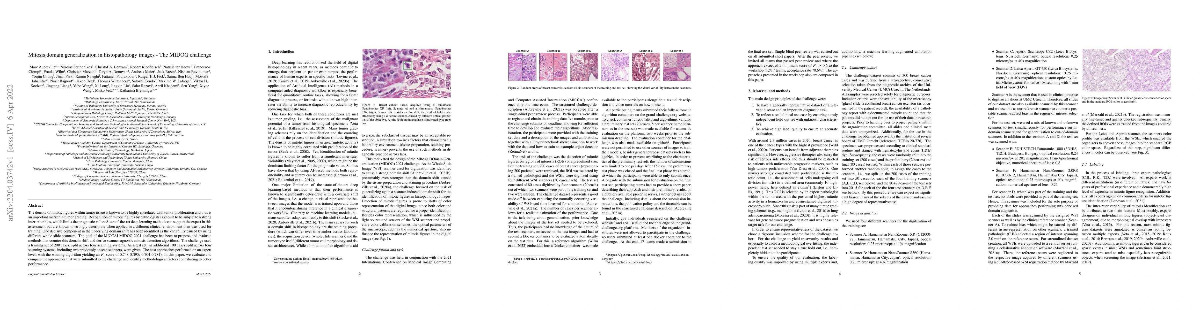
The density of mitotic figures within tumor tissue is known to be highly correlated with tumor proliferation and thus is an important marker in tumor grading. Recognition of mitotic figures by patho...
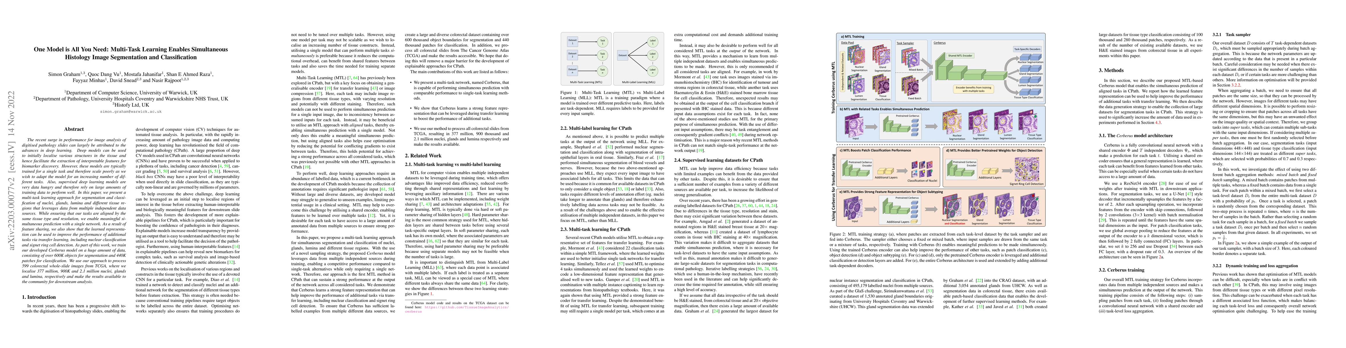
The recent surge in performance for image analysis of digitised pathology slides can largely be attributed to the advances in deep learning. Deep models can be used to initially localise various str...
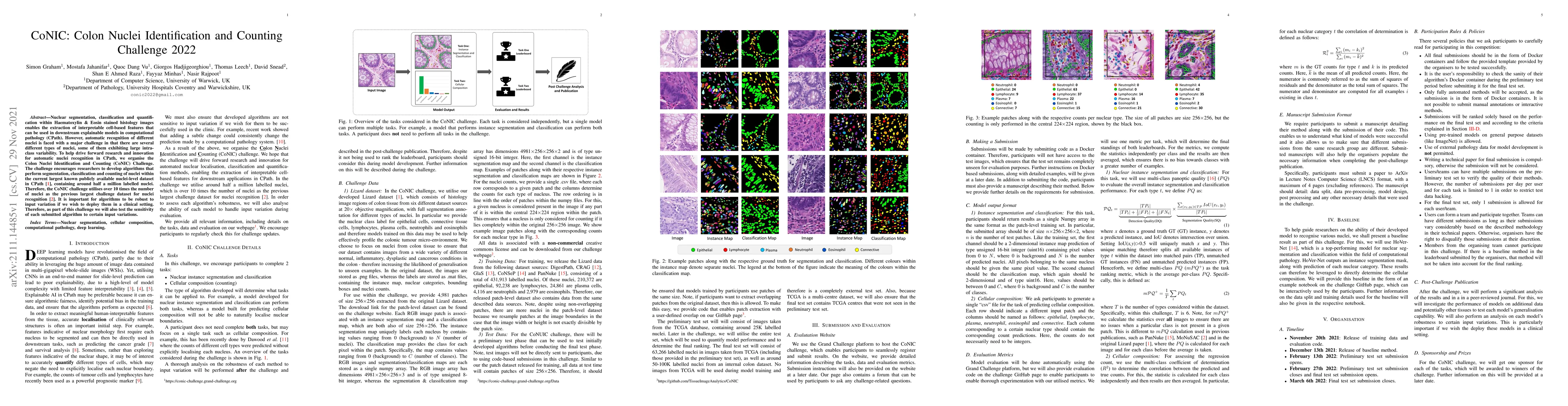
Nuclear segmentation, classification and quantification within Haematoxylin & Eosin stained histology images enables the extraction of interpretable cell-based features that can be used in downstrea...
From the simple measurement of tissue attributes in pathology workflow to designing an explainable diagnostic/prognostic AI tool, access to accurate semantic segmentation of tissue regions in histol...
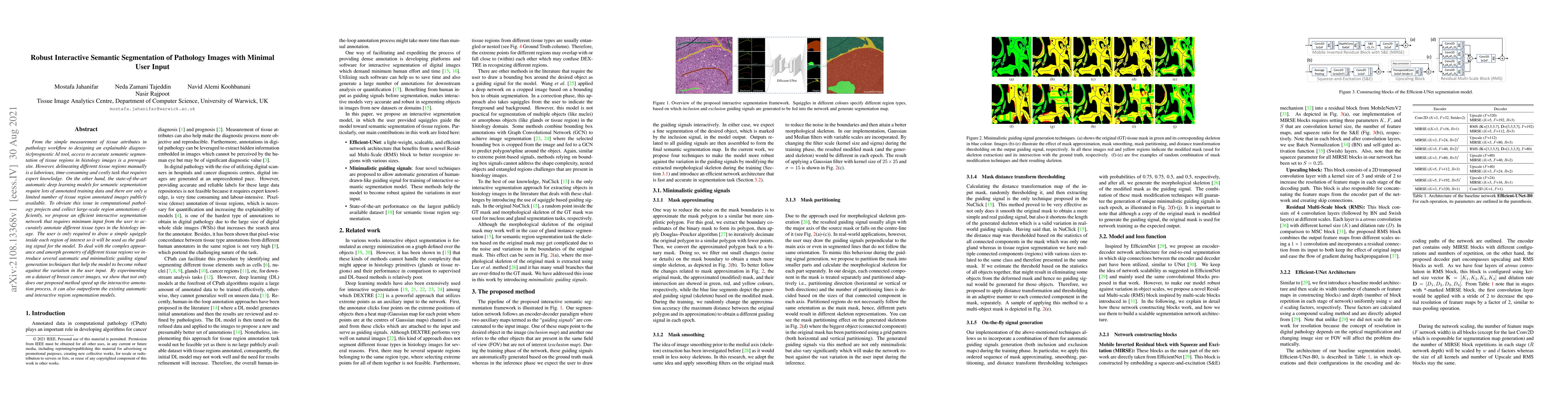
The development of deep segmentation models for computational pathology (CPath) can help foster the investigation of interpretable morphological biomarkers. Yet, there is a major bottleneck in the s...

Recent advances in whole slide imaging (WSI) technology have led to the development of a myriad of computer vision and artificial intelligence (AI) based diagnostic, prognostic, and predictive algor...
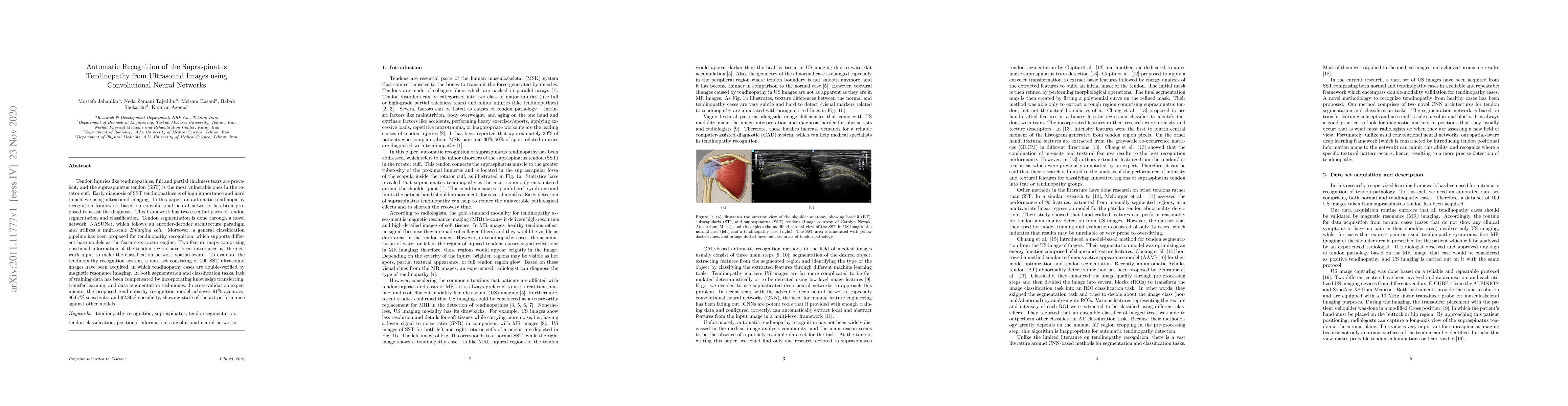
Tendon injuries like tendinopathies, full and partial thickness tears are prevalent, and the supraspinatus tendon (SST) is the most vulnerable ones in the rotator cuff. Early diagnosis of SST tendin...
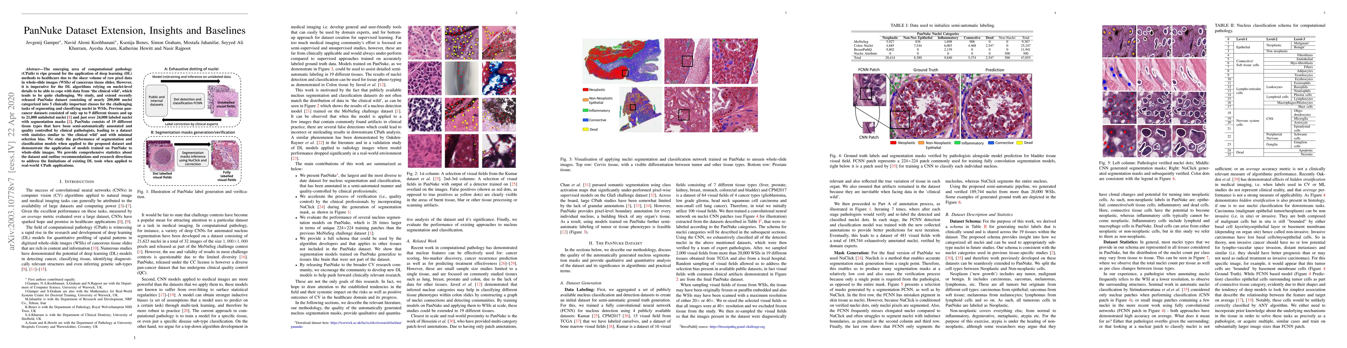
The emerging area of computational pathology (CPath) is ripe ground for the application of deep learning (DL) methods to healthcare due to the sheer volume of raw pixel data in whole-slide images (W...
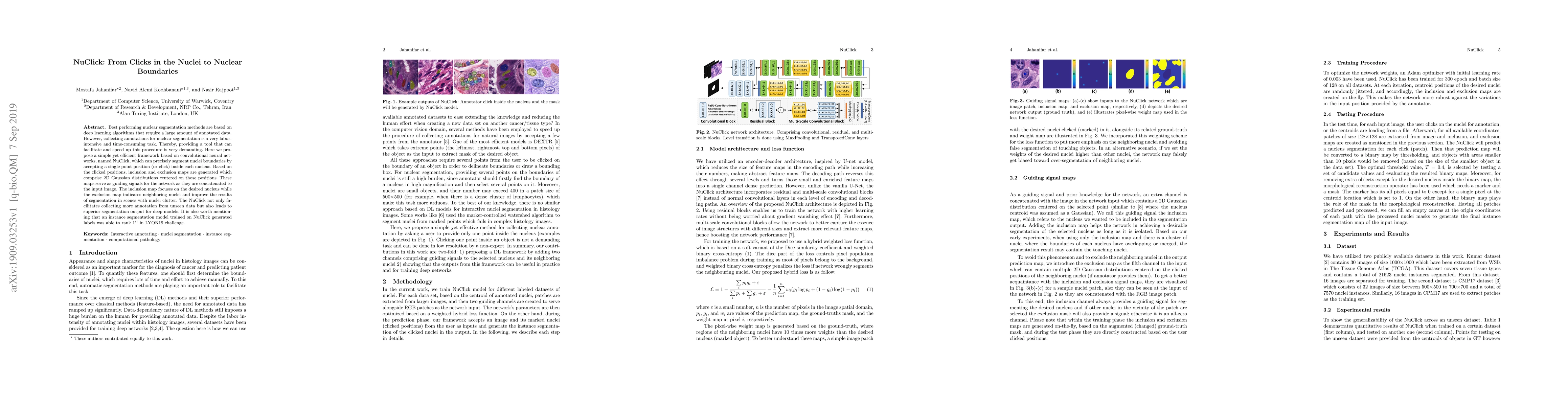
Best performing nuclear segmentation methods are based on deep learning algorithms that require a large amount of annotated data. However, collecting annotations for nuclear segmentation is a very l...
Nuclear segmentation in histology images is a challenging task due to significant variations in the shape and appearance of nuclei. One of the main hurdles in nuclear instance segmentation is overla...
Deep learning models have shown immense promise in computational pathology (CPath) tasks, but their performance often suffers when applied to unseen data due to domain shifts. Addressing this requires...
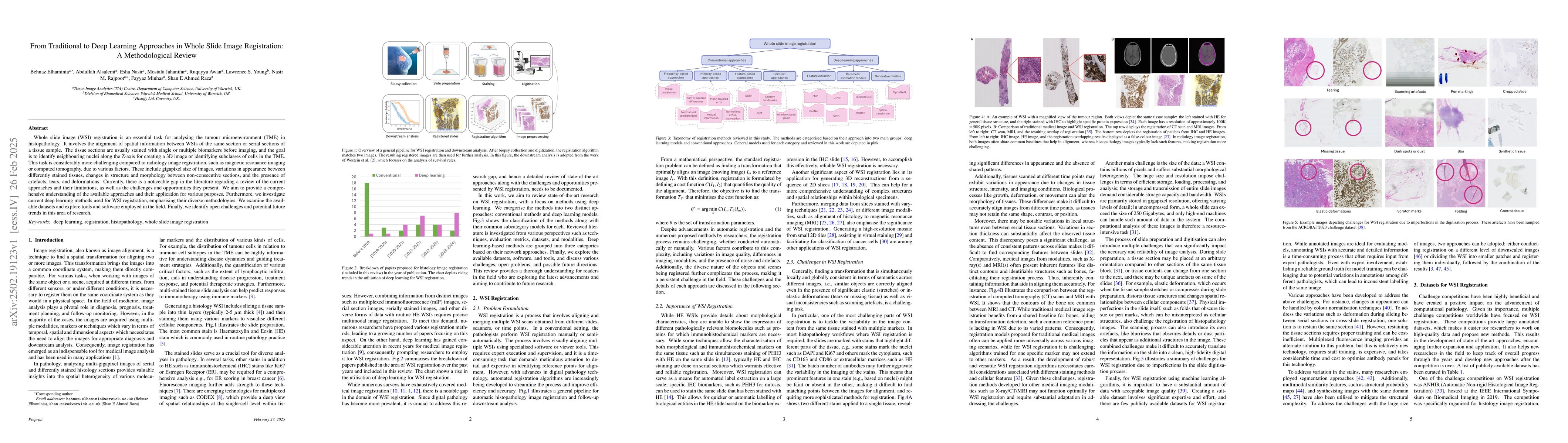
Whole slide image (WSI) registration is an essential task for analysing the tumour microenvironment (TME) in histopathology. It involves the alignment of spatial information between WSIs of the same s...
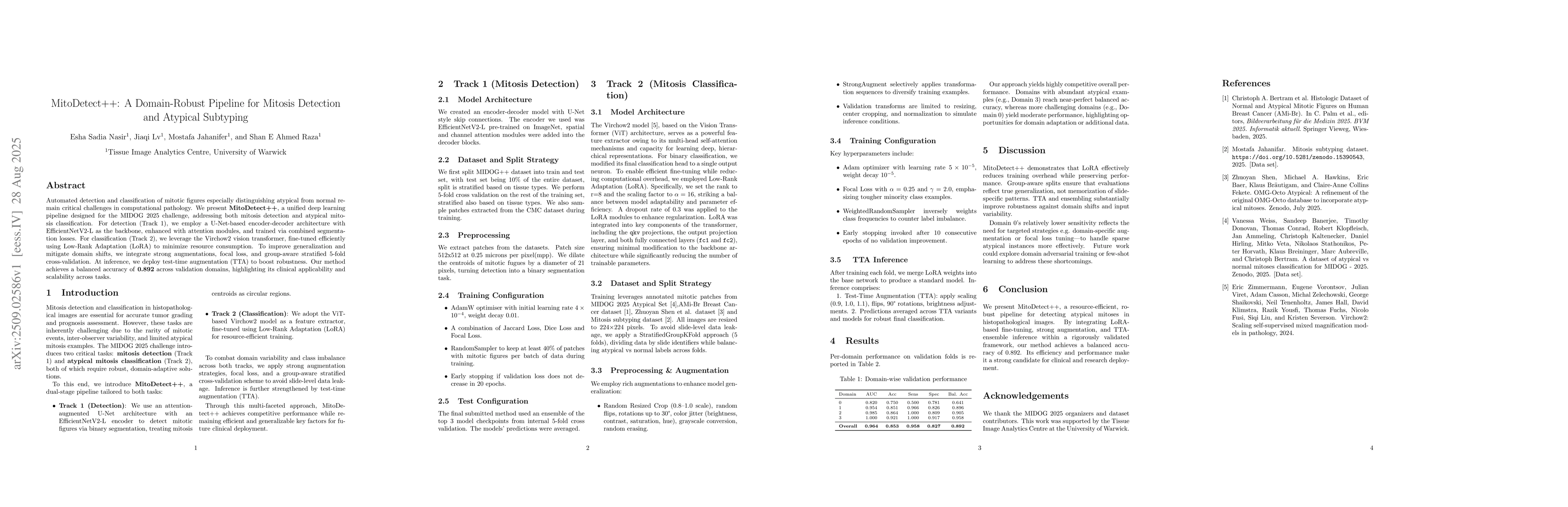
Automated detection and classification of mitotic figures especially distinguishing atypical from normal remain critical challenges in computational pathology. We present MitoDetect++, a unified deep ...
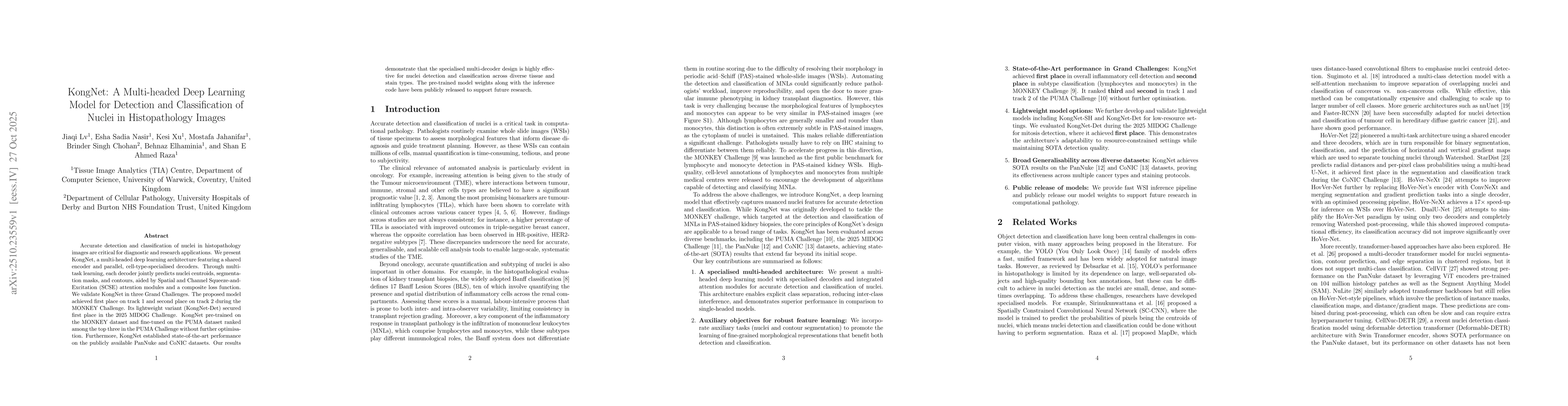
Accurate detection and classification of nuclei in histopathology images are critical for diagnostic and research applications. We present KongNet, a multi-headed deep learning architecture featuring ...
As the volume and complexity of nonclinical toxicology studies continue to increase, toxicologic pathology reporting faces persistent challenges, including fragmented sources of data (e.g., histopatho...
Automated mitosis detection is a well-established task in computational pathology. While previous benchmarks focused on scanner-induced domain shift, clinical "real-world" application requires models ...