Academic Profile
Statistics
Similar Authors
Papers on arXiv
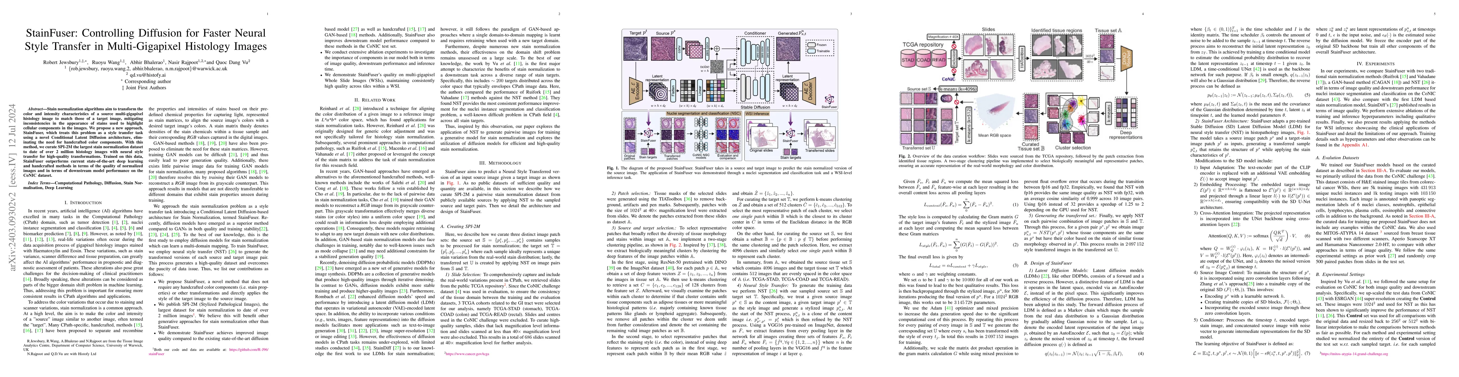
Stain normalization algorithms aim to transform the color and intensity characteristics of a source multi-gigapixel histology image to match those of a target image, mitigating inconsistencies in the ...
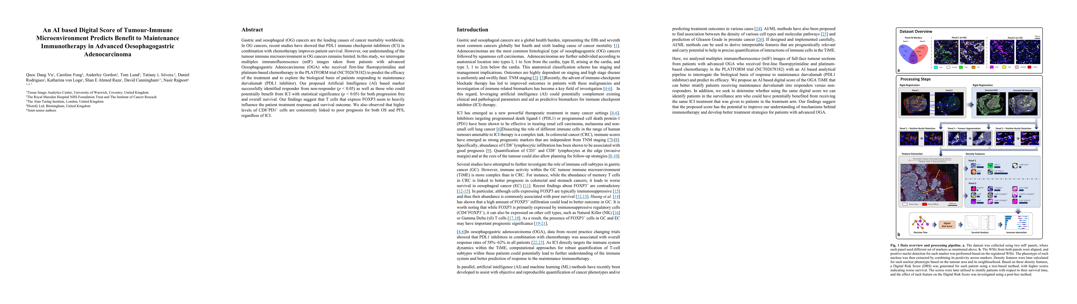
Gastric and oesophageal (OG) cancers are the leading causes of cancer mortality worldwide. In OG cancers, recent studies have showed that PDL1 immune checkpoint inhibitors (ICI) in combination with ...
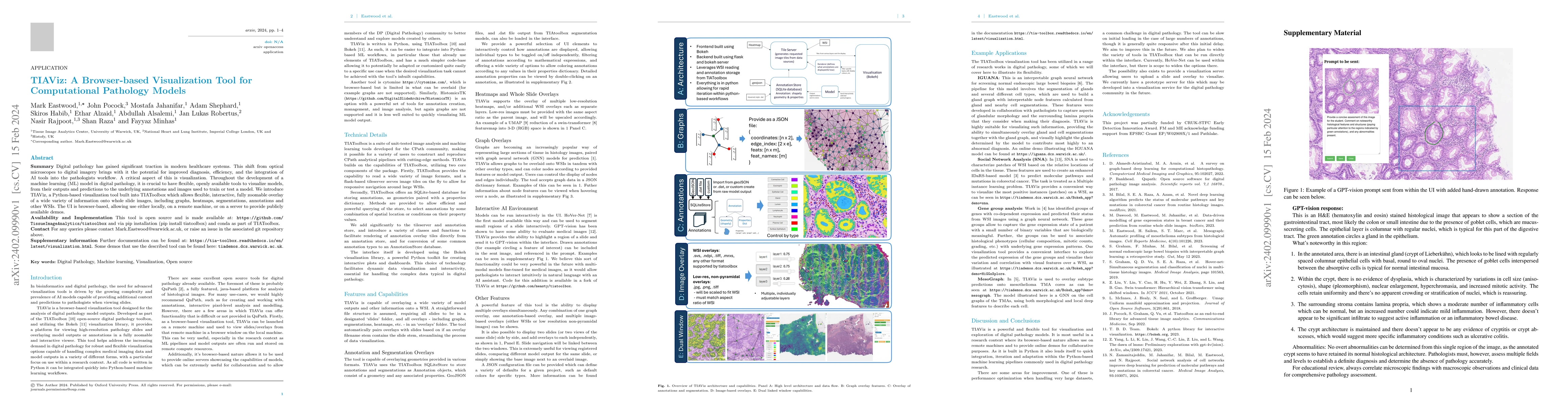
Digital pathology has gained significant traction in modern healthcare systems. This shift from optical microscopes to digital imagery brings with it the potential for improved diagnosis, efficiency...
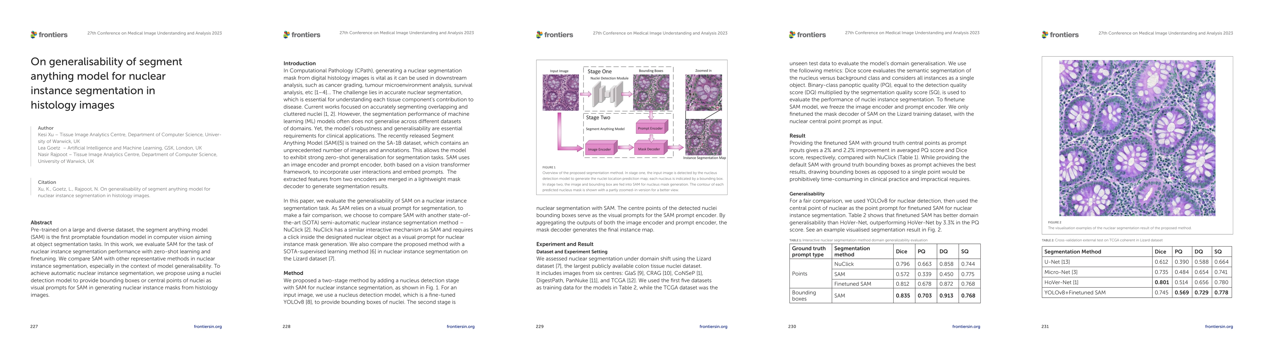
Pre-trained on a large and diverse dataset, the segment anything model (SAM) is the first promptable foundation model in computer vision aiming at object segmentation tasks. In this work, we evaluat...
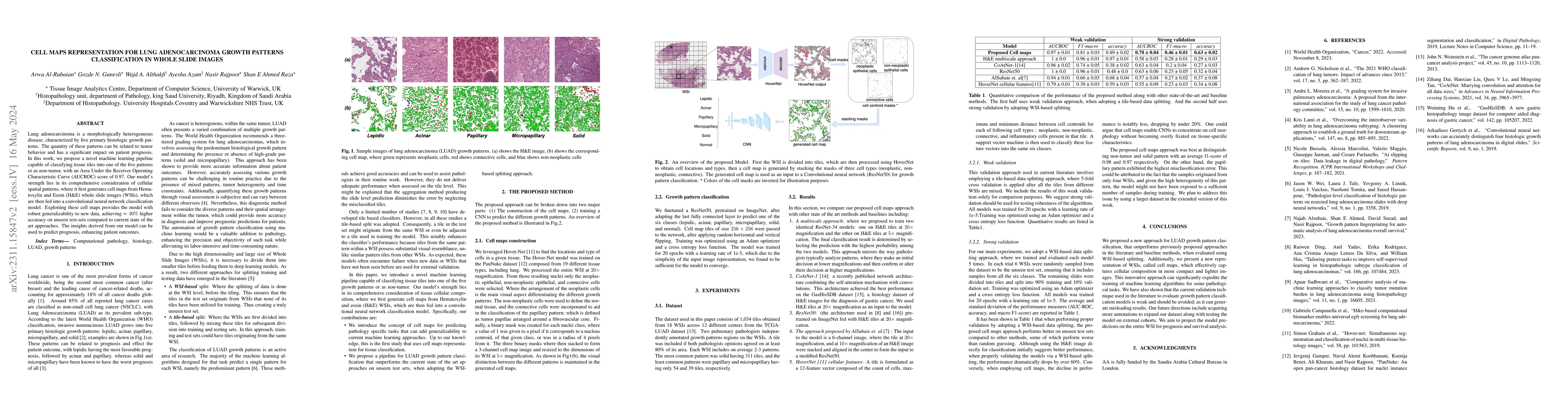
Lung adenocarcinoma is a morphologically heterogeneous disease, characterized by five primary histologic growth patterns. The quantity of these patterns can be related to tumor behavior and has a si...

Deep learning models have exhibited exceptional effectiveness in Computational Pathology (CPath) by tackling intricate tasks across an array of histology image analysis applications. Nevertheless, t...
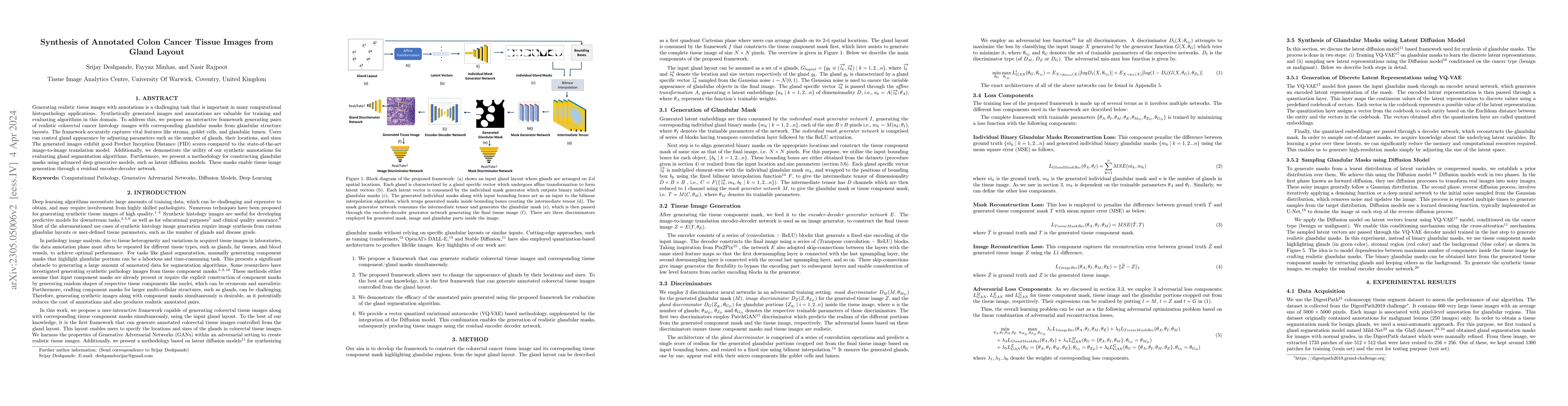
Generating realistic tissue images with annotations is a challenging task that is important in many computational histopathology applications. Synthetically generated images and annotations are valu...
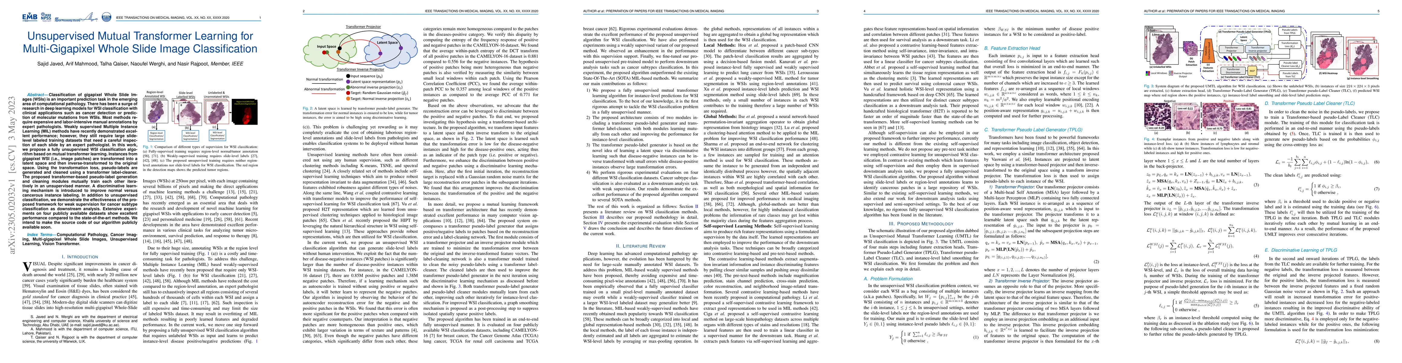
Classification of gigapixel Whole Slide Images (WSIs) is an important prediction task in the emerging area of computational pathology. There has been a surge of research in deep learning models for ...
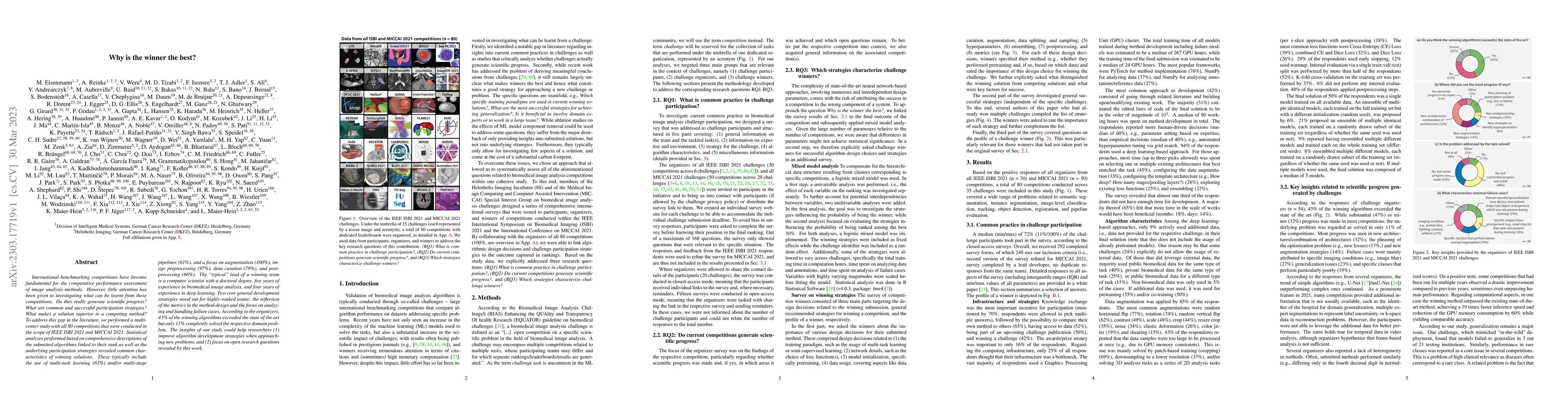
International benchmarking competitions have become fundamental for the comparative performance assessment of image analysis methods. However, little attention has been given to investigating what c...
Validation metrics are key for the reliable tracking of scientific progress and for bridging the current chasm between artificial intelligence (AI) research and its translation into practice. Howeve...
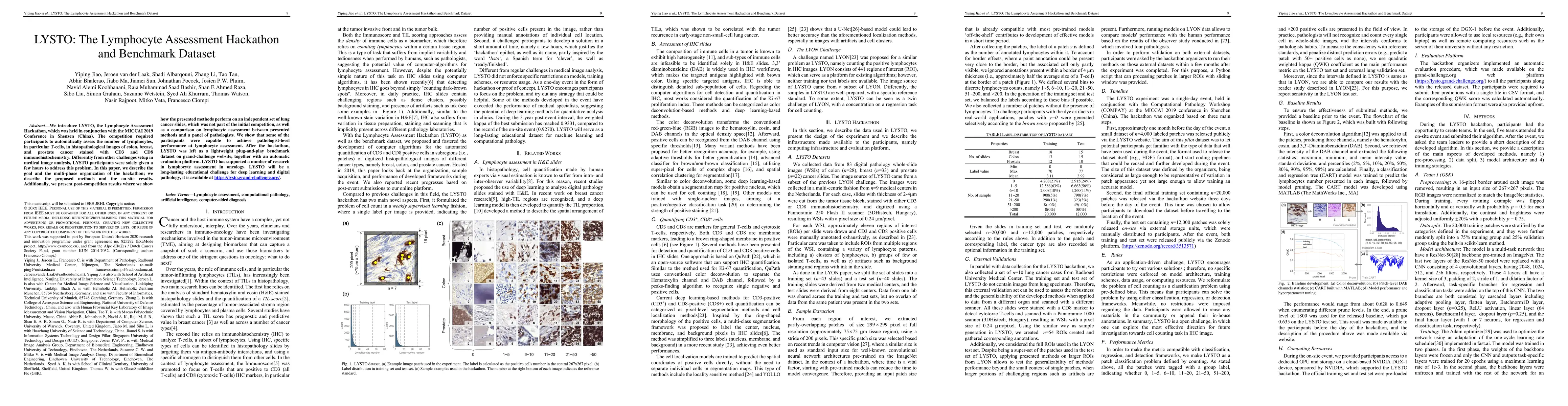
We introduce LYSTO, the Lymphocyte Assessment Hackathon, which was held in conjunction with the MICCAI 2019 Conference in Shenzen (China). The competition required participants to automatically asse...
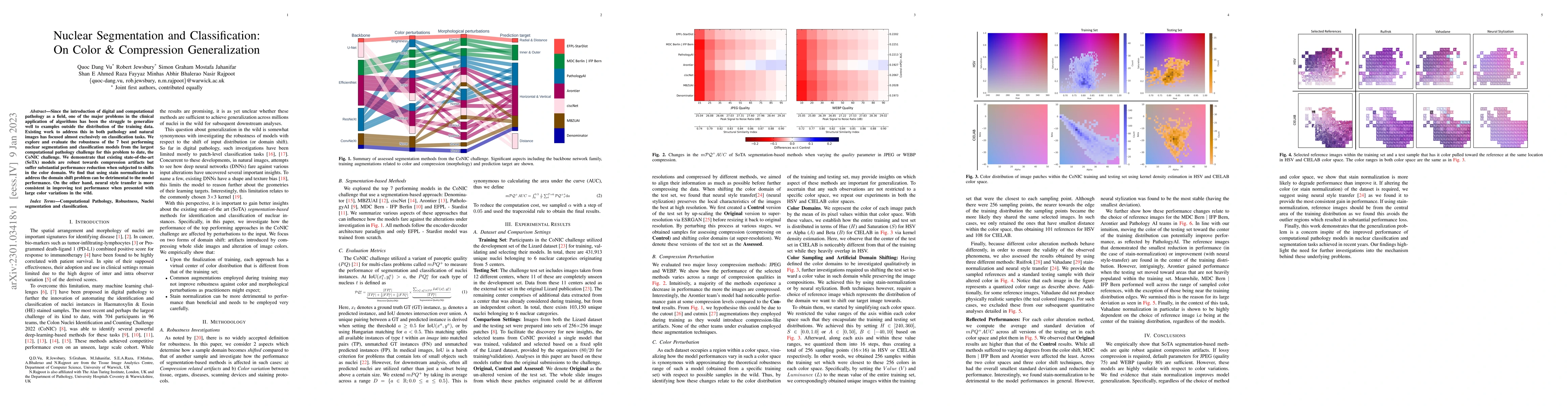
Since the introduction of digital and computational pathology as a field, one of the major problems in the clinical application of algorithms has been the struggle to generalize well to examples out...
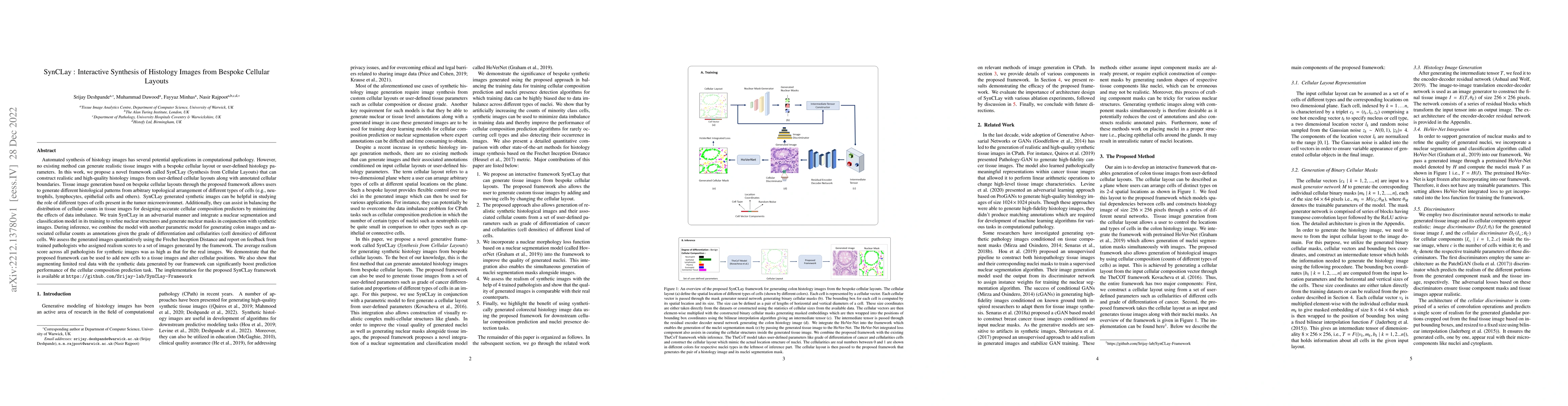
Automated synthesis of histology images has several potential applications in computational pathology. However, no existing method can generate realistic tissue images with a bespoke cellular layout...
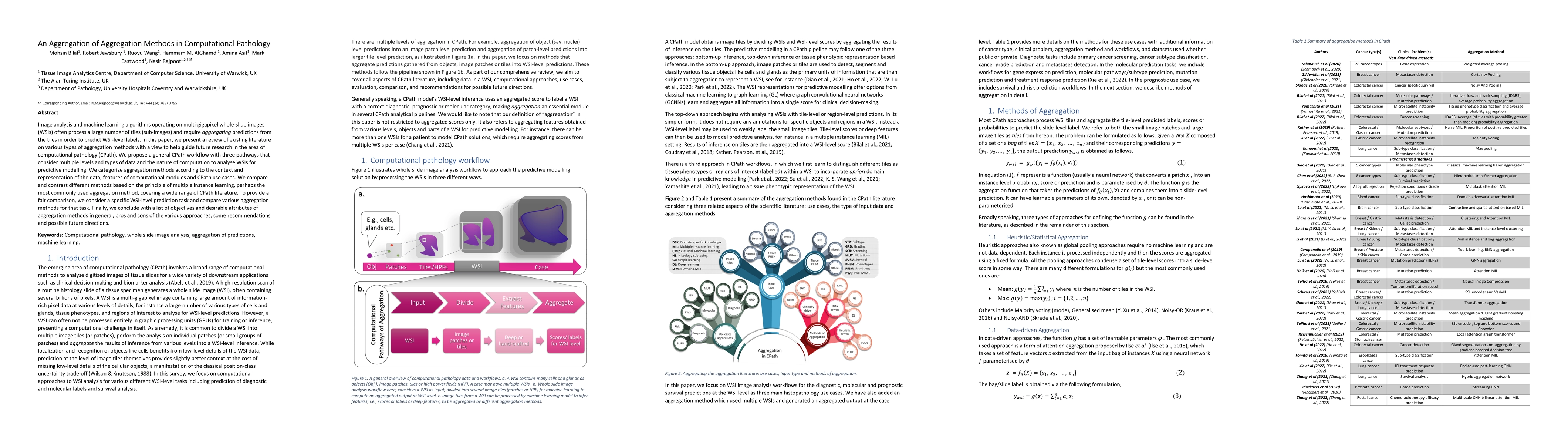
Image analysis and machine learning algorithms operating on multi-gigapixel whole-slide images (WSIs) often process a large number of tiles (sub-images) and require aggregating predictions from the ...
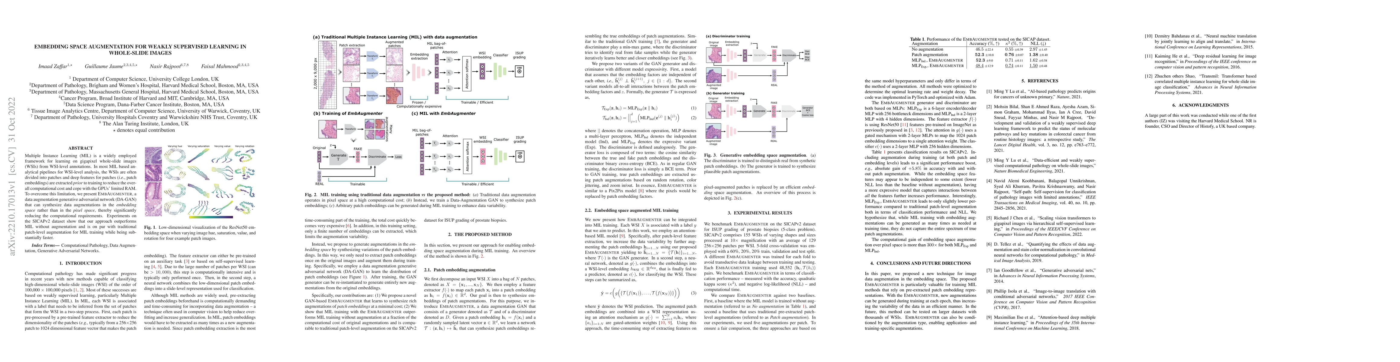
Multiple Instance Learning (MIL) is a widely employed framework for learning on gigapixel whole-slide images (WSIs) from WSI-level annotations. In most MIL based analytical pipelines for WSI-level a...
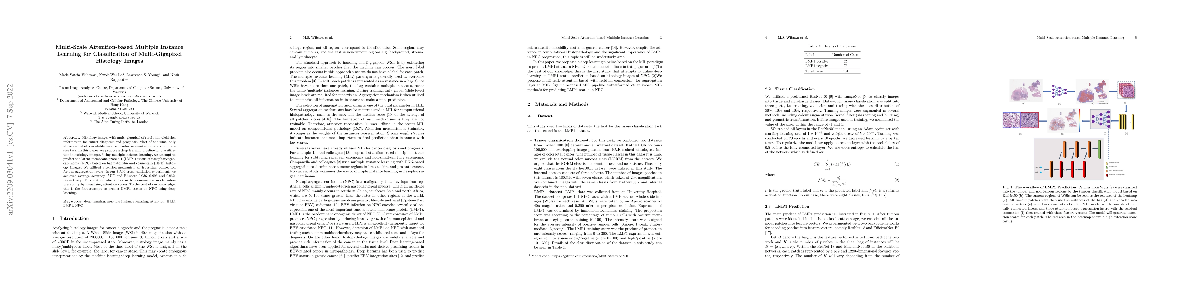
Histology images with multi-gigapixel of resolution yield rich information for cancer diagnosis and prognosis. Most of the time, only slide-level label is available because pixel-wise annotation is ...
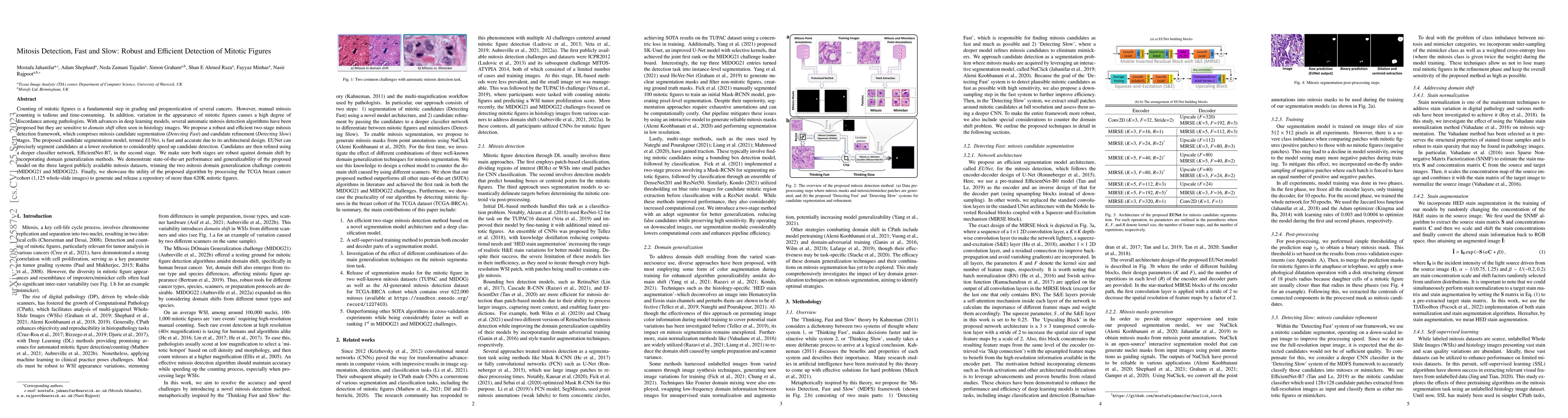
Counting of mitotic figures is a fundamental step in grading and prognostication of several cancers. However, manual mitosis counting is tedious and time-consuming. In addition, variation in the app...
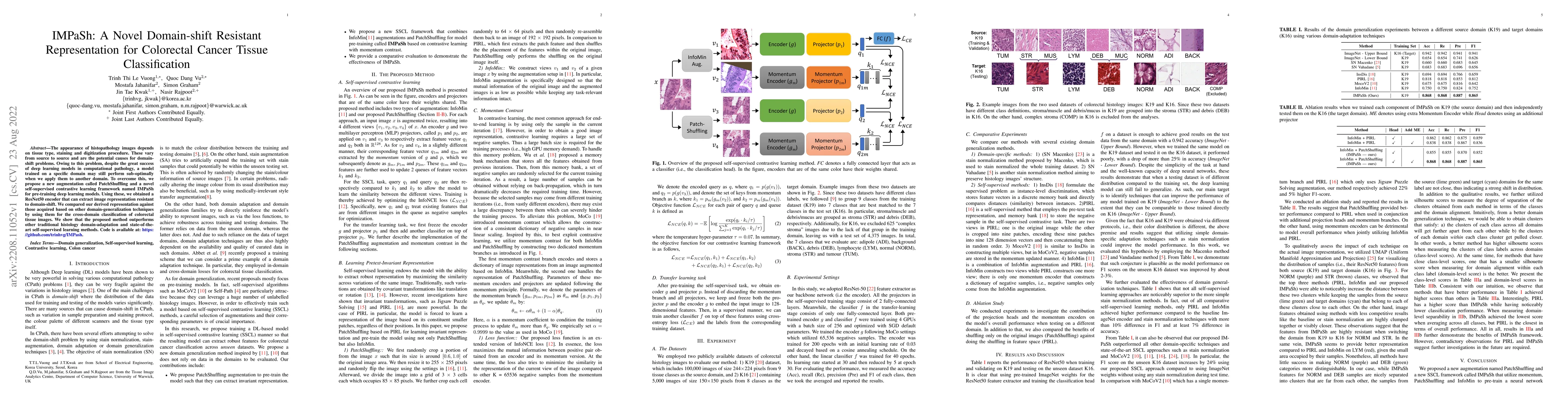
The appearance of histopathology images depends on tissue type, staining and digitization procedure. These vary from source to source and are the potential causes for domain-shift problems. Owing to...
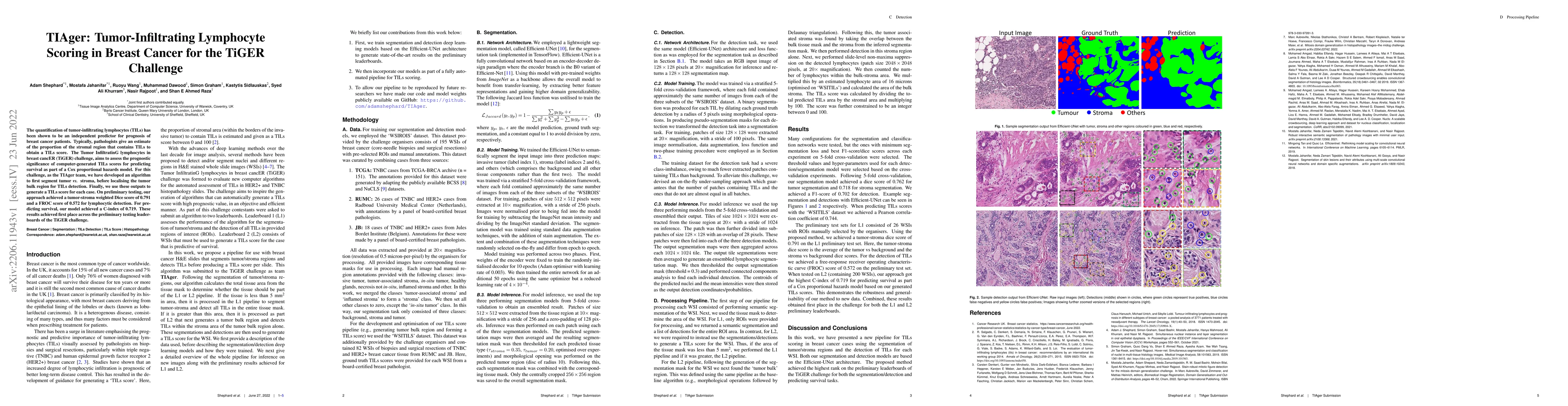
The quantification of tumor-infiltrating lymphocytes (TILs) has been shown to be an independent predictor for prognosis of breast cancer patients. Typically, pathologists give an estimate of the pro...
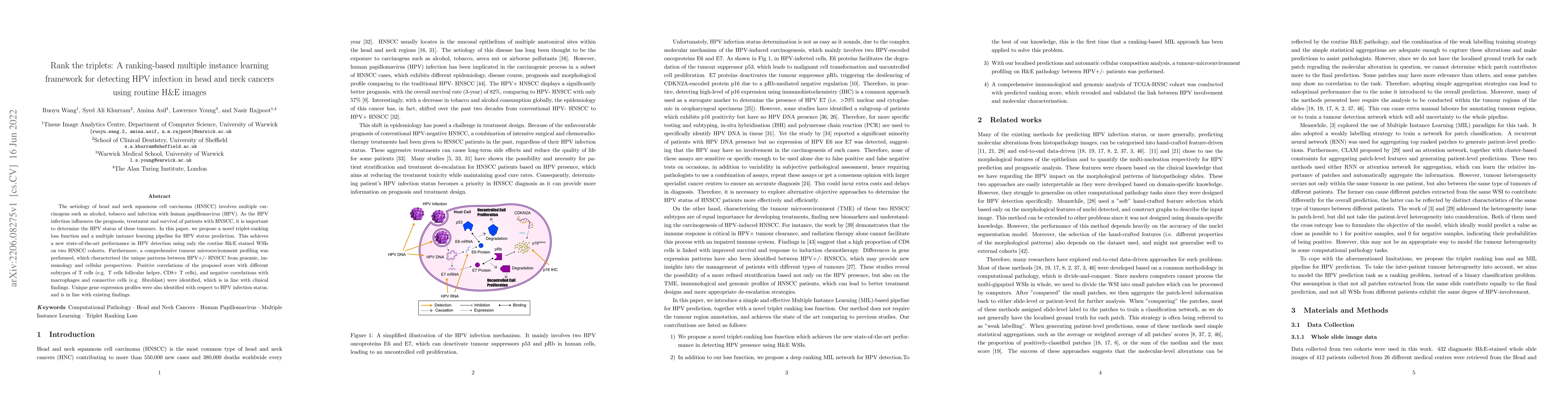
The aetiology of head and neck squamous cell carcinoma (HNSCC) involves multiple carcinogens such as alcohol, tobacco and infection with human papillomavirus (HPV). As the HPV infection influences t...
Increasing evidence shows that flaws in machine learning (ML) algorithm validation are an underestimated global problem. Particularly in automatic biomedical image analysis, chosen performance metri...
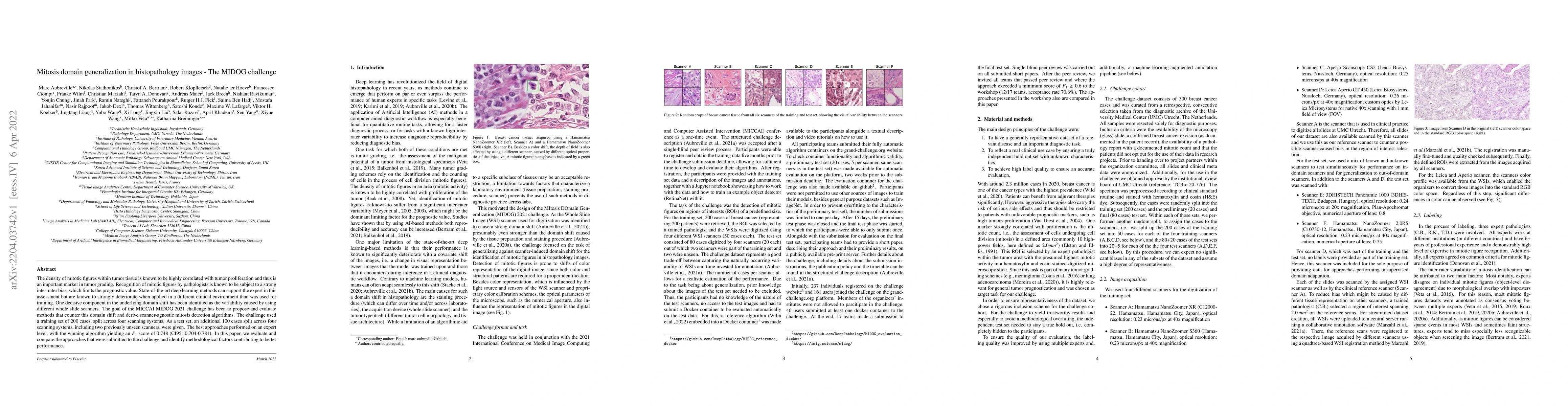
The density of mitotic figures within tumor tissue is known to be highly correlated with tumor proliferation and thus is an important marker in tumor grading. Recognition of mitotic figures by patho...
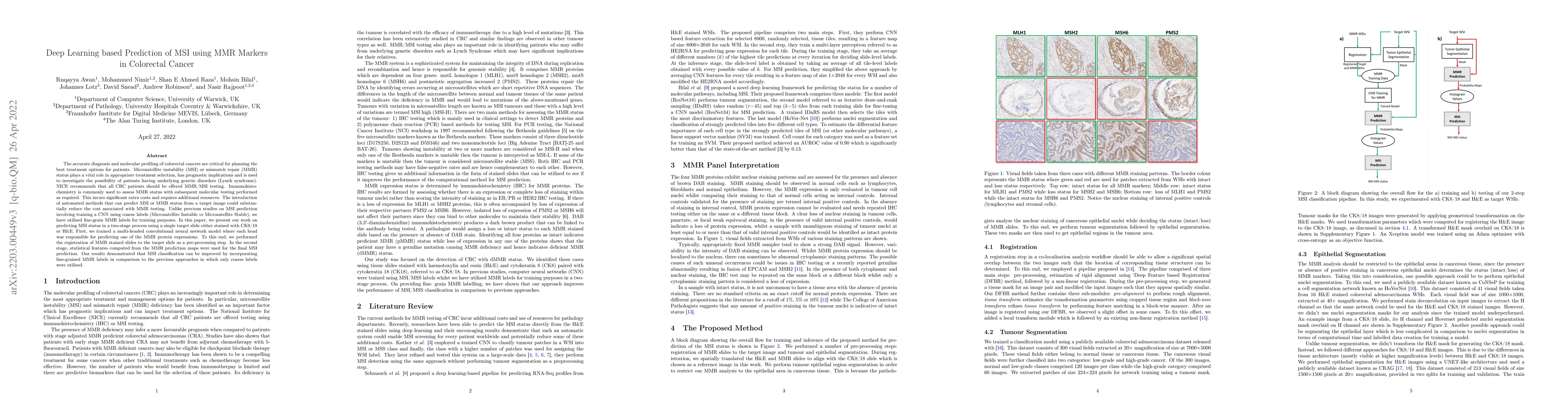
The accurate diagnosis and molecular profiling of colorectal cancers are critical for planning the best treatment options for patients. Microsatellite instability (MSI) or mismatch repair (MMR) stat...
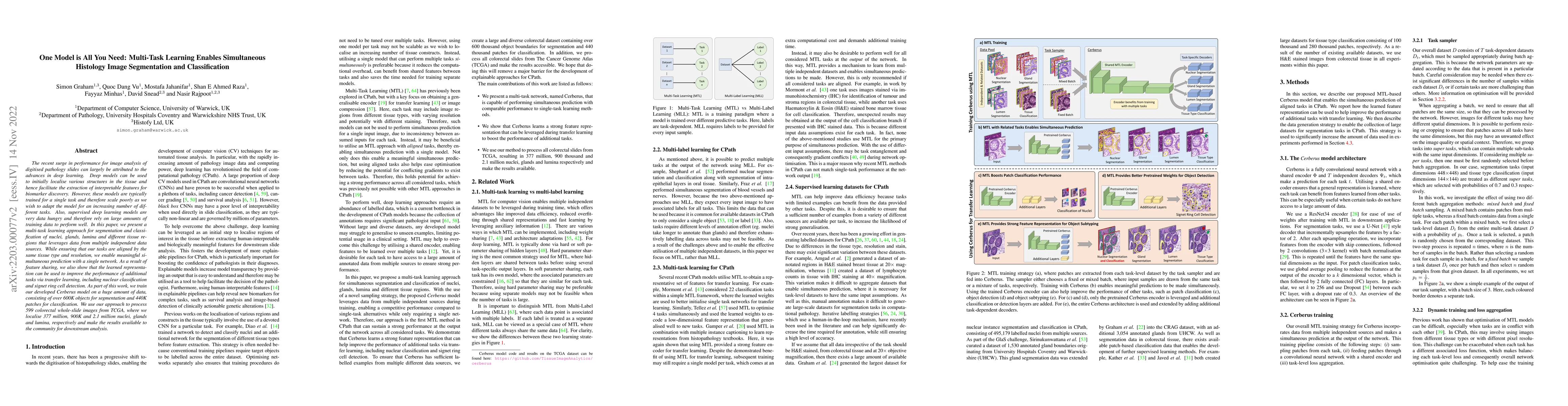
The recent surge in performance for image analysis of digitised pathology slides can largely be attributed to the advances in deep learning. Deep models can be used to initially localise various str...

Cross-slide image analysis provides additional information by analysing the expression of different biomarkers as compared to a single slide analysis. These biomarker stained slides are analysed sid...
Diagnostic, prognostic and therapeutic decision-making of cancer in pathology clinics can now be carried out based on analysis of multi-gigapixel tissue images, also known as whole-slide images (WSI...
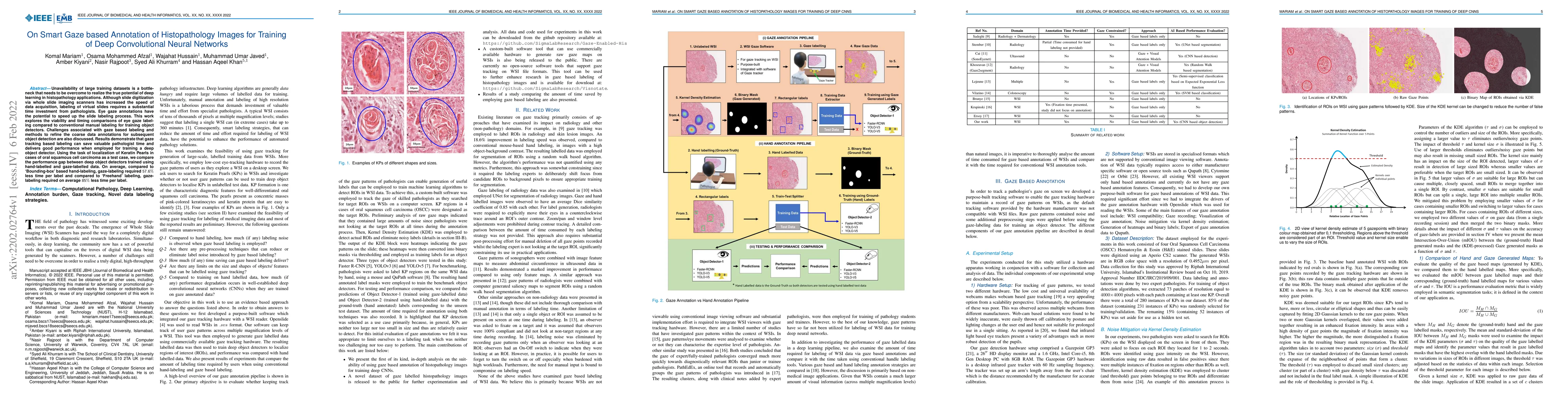
Unavailability of large training datasets is a bottleneck that needs to be overcome to realize the true potential of deep learning in histopathology applications. Although slide digitization via who...
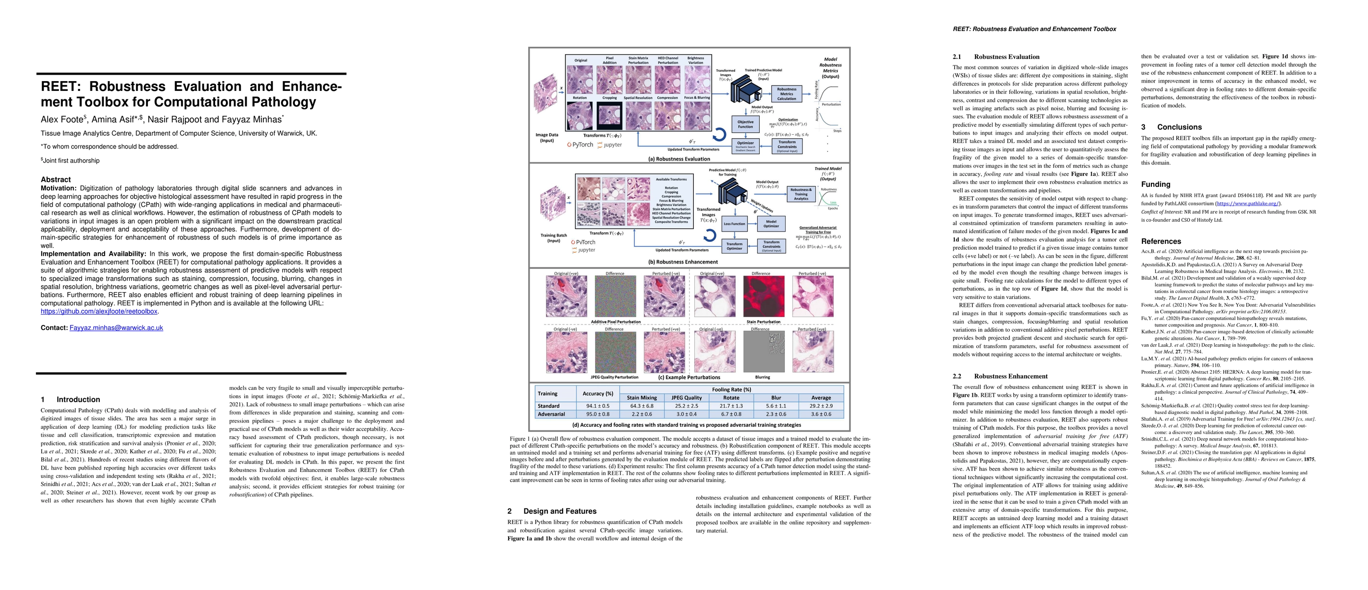
Motivation: Digitization of pathology laboratories through digital slide scanners and advances in deep learning approaches for objective histological assessment have resulted in rapid progress in th...
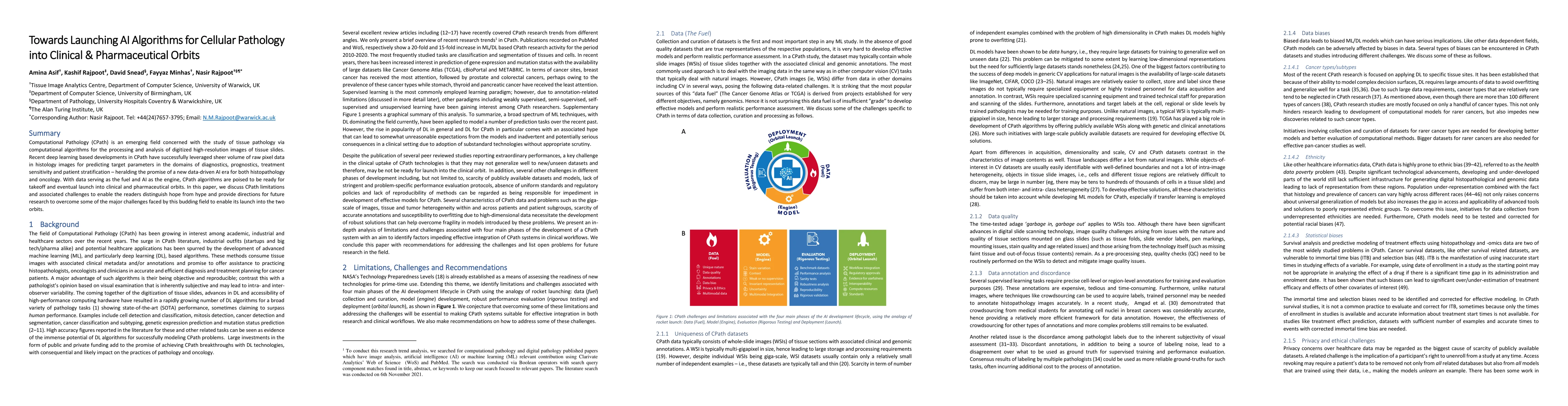
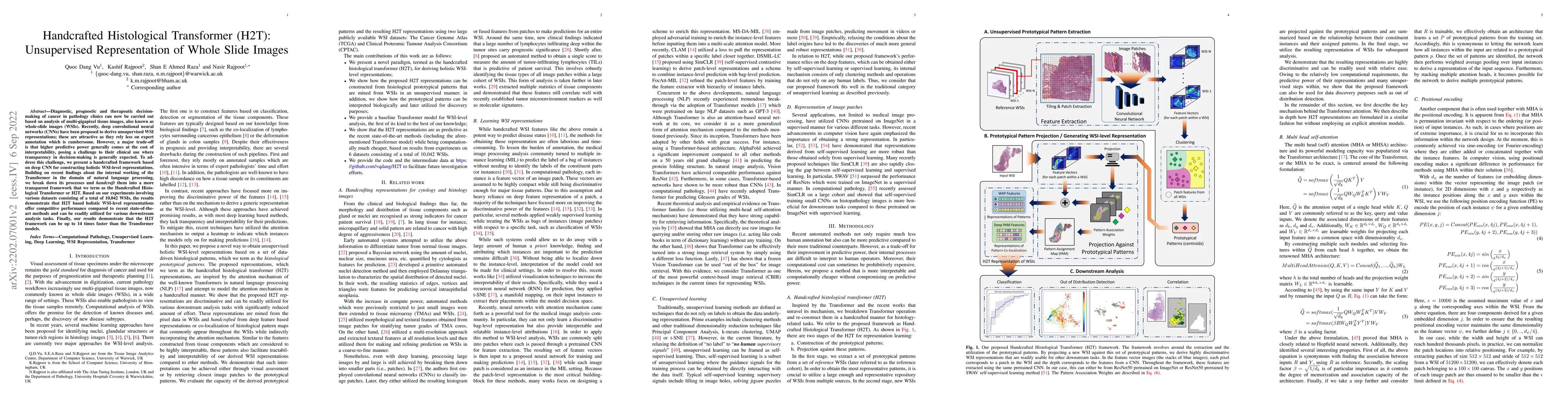
Computational Pathology (CPath) is an emerging field concerned with the study of tissue pathology via computational algorithms for the processing and analysis of digitized high-resolution images of ...
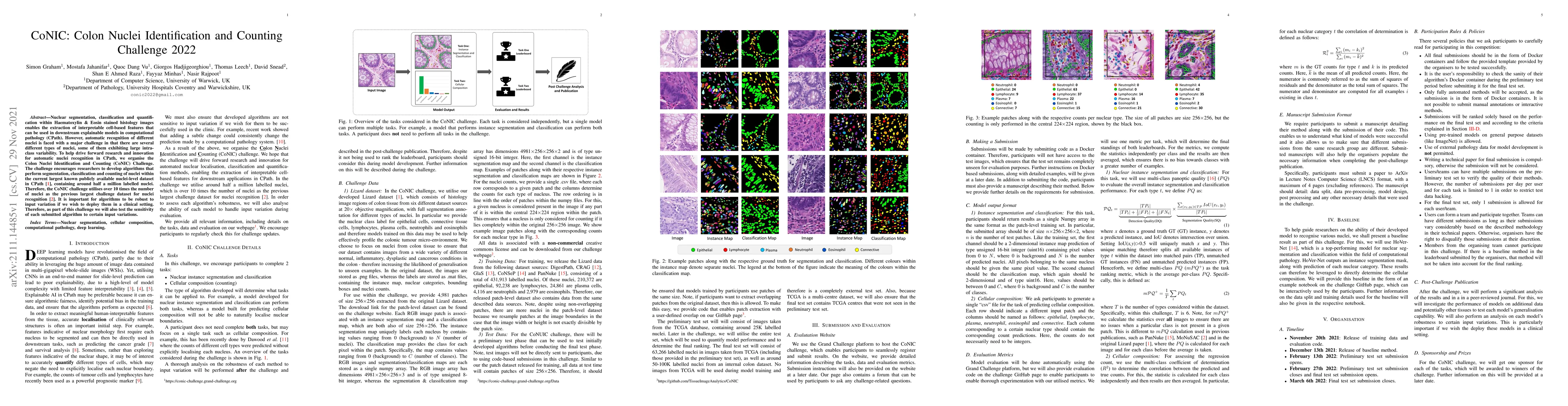
Nuclear segmentation, classification and quantification within Haematoxylin & Eosin stained histology images enables the extraction of interpretable cell-based features that can be used in downstrea...
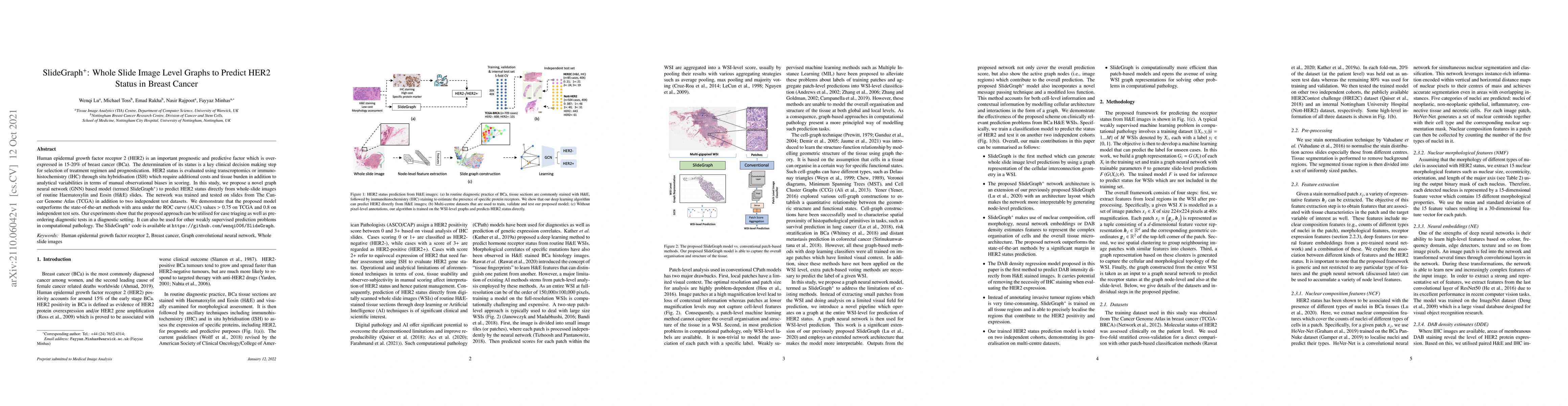
Human epidermal growth factor receptor 2 (HER2) is an important prognostic and predictive factor which is overexpressed in 15-20% of breast cancer (BCa). The determination of its status is a key cli...
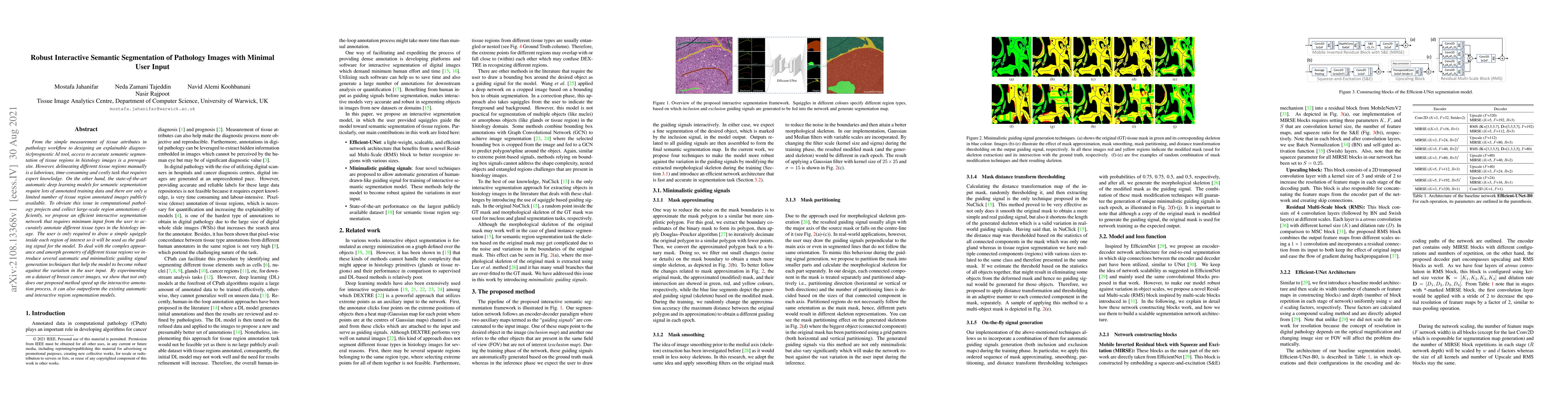
From the simple measurement of tissue attributes in pathology workflow to designing an explainable diagnostic/prognostic AI tool, access to accurate semantic segmentation of tissue regions in histol...
The development of deep segmentation models for computational pathology (CPath) can help foster the investigation of interpretable morphological biomarkers. Yet, there is a major bottleneck in the s...
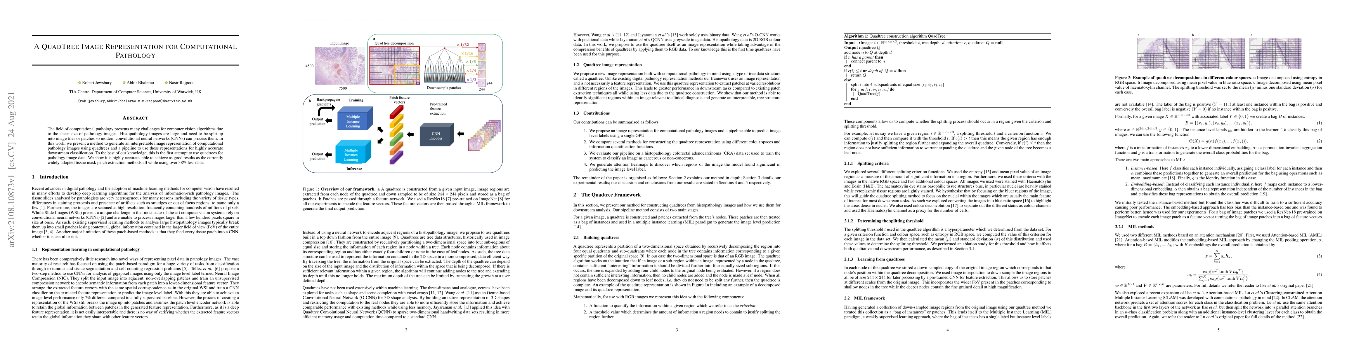
The field of computational pathology presents many challenges for computer vision algorithms due to the sheer size of pathology images. Histopathology images are large and need to be split up into i...

Recent advances in whole slide imaging (WSI) technology have led to the development of a myriad of computer vision and artificial intelligence (AI) based diagnostic, prognostic, and predictive algor...
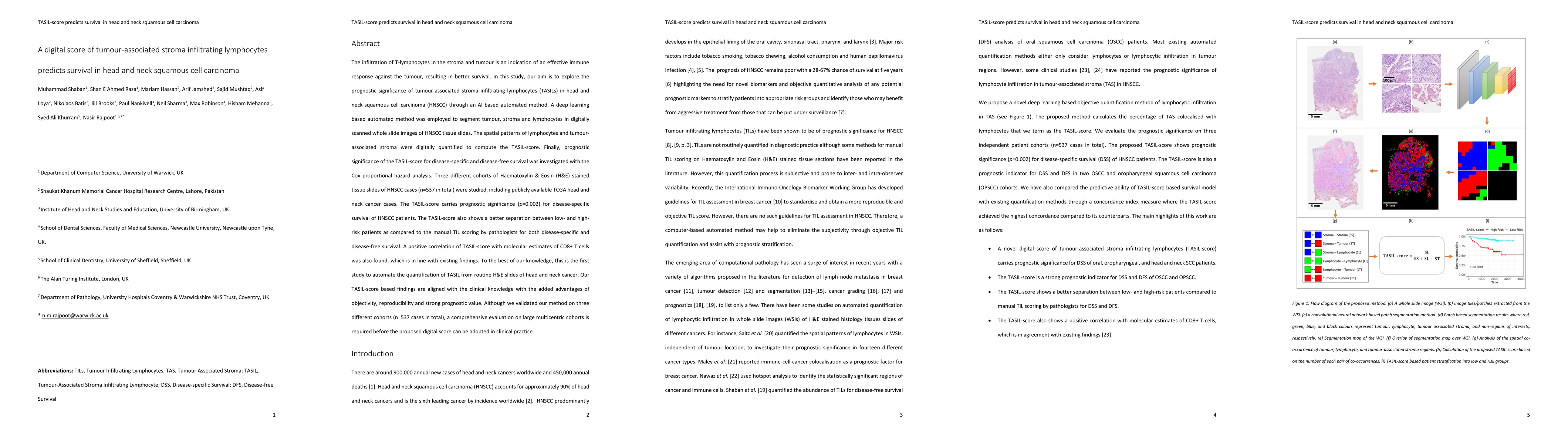
The infiltration of T-lymphocytes in the stroma and tumour is an indication of an effective immune response against the tumour, resulting in better survival. In this study, our aim is to explore the...
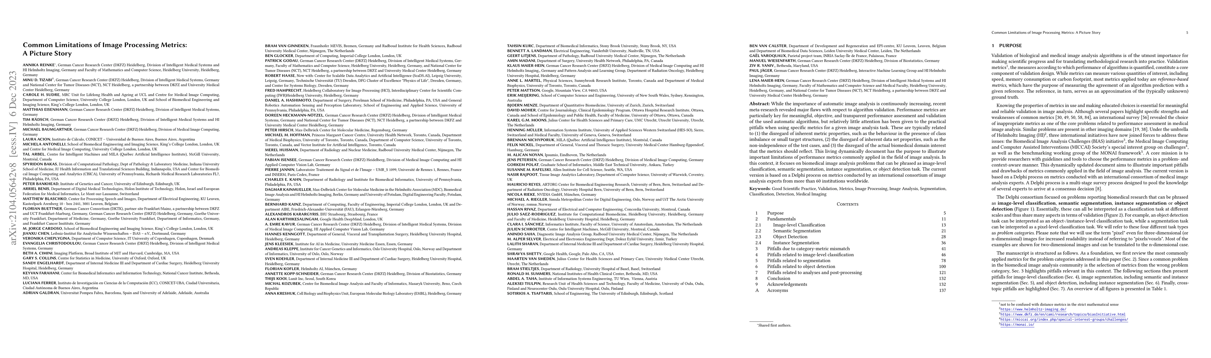
While the importance of automatic image analysis is continuously increasing, recent meta-research revealed major flaws with respect to algorithm validation. Performance metrics are particularly key ...
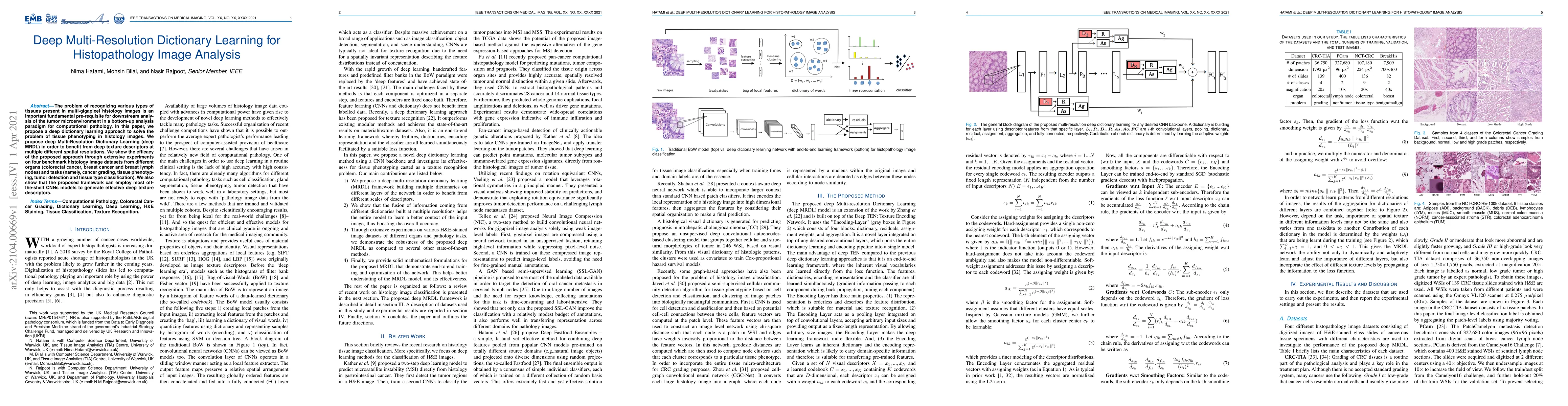
The problem of recognizing various types of tissues present in multi-gigapixel histology images is an important fundamental pre-requisite for downstream analysis of the tumor microenvironment in a b...
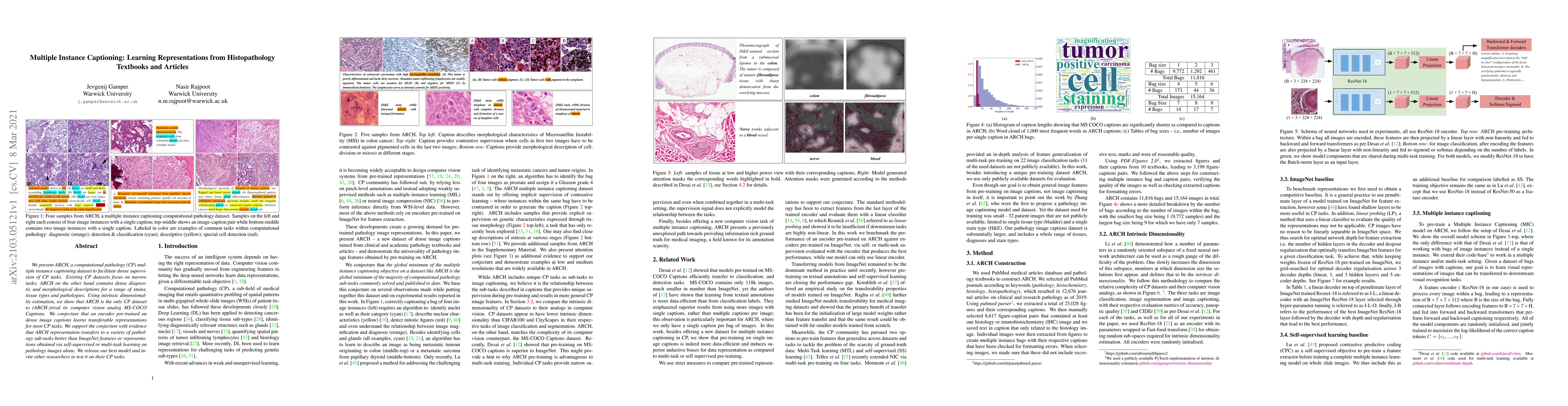
We present ARCH, a computational pathology (CP) multiple instance captioning dataset to facilitate dense supervision of CP tasks. Existing CP datasets focus on narrow tasks; ARCH on the other hand c...
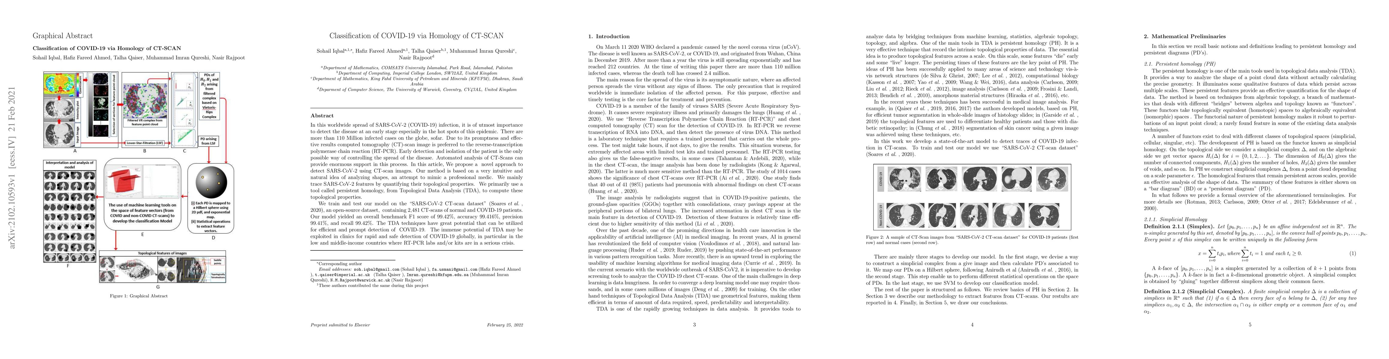
In this worldwide spread of SARS-CoV-2 (COVID-19) infection, it is of utmost importance to detect the disease at an early stage especially in the hot spots of this epidemic. There are more than 110 ...
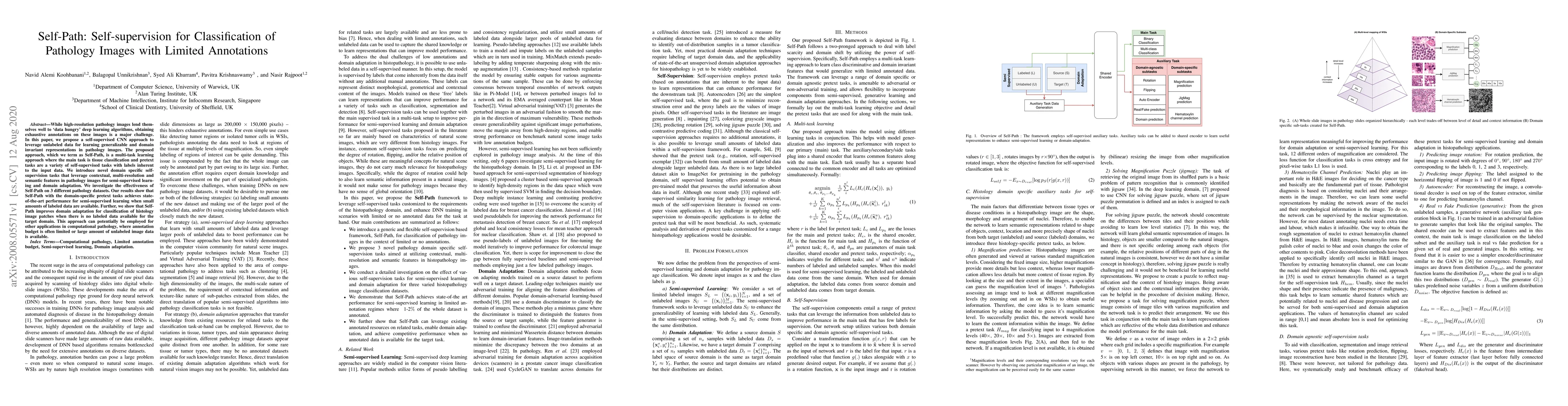
While high-resolution pathology images lend themselves well to `data hungry' deep learning algorithms, obtaining exhaustive annotations on these images is a major challenge. In this paper, we propos...
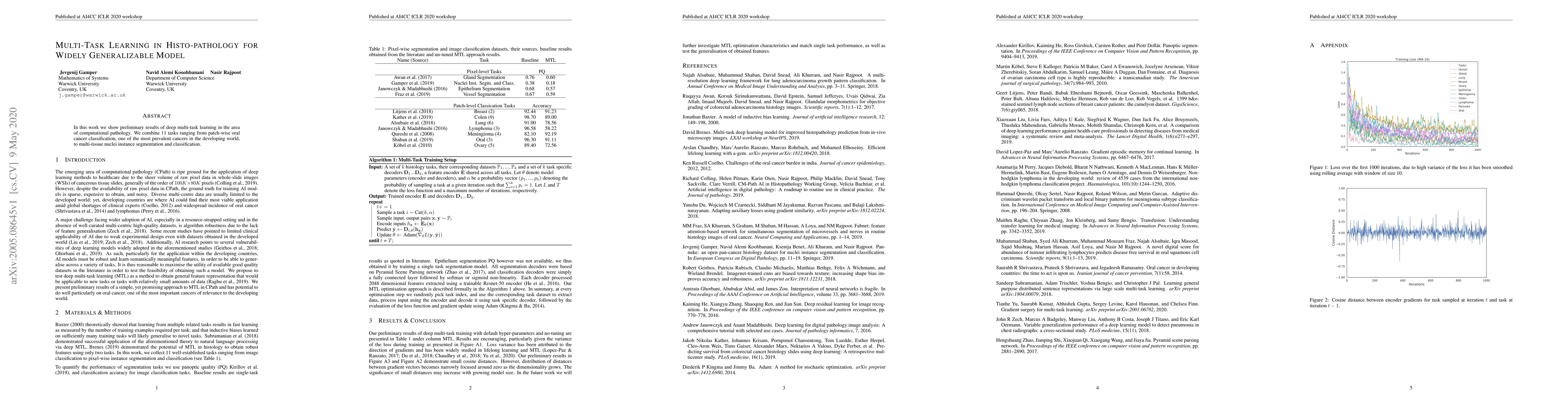
In this work we show preliminary results of deep multi-task learning in the area of computational pathology. We combine 11 tasks ranging from patch-wise oral cancer classification, one of the most p...
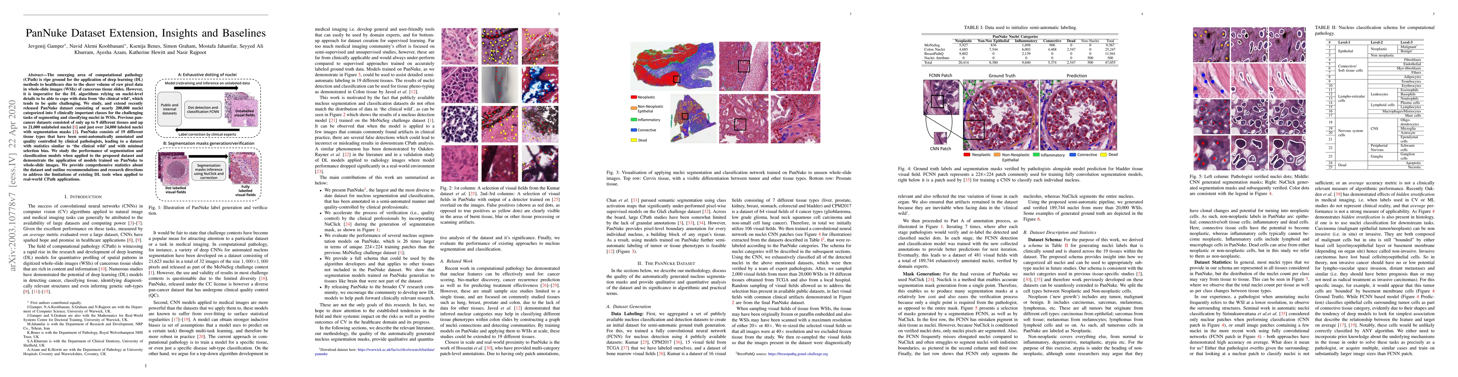
The emerging area of computational pathology (CPath) is ripe ground for the application of deep learning (DL) methods to healthcare due to the sheer volume of raw pixel data in whole-slide images (W...
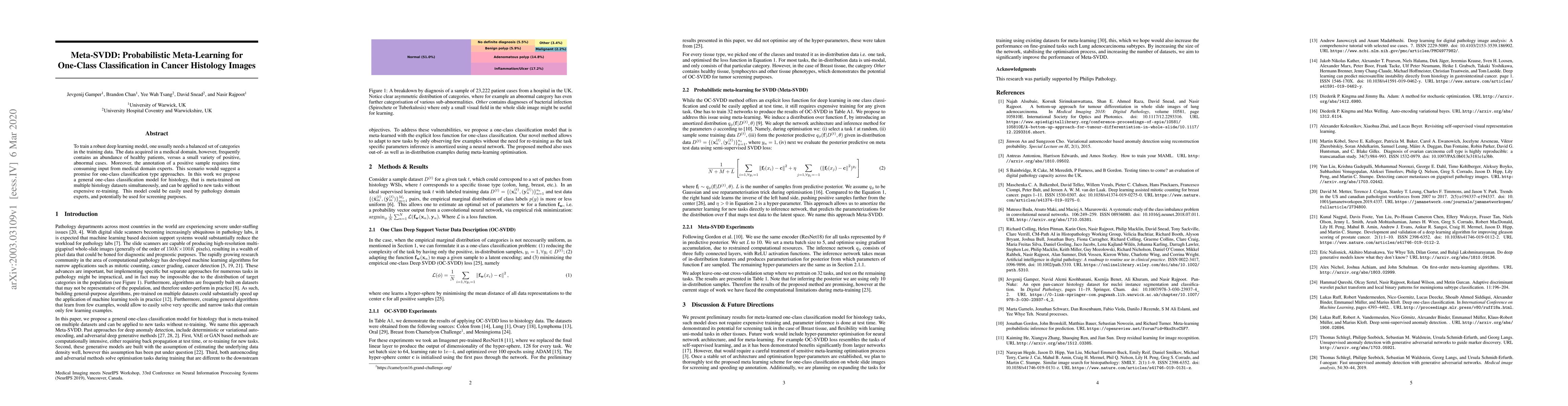
To train a robust deep learning model, one usually needs a balanced set of categories in the training data. The data acquired in a medical domain, however, frequently contains an abundance of health...
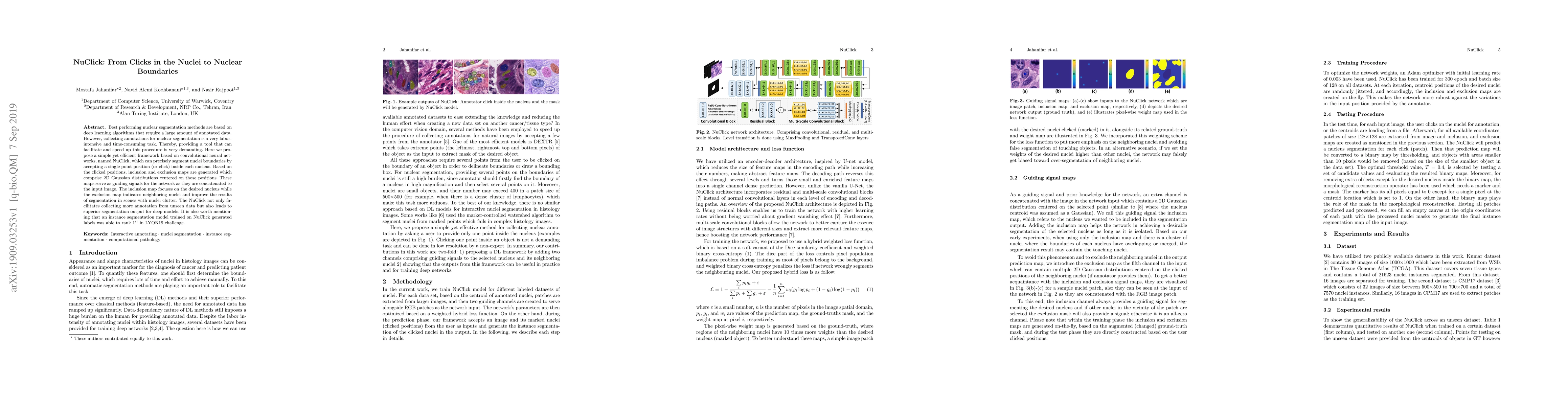
Best performing nuclear segmentation methods are based on deep learning algorithms that require a large amount of annotated data. However, collecting annotations for nuclear segmentation is a very l...
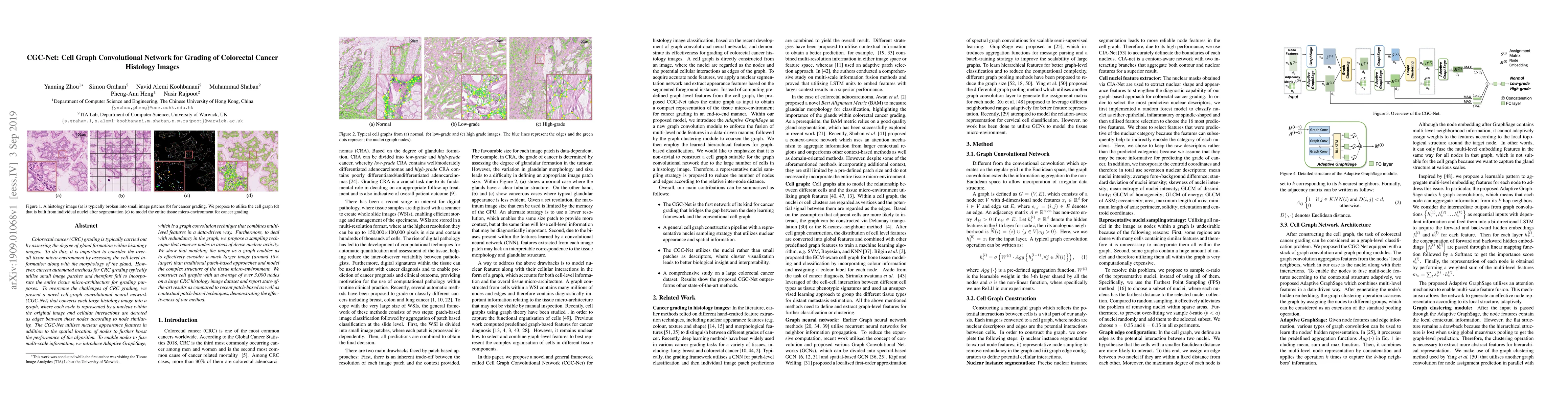
Colorectal cancer (CRC) grading is typically carried out by assessing the degree of gland formation within histology images. To do this, it is important to consider the overall tissue micro-environm...
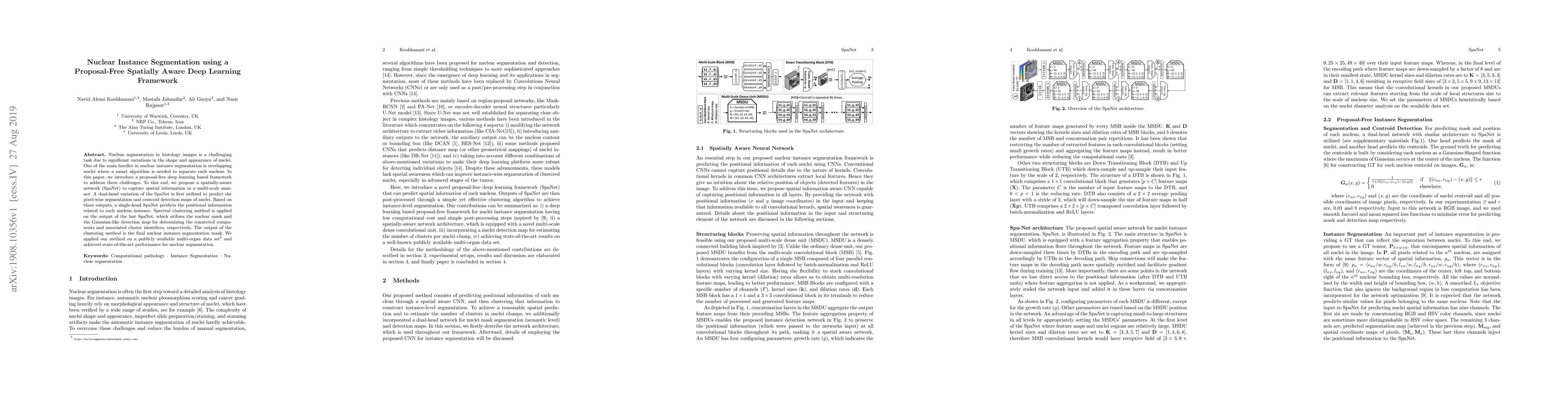
Nuclear segmentation in histology images is a challenging task due to significant variations in the shape and appearance of nuclei. One of the main hurdles in nuclear instance segmentation is overla...
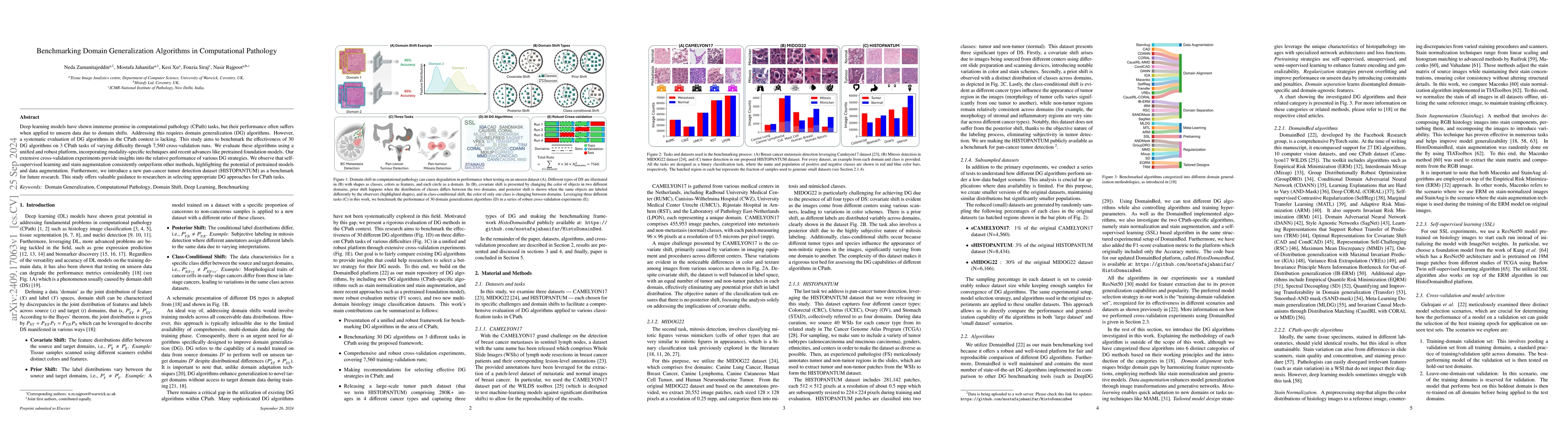
Deep learning models have shown immense promise in computational pathology (CPath) tasks, but their performance often suffers when applied to unseen data due to domain shifts. Addressing this requires...
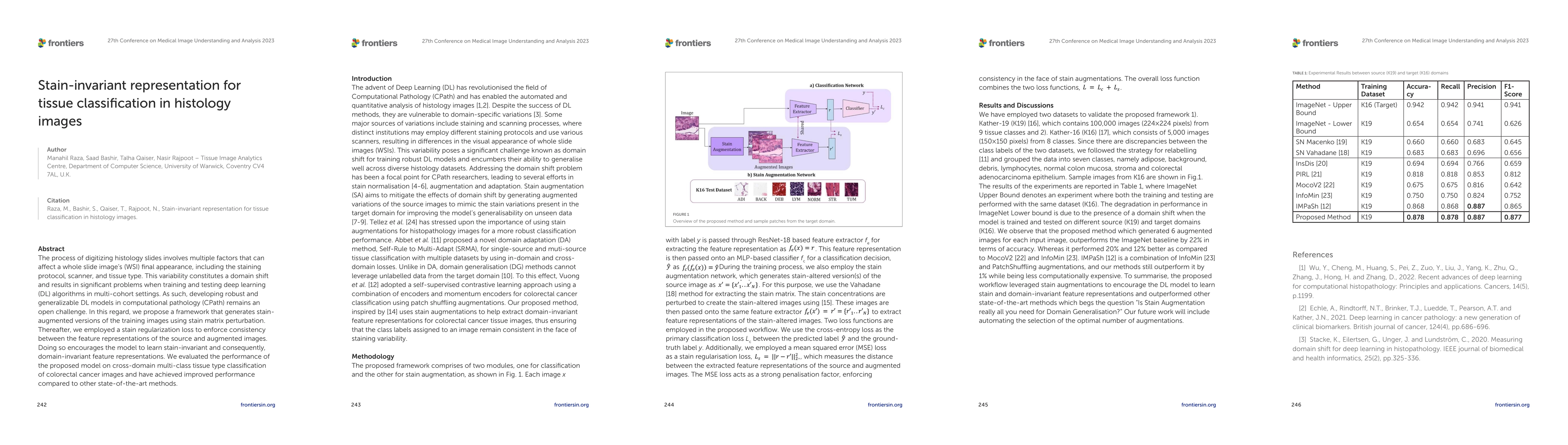
The process of digitising histology slides involves multiple factors that can affect a whole slide image's (WSI) final appearance, including the staining protocol, scanner, and tissue type. This varia...

From self-supervised, vision-only models to contrastive visual-language frameworks, computational pathology has rapidly evolved in recent years. Generative AI "co-pilots" now demonstrate the ability t...
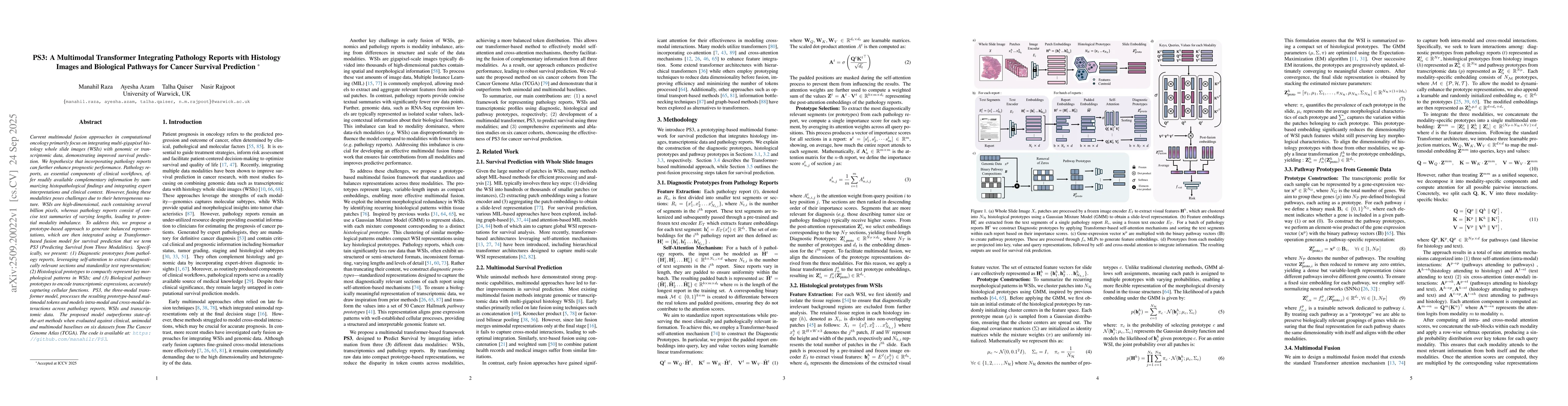
Current multimodal fusion approaches in computational oncology primarily focus on integrating multi-gigapixel histology whole slide images (WSIs) with genomic or transcriptomic data, demonstrating imp...
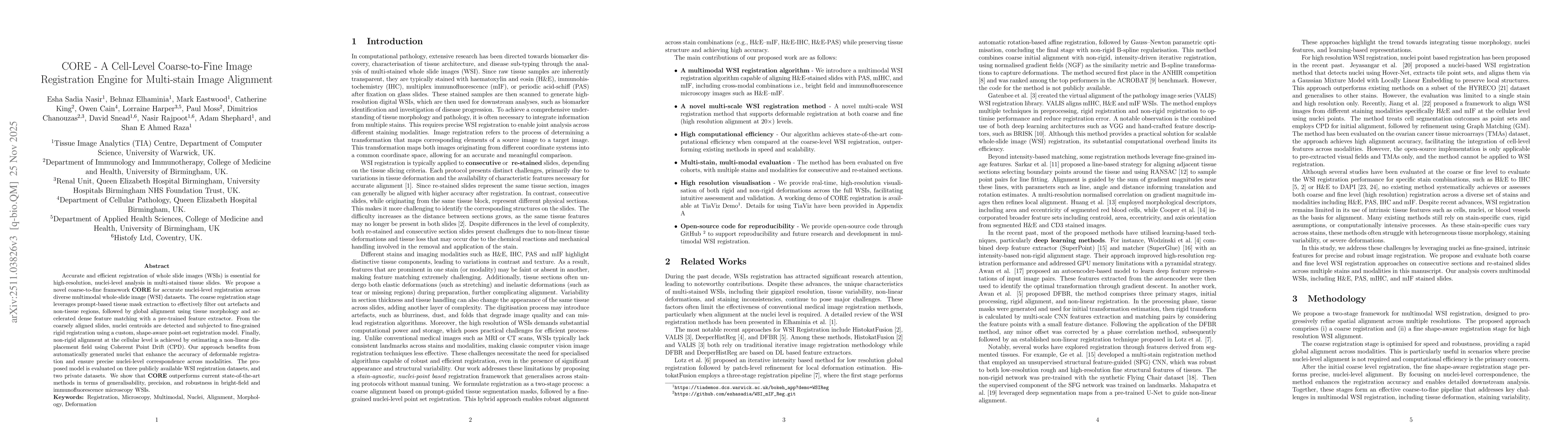
Accurate and efficient registration of whole slide images (WSIs) is essential for high-resolution, nuclei-level analysis in multi-stained tissue slides. We propose a novel coarse-to-fine framework COR...
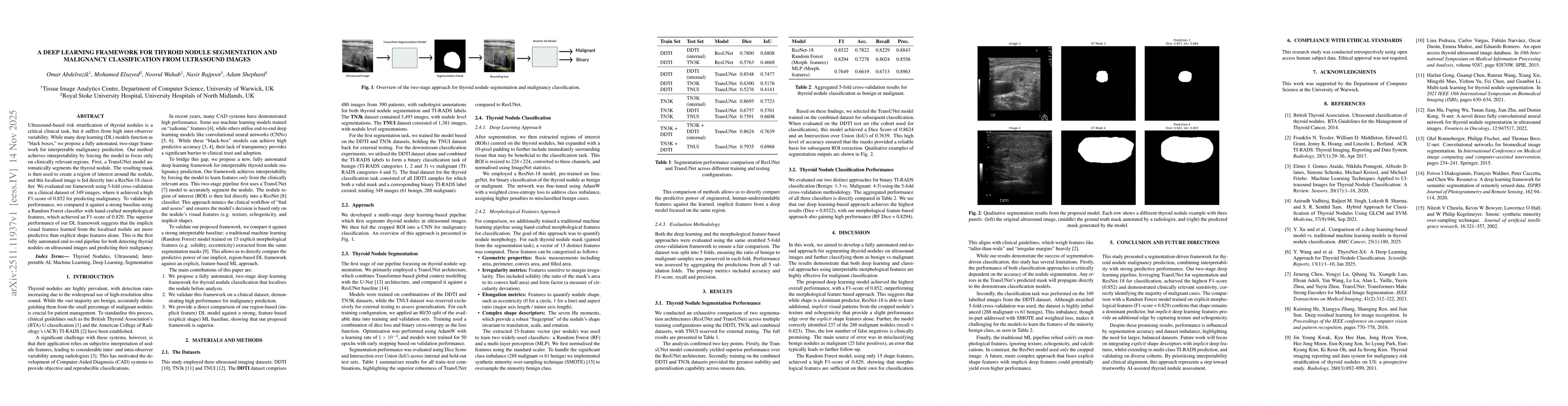
Ultrasound-based risk stratification of thyroid nodules is a critical clinical task, but it suffers from high inter-observer variability. While many deep learning (DL) models function as "black boxes,...
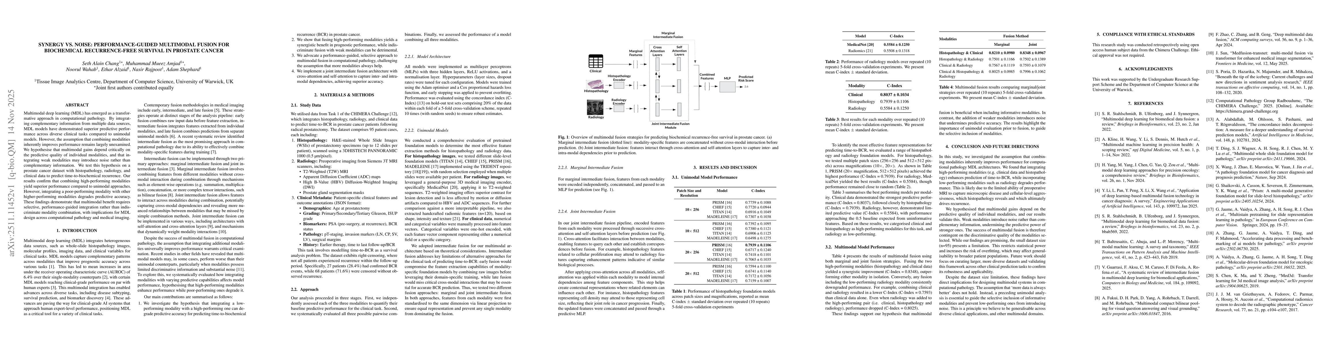
Multimodal deep learning (MDL) has emerged as a transformative approach in computational pathology. By integrating complementary information from multiple data sources, MDL models have demonstrated su...
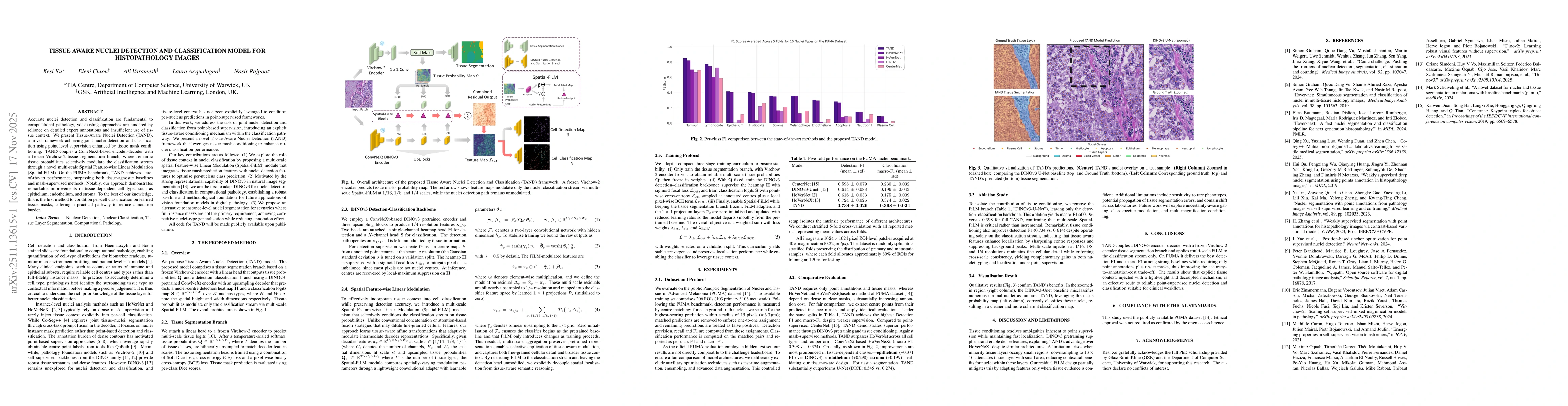
Accurate nuclei detection and classification are fundamental to computational pathology, yet existing approaches are hindered by reliance on detailed expert annotations and insufficient use of tissue ...
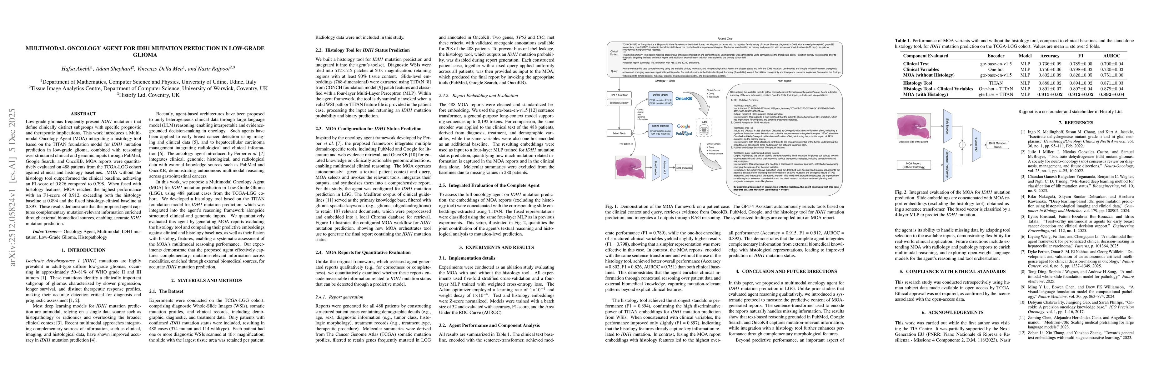
Low-grade gliomas frequently present IDH1 mutations that define clinically distinct subgroups with specific prognostic and therapeutic implications. This work introduces a Multimodal Oncology Agent (M...
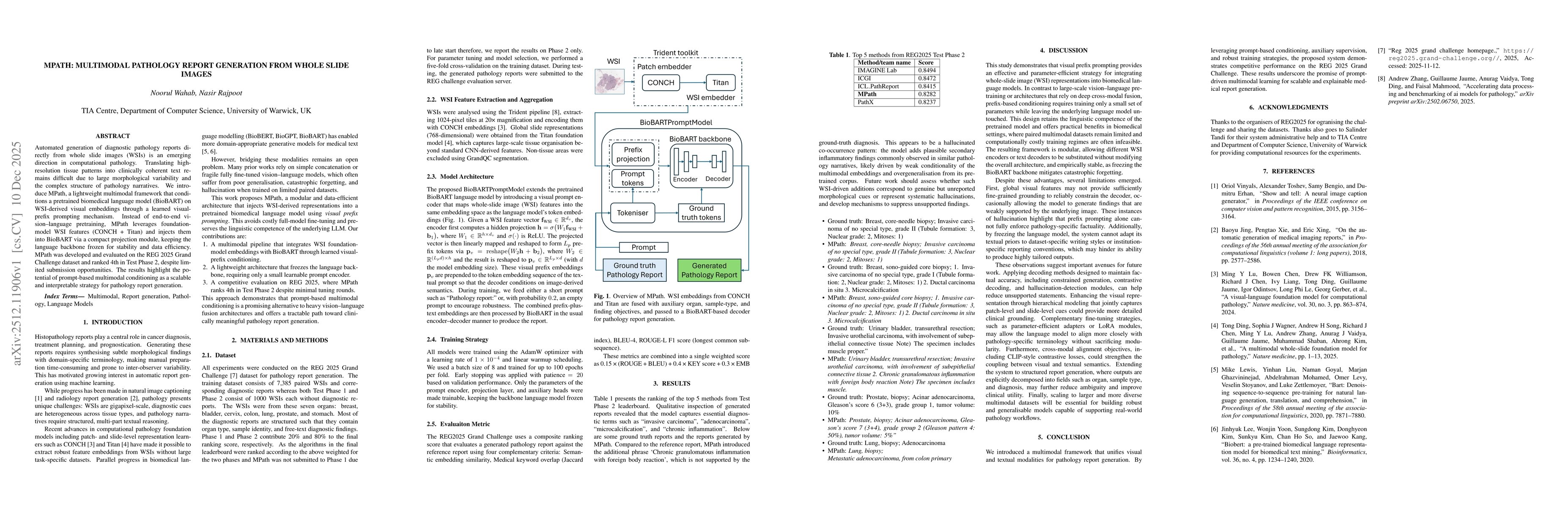
Automated generation of diagnostic pathology reports directly from whole slide images (WSIs) is an emerging direction in computational pathology. Translating high-resolution tissue patterns into clini...
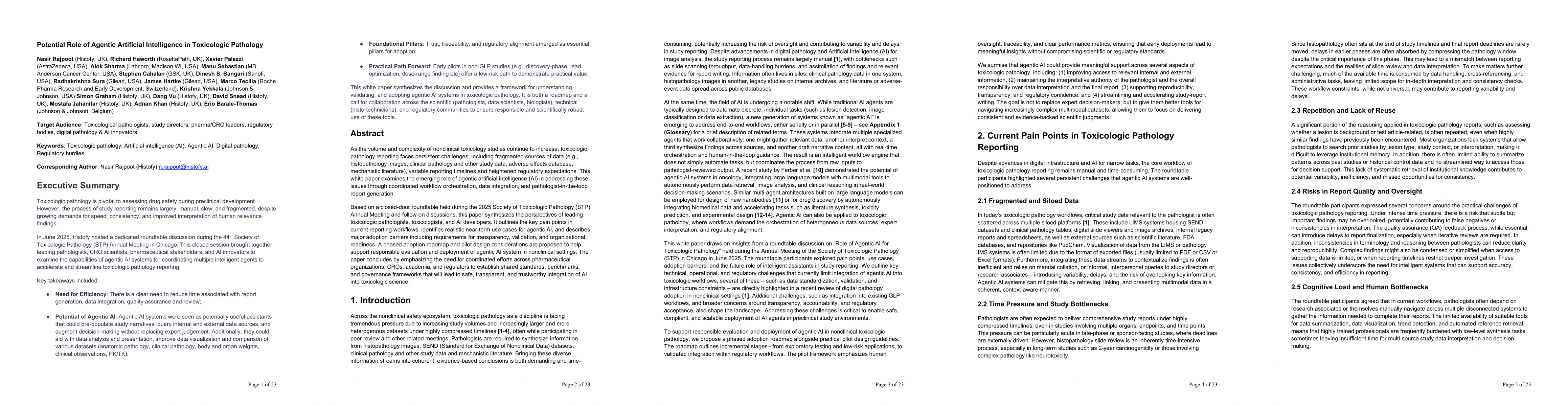
As the volume and complexity of nonclinical toxicology studies continue to increase, toxicologic pathology reporting faces persistent challenges, including fragmented sources of data (e.g., histopatho...