Academic Profile
Statistics
Similar Authors
Papers on arXiv
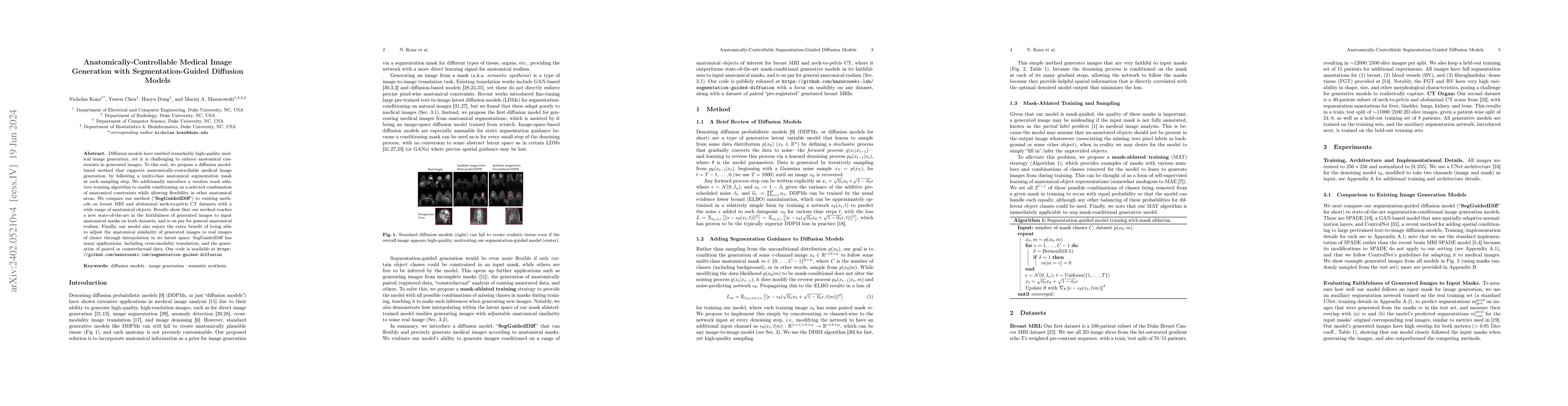
Diffusion models have enabled remarkably high-quality medical image generation, yet it is challenging to enforce anatomical constraints in generated images. To this end, we propose a diffusion model-b...
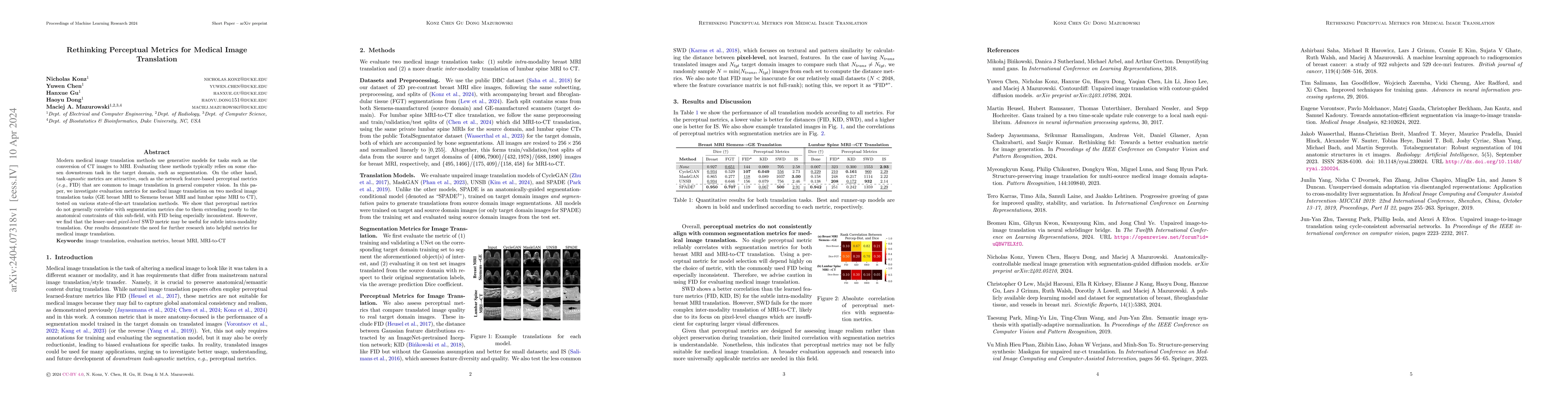
Modern medical image translation methods use generative models for tasks such as the conversion of CT images to MRI. Evaluating these methods typically relies on some chosen downstream task in the t...
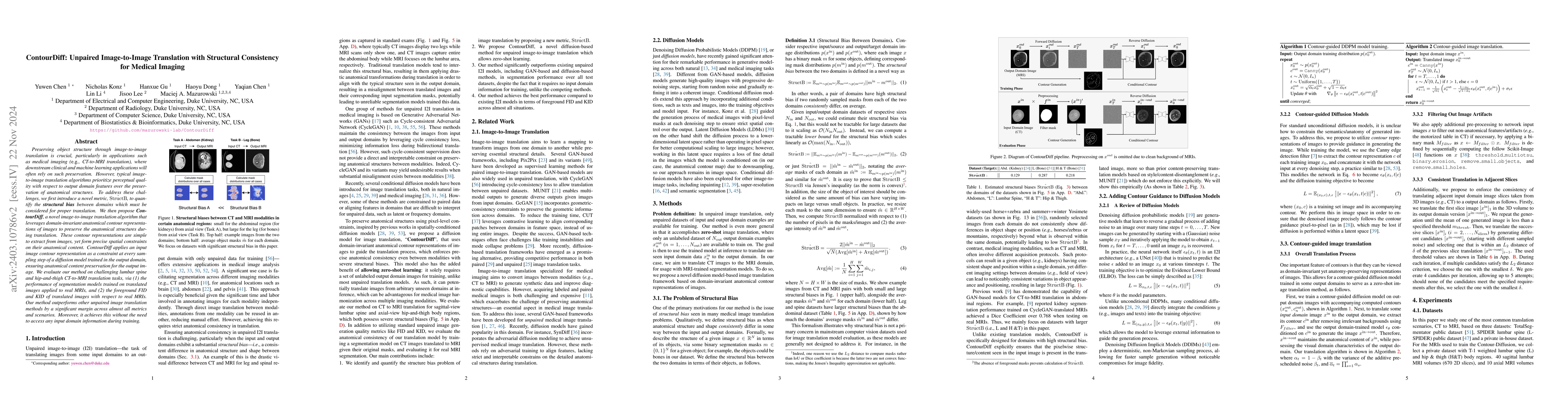
Accurately translating medical images across different modalities (e.g., CT to MRI) has numerous downstream clinical and machine learning applications. While several methods have been proposed to ac...
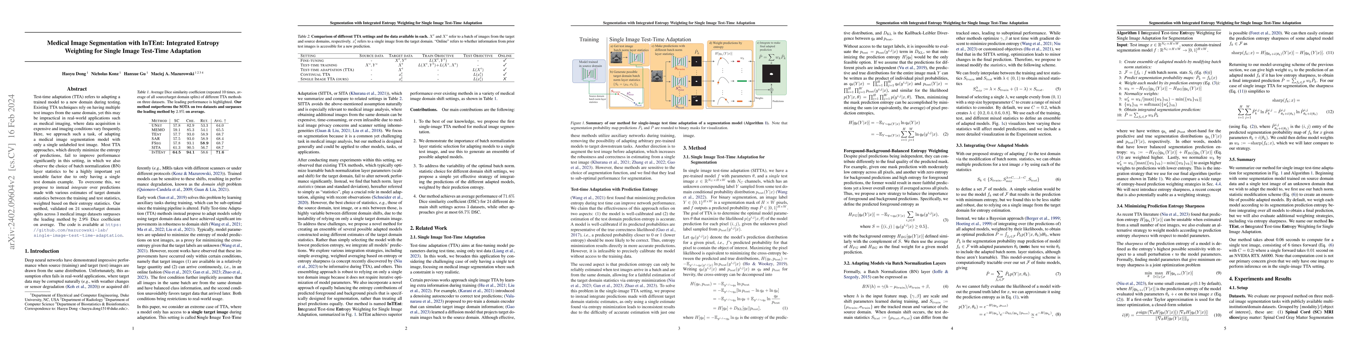
Test-time adaptation (TTA) refers to adapting a trained model to a new domain during testing. Existing TTA techniques rely on having multiple test images from the same domain, yet this may be imprac...
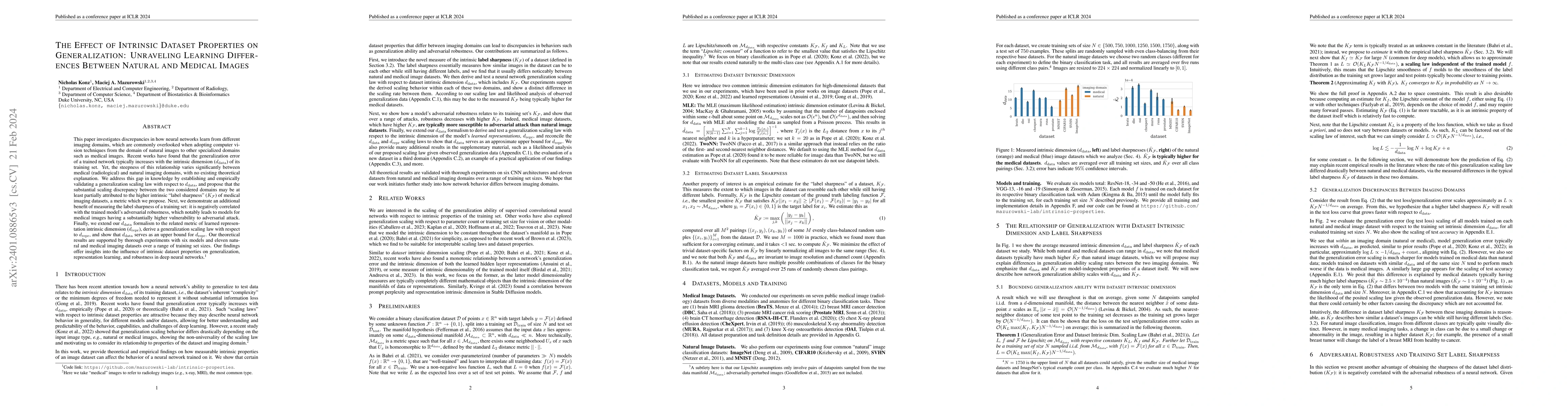
This paper investigates discrepancies in how neural networks learn from different imaging domains, which are commonly overlooked when adopting computer vision techniques from the domain of natural i...
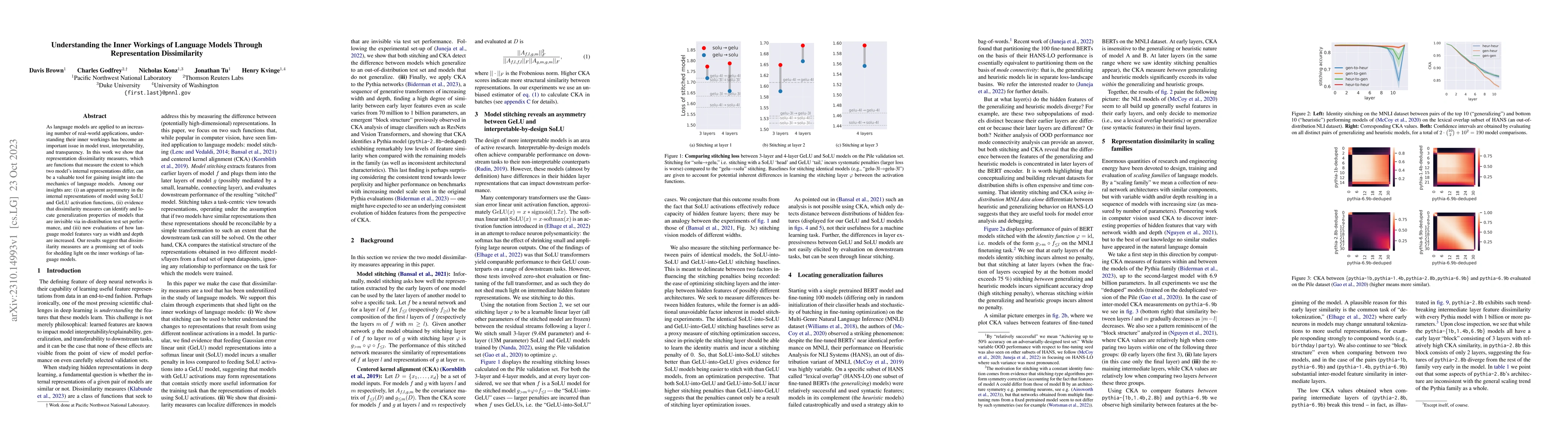
As language models are applied to an increasing number of real-world applications, understanding their inner workings has become an important issue in model trust, interpretability, and transparency...
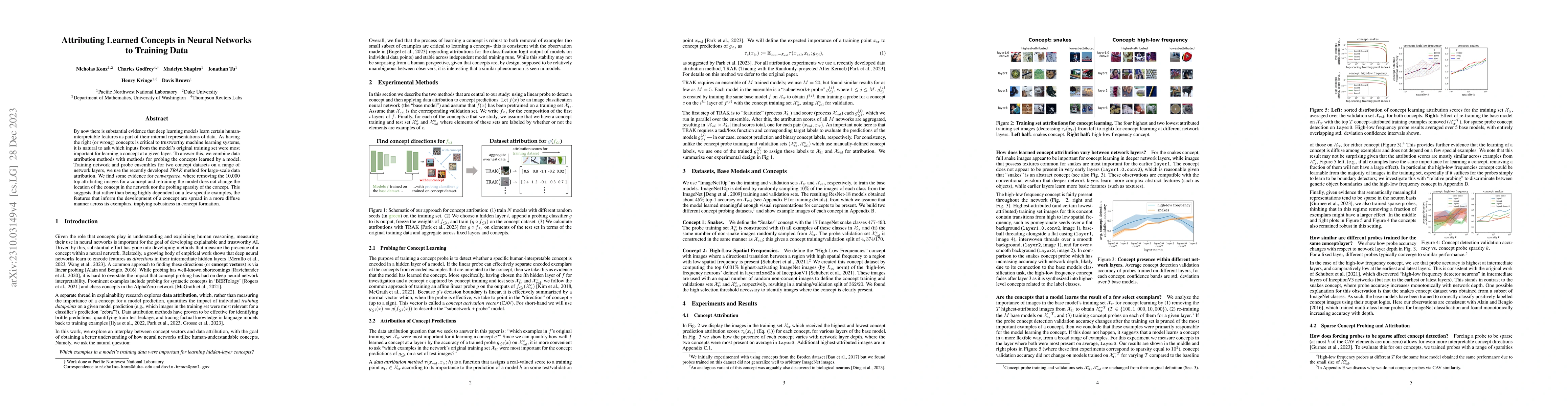
By now there is substantial evidence that deep learning models learn certain human-interpretable features as part of their internal representations of data. As having the right (or wrong) concepts i...
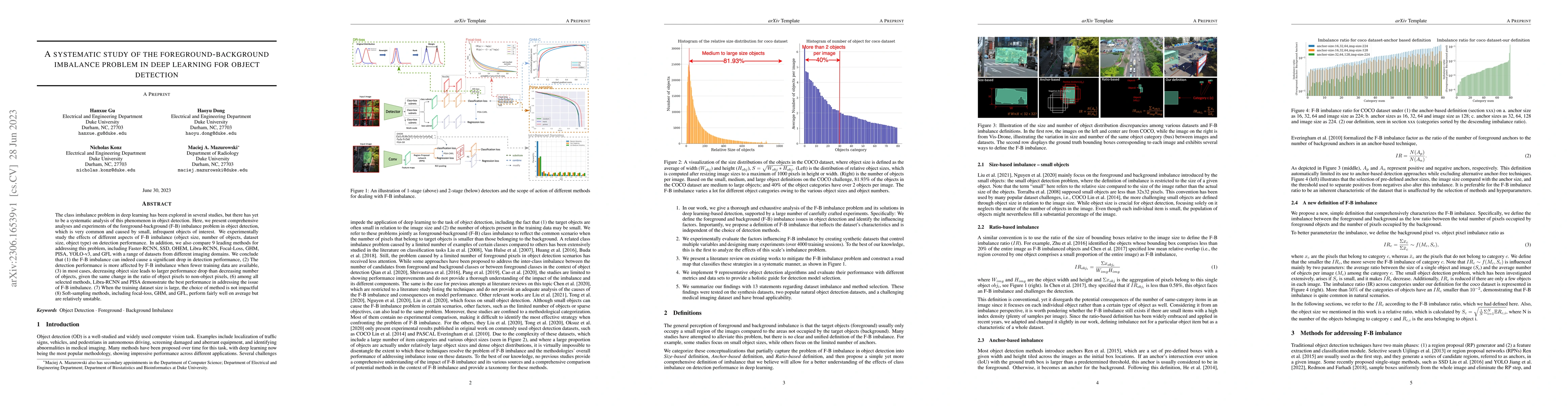
The class imbalance problem in deep learning has been explored in several studies, but there has yet to be a systematic analysis of this phenomenon in object detection. Here, we present comprehensiv...
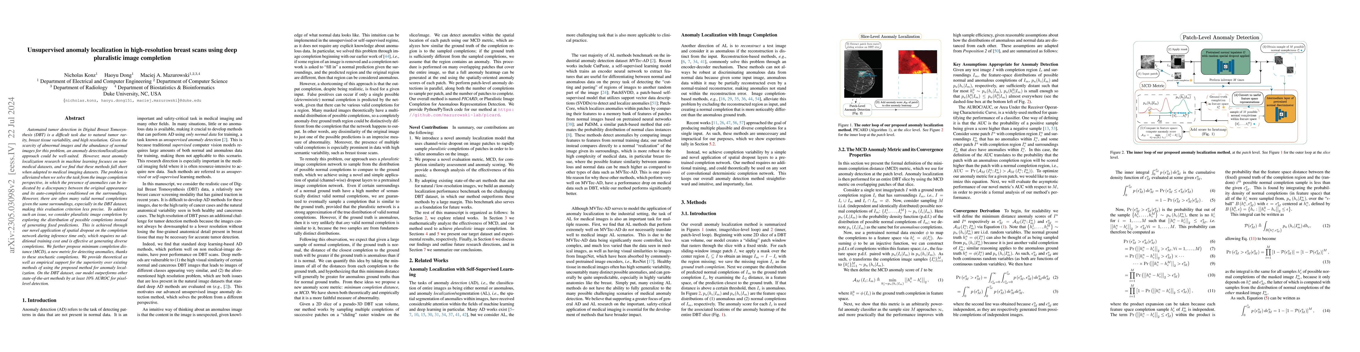
Automated tumor detection in Digital Breast Tomosynthesis (DBT) is a difficult task due to natural tumor rarity, breast tissue variability, and high resolution. Given the scarcity of abnormal images...
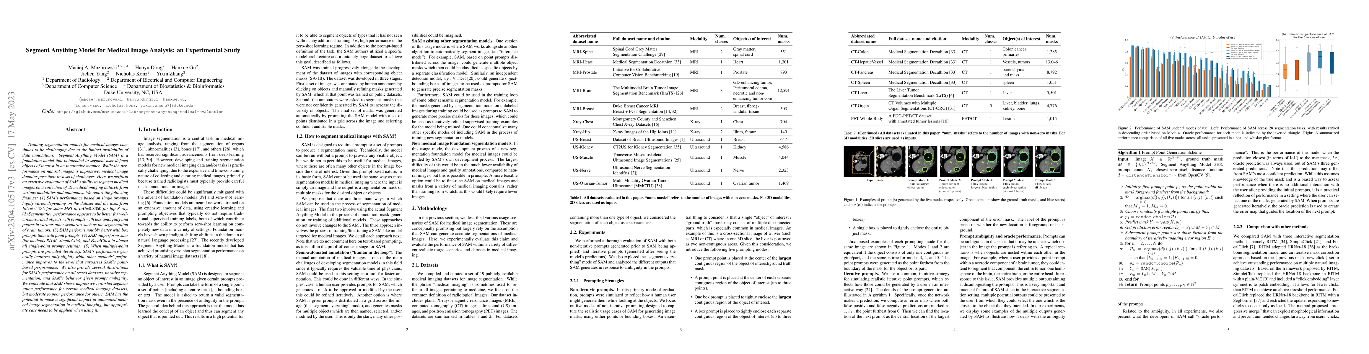
Training segmentation models for medical images continues to be challenging due to the limited availability of data annotations. Segment Anything Model (SAM) is a foundation model that is intended t...
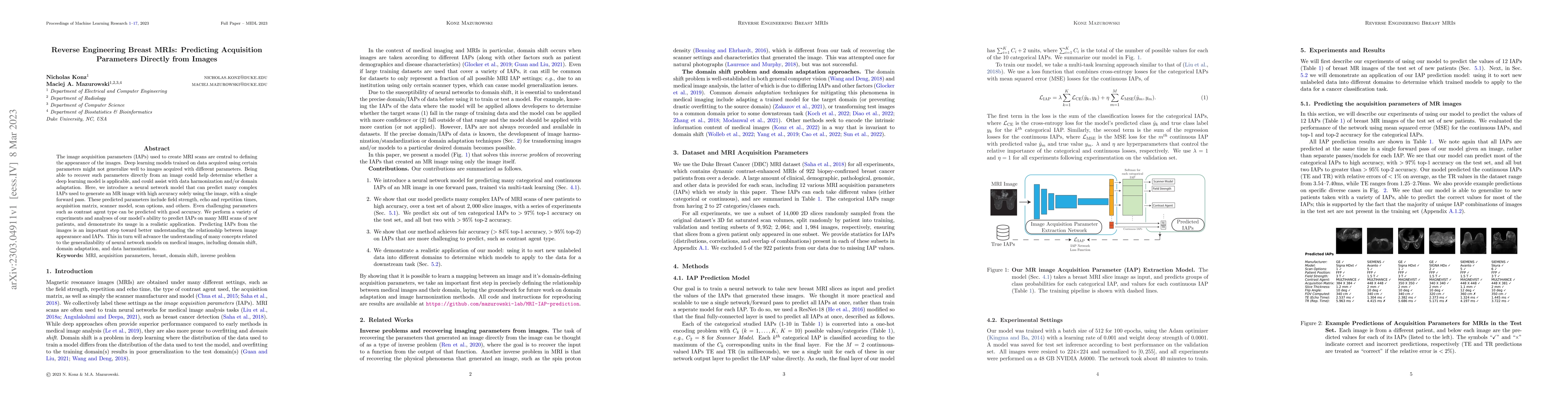
The image acquisition parameters (IAPs) used to create MRI scans are central to defining the appearance of the images. Deep learning models trained on data acquired using certain parameters might no...
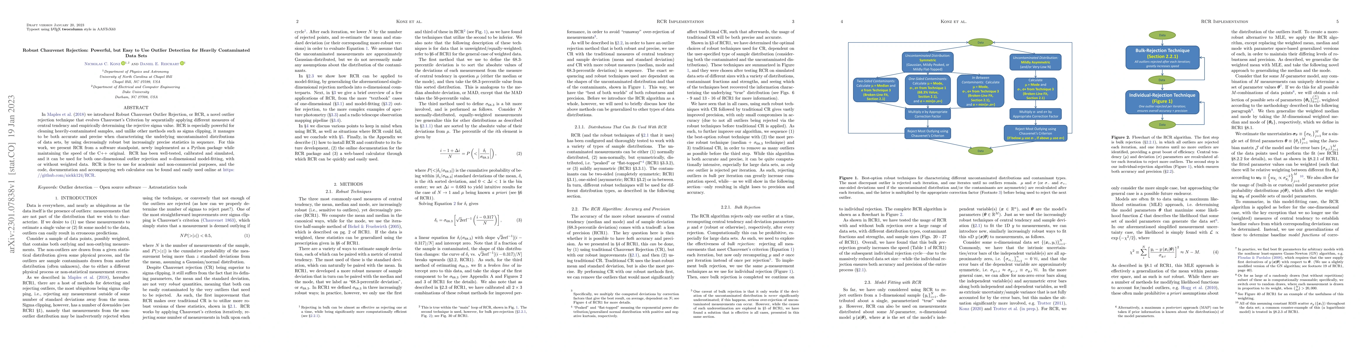
In Maples et al. (2018) we introduced Robust Chauvenet Outlier Rejection, or RCR, a novel outlier rejection technique that evolves Chauvenet's Criterion by sequentially applying different measures o...
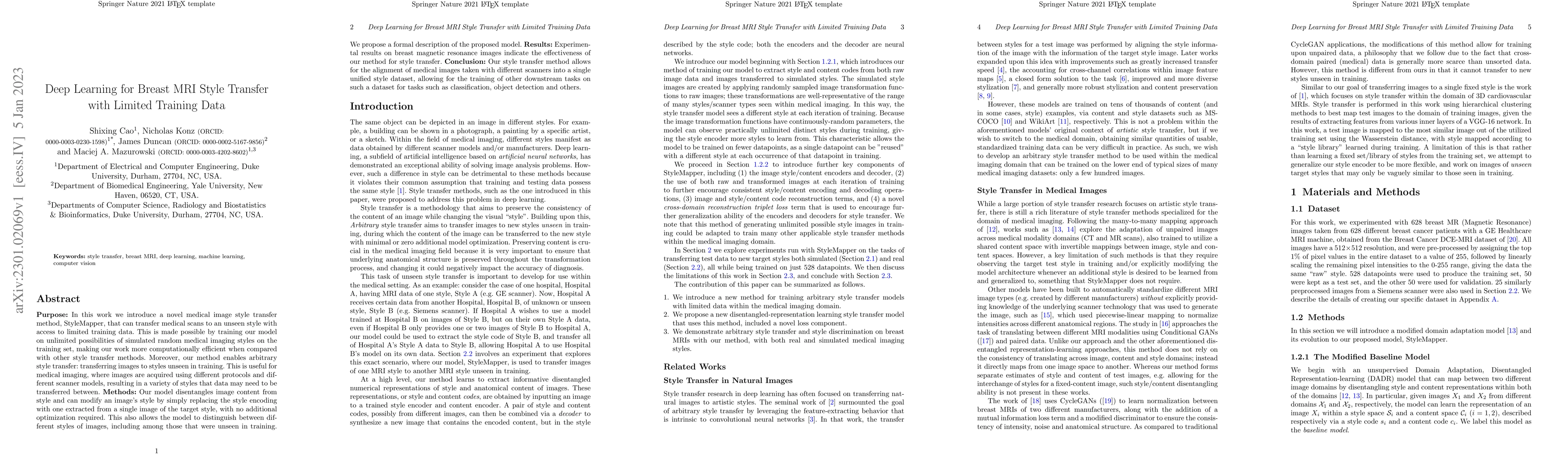
In this work we introduce a novel medical image style transfer method, StyleMapper, that can transfer medical scans to an unseen style with access to limited training data. This is made possible by ...
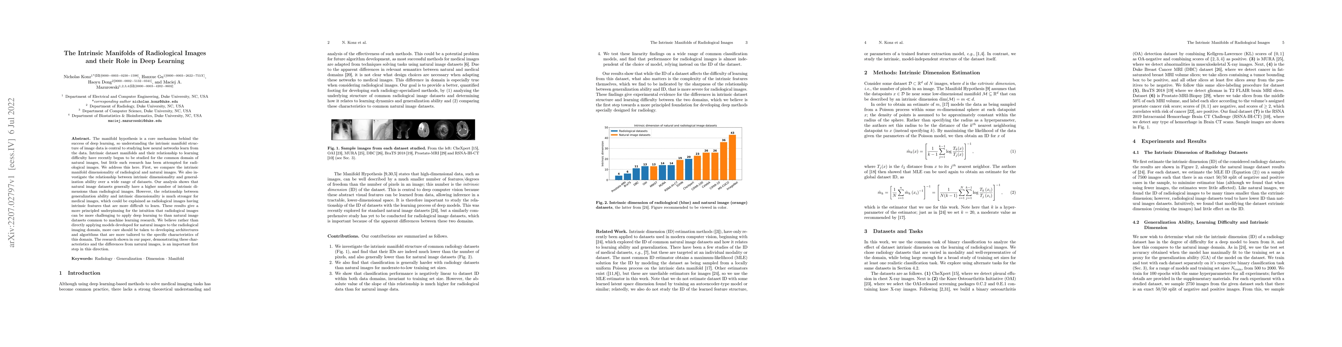
The manifold hypothesis is a core mechanism behind the success of deep learning, so understanding the intrinsic manifold structure of image data is central to studying how neural networks learn from...
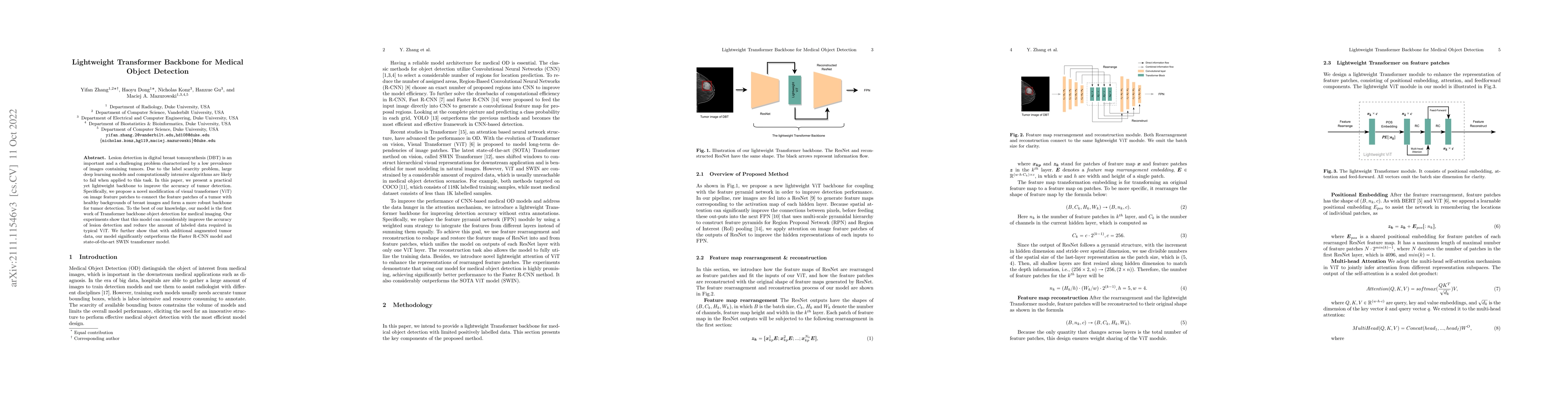
Lesion detection in digital breast tomosynthesis (DBT) is an important and a challenging problem characterized by a low prevalence of images containing tumors. Due to the label scarcity problem, lar...
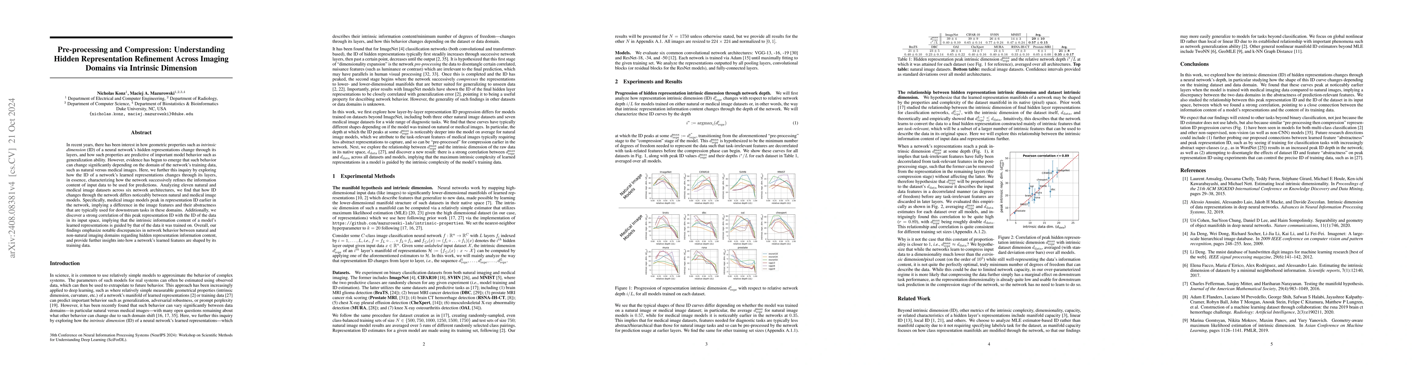
In recent years, there has been interest in how geometric properties such as intrinsic dimension (ID) of a neural network's hidden representations evolve through its layers, and how such properties ar...
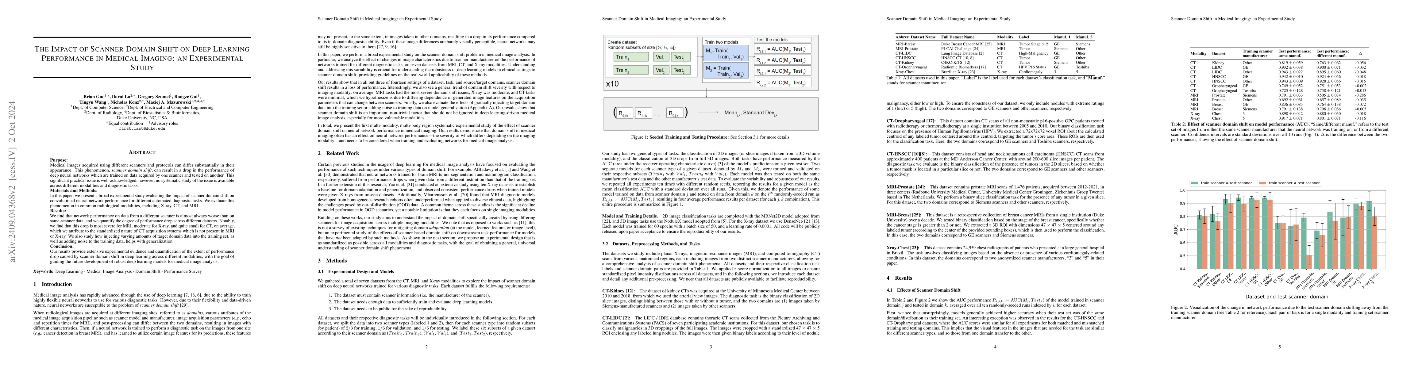
Purpose: Medical images acquired using different scanners and protocols can differ substantially in their appearance. This phenomenon, scanner domain shift, can result in a drop in the performance of ...
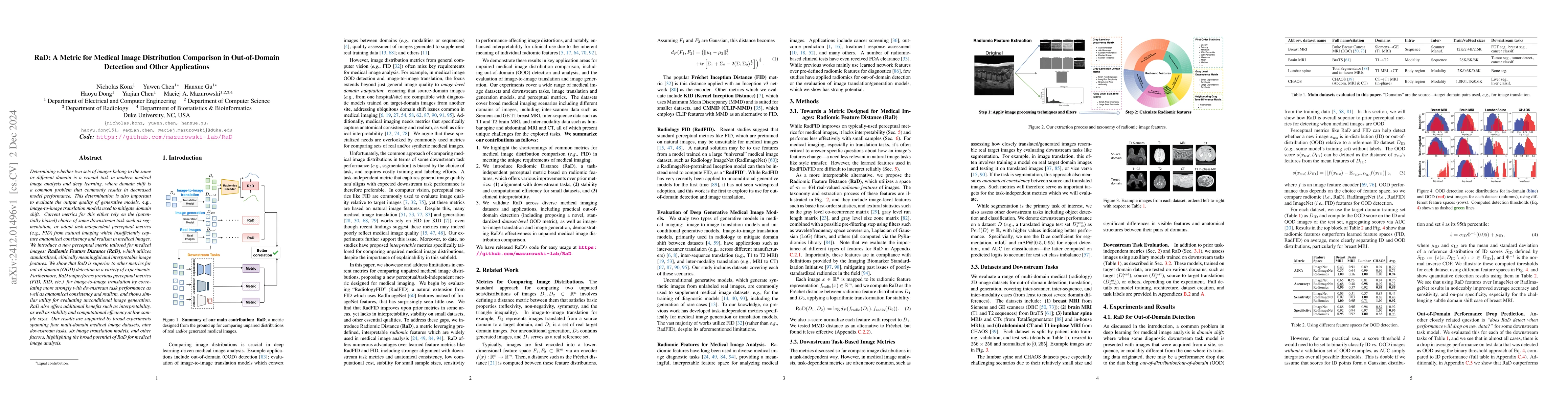
Determining whether two sets of images belong to the same or different domain is a crucial task in modern medical image analysis and deep learning, where domain shift is a common problem that commonly...
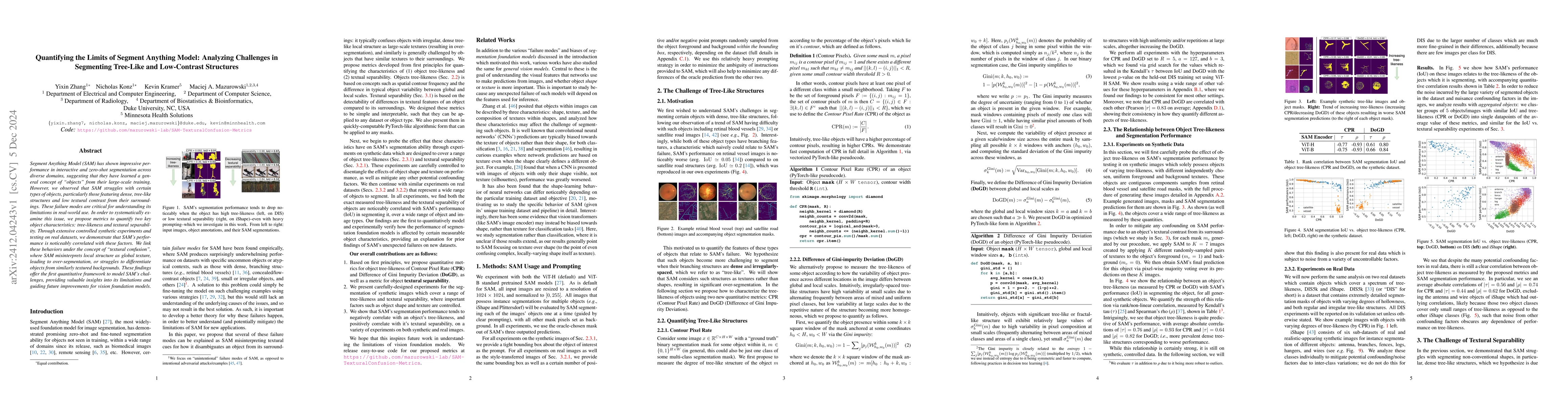
Segment Anything Model (SAM) has shown impressive performance in interactive and zero-shot segmentation across diverse domains, suggesting that they have learned a general concept of "objects" from th...
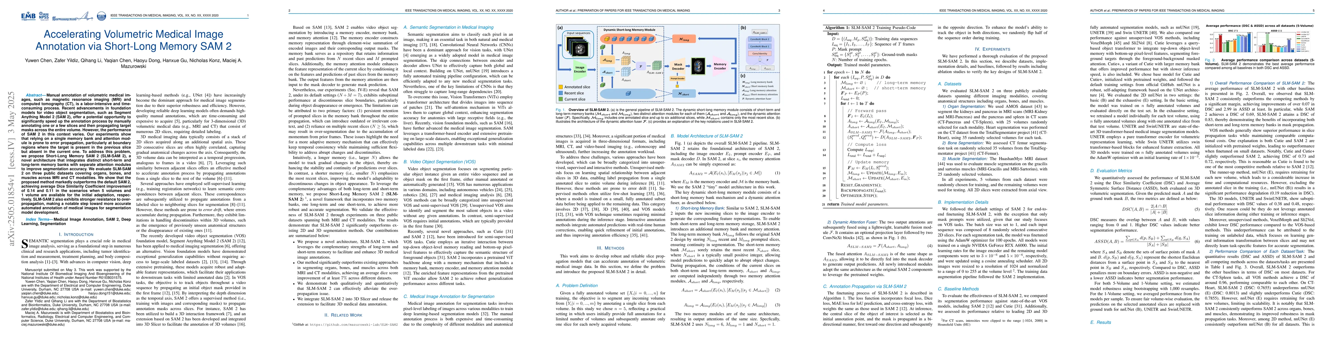
Manual annotation of volumetric medical images, such as magnetic resonance imaging (MRI) and computed tomography (CT), is a labor-intensive and time-consuming process. Recent advancements in foundatio...
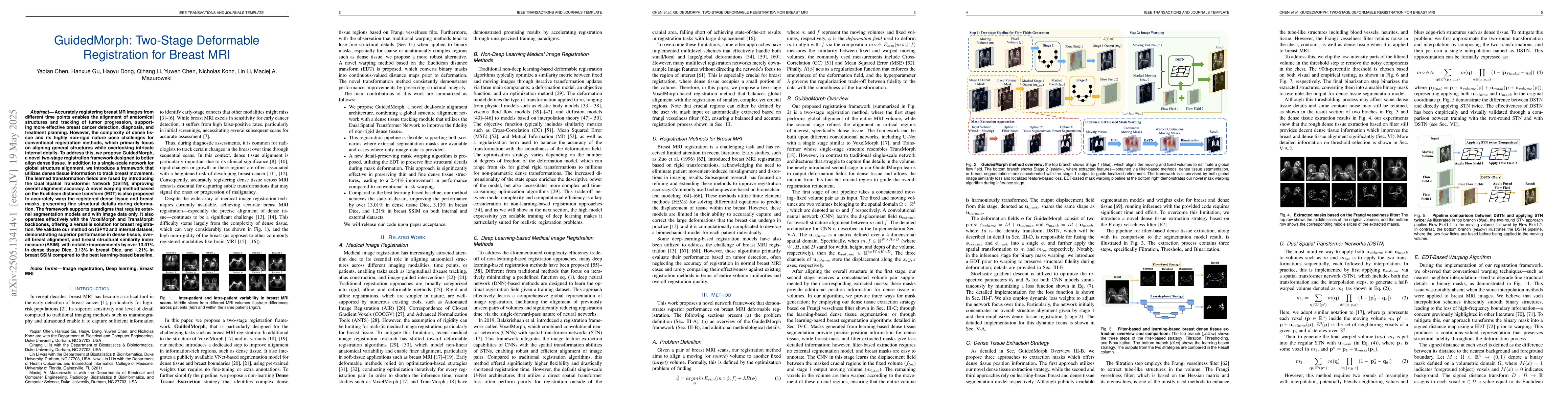
Accurately registering breast MR images from different time points enables the alignment of anatomical structures and tracking of tumor progression, supporting more effective breast cancer detection, ...
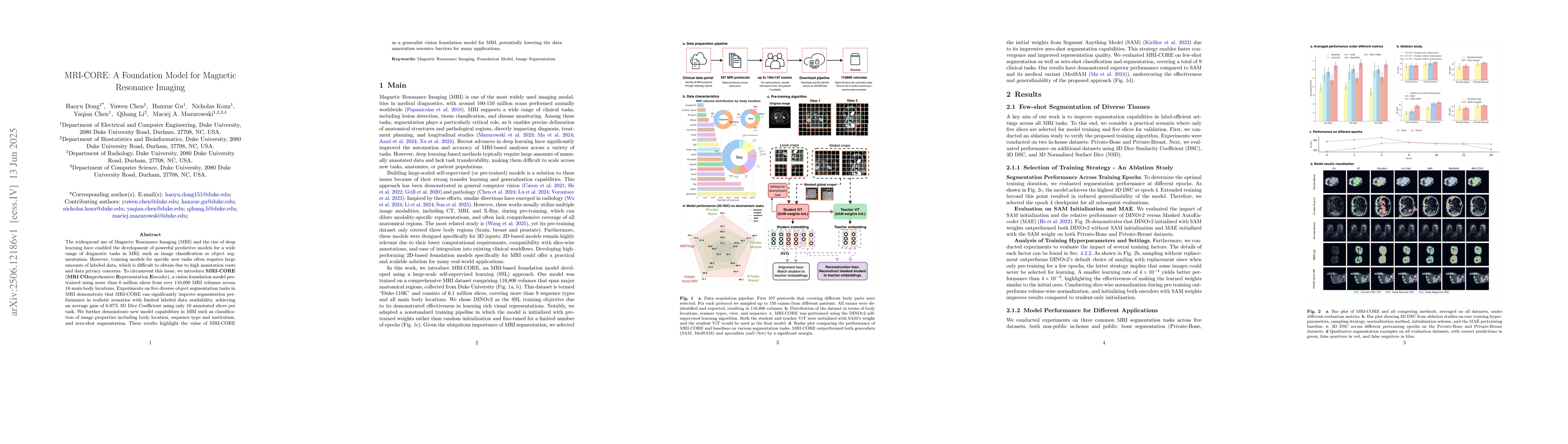
The widespread use of Magnetic Resonance Imaging (MRI) and the rise of deep learning have enabled the development of powerful predictive models for a wide range of diagnostic tasks in MRI, such as ima...
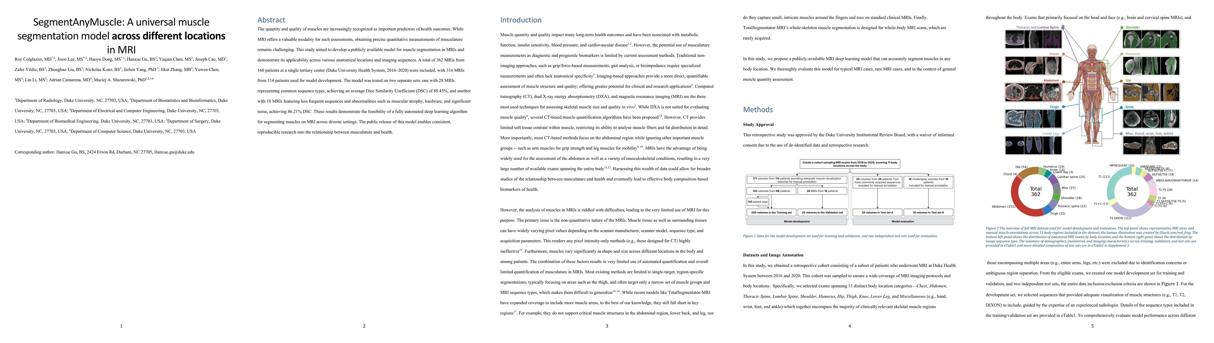
The quantity and quality of muscles are increasingly recognized as important predictors of health outcomes. While MRI offers a valuable modality for such assessments, obtaining precise quantitative me...
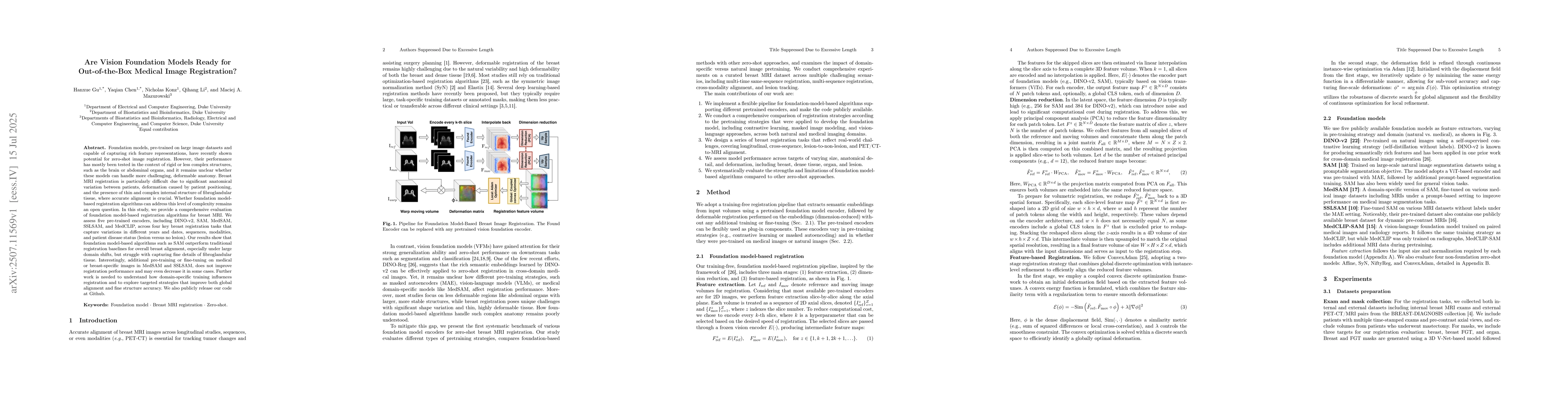
Foundation models, pre-trained on large image datasets and capable of capturing rich feature representations, have recently shown potential for zero-shot image registration. However, their performance...
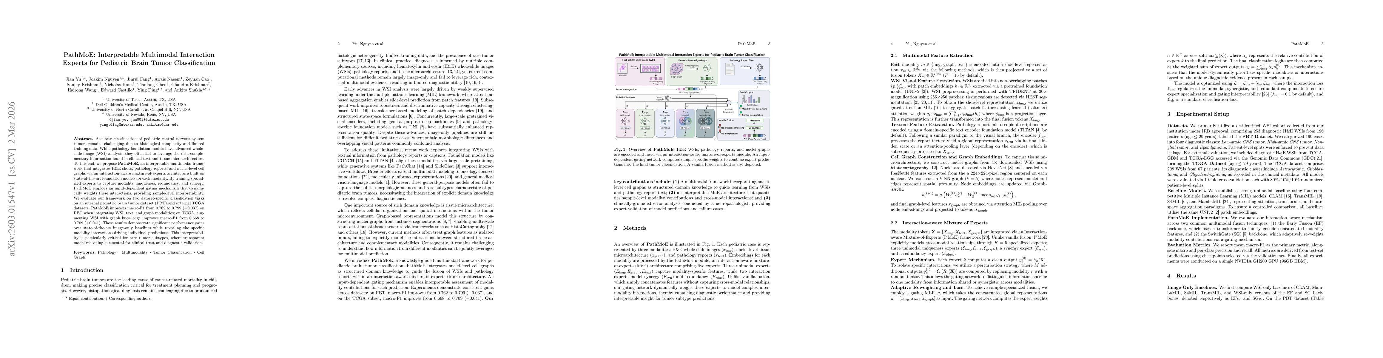
Accurate classification of pediatric central nervous system tumors remains challenging due to histological complexity and limited training data. While pathology foundation models have advanced whole-s...
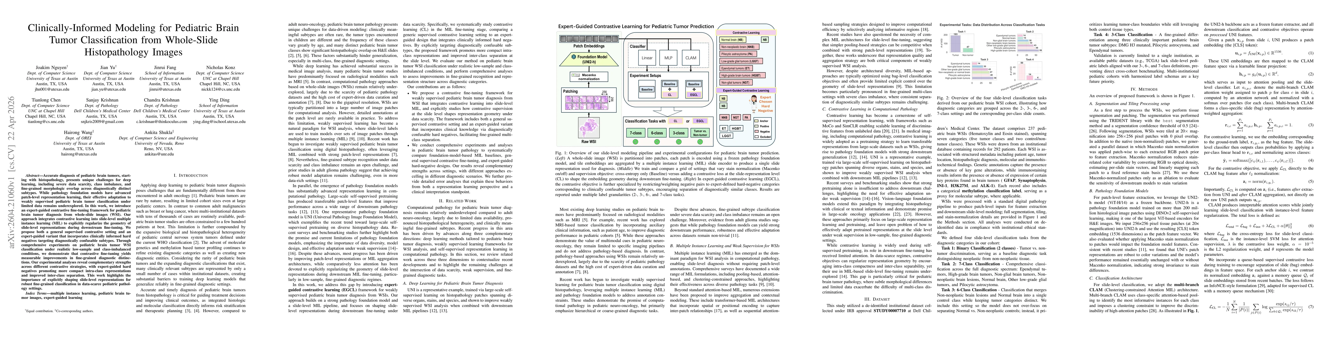
Accurate diagnosis of pediatric brain tumors, starting with histopathology, presents unique challenges for deep learning, including severe data scarcity, class imbalance, and fine-grained morphologic ...
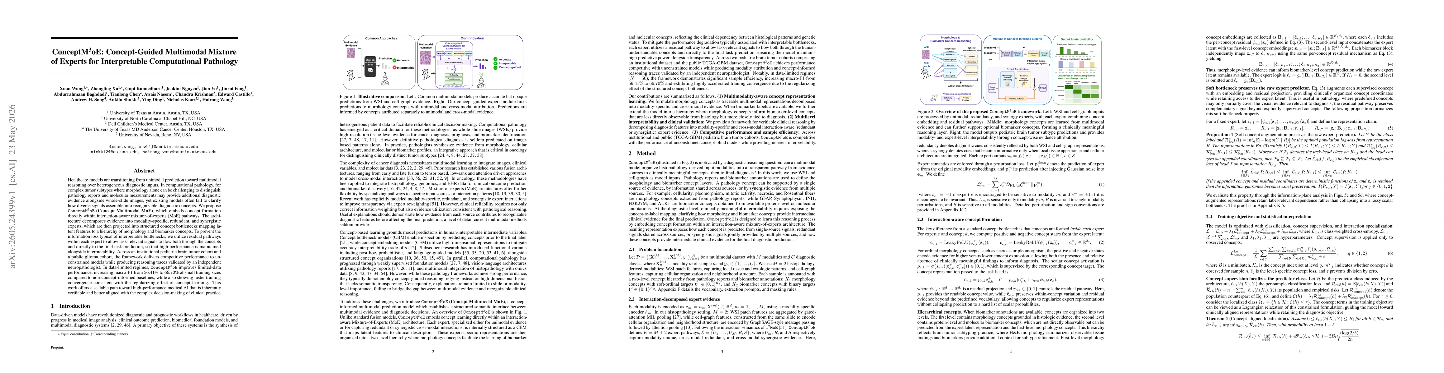
Healthcare models are transitioning from unimodal prediction toward multimodal reasoning over heterogeneous diagnostic inputs. In computational pathology, for complex tumor subtypes where morphology a...