ArxivLens
O
Researcher

Oana Dumitrascu

9 papers on arXiv

9 arXiv Papers
21 Total Publications
Profile

Academic Profile

Name Oana M. Dumitrascu
Total Papers 21
Scholar Profile View on Semantic Scholar
Metrics

Statistics

9 arXiv Papers
21 Total Publications
Publications

Papers on arXiv

Thumbnail for RBAD: A Dataset and Benchmark for Retinal Vessels Branching Angle
  Detection
arXiv
RBAD: A Dataset and Benchmark for Retinal Vessels Branching Angle Detection

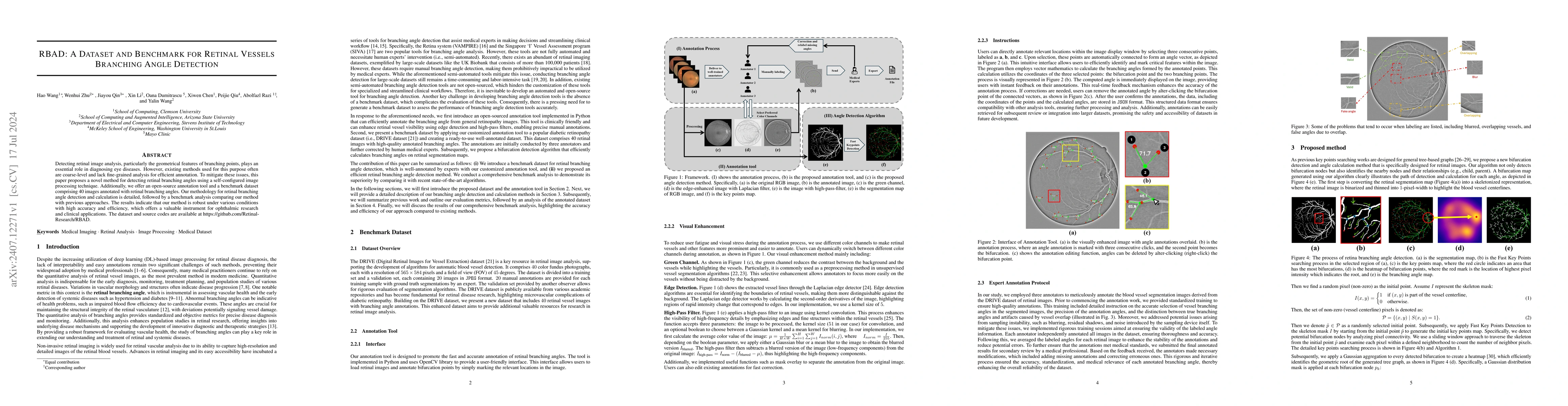
Detecting retinal image analysis, particularly the geometrical features of branching points, plays an essential role in diagnosing eye diseases. However, existing methods used for this purpose often a...

Thumbnail for Context-Aware Optimal Transport Learning for Retinal Fundus Image
  Enhancement
arXiv
Context-Aware Optimal Transport Learning for Retinal Fundus Image Enhancement

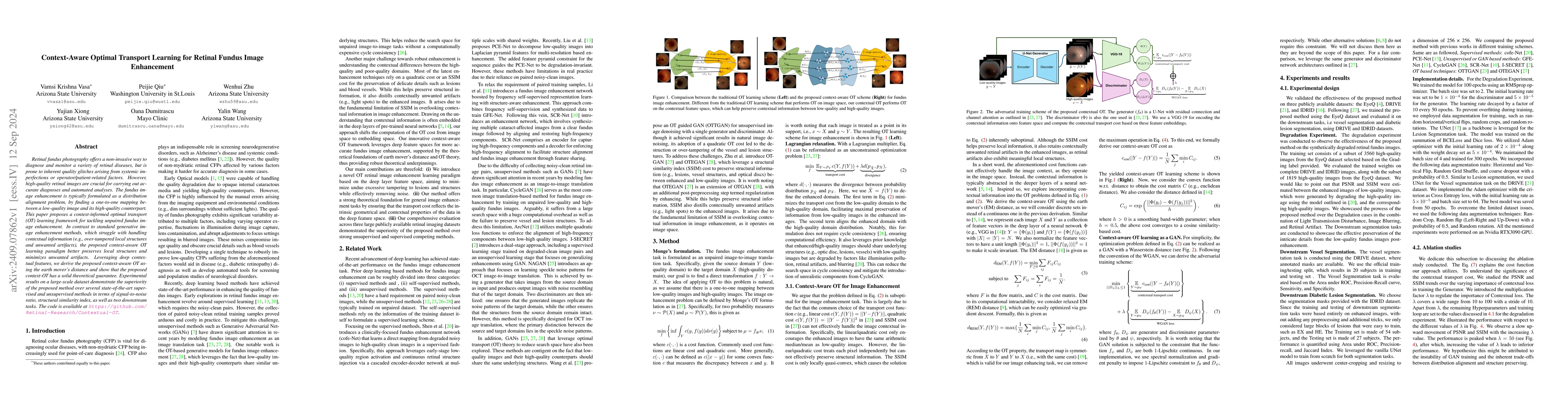
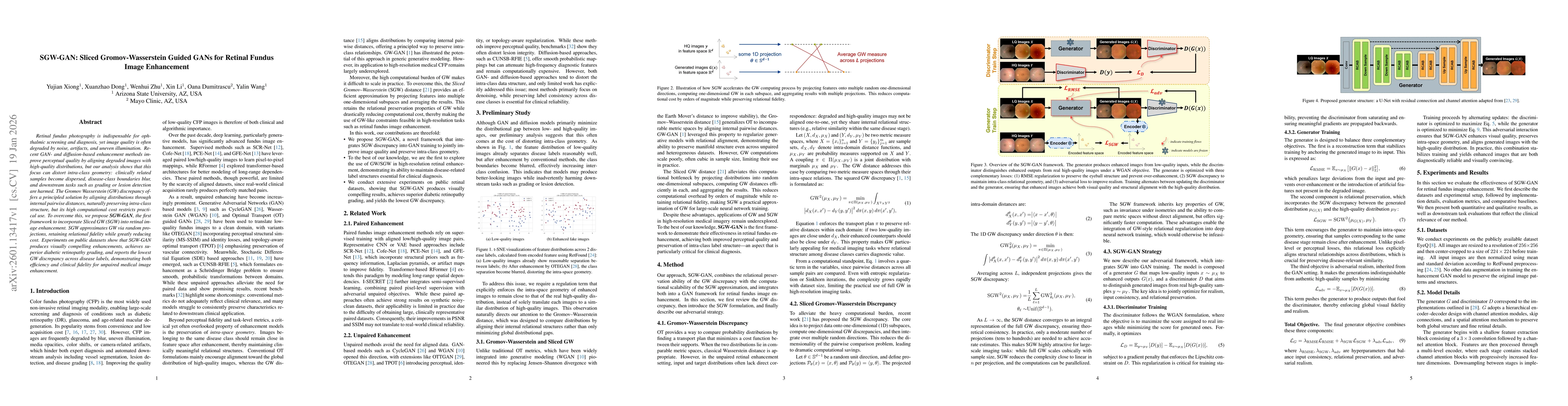
Retinal fundus photography offers a non-invasive way to diagnose and monitor a variety of retinal diseases, but is prone to inherent quality glitches arising from systemic imperfections or operator/pa...

Thumbnail for EyeBench: A Call for More Rigorous Evaluation of Retinal Image
  Enhancement
arXiv
EyeBench: A Call for More Rigorous Evaluation of Retinal Image Enhancement

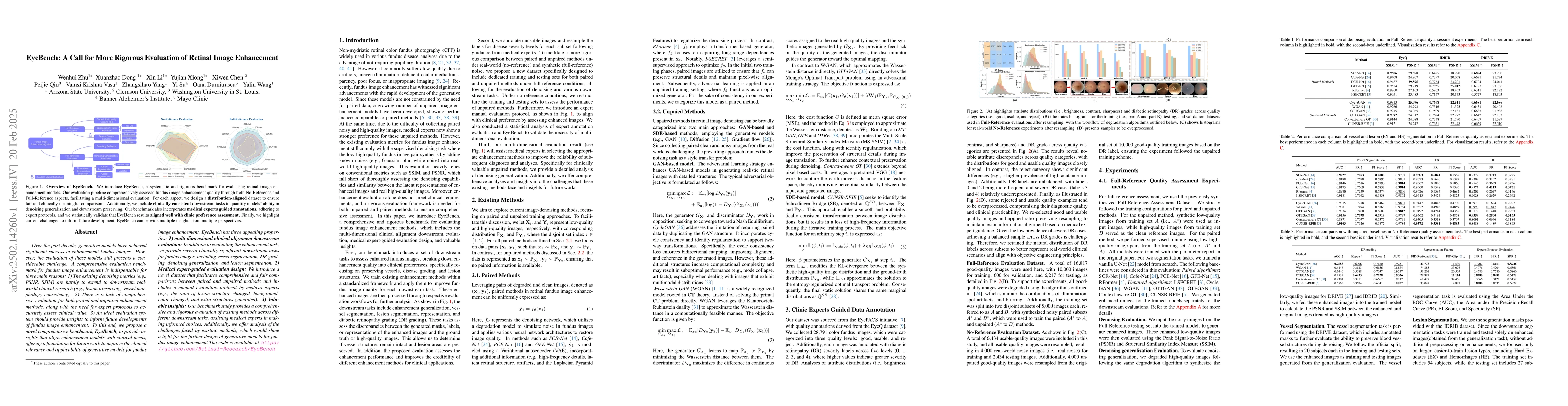
Over the past decade, generative models have achieved significant success in enhancement fundus images.However, the evaluation of these models still presents a considerable challenge. A comprehensive ...

Thumbnail for RetinalGPT: A Retinal Clinical Preference Conversational Assistant
  Powered by Large Vision-Language Models
arXiv
RetinalGPT: A Retinal Clinical Preference Conversational Assistant Powered by Large Vision-Language Models

Recently, Multimodal Large Language Models (MLLMs) have gained significant attention for their remarkable ability to process and analyze non-textual data, such as images, videos, and audio. Notably, s...

Thumbnail for VAOT: Vessel-Aware Optimal Transport for Retinal Fundus Enhancement
arXiv
VAOT: Vessel-Aware Optimal Transport for Retinal Fundus Enhancement

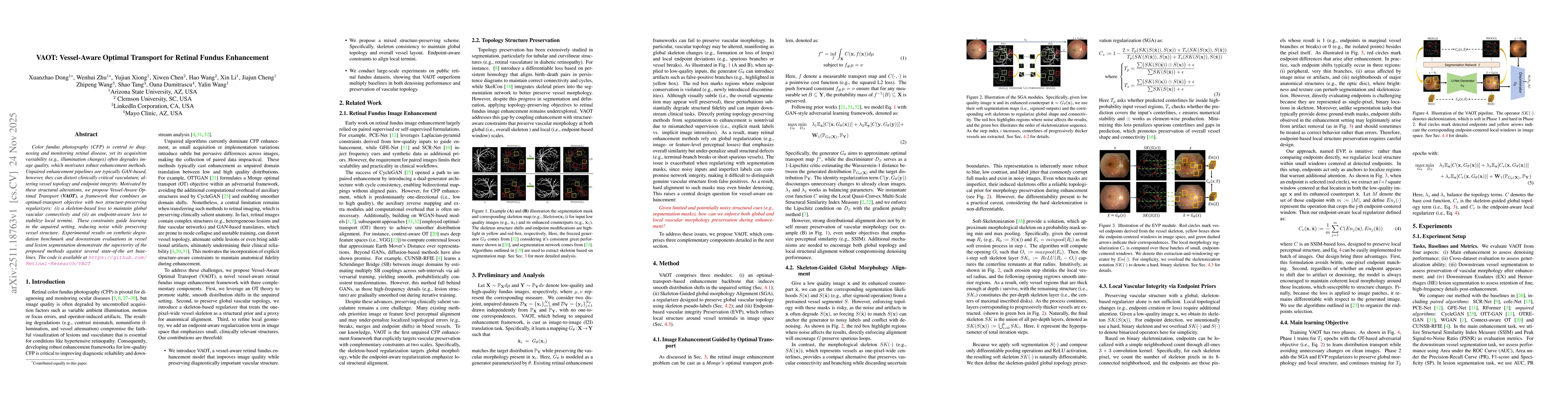
Color fundus photography (CFP) is central to diagnosing and monitoring retinal disease, yet its acquisition variability (e.g., illumination changes) often degrades image quality, which motivates robus...

Thumbnail for nnMobileNet++: Towards Efficient Hybrid Networks for Retinal Image Analysis
arXiv
nnMobileNet++: Towards Efficient Hybrid Networks for Retinal Image Analysis

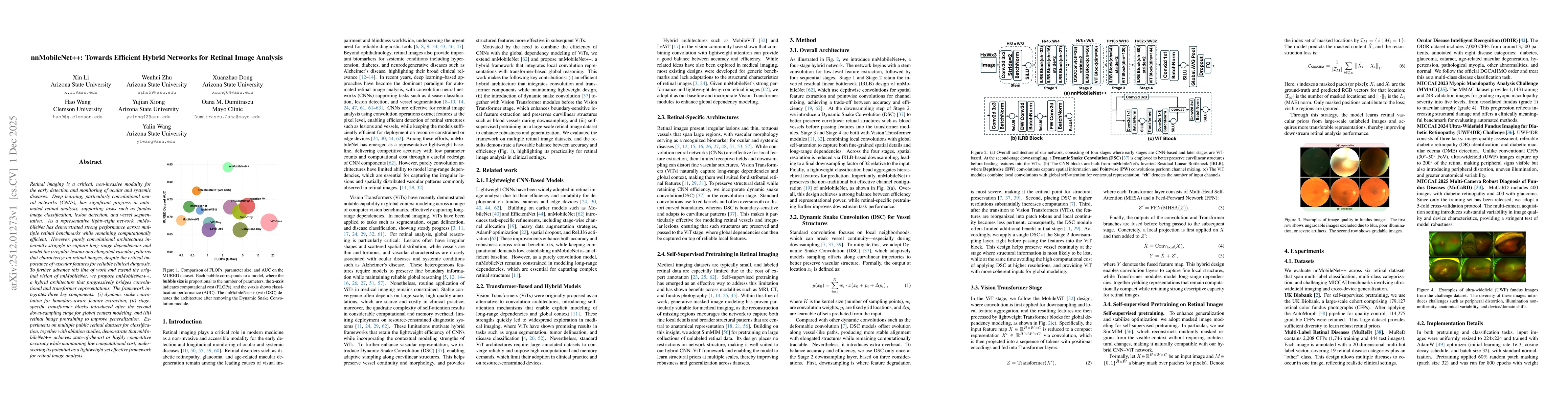
Retinal imaging is a critical, non-invasive modality for the early detection and monitoring of ocular and systemic diseases. Deep learning, particularly convolutional neural networks (CNNs), has signi...

Thumbnail for SGW-GAN: Sliced Gromov-Wasserstein Guided GANs for Retinal Fundus Image Enhancement
arXiv
SGW-GAN: Sliced Gromov-Wasserstein Guided GANs for Retinal Fundus Image Enhancement

Retinal fundus photography is indispensable for ophthalmic screening and diagnosis, yet image quality is often degraded by noise, artifacts, and uneven illumination. Recent GAN- and diffusion-based en...

Thumbnail for Bridging Restoration and Diagnosis: A Comprehensive Benchmark for Retinal Fundus Enhancement
arXiv
Bridging Restoration and Diagnosis: A Comprehensive Benchmark for Retinal Fundus Enhancement

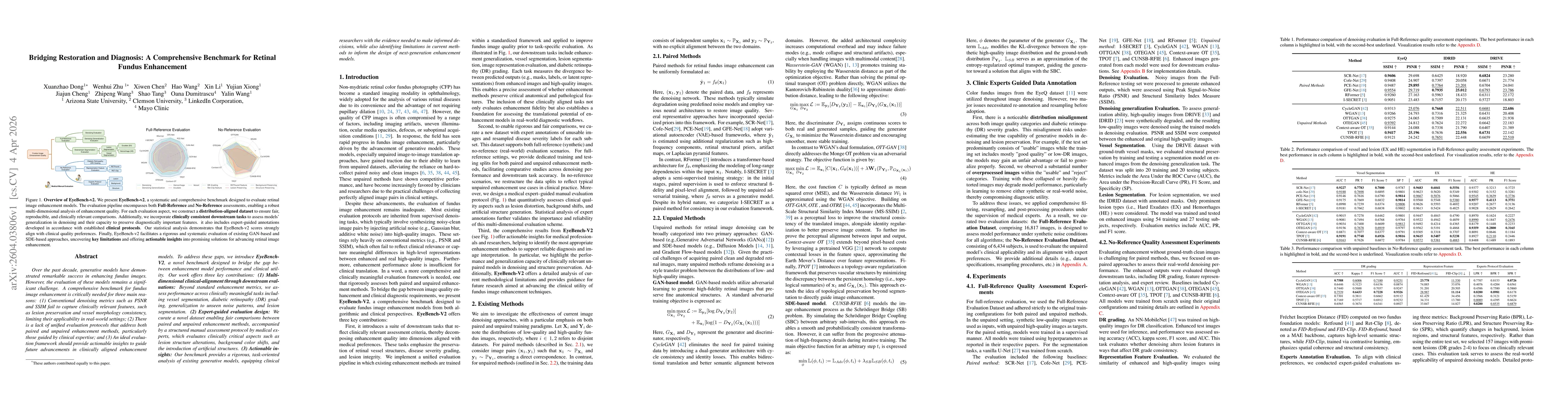
Over the past decade, generative models have demonstrated success in enhancing fundus images. However, the evaluation of these models remains a challenge. A benchmark for fundus image enhancement is n...

Thumbnail for OphIn-500K: Curating Web-Scale Visual Instructions for Scaling Ophthalmic Multimodal Large Language Models
arXiv
OphIn-500K: Curating Web-Scale Visual Instructions for Scaling Ophthalmic Multimodal Large Language Models

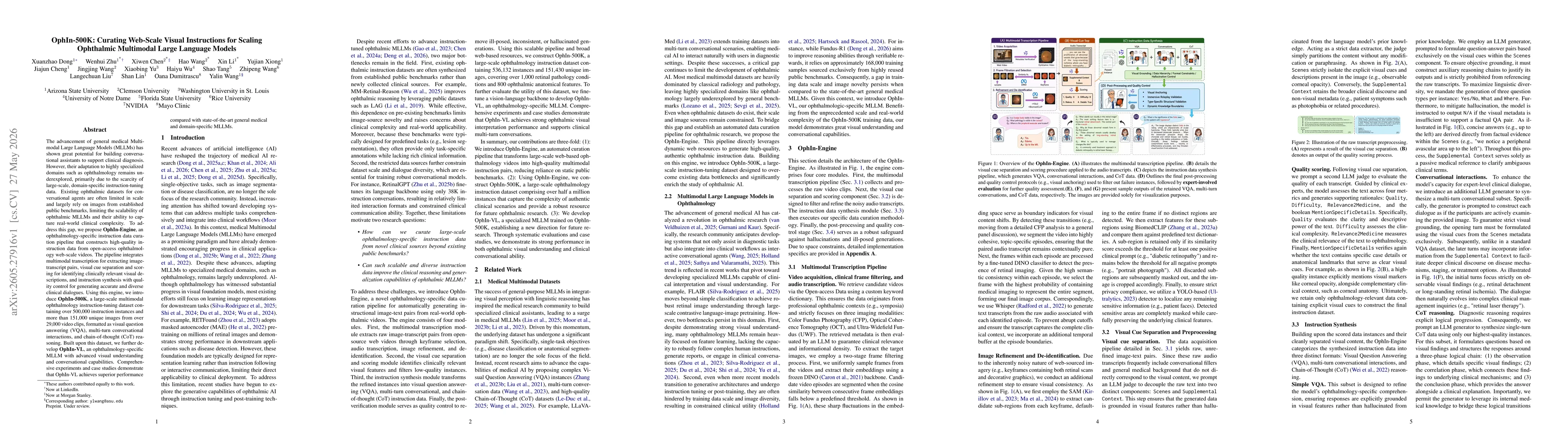
The advancement of general medical Multimodal Large Language Models (MLLMs) has shown great potential for building conversational assistants to support clinical diagnosis. However, their adaptation to...

This website uses cookies to ensure you get the best experience. Learn more