Ole Solheim

8 papers on arXiv

Academic Profile

Name: O. Solheim
Total Papers: 4

Statistics

arXiv Papers 8
Total Publications 4

Papers on arXiv

Thumbnail for Postoperative glioblastoma segmentation: Development of a fully
  automated pipeline using deep convolutional neural networks and comparison
  with currently available models

Postoperative glioblastoma segmentation: Development of a fully automated pipeline using deep convolutional neural networks and comparison with currently available models

Accurately assessing tumor removal is paramount in the management of glioblastoma. We developed a pipeline using MRI scans and neural networks to segment tumor subregions and the surgical cavity in ...

Thumbnail for Raidionics: an open software for pre- and postoperative central nervous
  system tumor segmentation and standardized reporting

Raidionics: an open software for pre- and postoperative central nervous system tumor segmentation and standardized reporting

For patients suffering from central nervous system tumors, prognosis estimation, treatment decisions, and postoperative assessments are made from the analysis of a set of magnetic resonance (MR) sca...

Thumbnail for Segmentation of glioblastomas in early post-operative multi-modal MRI
  with deep neural networks

Segmentation of glioblastomas in early post-operative multi-modal MRI with deep neural networks

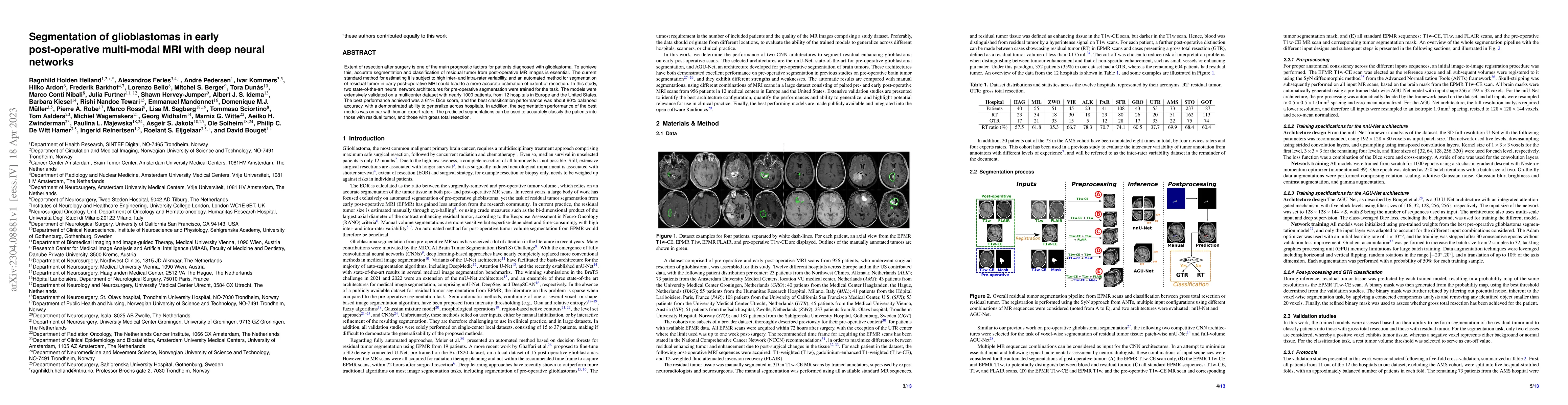
Extent of resection after surgery is one of the main prognostic factors for patients diagnosed with glioblastoma. To achieve this, accurate segmentation and classification of residual tumor from pos...

Thumbnail for RESECT-SEG: Open access annotations of intra-operative brain tumor
  ultrasound images

RESECT-SEG: Open access annotations of intra-operative brain tumor ultrasound images

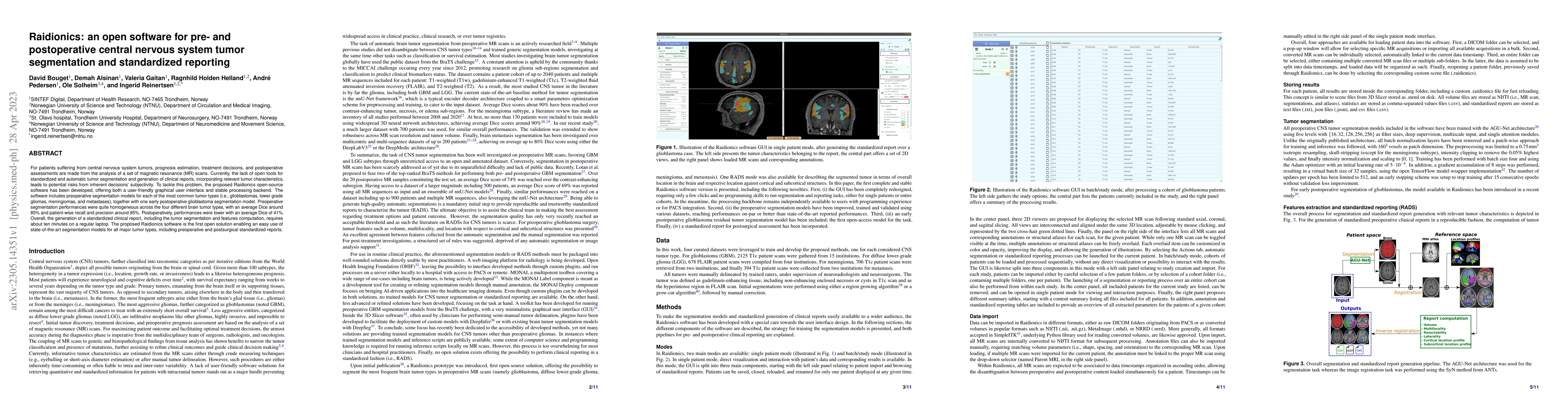
Purpose: Registration and segmentation of magnetic resonance (MR) and ultrasound (US) images play an essential role in surgical planning and resection of brain tumors. However, validating these tech...

Thumbnail for Meningioma segmentation in T1-weighted MRI leveraging global context and
  attention mechanisms

Meningioma segmentation in T1-weighted MRI leveraging global context and attention mechanisms

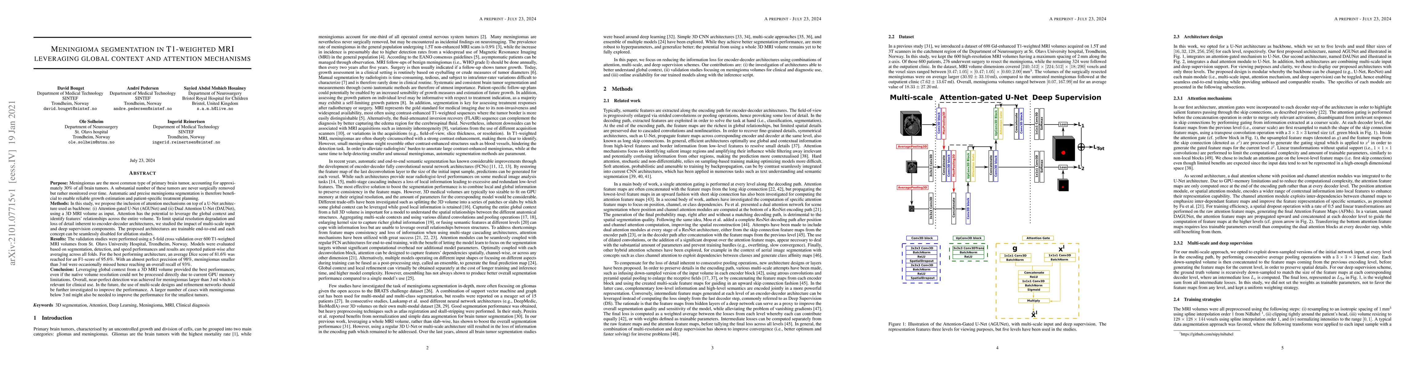
Meningiomas are the most common type of primary brain tumor, accounting for approximately 30% of all brain tumors. A substantial number of these tumors are never surgically removed but rather monito...

Thumbnail for Fast meningioma segmentation in T1-weighted MRI volumes using a
  lightweight 3D deep learning architecture

Fast meningioma segmentation in T1-weighted MRI volumes using a lightweight 3D deep learning architecture

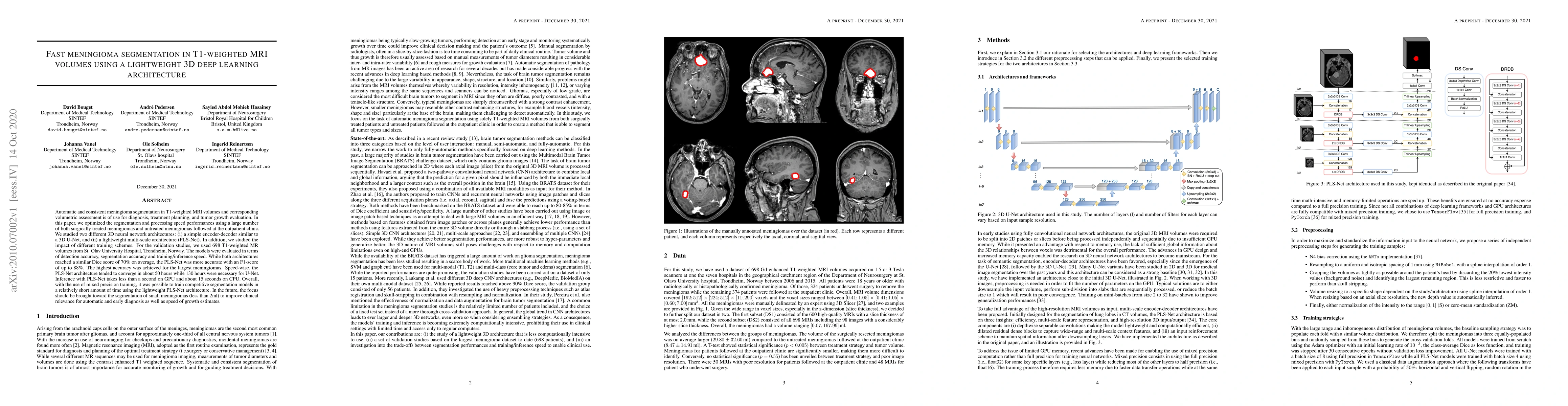
Automatic and consistent meningioma segmentation in T1-weighted MRI volumes and corresponding volumetric assessment is of use for diagnosis, treatment planning, and tumor growth evaluation. In this ...

Thumbnail for Automatic brain tumor segmentation in 2D intra-operative ultrasound
  images using MRI tumor annotations

Automatic brain tumor segmentation in 2D intra-operative ultrasound images using MRI tumor annotations

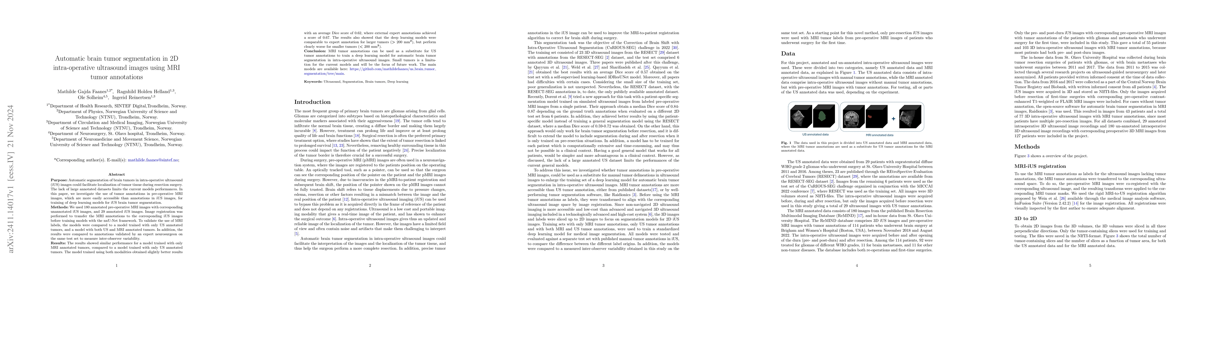
Automatic segmentation of brain tumors in intra-operative ultrasound (iUS) images could facilitate localization of tumor tissue during resection surgery. The lack of large annotated datasets limits th...

Thumbnail for A unified FLAIR hyperintensity segmentation model for various CNS tumor types and acquisition time points

A unified FLAIR hyperintensity segmentation model for various CNS tumor types and acquisition time points

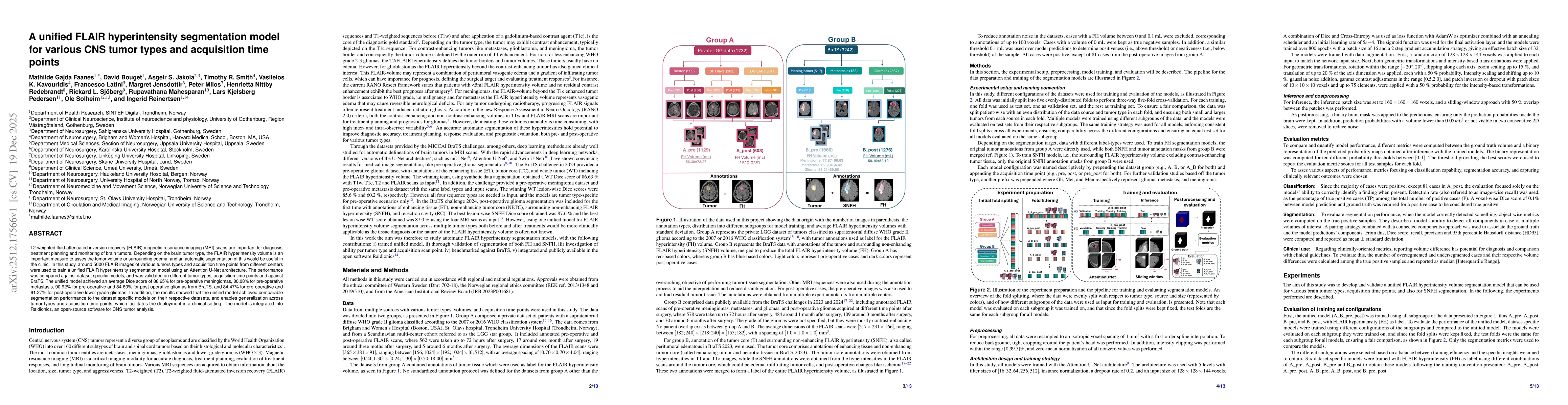
T2-weighted fluid-attenuated inversion recovery (FLAIR) magnetic resonance imaging (MRI) scans are important for diagnosis, treatment planning and monitoring of brain tumors. Depending on the brain tu...

This website uses cookies to ensure you get the best experience. Learn more