Academic Profile
Statistics
Similar Authors
Papers on arXiv
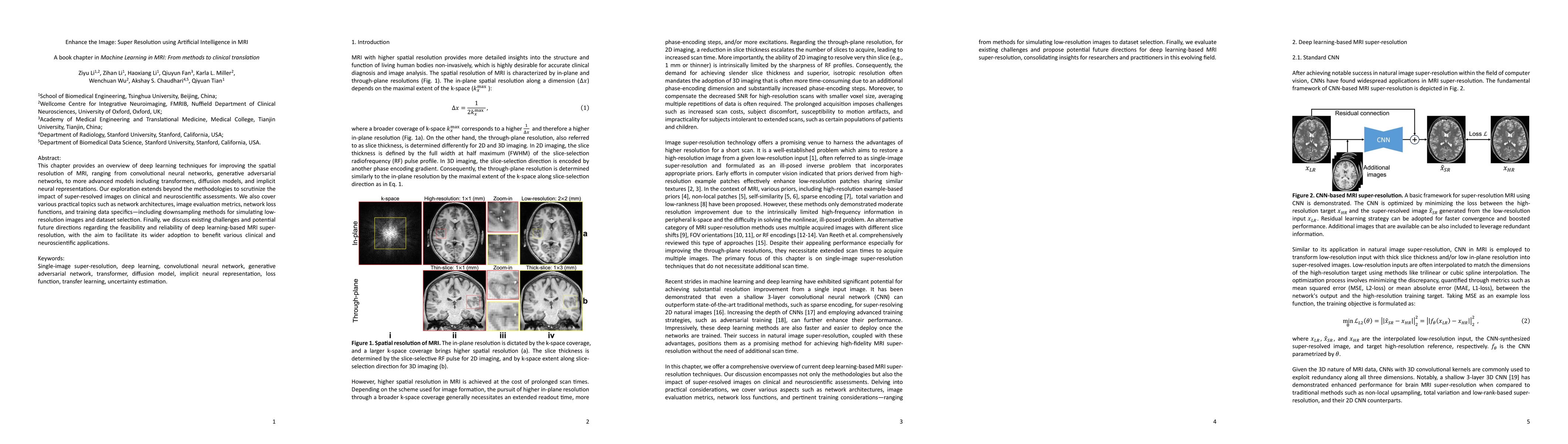
This chapter provides an overview of deep learning techniques for improving the spatial resolution of MRI, ranging from convolutional neural networks, generative adversarial networks, to more advanced...
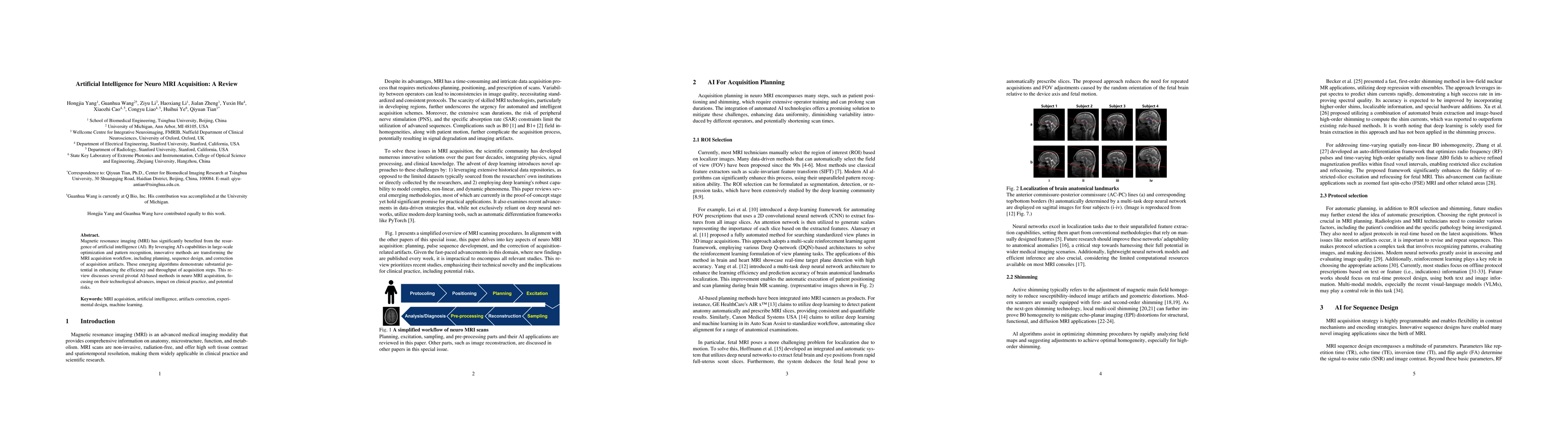
Magnetic resonance imaging (MRI) has significantly benefited from the resurgence of artificial intelligence (AI). By leveraging AI's capabilities in large-scale optimization and pattern recognition,...

Tractography traces the peak directions extracted from fiber orientation distribution (FOD) suffering from ambiguous spatial correspondences between diffusion directions and fiber geometry, which is...
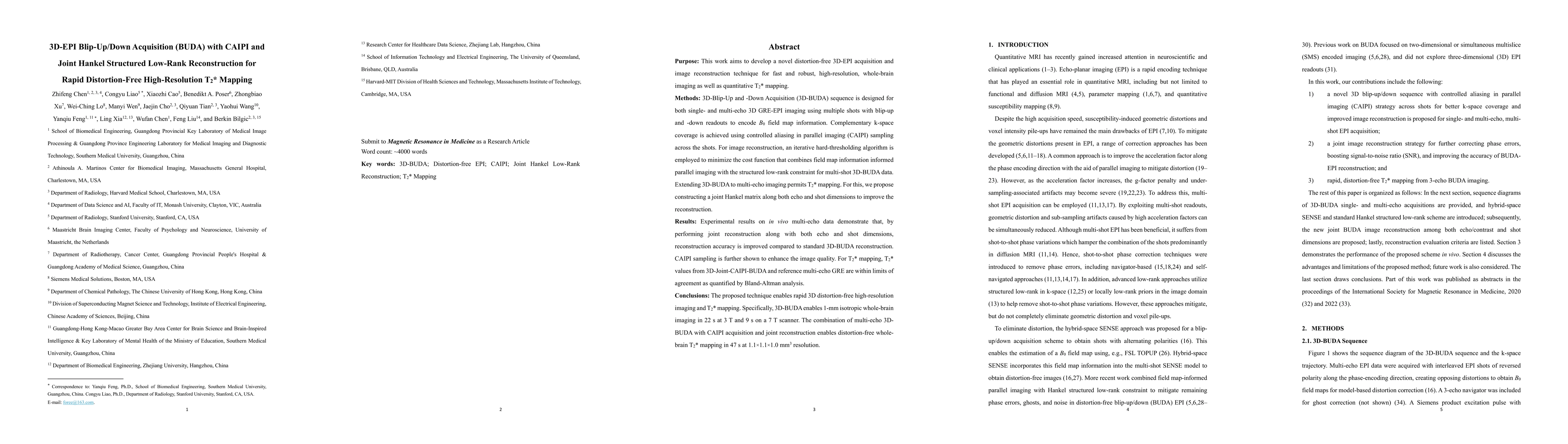
Purpose: This work aims to develop a novel distortion-free 3D-EPI acquisition and image reconstruction technique for fast and robust, high-resolution, whole-brain imaging as well as quantitative T2*...
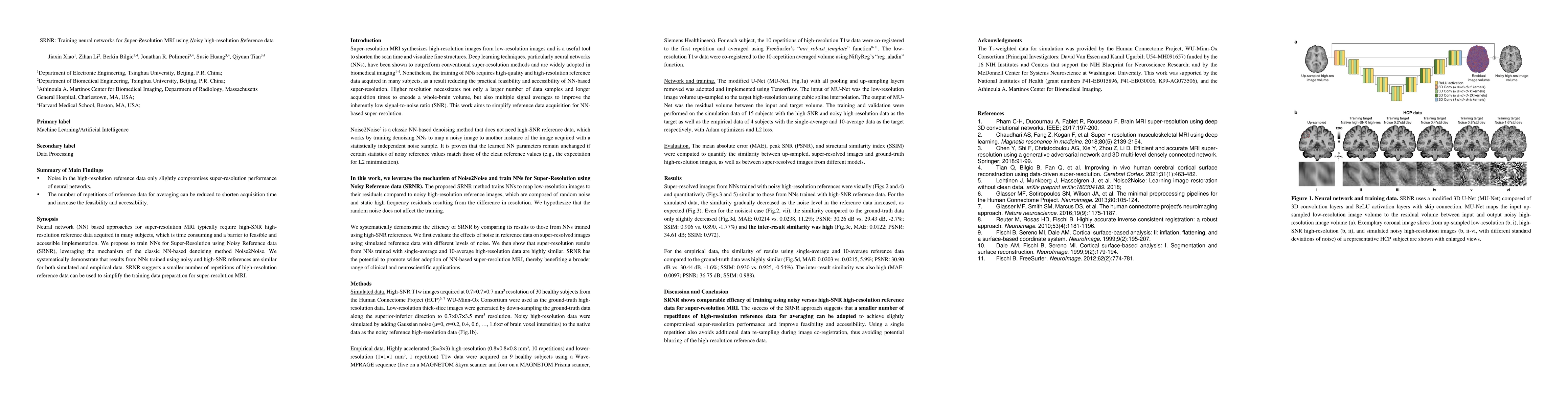
Neural network (NN) based approaches for super-resolution MRI typically require high-SNR high-resolution reference data acquired in many subjects, which is time consuming and a barrier to feasible a...
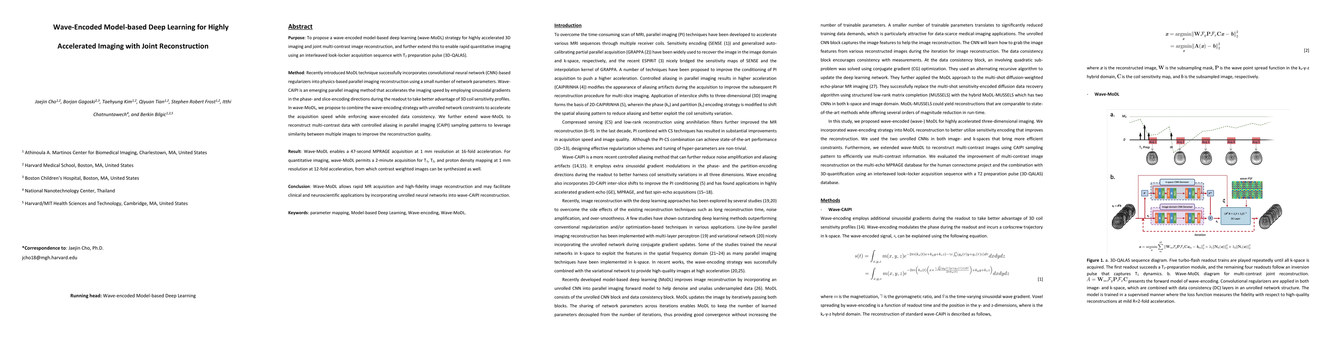
Purpose: To propose a wave-encoded model-based deep learning (wave-MoDL) strategy for highly accelerated 3D imaging and joint multi-contrast image reconstruction, and further extend this to enable r...
Dropout is conventionally used during the training phase as regularization method and for quantifying uncertainty in deep learning. We propose to use dropout during training as well as inference ste...
The noise in diffusion-weighted images (DWIs) decreases the accuracy and precision of diffusion tensor magnetic resonance imaging (DTI) derived microstructural parameters and leads to prolonged acqu...
We introduce wave encoded acquisition and reconstruction techniques for highly accelerated echo planar imaging (EPI) with reduced g-factor penalty and image artifacts. Wave-EPI involves playing sinu...
High-resolution diffusion tensor imaging (DTI) is beneficial for probing tissue microstructure in fine neuroanatomical structures, but long scan times and limited signal-to-noise ratio pose signific...
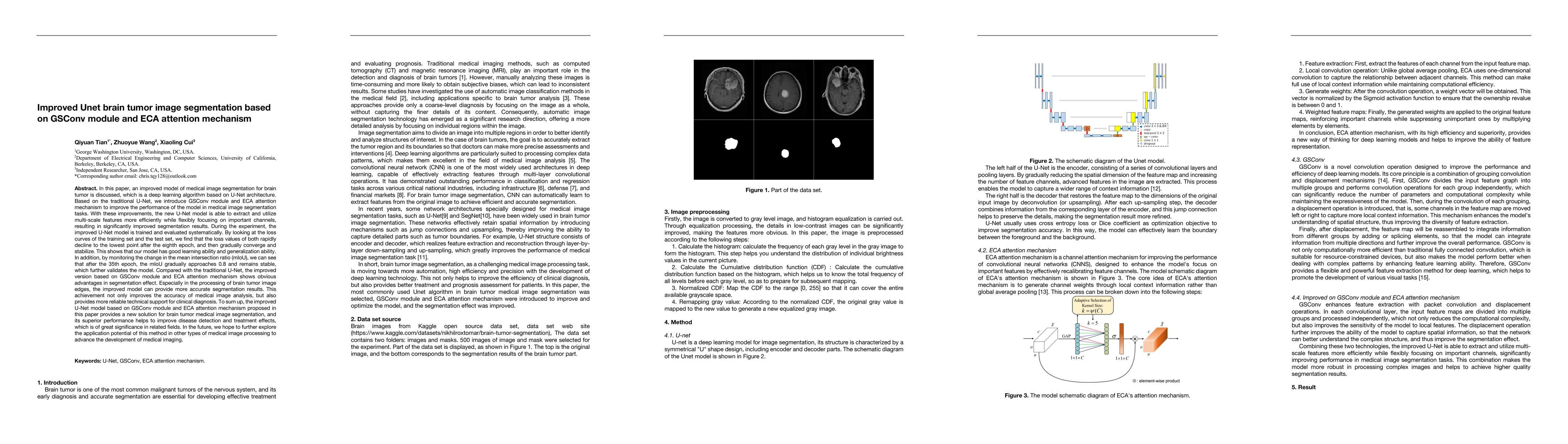
An improved model of medical image segmentation for brain tumor is discussed, which is a deep learning algorithm based on U-Net architecture. Based on the traditional U-Net, we introduce GSConv module...
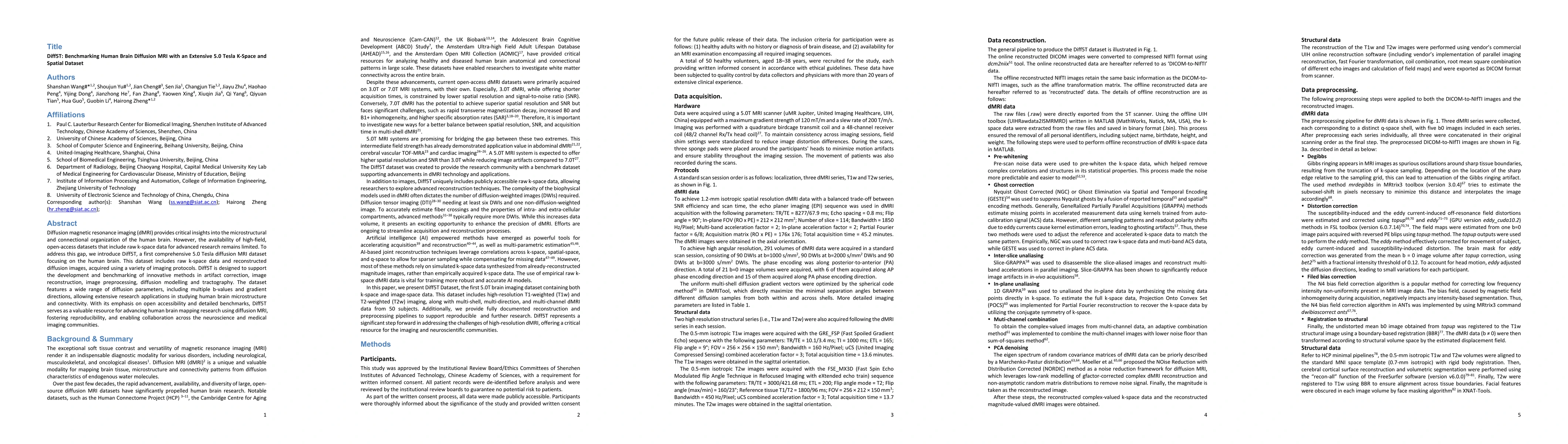
Diffusion magnetic resonance imaging (dMRI) provides critical insights into the microstructural and connectional organization of the human brain. However, the availability of high-field, open-access d...
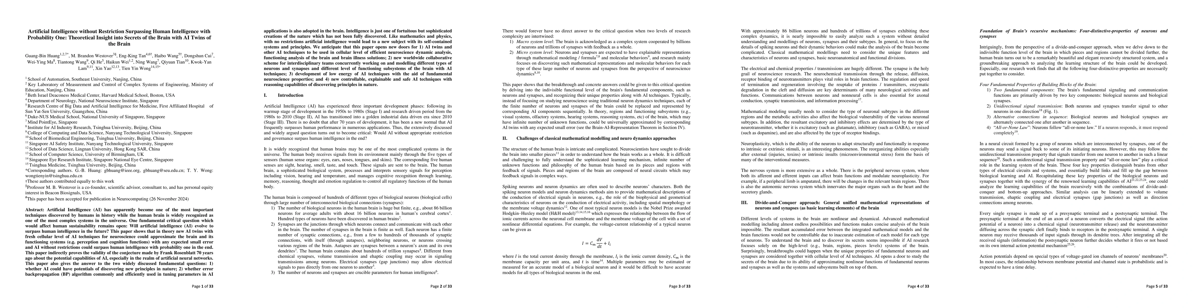
Artificial Intelligence (AI) has apparently become one of the most important techniques discovered by humans in history while the human brain is widely recognized as one of the most complex systems in...
Lesion Segmentation in PET/CT scans is an essential part of modern oncological workflows. To address the challenges of time-intensive manual annotation and high inter-observer variability, the autoPET...
Reliable brain tumor segmentation in MRI is indispensable for treatment planning and outcome monitoring, yet models trained on curated benchmarks often fail under domain shifts arising from scanner an...
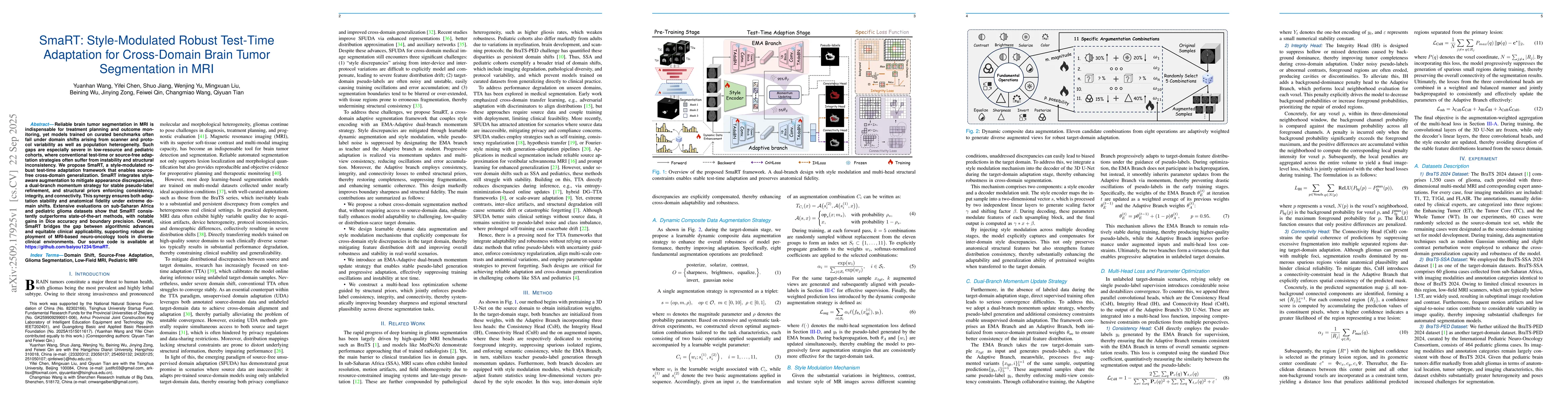
Accurate brain tumor segmentation is essential for preoperative evaluation and personalized treatment. Multi-modal MRI is widely used due to its ability to capture complementary tumor features across ...
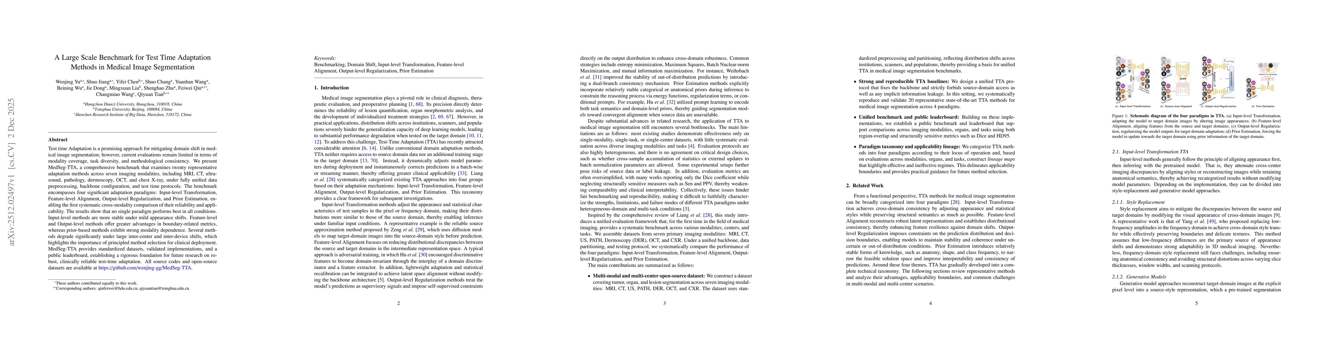
Test time Adaptation is a promising approach for mitigating domain shift in medical image segmentation; however, current evaluations remain limited in terms of modality coverage, task diversity, and m...
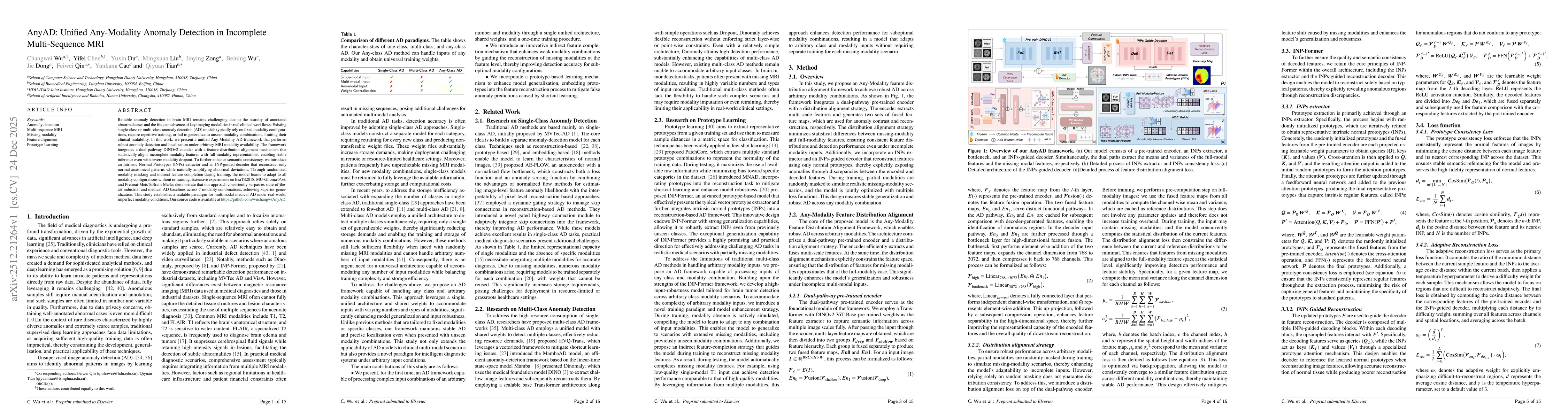
Reliable anomaly detection in brain MRI remains challenging due to the scarcity of annotated abnormal cases and the frequent absence of key imaging modalities in real clinical workflows. Existing sing...
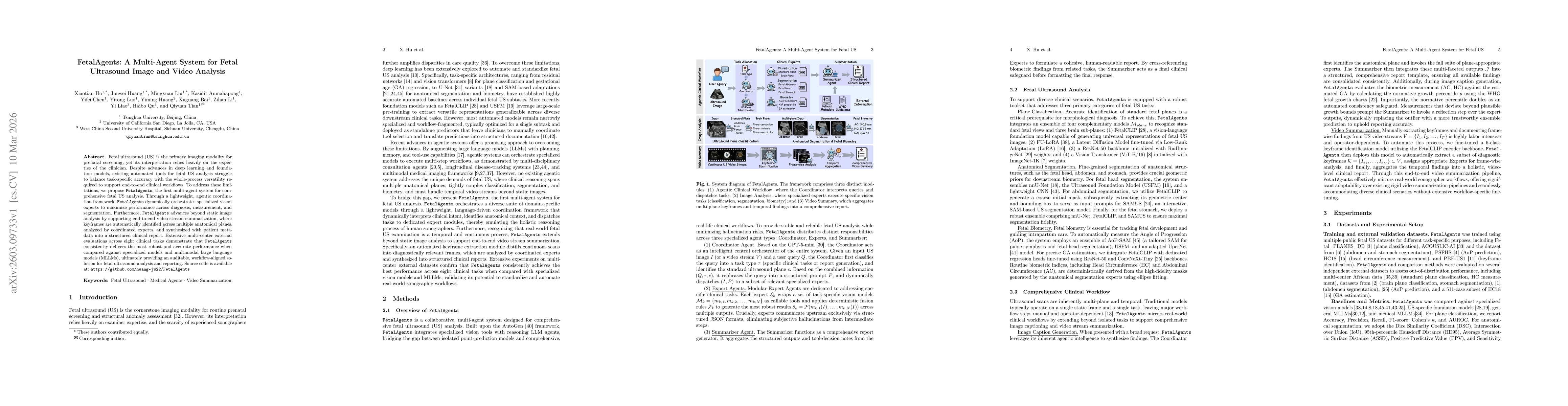
Fetal ultrasound (US) is the primary imaging modality for prenatal screening, yet its interpretation relies heavily on the expertise of the clinician. Despite advances in deep learning and foundation ...
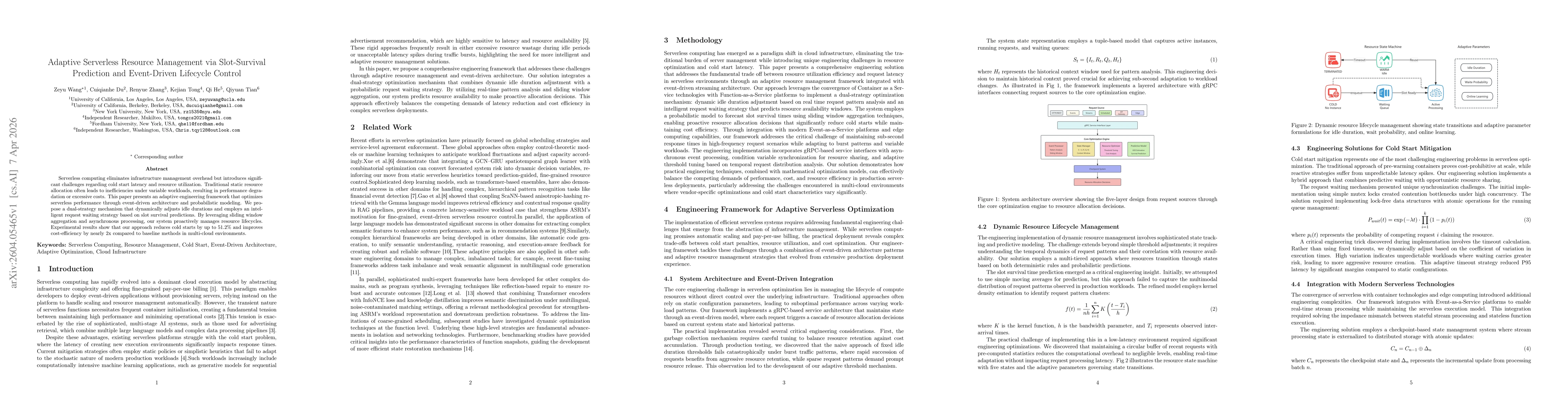
Serverless computing eliminates infrastructure management overhead but introduces significant challenges regarding cold start latency and resource utilization. Traditional static resource allocation o...
Chest computed tomography (CT) is central to the detection and management of thoracic disease, yet the growing scale and complexity of volumetric imaging increasingly exceed what can be addressed by s...
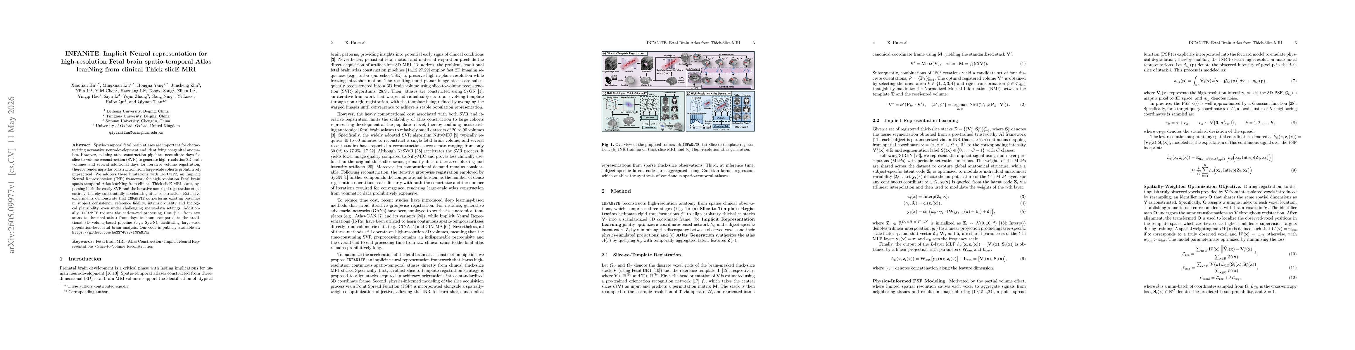
Spatio-temporal fetal brain atlases are important for characterizing normative neurodevelopment and identifying congenital anomalies. However, existing atlas construction pipelines necessitate days fo...
Background: Prenatal germinal matrix-intraventricular hemorrhage (GMH-IVH) is a leading cause of infant mortality and neurodevelopmental impairment. Manual diagnosis and lesion segmentation are labor-...
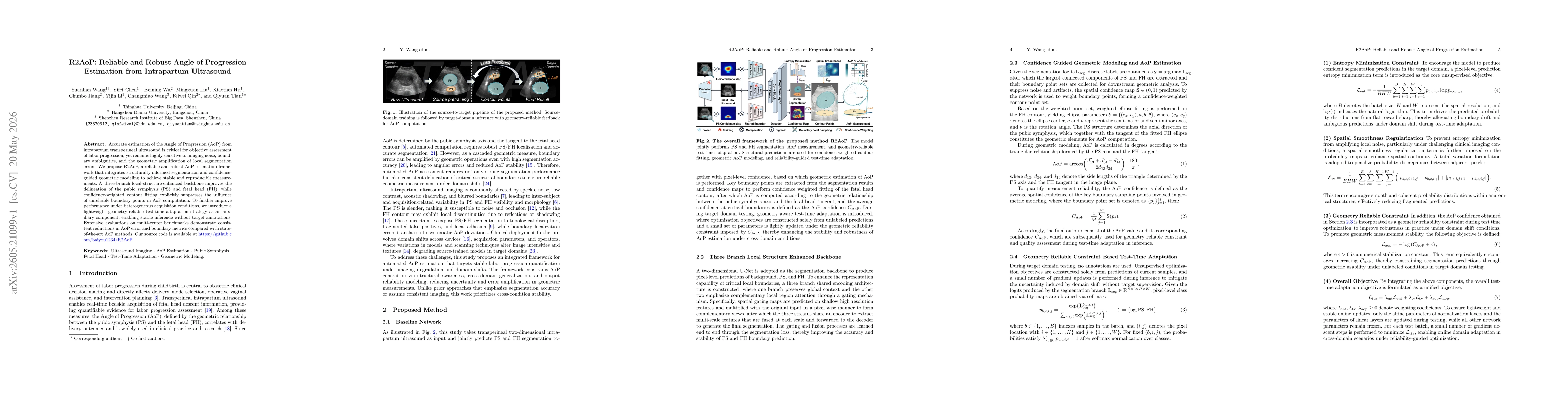
Accurate estimation of the Angle of Progression (AoP) from intrapartum transperineal ultrasound is critical for objective assessment of labor progression, yet remains highly sensitive to imaging noise...
Automated fetal ultrasound interpretation requires a workflow from visual perception, including plane recognition and anatomical segmentation, to clinical understanding, including biometric measuremen...
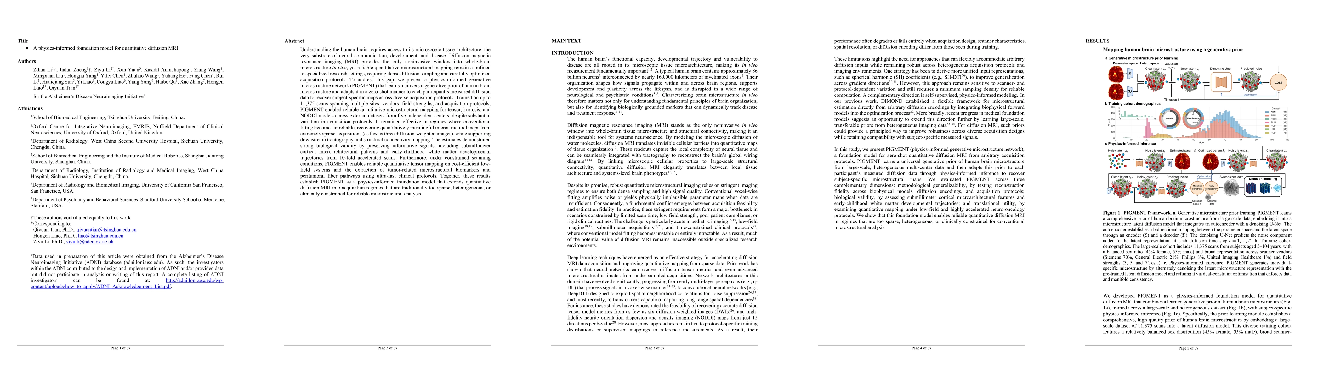
Understanding the human brain requires access to its microscopic tissue architecture. Diffusion magnetic resonance imaging (MRI) provides the only noninvasive window into whole-brain microstructure in...
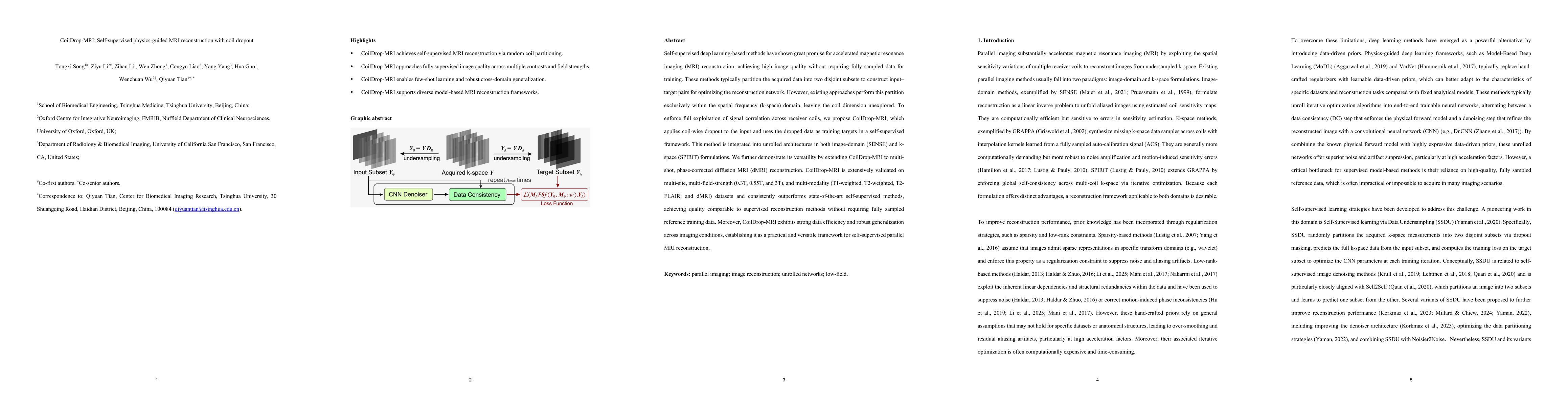
Self-supervised deep learning-based methods have shown great promise for accelerated magnetic resonance imaging (MRI) reconstruction, achieving high image quality without requiring fully sampled data ...