Academic Profile
Statistics
Similar Authors
Papers on arXiv
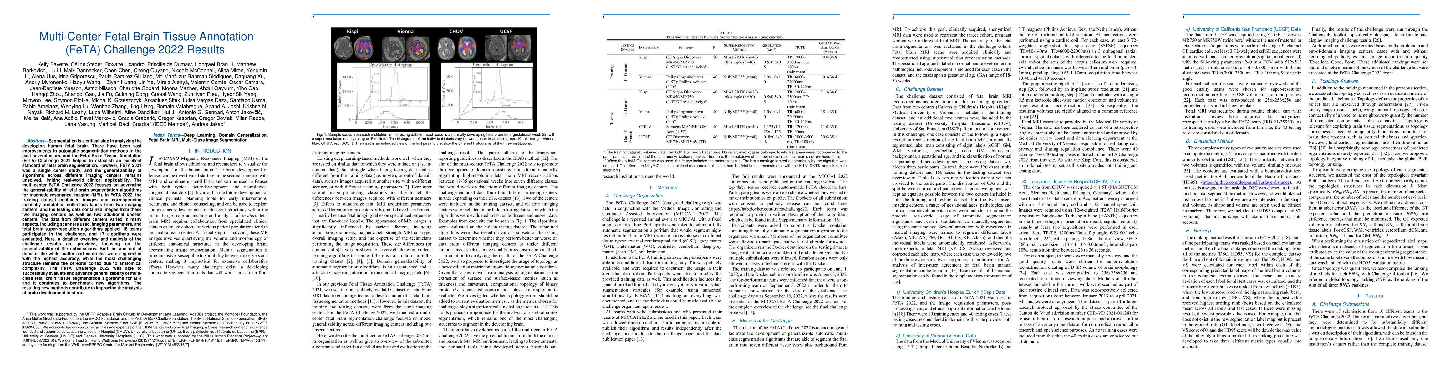
Segmentation is a critical step in analyzing the developing human fetal brain. There have been vast improvements in automatic segmentation methods in the past several years, and the Fetal Brain Tiss...
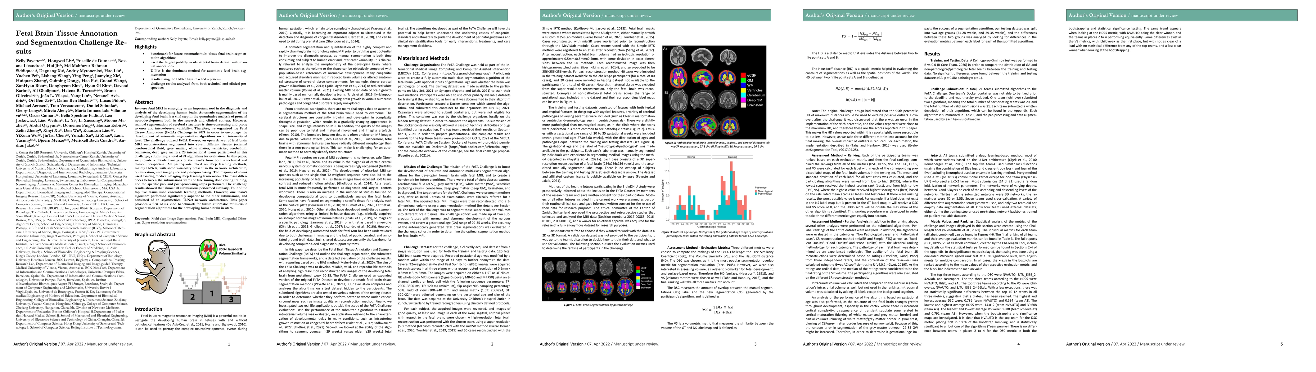
In-utero fetal MRI is emerging as an important tool in the diagnosis and analysis of the developing human brain. Automatic segmentation of the developing fetal brain is a vital step in the quantitat...
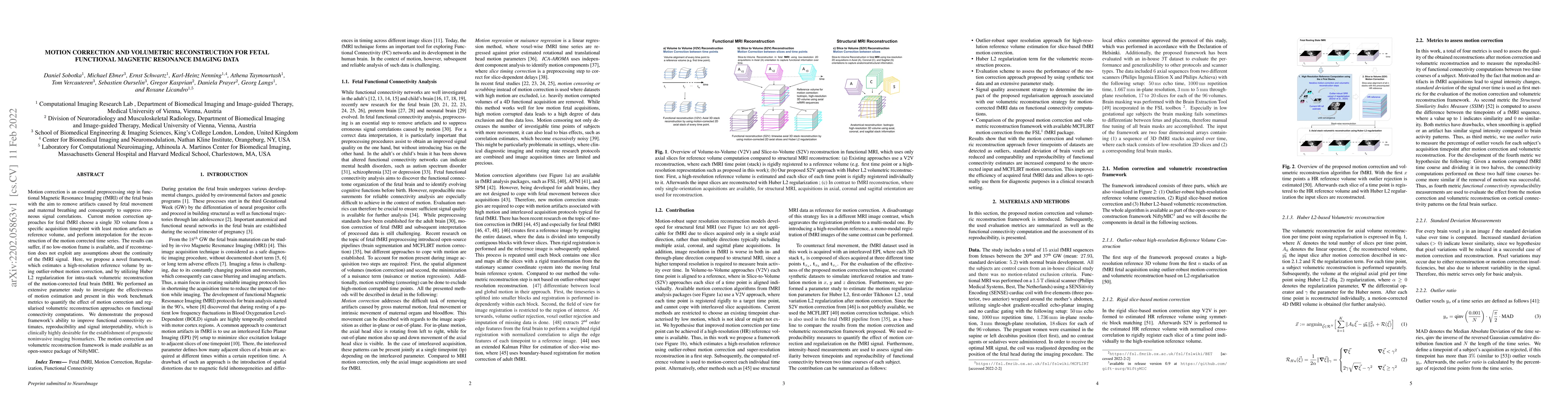
Motion correction is an essential preprocessing step in functional Magnetic Resonance Imaging (fMRI) of the fetal brain with the aim to remove artifacts caused by fetal movement and maternal breathi...
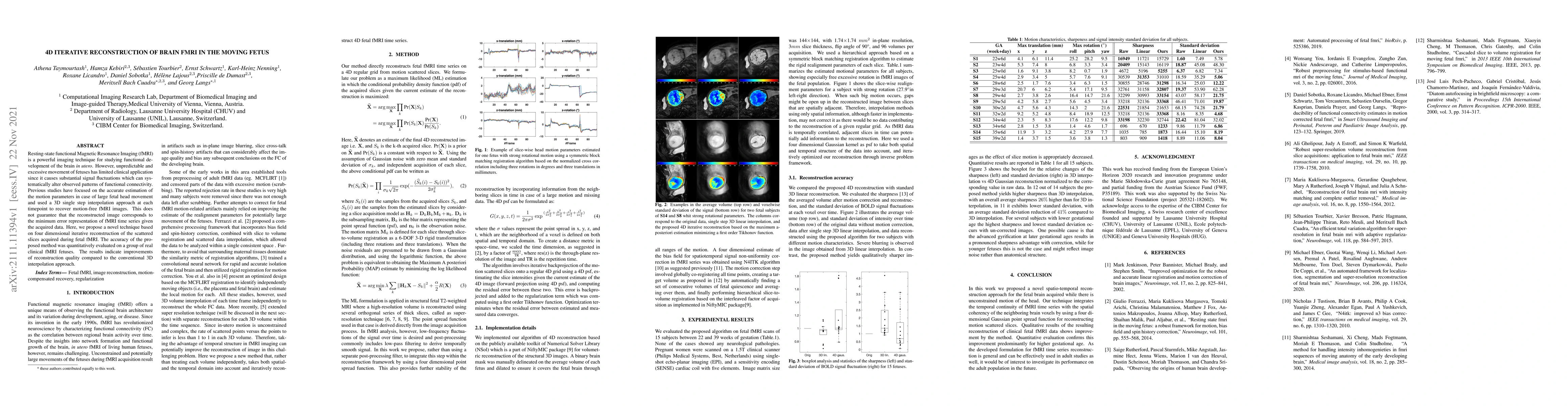
Resting-state functional Magnetic Resonance Imaging (fMRI) is a powerful imaging technique for studying functional development of the brain in utero. However, unpredictable and excessive movement of...
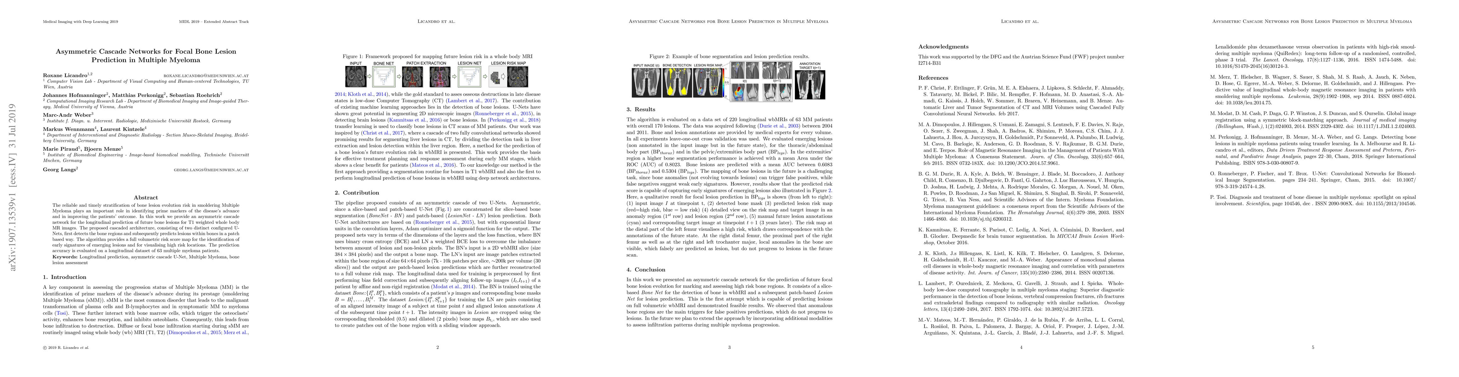
The reliable and timely stratification of bone lesion evolution risk in smoldering Multiple Myeloma plays an important role in identifying prime markers of the disease's advance and in improving the...
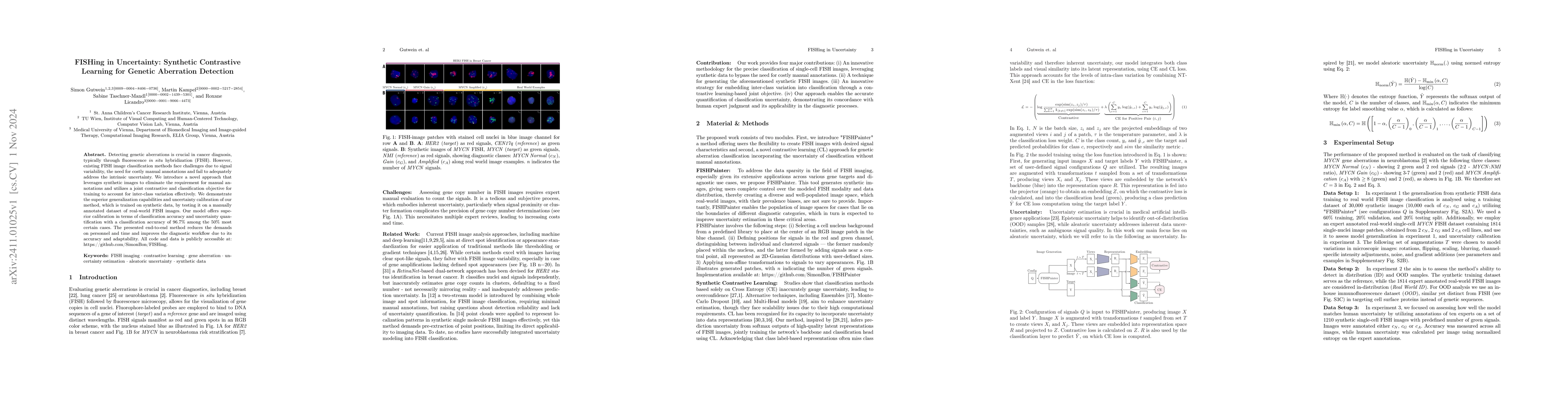
Detecting genetic aberrations is crucial in cancer diagnosis, typically through fluorescence in situ hybridization (FISH). However, existing FISH image classification methods face challenges due to si...
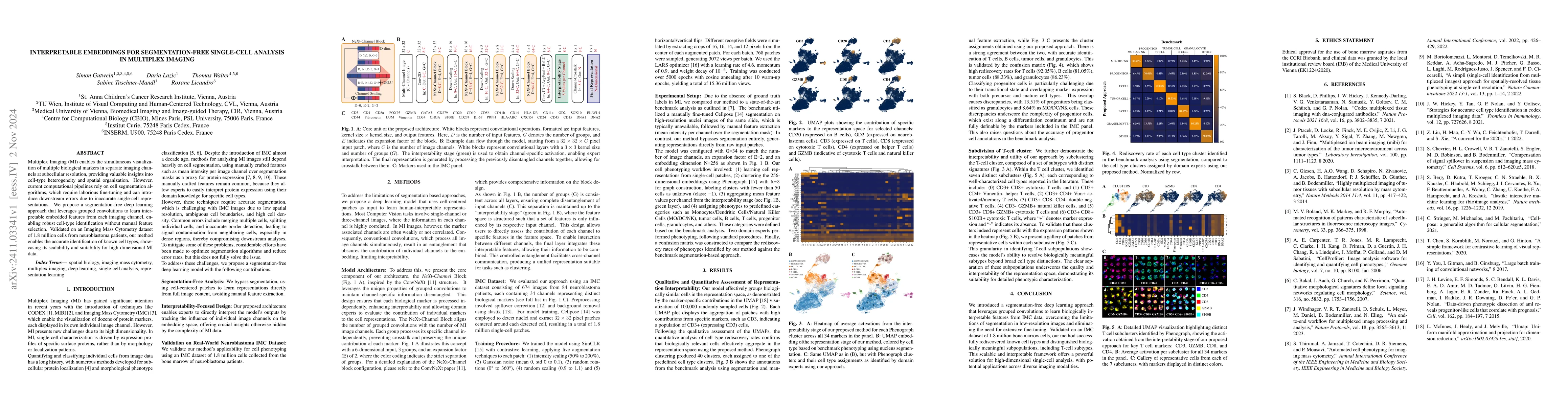
Multiplex Imaging (MI) enables the simultaneous visualization of multiple biological markers in separate imaging channels at subcellular resolution, providing valuable insights into cell-type heteroge...
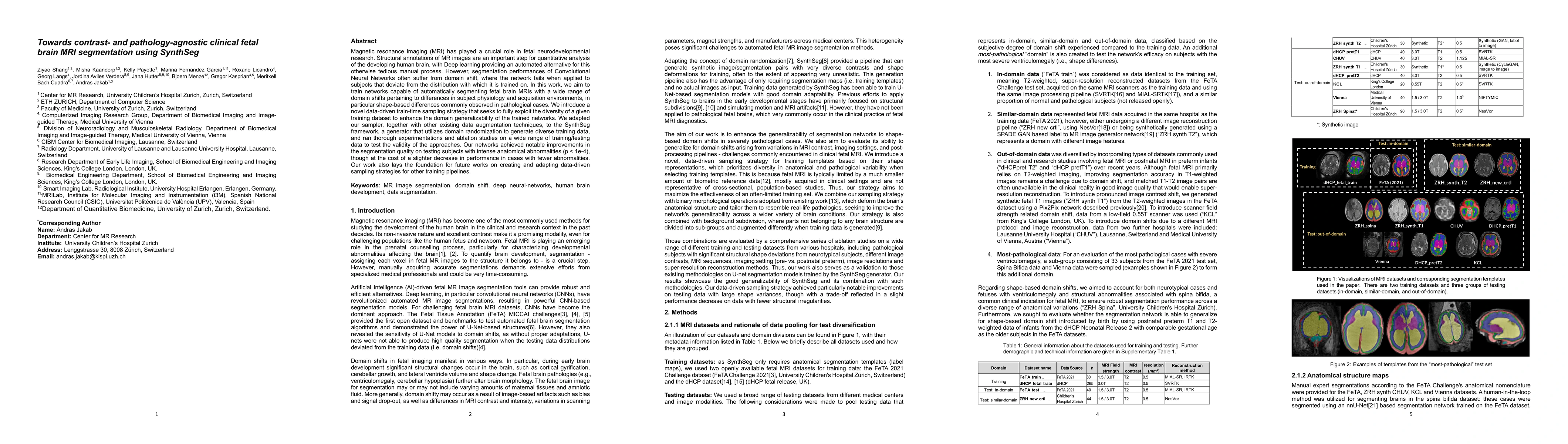
Magnetic resonance imaging (MRI) has played a crucial role in fetal neurodevelopmental research. Structural annotations of MR images are an important step for quantitative analysis of the developing h...
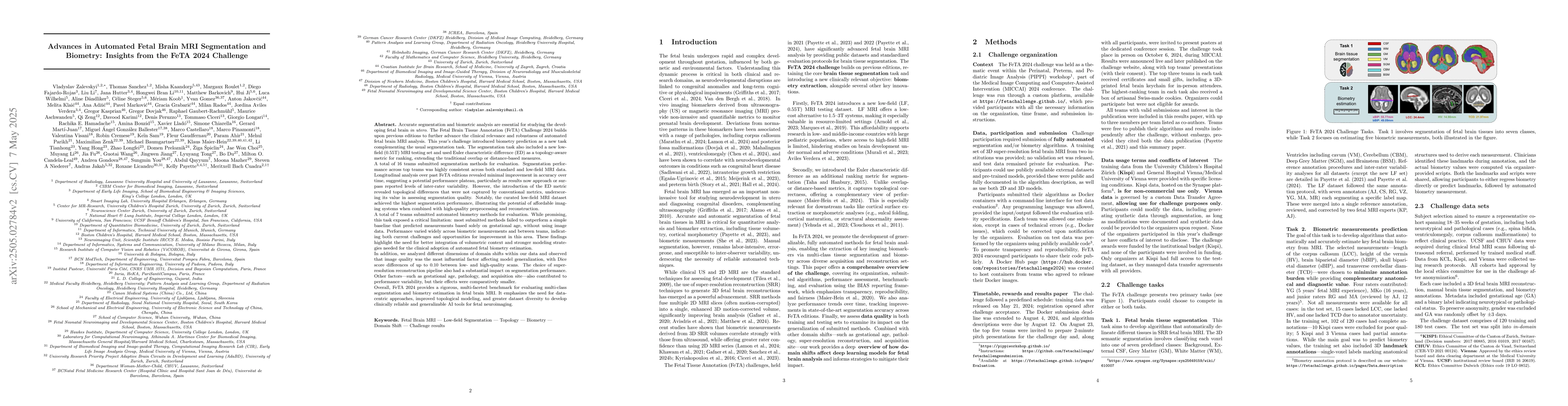
Accurate fetal brain tissue segmentation and biometric analysis are essential for studying brain development in utero. The FeTA Challenge 2024 advanced automated fetal brain MRI analysis by introducin...
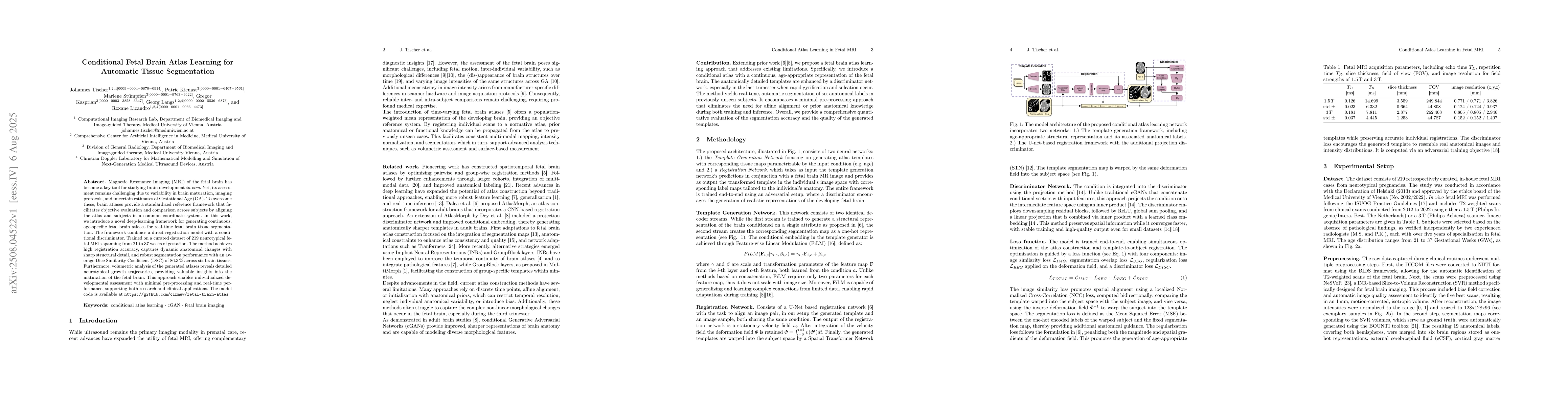
Magnetic Resonance Imaging (MRI) of the fetal brain has become a key tool for studying brain development in vivo. Yet, its assessment remains challenging due to variability in brain maturation, imagin...
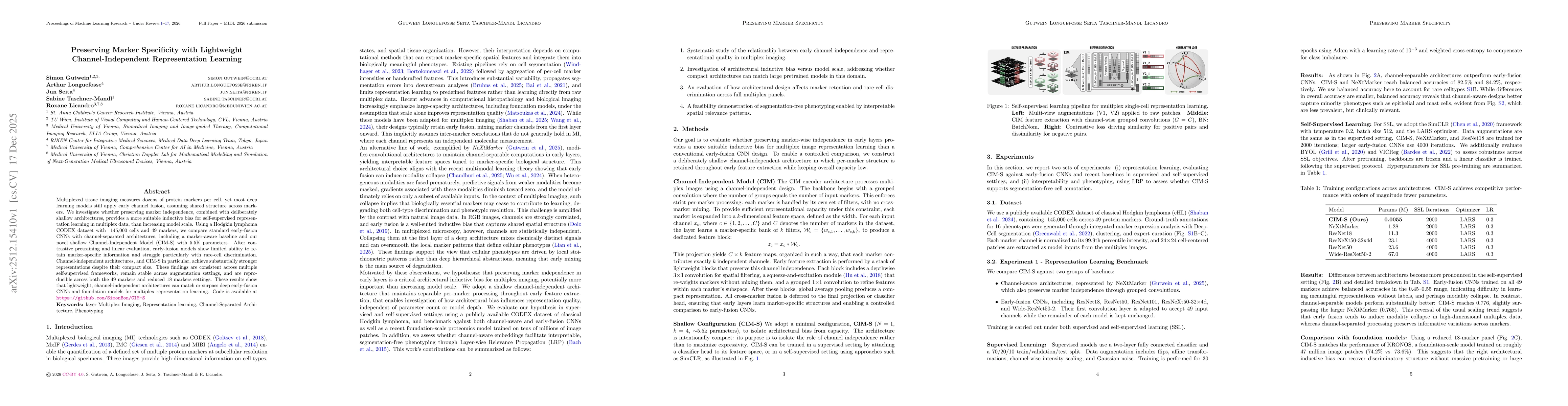
Multiplexed tissue imaging measures dozens of protein markers per cell, yet most deep learning models still apply early channel fusion, assuming shared structure across markers. We investigate whether...