ArxivLens
R
Researcher

Rudolf L. M. van Herten

5 papers on arXiv

5 arXiv Papers
3 Total Publications
Profile

Academic Profile

Name R. L. van Herten
Total Papers 3
Scholar Profile View on Semantic Scholar
Metrics

Statistics

5 arXiv Papers
3 Total Publications
Publications

Papers on arXiv

Thumbnail for Automatic Coronary Artery Plaque Quantification and CAD-RADS Prediction
  using Mesh Priors
arXiv
Automatic Coronary Artery Plaque Quantification and CAD-RADS Prediction using Mesh Priors

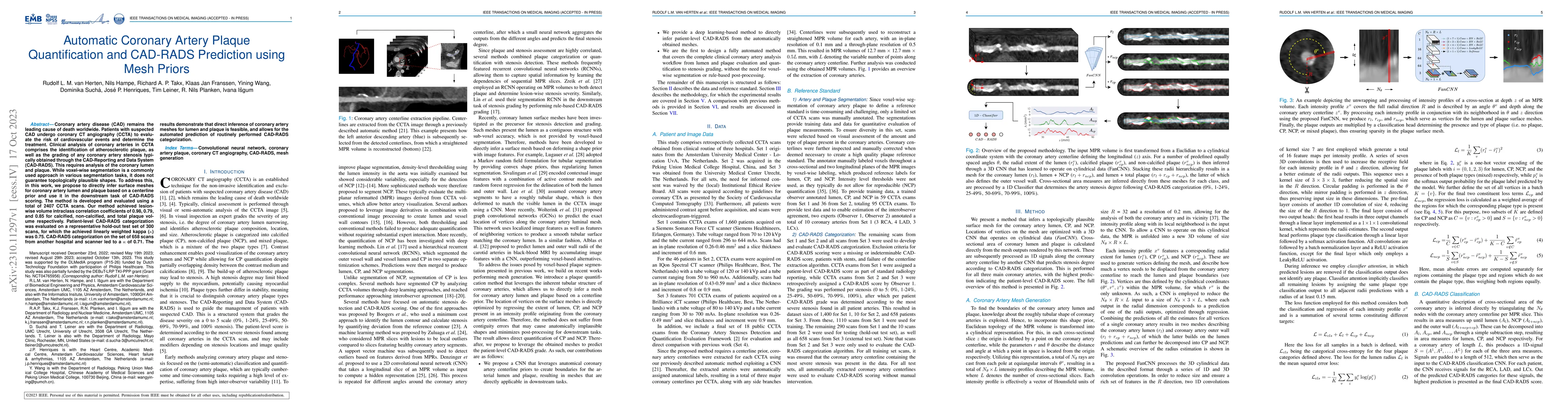
Coronary artery disease (CAD) remains the leading cause of death worldwide. Patients with suspected CAD undergo coronary CT angiography (CCTA) to evaluate the risk of cardiovascular events and deter...

Thumbnail for Physics-informed neural networks for myocardial perfusion MRI
  quantification
arXiv
Physics-informed neural networks for myocardial perfusion MRI quantification

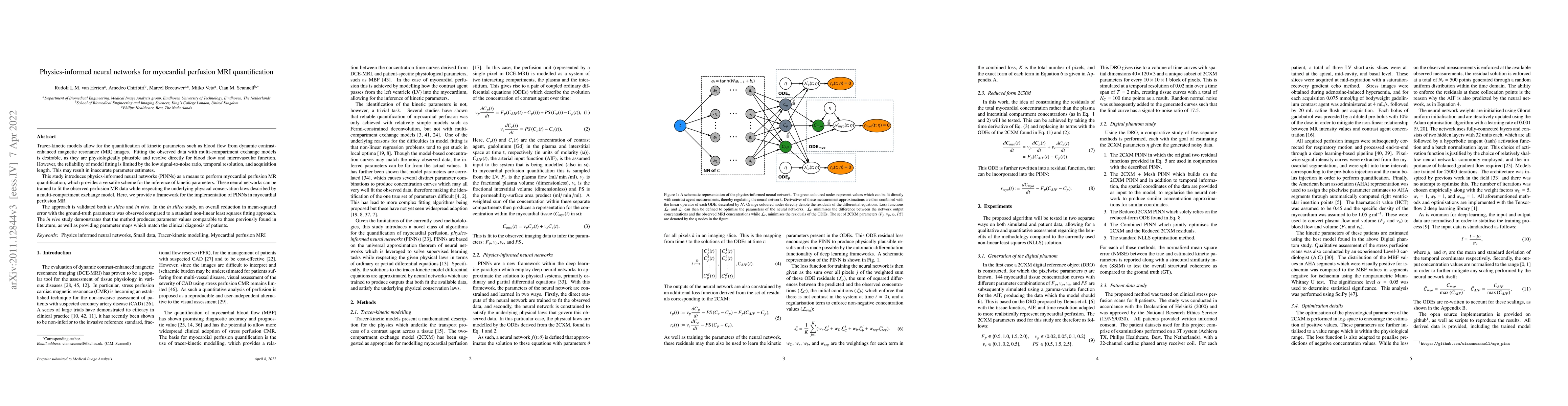
Tracer-kinetic models allow for the quantification of kinetic parameters such as blood flow from dynamic contrast-enhanced magnetic resonance (MR) images. Fitting the observed data with multi-compar...

Thumbnail for World of Forms: Deformable Geometric Templates for One-Shot Surface
  Meshing in Coronary CT Angiography
arXiv
World of Forms: Deformable Geometric Templates for One-Shot Surface Meshing in Coronary CT Angiography

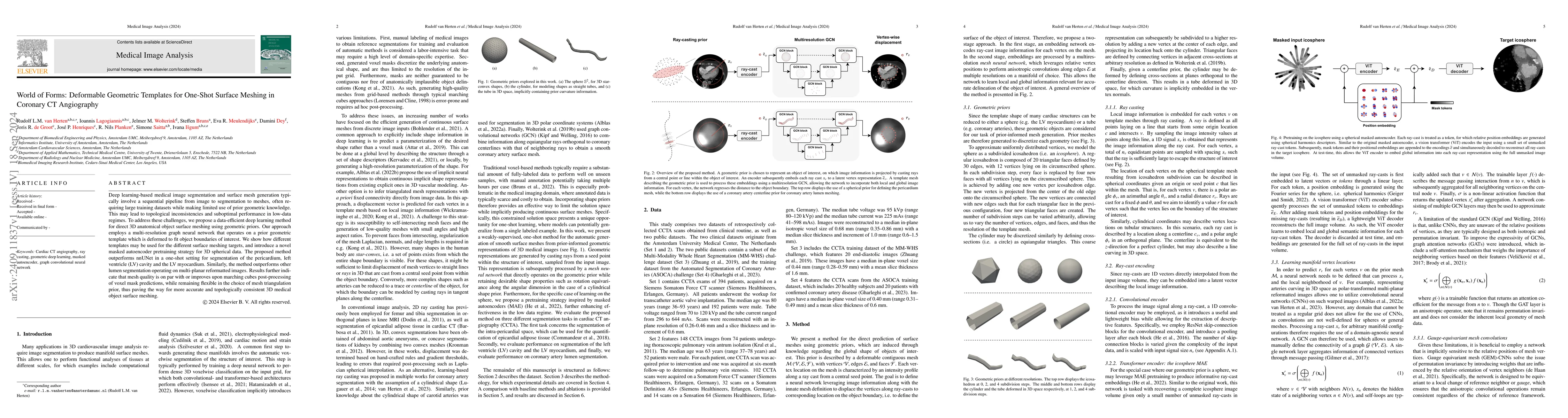
Deep learning-based medical image segmentation and surface mesh generation typically involve a sequential pipeline from image to segmentation to meshes, often requiring large training datasets while m...

Thumbnail for Attenuation artifact detection and severity classification in
  intracoronary OCT using mixed image representations
arXiv
Attenuation artifact detection and severity classification in intracoronary OCT using mixed image representations

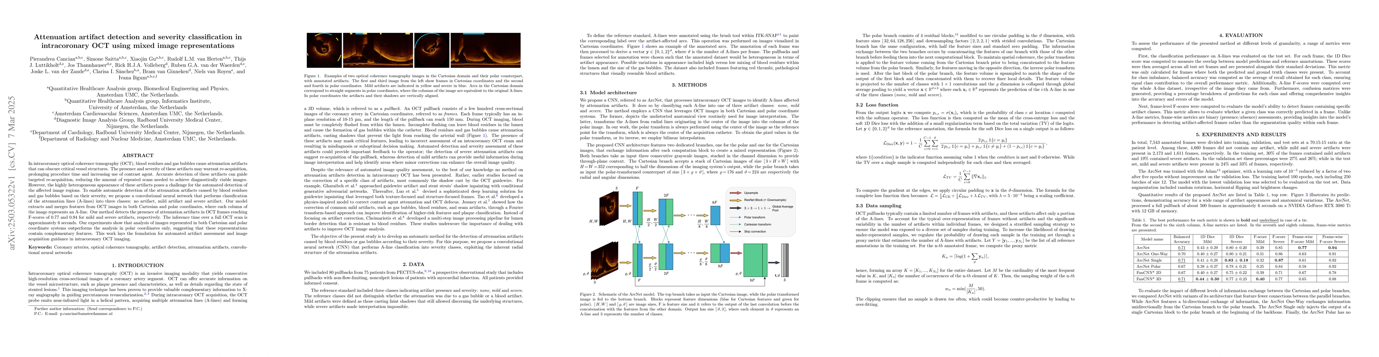
In intracoronary optical coherence tomography (OCT), blood residues and gas bubbles cause attenuation artifacts that can obscure critical vessel structures. The presence and severity of these artifact...

Thumbnail for A Multimodal 3D Foundation Model for Light Sheet Fluorescence Microscopy Enables Few-Shot Segmentation, Classification, and Deblurring
arXiv
A Multimodal 3D Foundation Model for Light Sheet Fluorescence Microscopy Enables Few-Shot Segmentation, Classification, and Deblurring

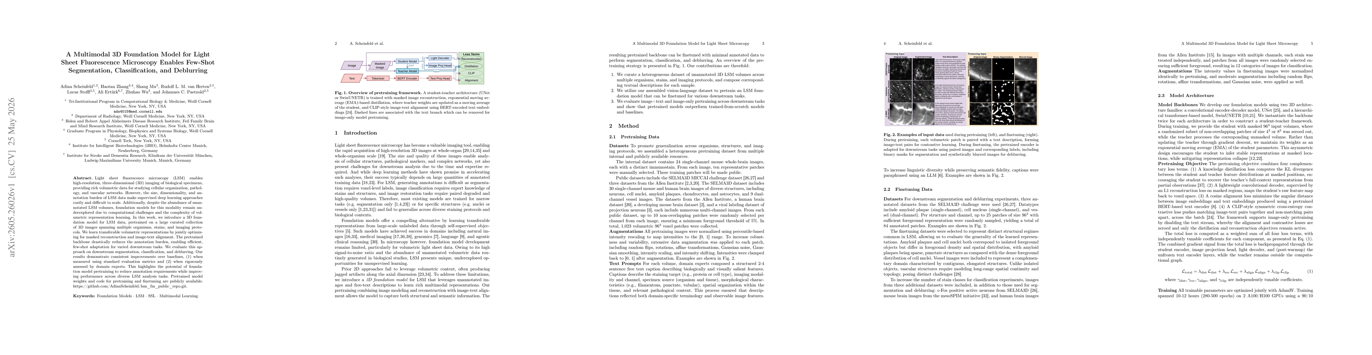
Light sheet fluorescence microscopy (LSM) enables high-resolution, three-dimensional (3D) imaging of biological specimens, providing rich volumetric data for studying cellular organization, pathology,...

This website uses cookies to ensure you get the best experience. Learn more