Academic Profile
Statistics
Similar Authors
Papers on arXiv
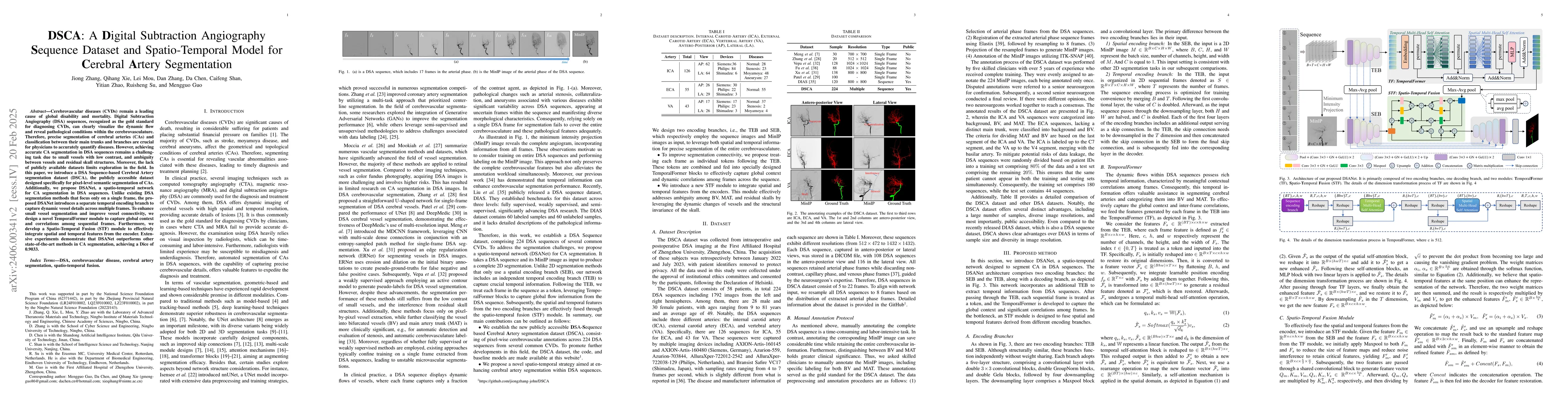
Cerebrovascular diseases (CVDs) remain a leading cause of global disability and mortality. Digital Subtraction Angiography (DSA) sequences, recognized as the golden standard for diagnosing CVDs, can...
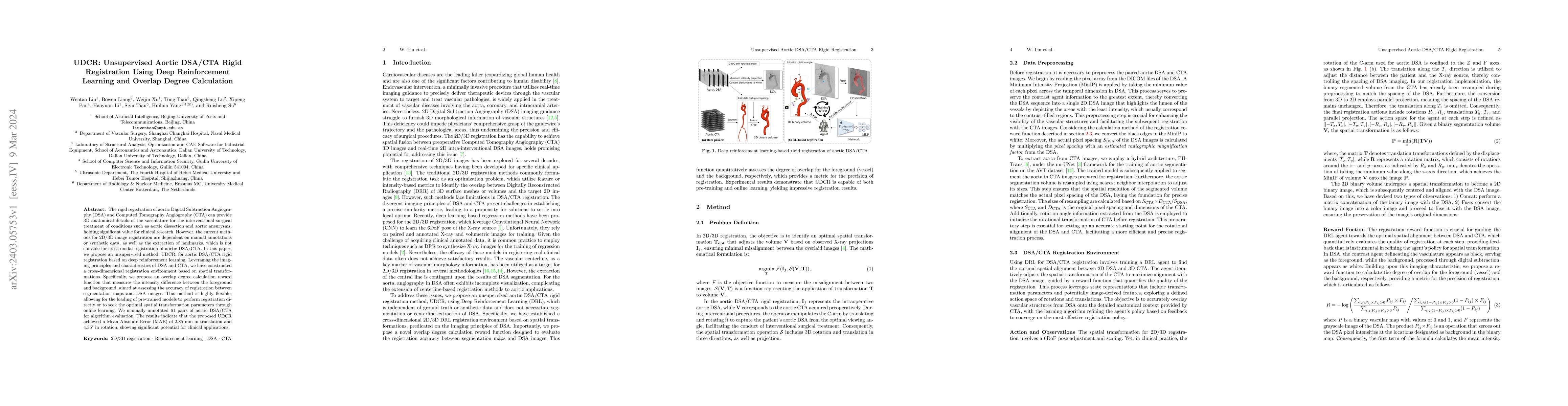
The rigid registration of aortic Digital Subtraction Angiography (DSA) and Computed Tomography Angiography (CTA) can provide 3D anatomical details of the vasculature for the interventional surgical ...
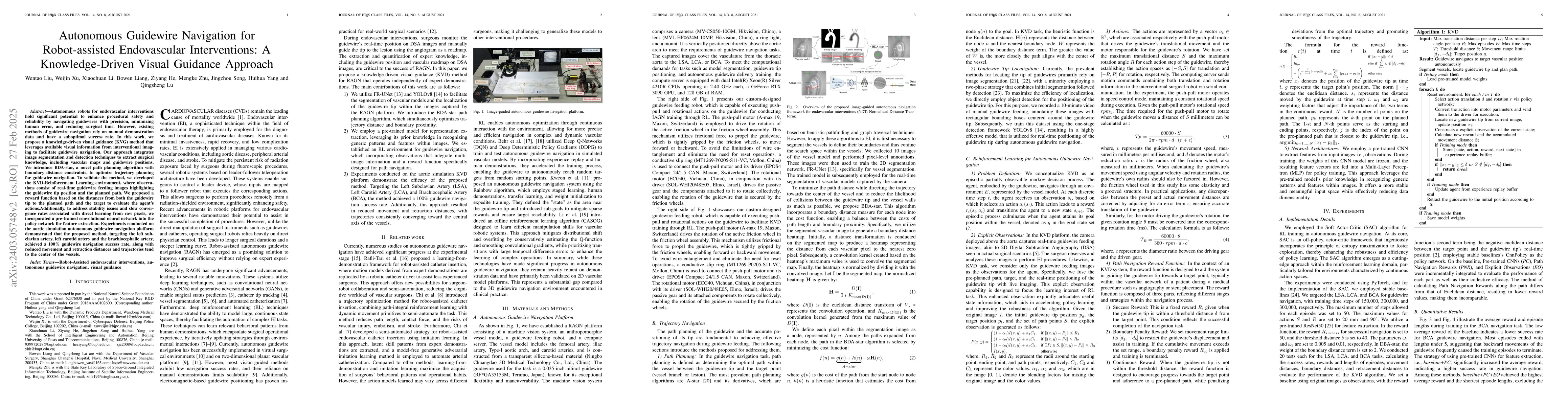
Autonomous robots in endovascular interventions possess the potential to navigate guidewires with safety and reliability, while reducing human error and shortening surgical time. However, current me...
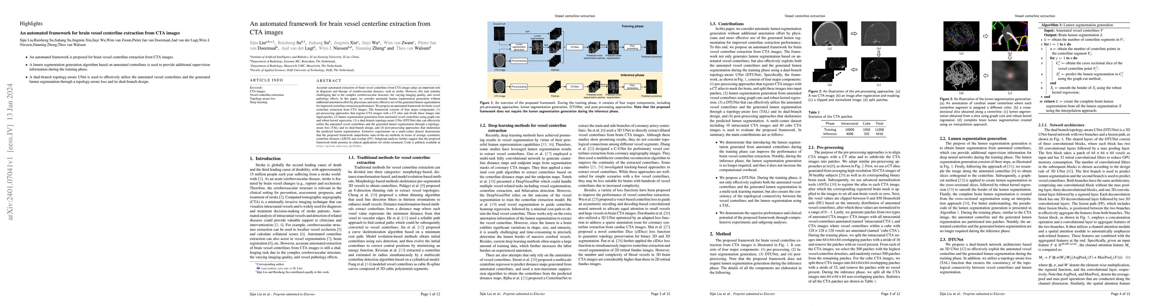
Accurate automated extraction of brain vessel centerlines from CTA images plays an important role in diagnosis and therapy of cerebrovascular diseases, such as stroke. However, this task remains cha...
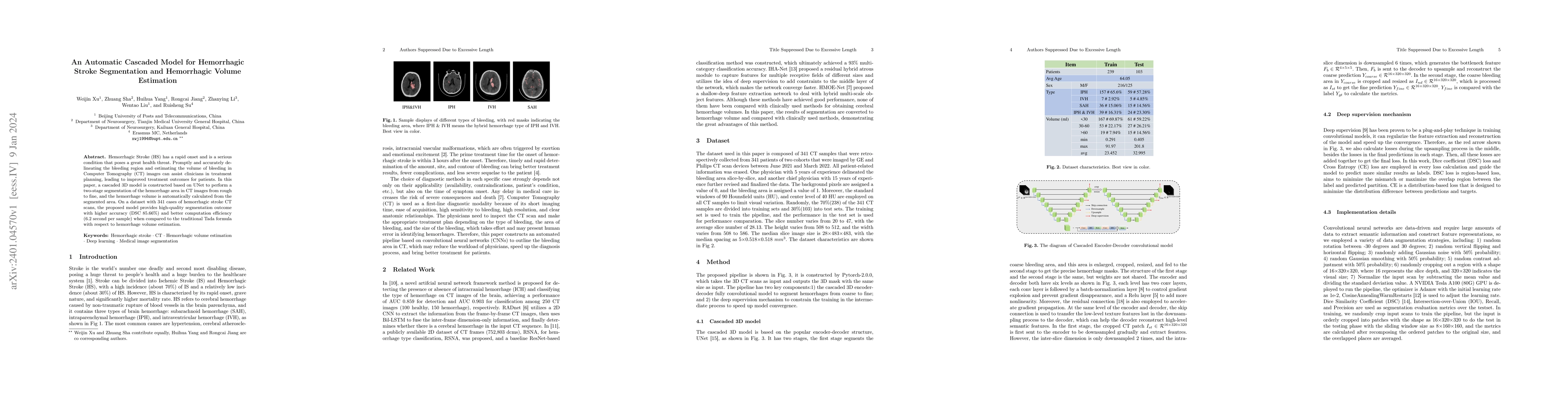
Hemorrhagic Stroke (HS) has a rapid onset and is a serious condition that poses a great health threat. Promptly and accurately delineating the bleeding region and estimating the volume of bleeding i...
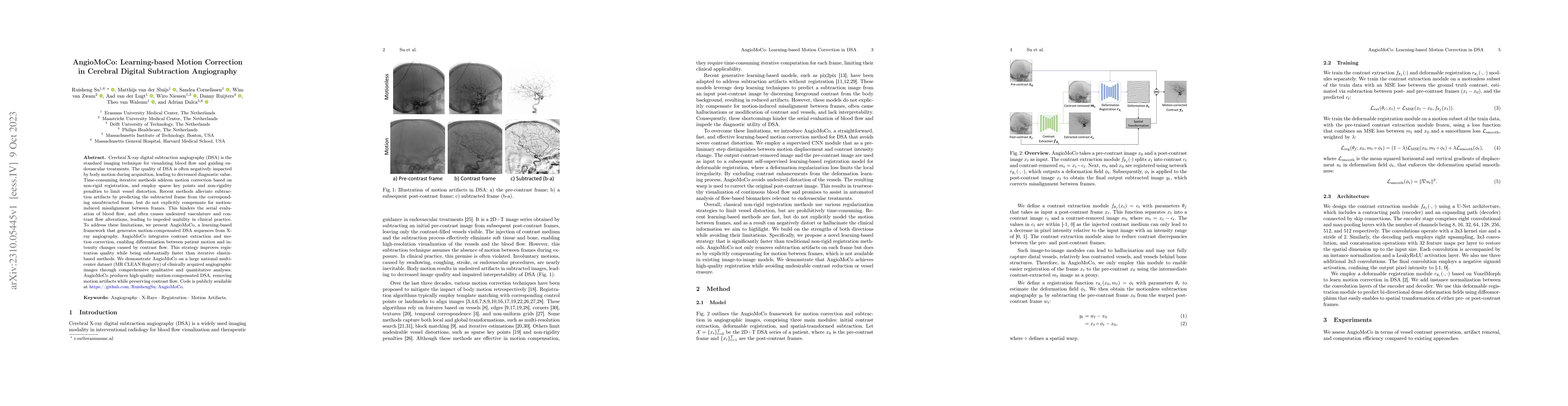
Cerebral X-ray digital subtraction angiography (DSA) is the standard imaging technique for visualizing blood flow and guiding endovascular treatments. The quality of DSA is often negatively impacted...
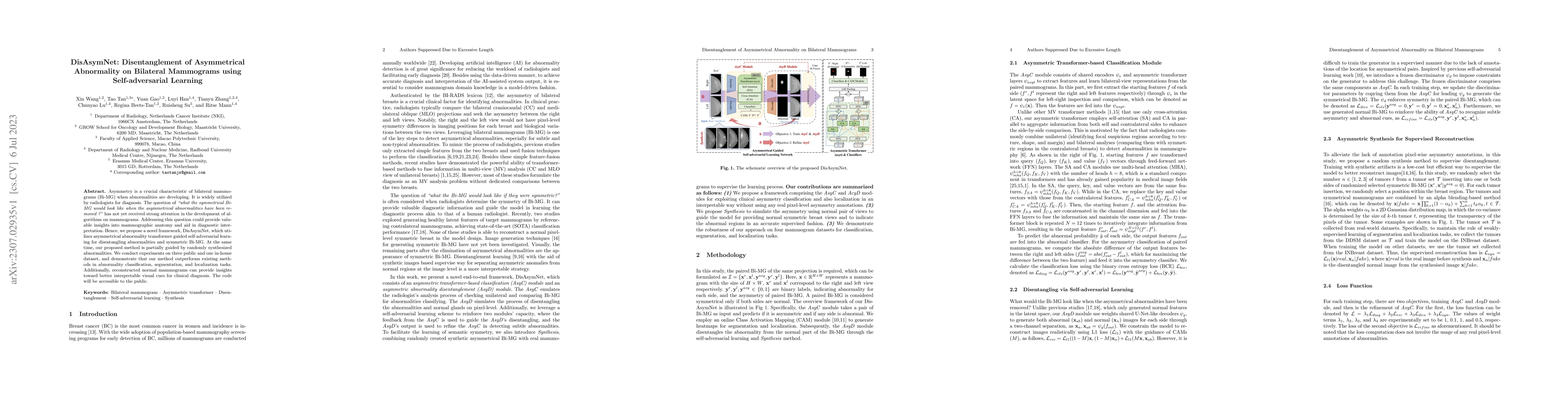
Asymmetry is a crucial characteristic of bilateral mammograms (Bi-MG) when abnormalities are developing. It is widely utilized by radiologists for diagnosis. The question of 'what the symmetrical Bi...
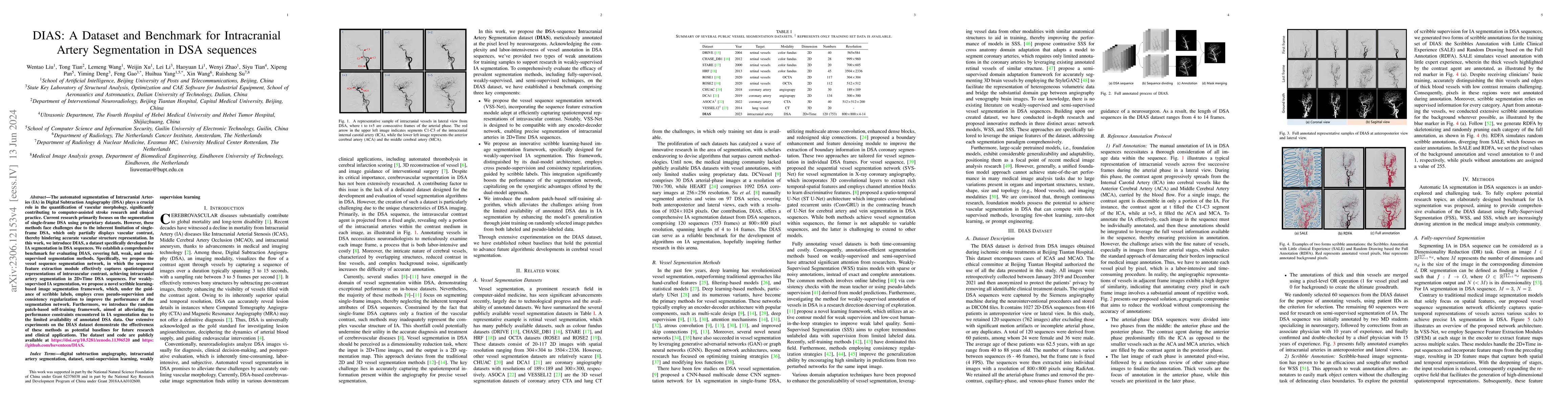
The automated segmentation of Intracranial Arteries (IA) in Digital Subtraction Angiography (DSA) plays a crucial role in the quantification of vascular morphology, significantly contributing to com...
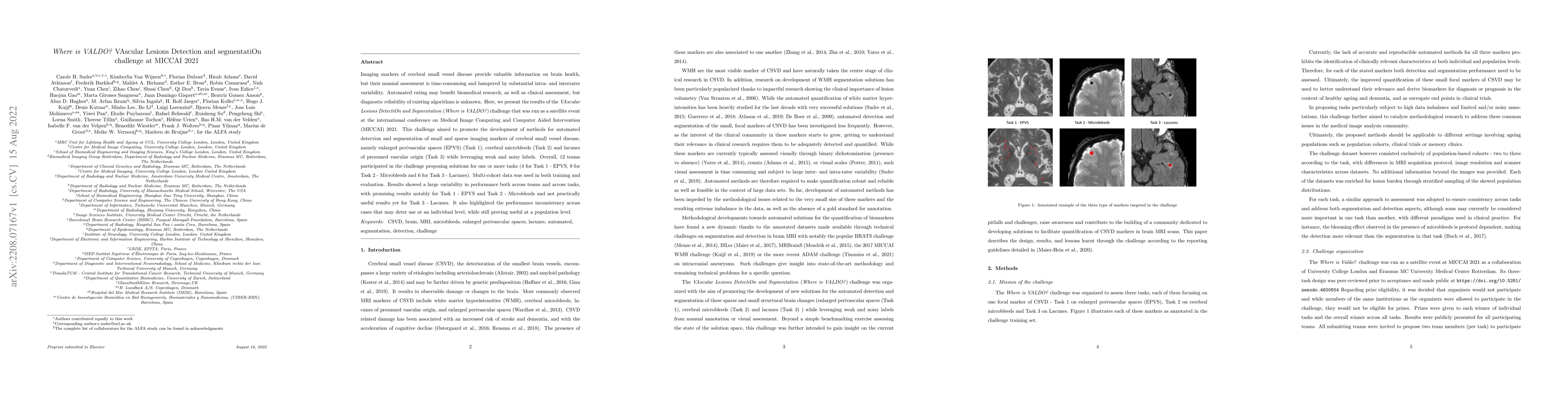
Imaging markers of cerebral small vessel disease provide valuable information on brain health, but their manual assessment is time-consuming and hampered by substantial intra- and interrater variabi...
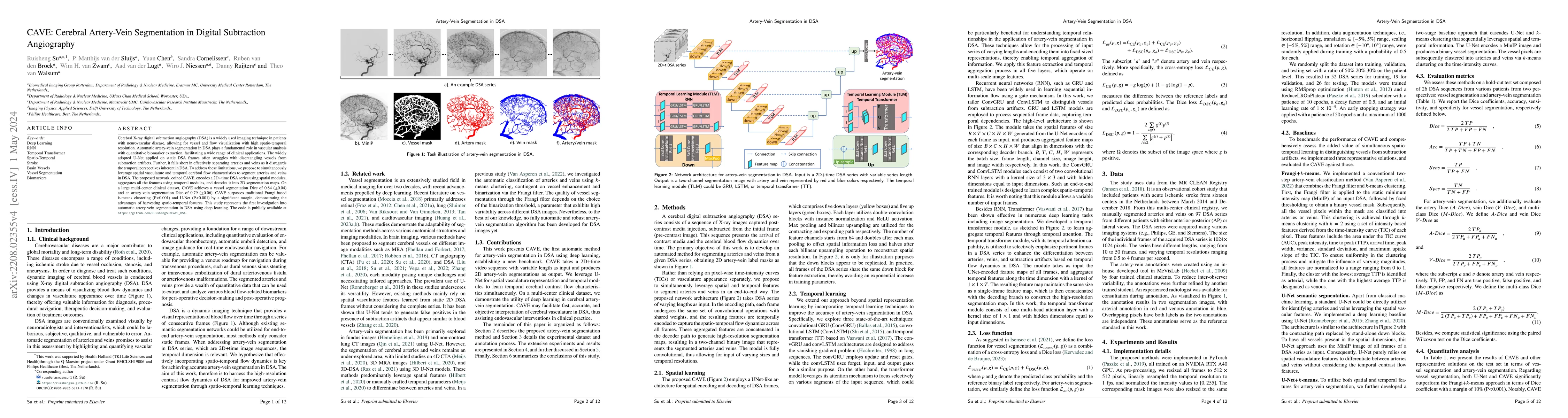
Cerebral X-ray digital subtraction angiography (DSA) is a widely used imaging technique in patients with neurovascular disease, allowing for vessel and flow visualization with high spatio-temporal r...
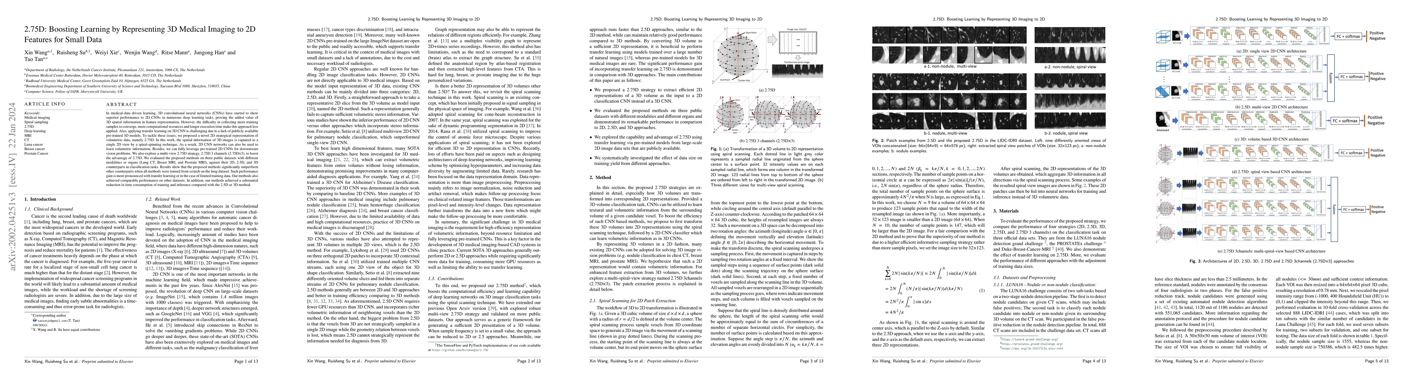
In medical-data driven learning, 3D convolutional neural networks (CNNs) have started to show superior performance to 2D CNNs in numerous deep learning tasks, proving the added value of 3D spatial i...
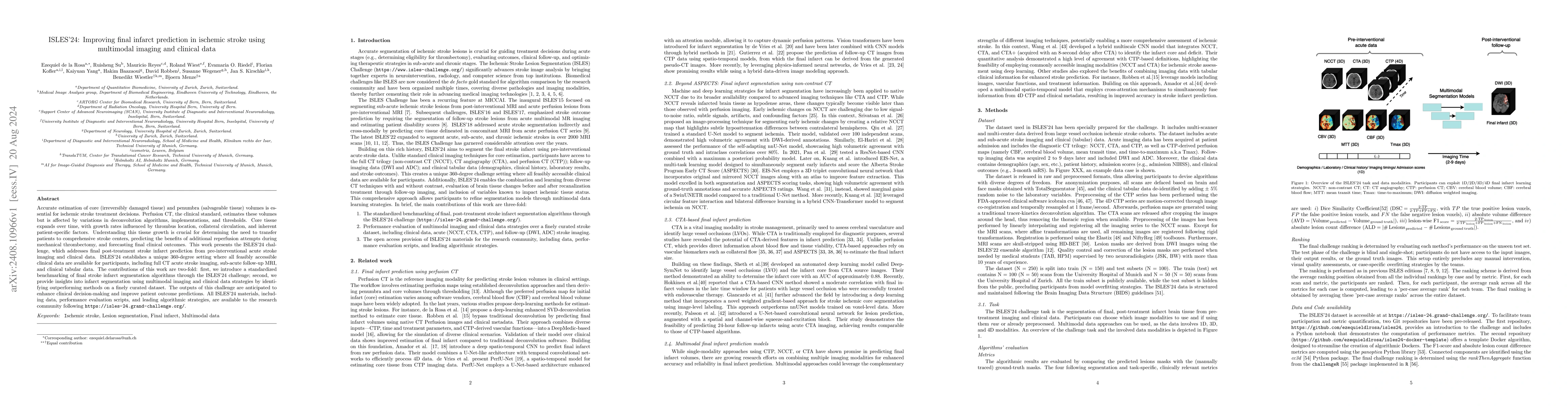
Accurate estimation of core (irreversibly damaged tissue) and penumbra (salvageable tissue) volumes is essential for ischemic stroke treatment decisions. Perfusion CT, the clinical standard, estimates...
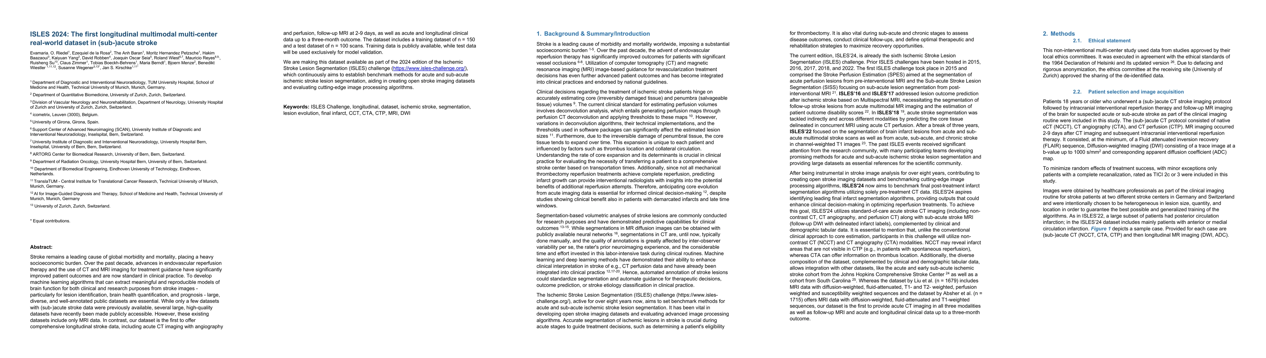
Stroke remains a leading cause of global morbidity and mortality, placing a heavy socioeconomic burden. Over the past decade, advances in endovascular reperfusion therapy and the use of CT and MRI ima...
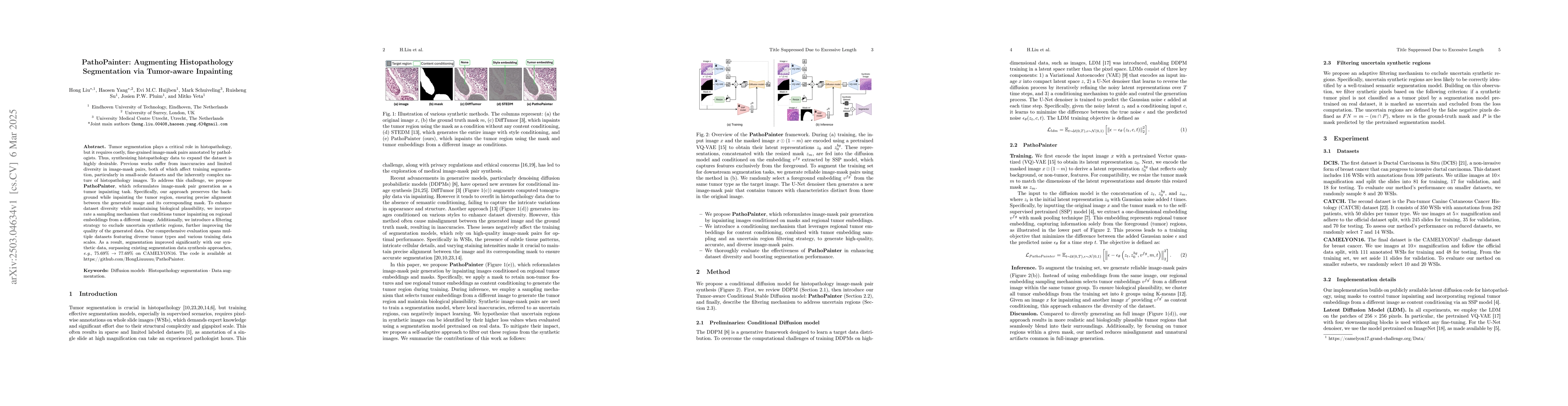
Tumor segmentation plays a critical role in histopathology, but it requires costly, fine-grained image-mask pairs annotated by pathologists. Thus, synthesizing histopathology data to expand the datase...

Distinguishing acute ischemic strokes (AIS) from stroke mimics (SMs), particularly in cases involving medium and small vessel occlusions, remains a significant diagnostic challenge. While computed tom...
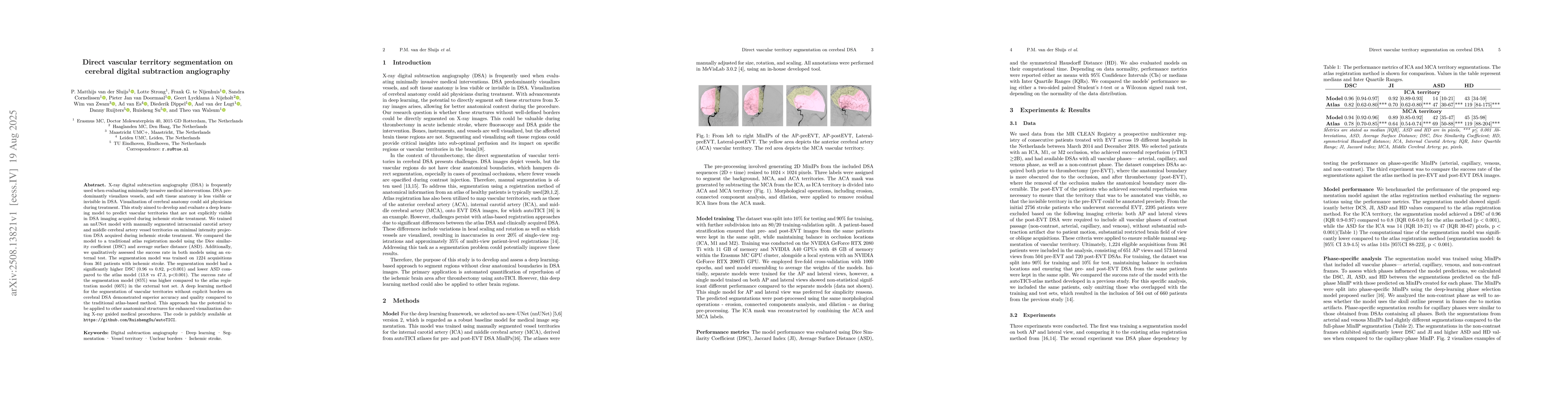
X-ray digital subtraction angiography (DSA) is frequently used when evaluating minimally invasive medical interventions. DSA predominantly visualizes vessels, and soft tissue anatomy is less visible o...
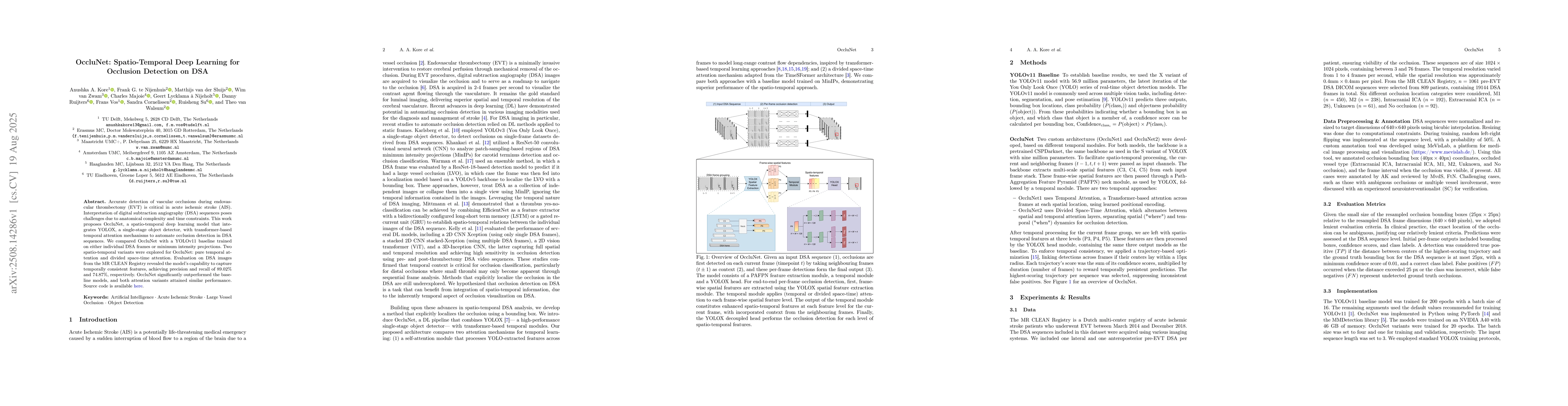
Accurate detection of vascular occlusions during endovascular thrombectomy (EVT) is critical in acute ischemic stroke (AIS). Interpretation of digital subtraction angiography (DSA) sequences poses cha...
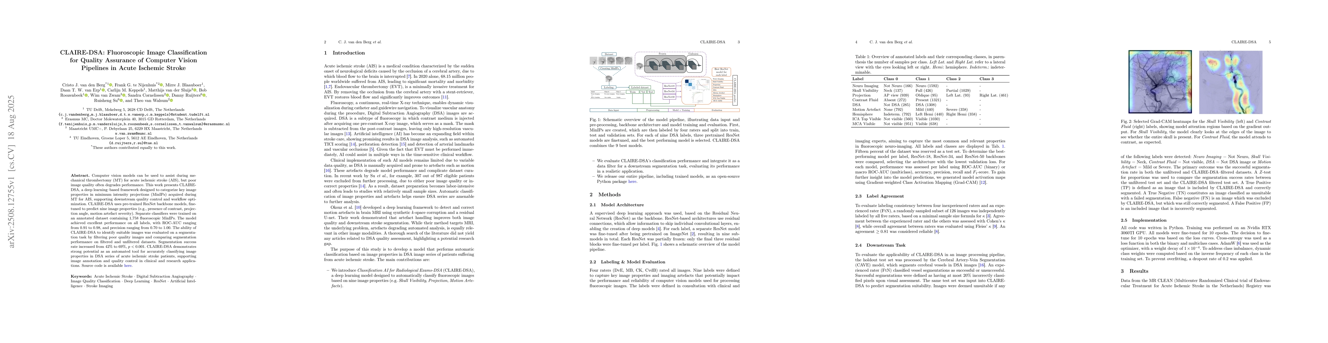
Computer vision models can be used to assist during mechanical thrombectomy (MT) for acute ischemic stroke (AIS), but poor image quality often degrades performance. This work presents CLAIRE-DSA, a de...
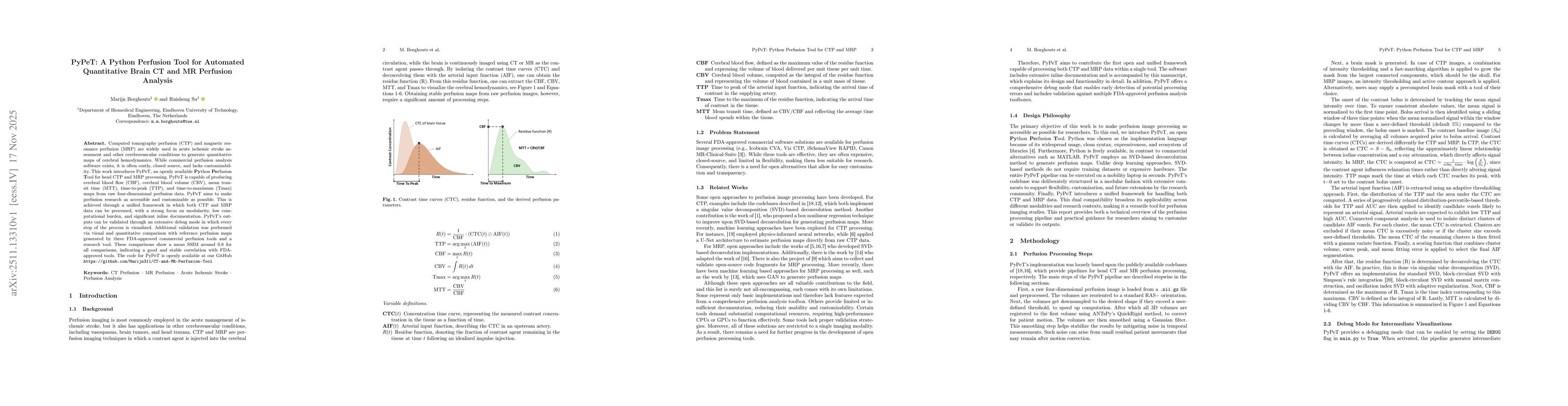
Computed tomography perfusion (CTP) and magnetic resonance perfusion (MRP) are widely used in acute ischemic stroke assessment and other cerebrovascular conditions to generate quantitative maps of cer...
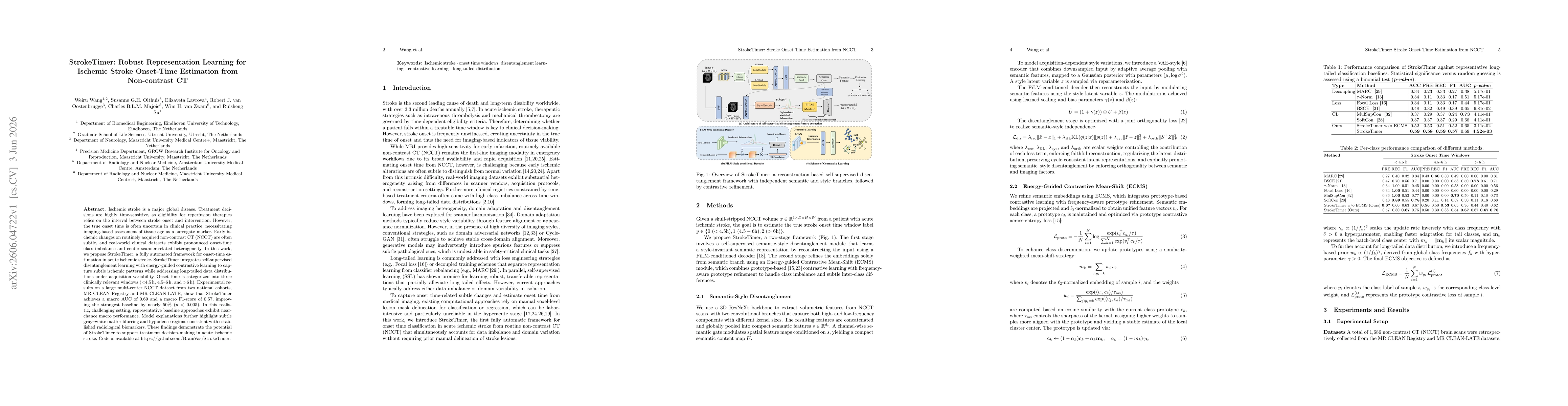
Ischemic stroke is a major global disease. Treatment decisions are highly time-sensitive, as eligibility for reperfusion therapies relies on the interval between stroke onset and intervention. However...