Academic Profile
Statistics
Similar Authors
Papers on arXiv
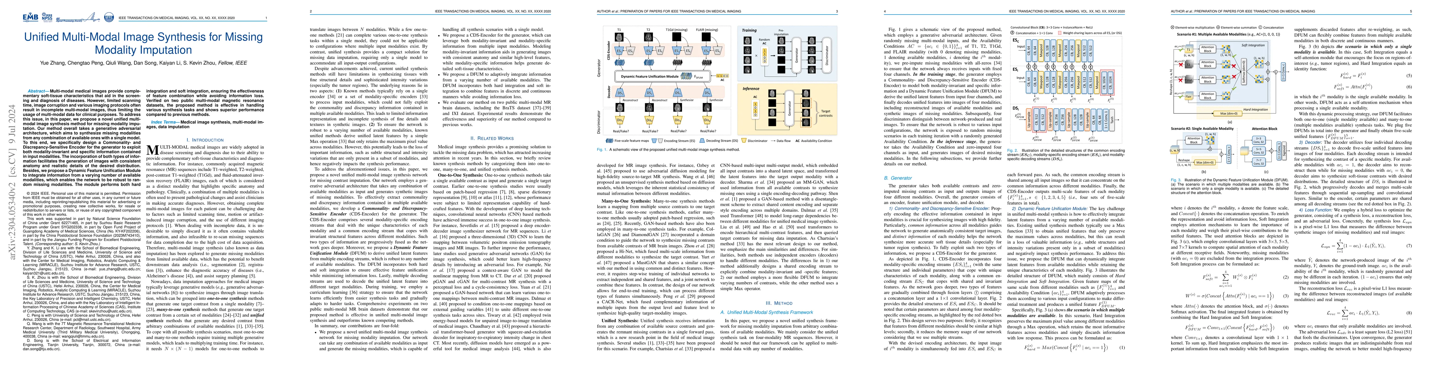
Multi-modal medical images provide complementary soft-tissue characteristics that aid in the screening and diagnosis of diseases. However, limited scanning time, image corruption and various imaging p...
Deep learning algorithms have demonstrated remarkable efficacy in various medical image analysis (MedIA) applications. However, recent research highlights a performance disparity in these algorithms w...
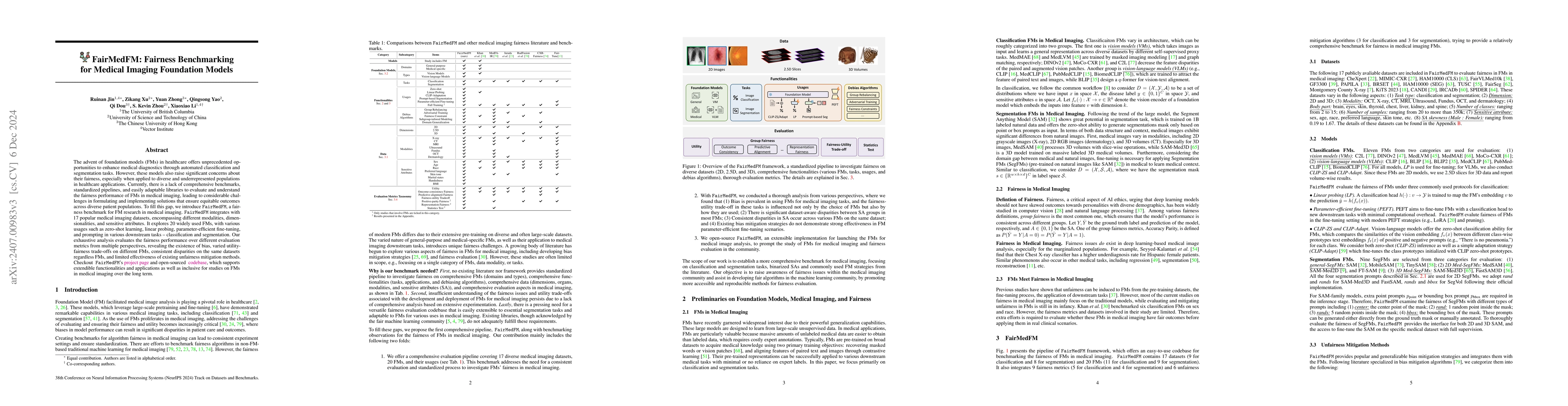
The advent of foundation models (FMs) in healthcare offers unprecedented opportunities to enhance medical diagnostics through automated classification and segmentation tasks. However, these models a...
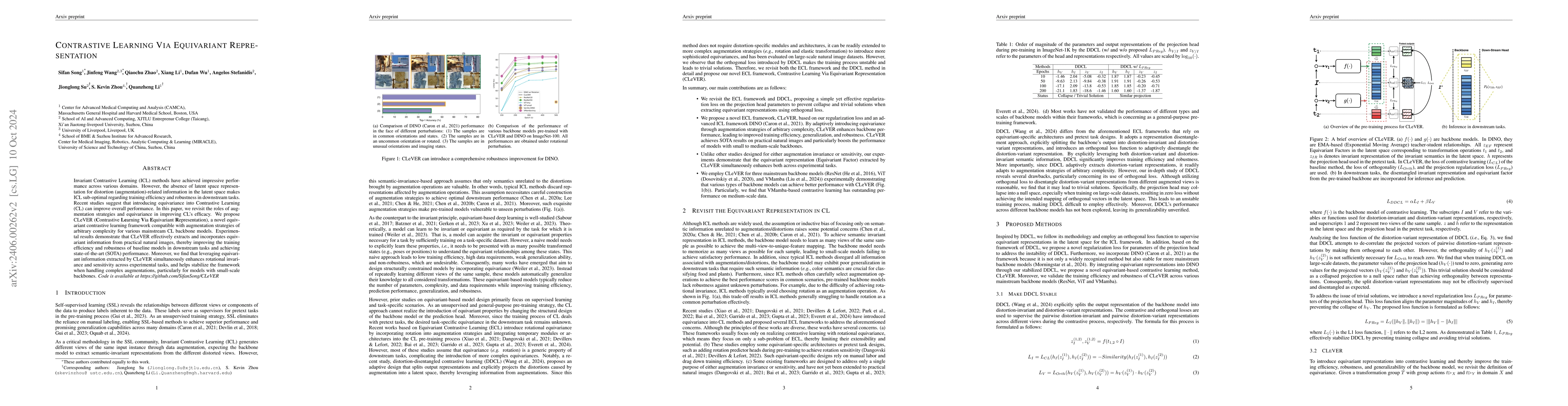
Invariant-based Contrastive Learning (ICL) methods have achieved impressive performance across various domains. However, the absence of latent space representation for distortion (augmentation)-rela...
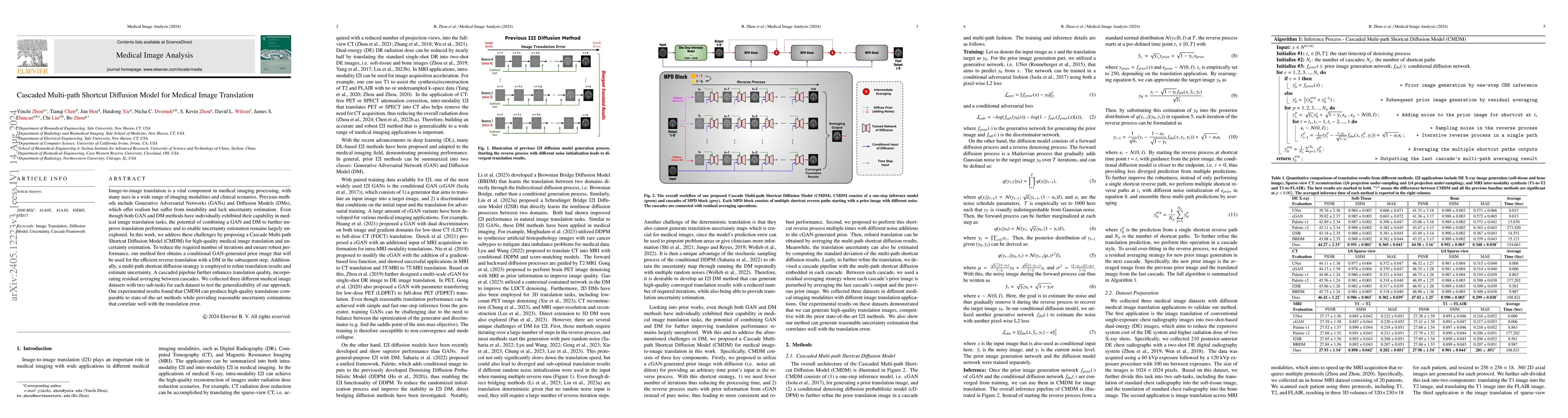
Image-to-image translation is a vital component in medical imaging processing, with many uses in a wide range of imaging modalities and clinical scenarios. Previous methods include Generative Advers...
Emerging unsupervised reconstruction techniques based on implicit neural representation (INR), such as NeRP, CoIL, and SCOPE, have shown unique capabilities in CT linear inverse imaging. In this wor...
Limited-angle and sparse-view computed tomography (LACT and SVCT) are crucial for expanding the scope of X-ray CT applications. However, they face challenges due to incomplete data acquisition, resu...
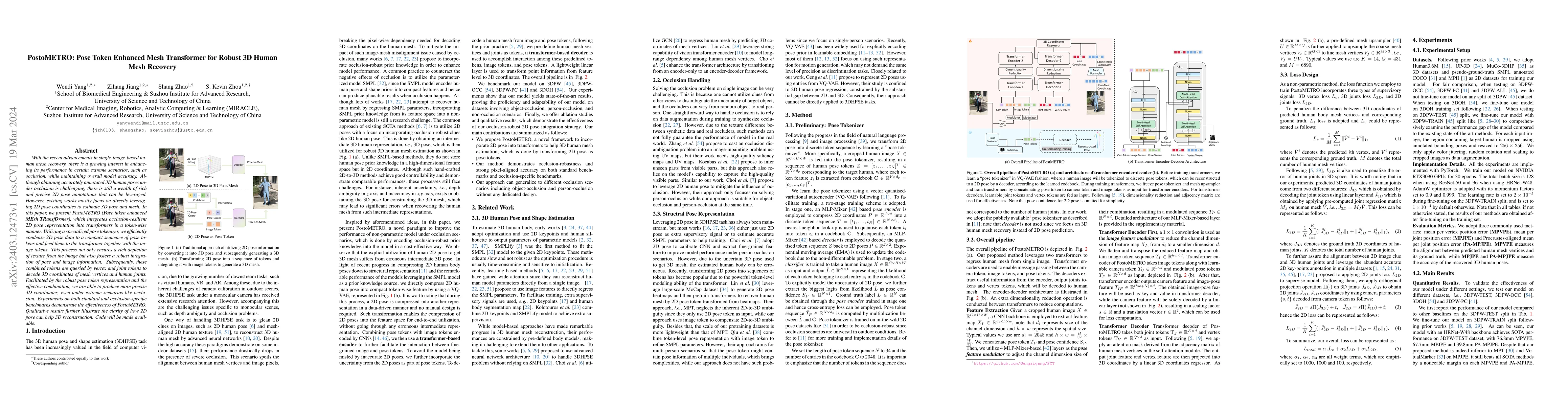
With the recent advancements in single-image-based human mesh recovery, there is a growing interest in enhancing its performance in certain extreme scenarios, such as occlusion, while maintaining ov...
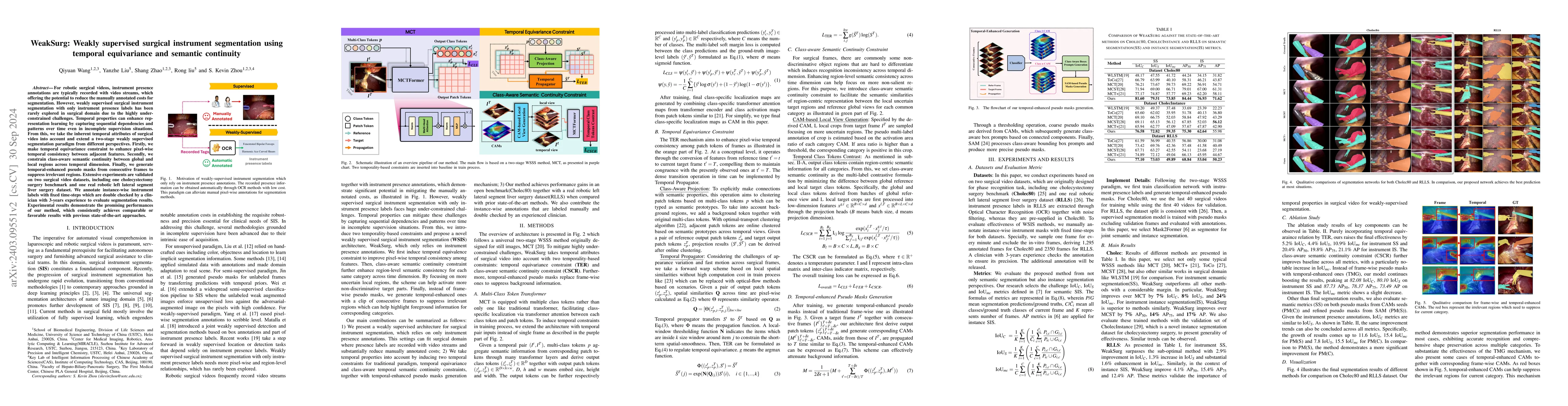
Weakly supervised surgical instrument segmentation with only instrument presence labels has been rarely explored in surgical domain. To mitigate the highly under-constrained challenges, we extend a ...
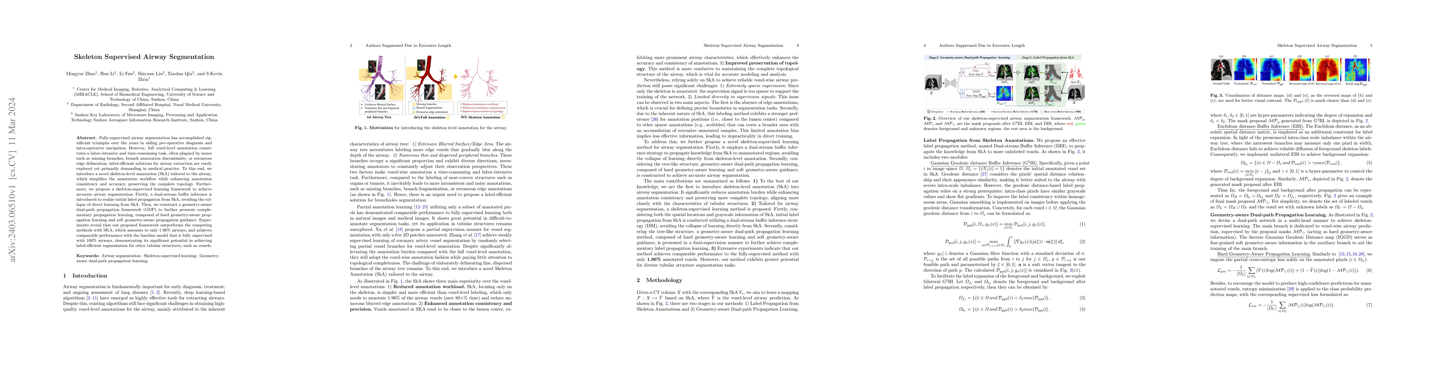
Fully-supervised airway segmentation has accomplished significant triumphs over the years in aiding pre-operative diagnosis and intra-operative navigation. However, full voxel-level annotation const...
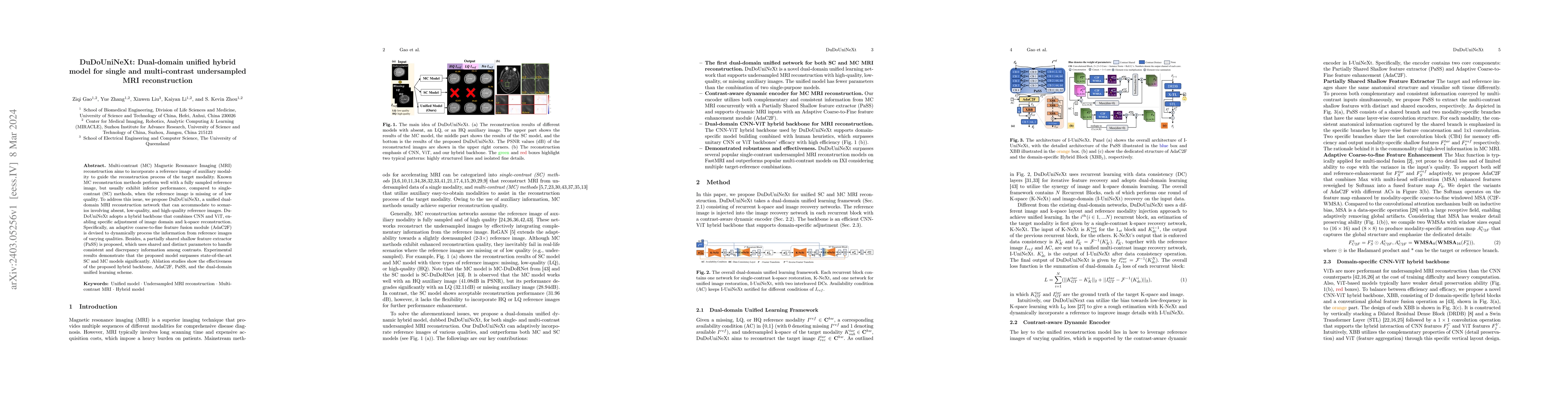
Multi-contrast (MC) Magnetic Resonance Imaging (MRI) reconstruction aims to incorporate a reference image of auxiliary modality to guide the reconstruction process of the target modality. Known MC r...
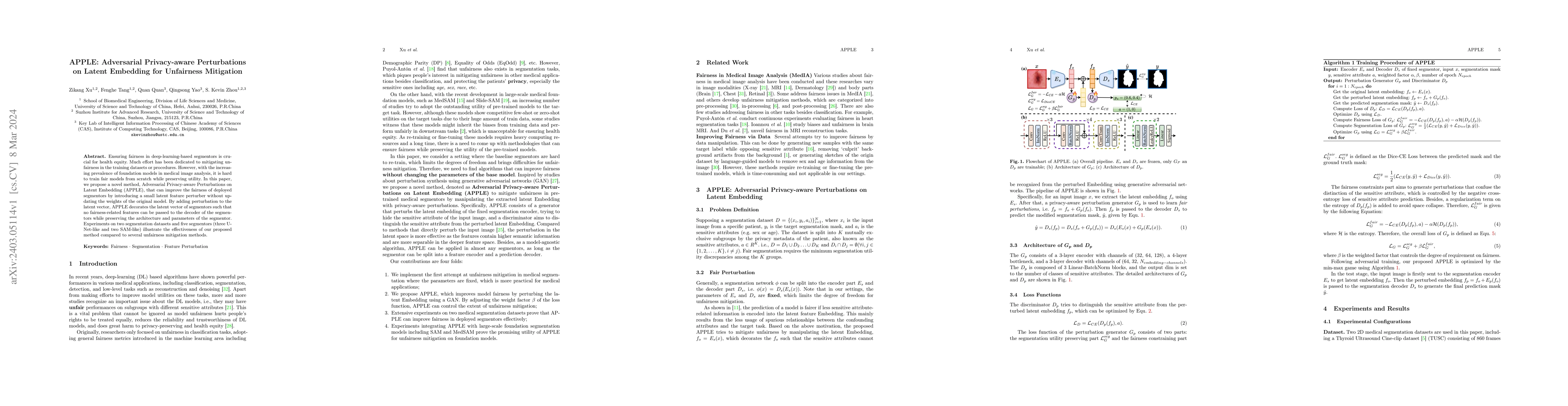
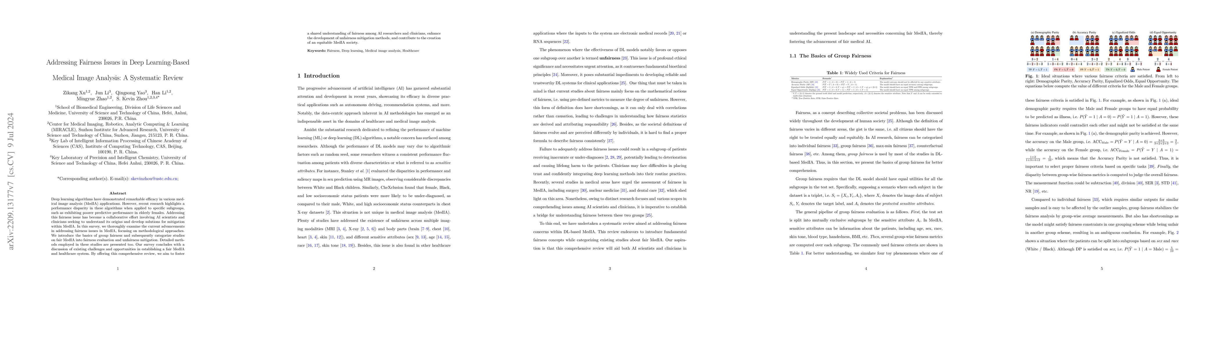
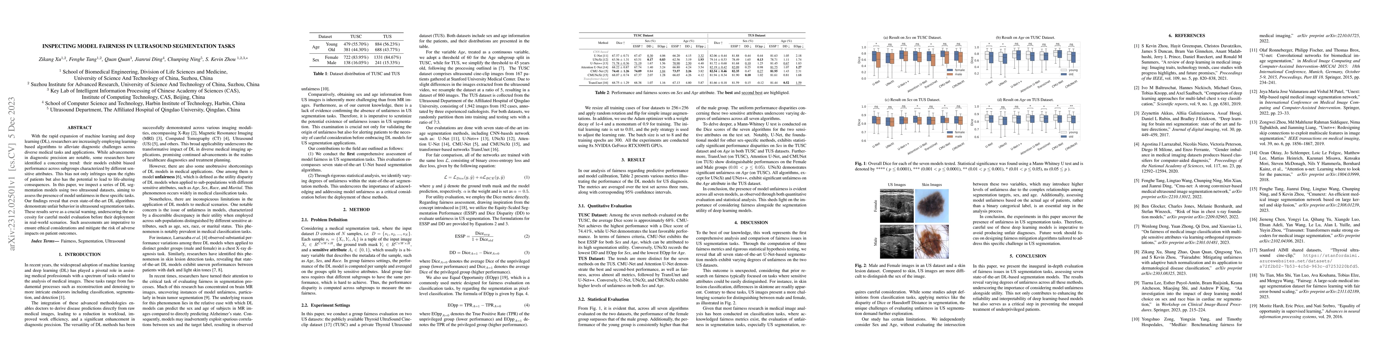
Ensuring fairness in deep-learning-based segmentors is crucial for health equity. Much effort has been dedicated to mitigating unfairness in the training datasets or procedures. However, with the in...
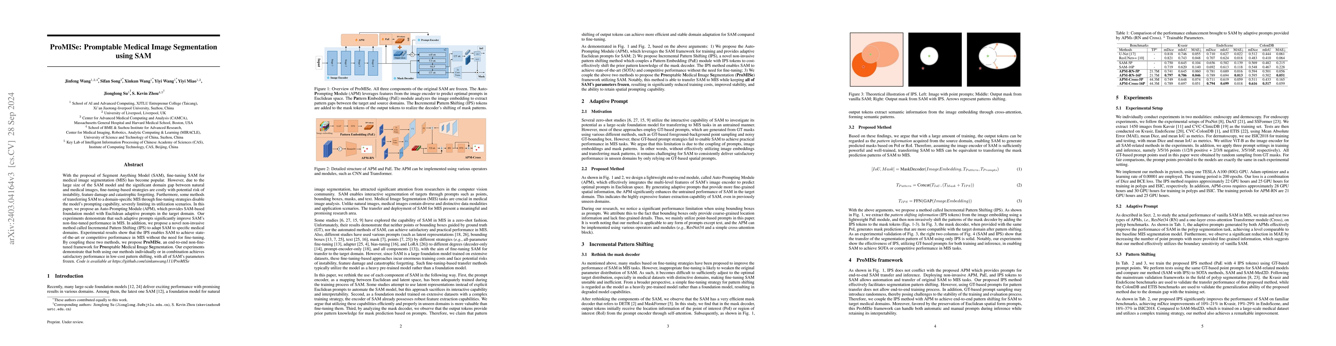
With the proposal of the Segment Anything Model (SAM), fine-tuning SAM for medical image segmentation (MIS) has become popular. However, due to the large size of the SAM model and the significant do...
In the realm of distributed systems tasked with managing and processing large-scale graph-structured data, optimizing graph partitioning stands as a pivotal challenge. The primary goal is to minimiz...
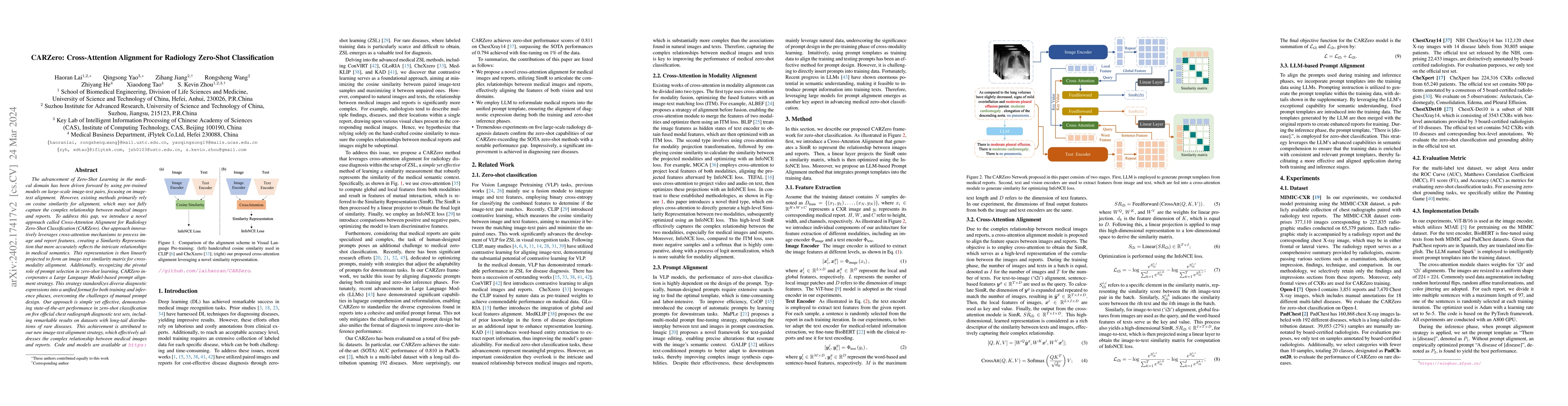
The advancement of Zero-Shot Learning in the medical domain has been driven forward by using pre-trained models on large-scale image-text pairs, focusing on image-text alignment. However, existing m...
When deploying a trained machine learning model in the real world, it is inevitable to receive inputs from out-of-distribution (OOD) sources. For instance, in continual learning settings, it is comm...
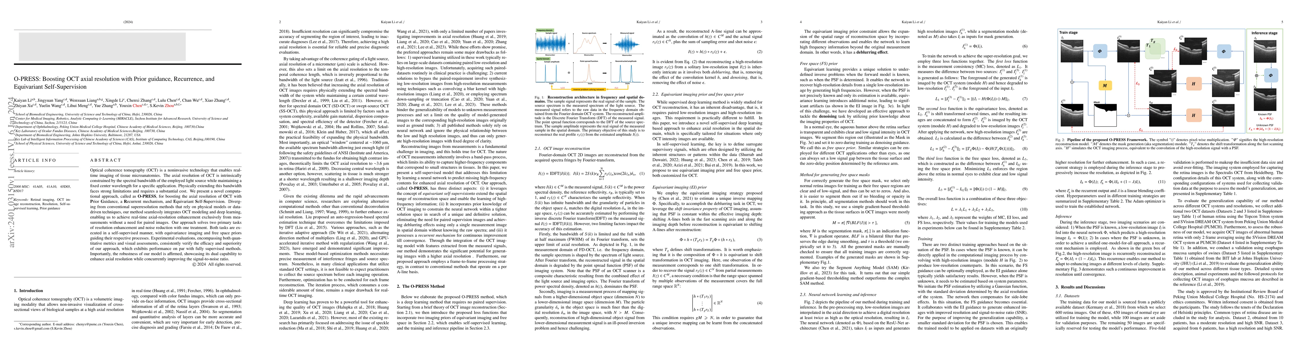
Optical coherence tomography (OCT) is a noninvasive technology that enables real-time imaging of tissue microanatomies. The axial resolution of OCT is intrinsically constrained by the spectral bandw...
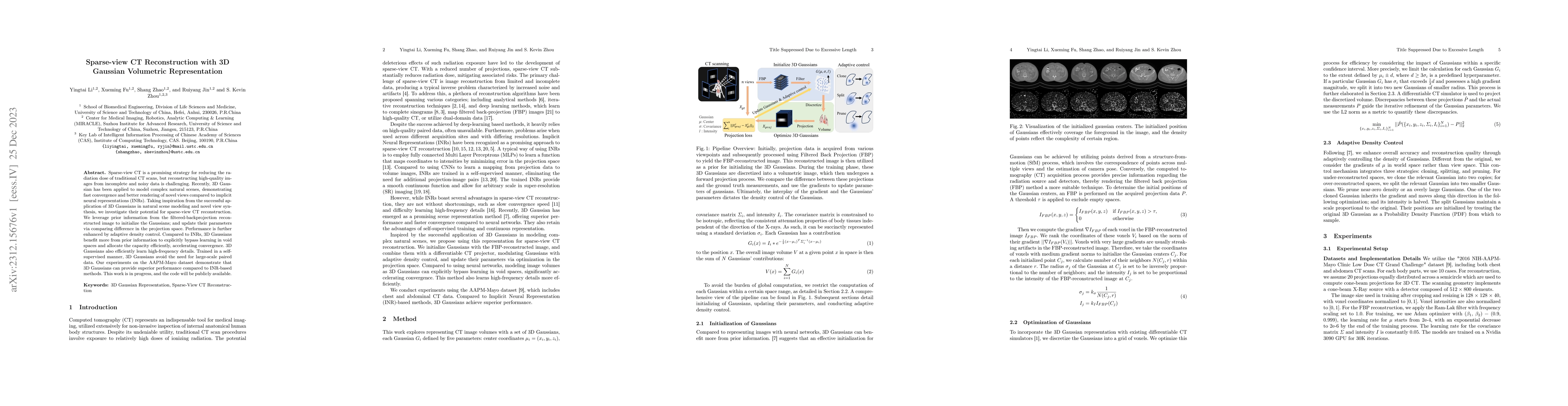
Sparse-view CT is a promising strategy for reducing the radiation dose of traditional CT scans, but reconstructing high-quality images from incomplete and noisy data is challenging. Recently, 3D Gau...
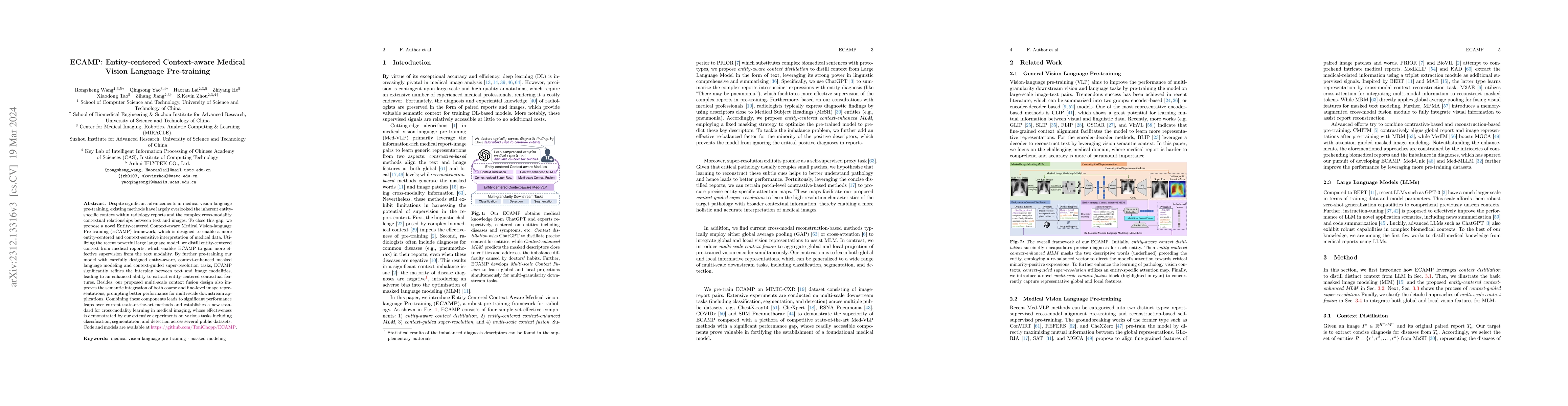
Despite significant advancements in medical vision-language pre-training, existing methods have largely overlooked the inherent entity-specific context within radiology reports and the complex cross...
With the rapid expansion of machine learning and deep learning (DL), researchers are increasingly employing learning-based algorithms to alleviate diagnostic challenges across diverse medical tasks ...
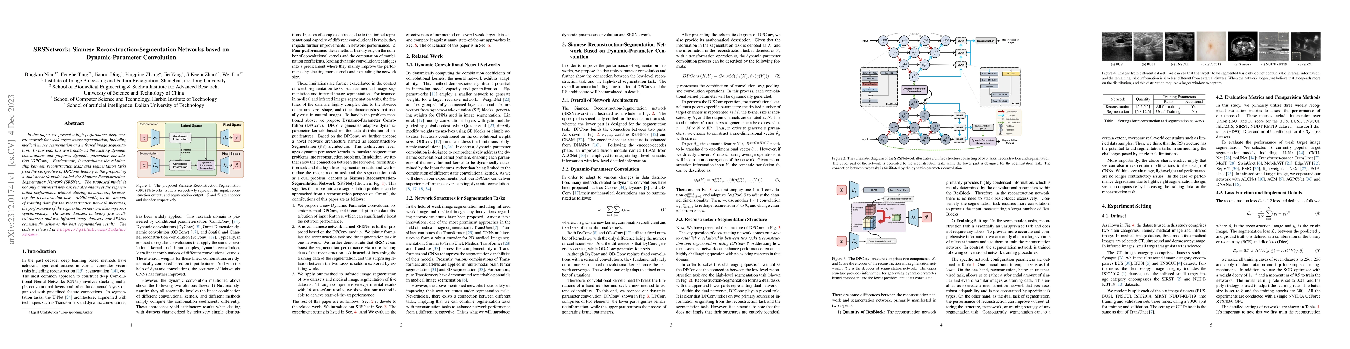
In this paper, we present a high-performance deep neural network for weak target image segmentation, including medical image segmentation and infrared image segmentation. To this end, this work anal...
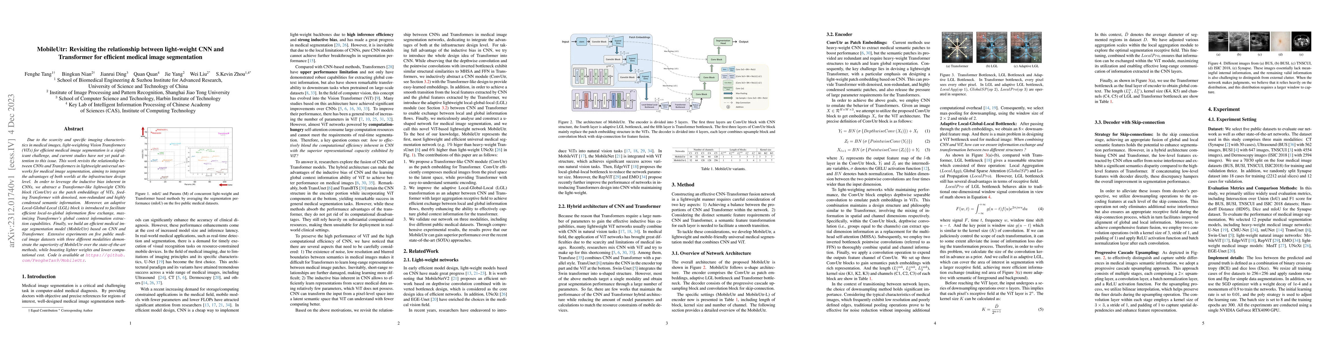
Due to the scarcity and specific imaging characteristics in medical images, light-weighting Vision Transformers (ViTs) for efficient medical image segmentation is a significant challenge, and curren...
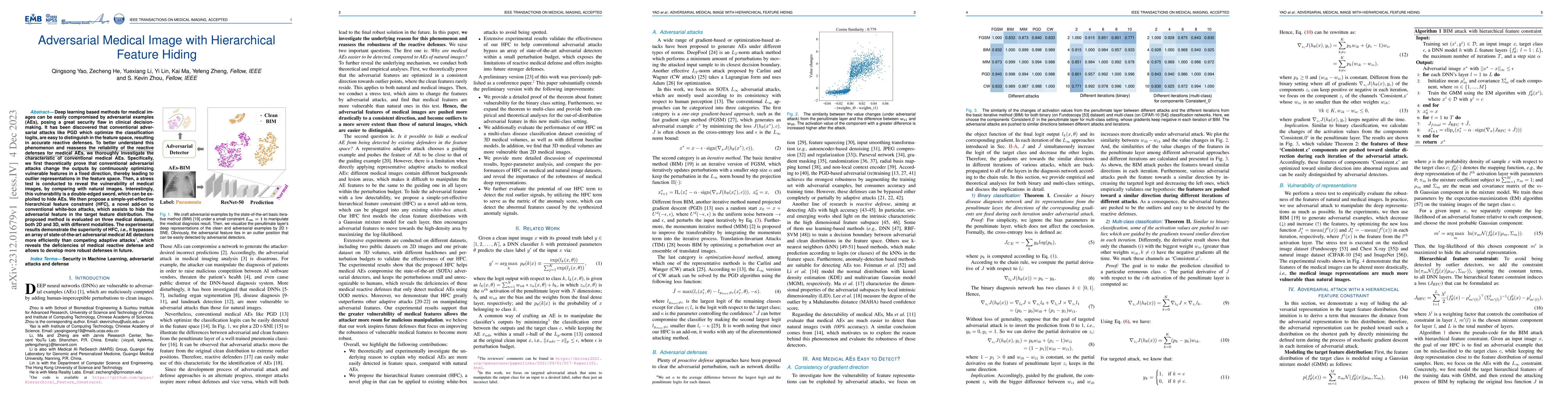
Deep learning based methods for medical images can be easily compromised by adversarial examples (AEs), posing a great security flaw in clinical decision-making. It has been discovered that conventi...
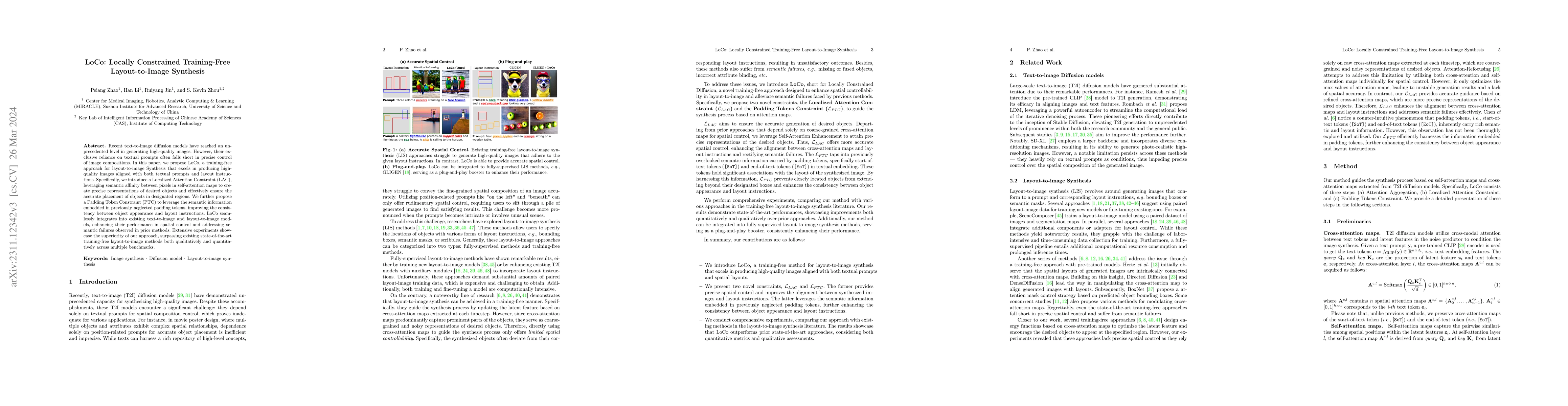
Recent text-to-image diffusion models have reached an unprecedented level in generating high-quality images. However, their exclusive reliance on textual prompts often falls short in precise control...
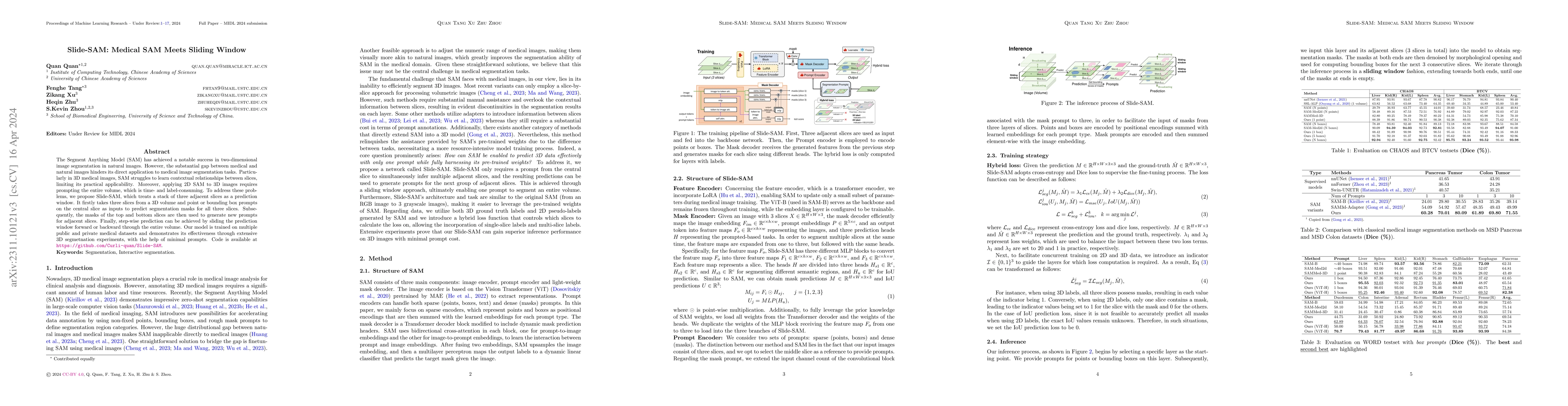
The Segment Anything Model (SAM) has achieved a notable success in two-dimensional image segmentation in natural images. However, the substantial gap between medical and natural images hinders its d...
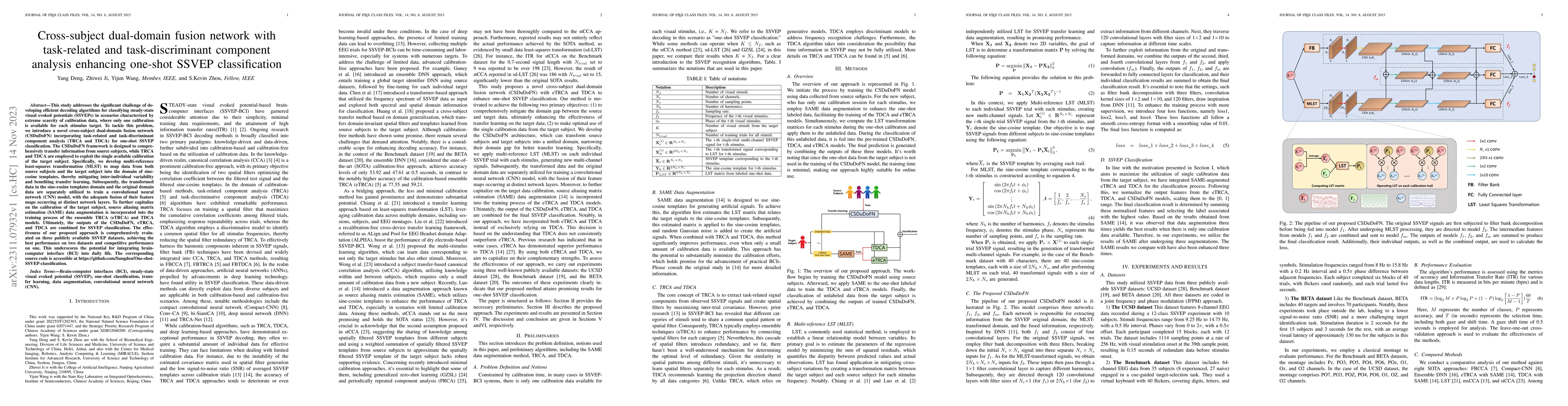
This study addresses the significant challenge of developing efficient decoding algorithms for classifying steady-state visual evoked potentials (SSVEPs) in scenarios characterized by extreme scarci...
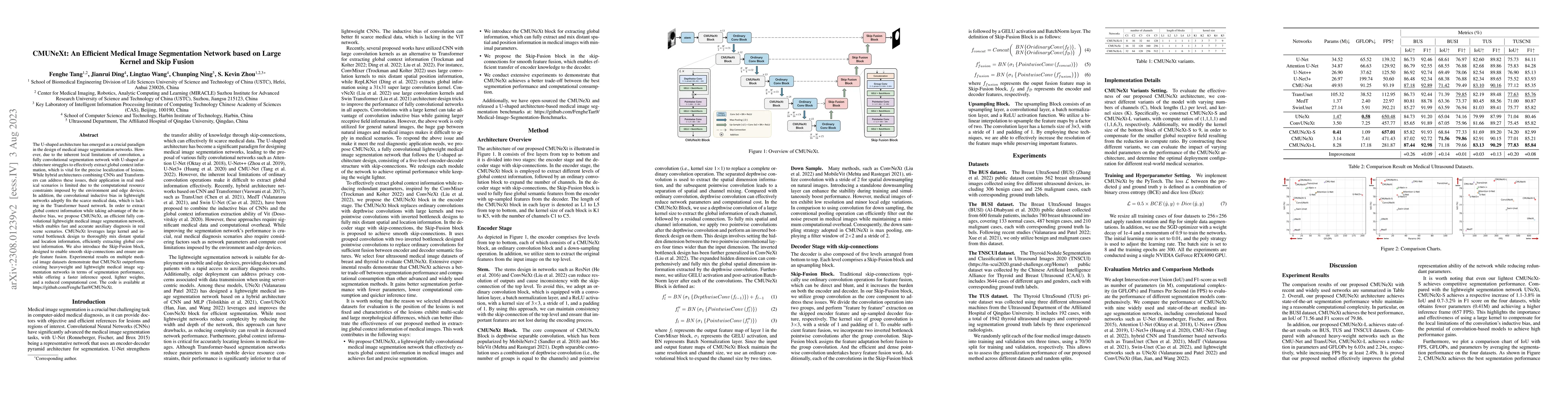
The U-shaped architecture has emerged as a crucial paradigm in the design of medical image segmentation networks. However, due to the inherent local limitations of convolution, a fully convolutional...
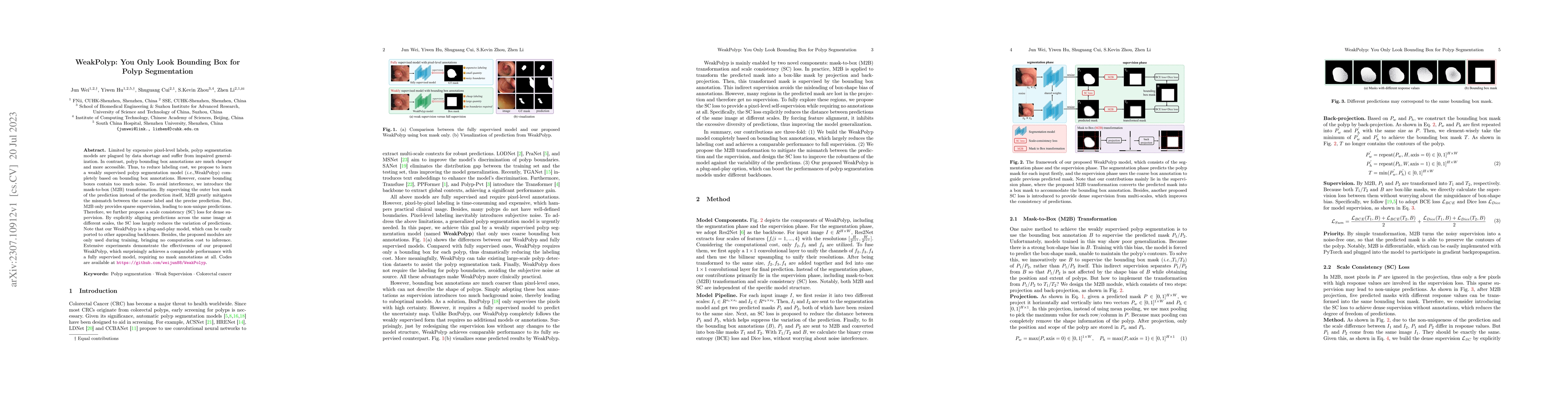
Limited by expensive pixel-level labels, polyp segmentation models are plagued by data shortage and suffer from impaired generalization. In contrast, polyp bounding box annotations are much cheaper ...
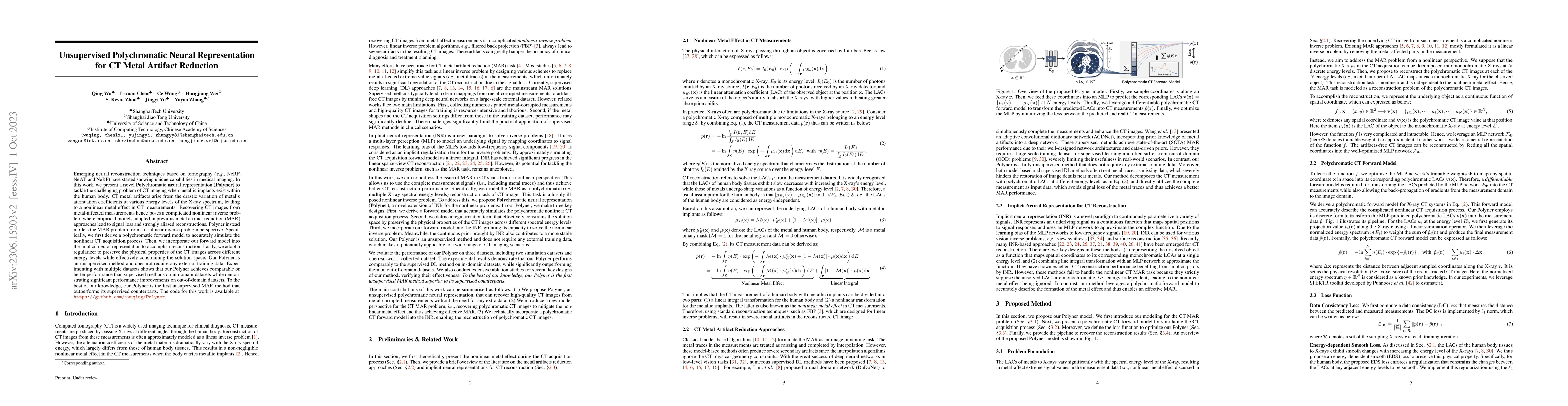
Emerging neural reconstruction techniques based on tomography (e.g., NeRF, NeAT, and NeRP) have started showing unique capabilities in medical imaging. In this work, we present a novel Polychromatic...
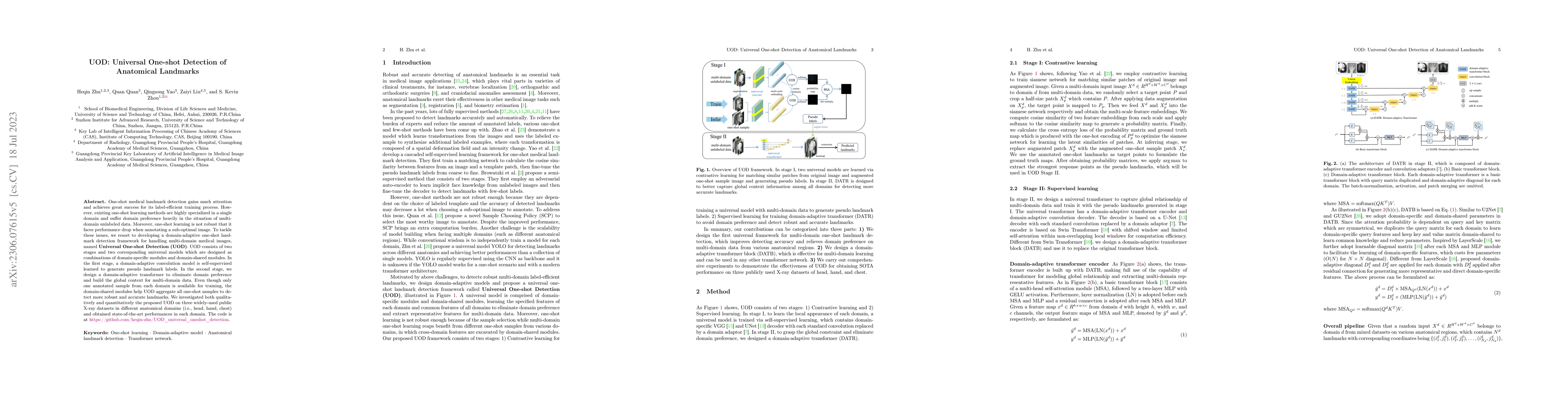
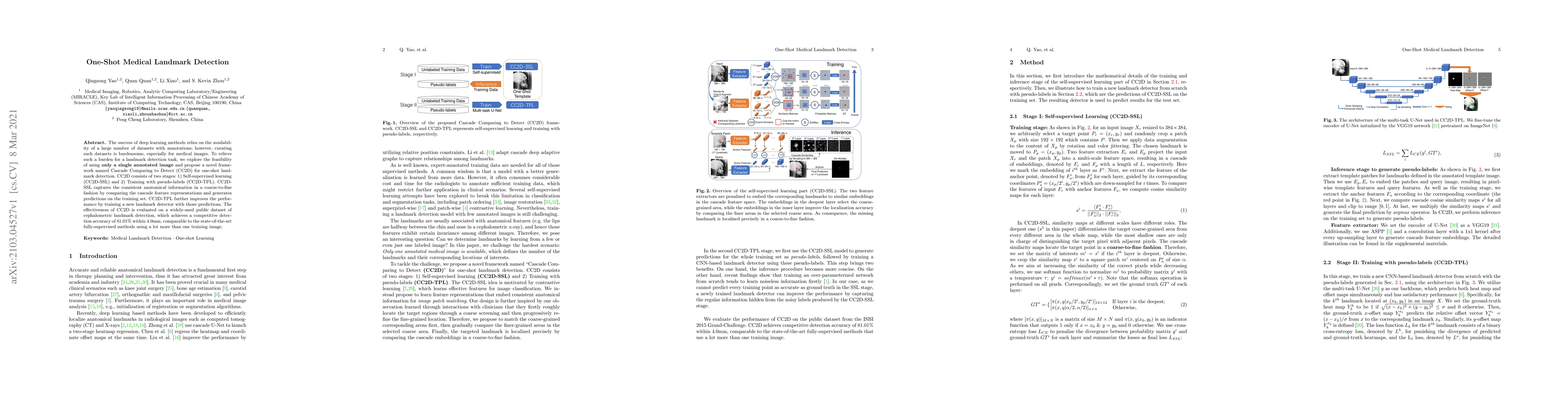
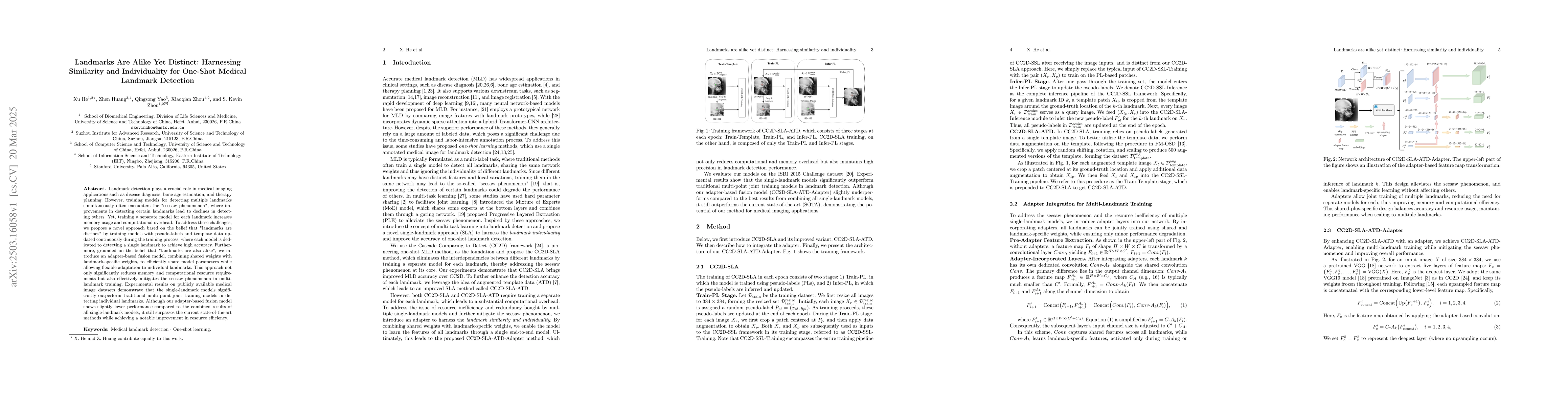
One-shot medical landmark detection gains much attention and achieves great success for its label-efficient training process. However, existing one-shot learning methods are highly specialized in a ...
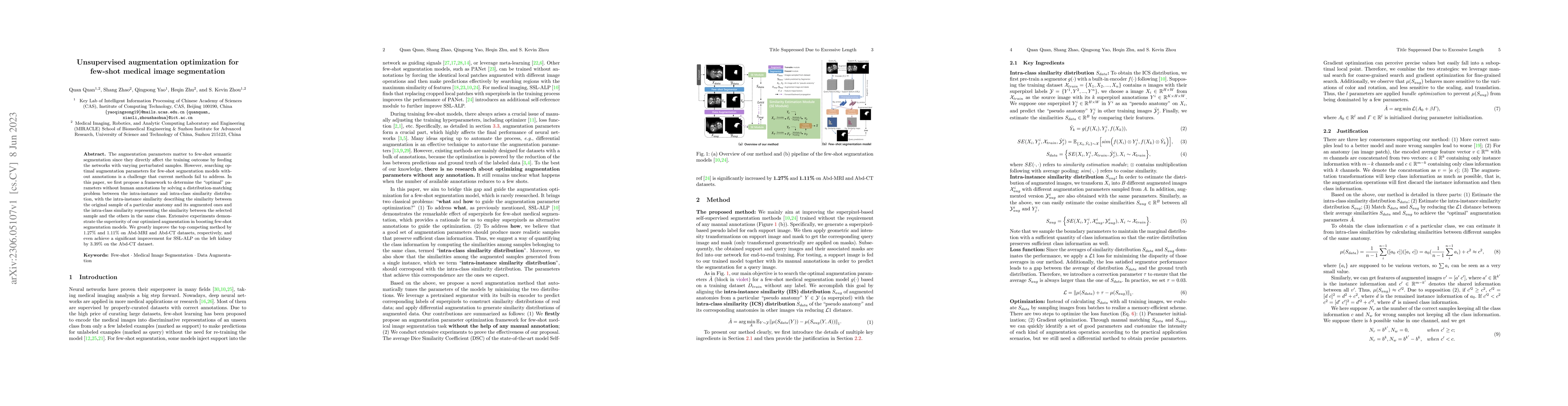
The augmentation parameters matter to few-shot semantic segmentation since they directly affect the training outcome by feeding the networks with varying perturbated samples. However, searching opti...

Domain shift and label scarcity heavily limit deep learning applications to various medical image analysis tasks. Unsupervised domain adaptation (UDA) techniques have recently achieved promising cro...
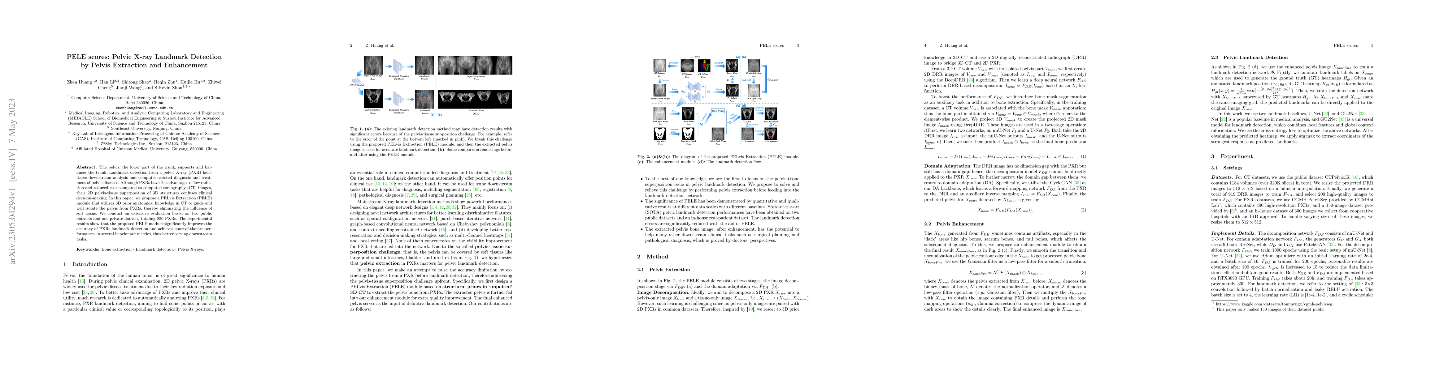
The pelvis, the lower part of the trunk, supports and balances the trunk. Landmark detection from a pelvic X-ray (PXR) facilitates downstream analysis and computer-assisted diagnosis and treatment o...
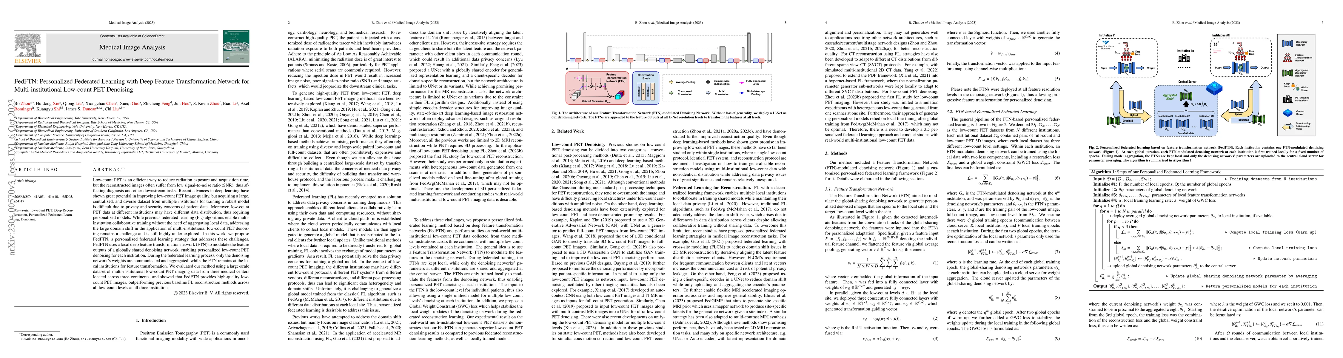
Low-count PET is an efficient way to reduce radiation exposure and acquisition time, but the reconstructed images often suffer from low signal-to-noise ratio (SNR), thus affecting diagnosis and othe...
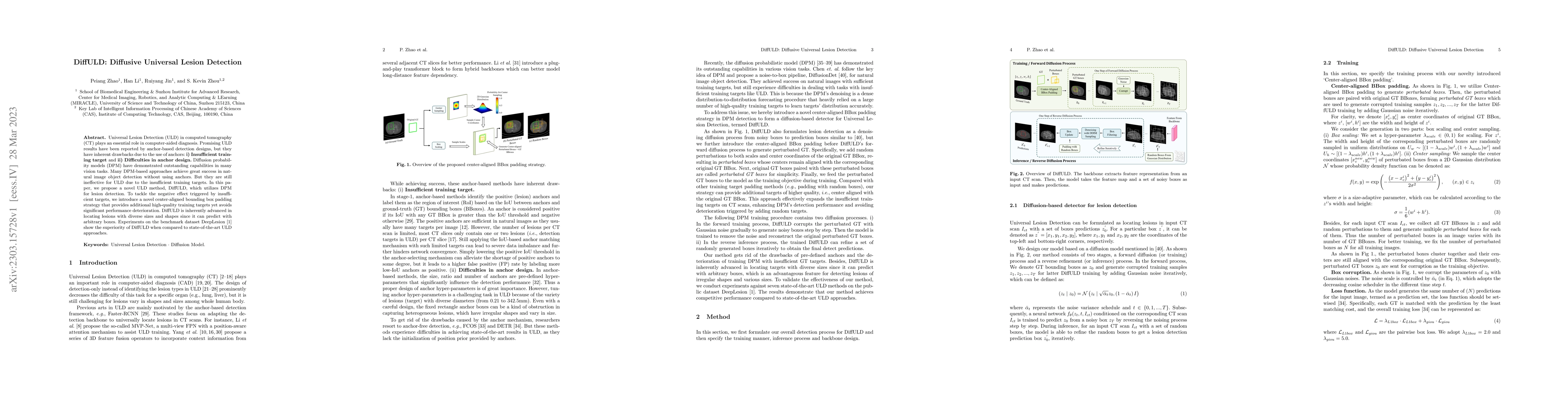
Universal Lesion Detection (ULD) in computed tomography (CT) plays an essential role in computer-aided diagnosis. Promising ULD results have been reported by anchor-based detection designs, but they...
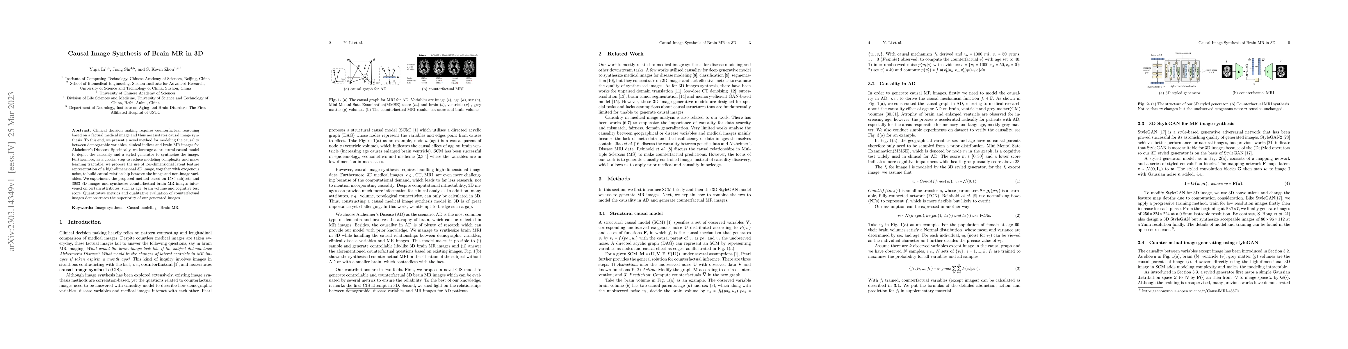
Clinical decision making requires counterfactual reasoning based on a factual medical image and thus necessitates causal image synthesis. To this end, we present a novel method for modeling the caus...
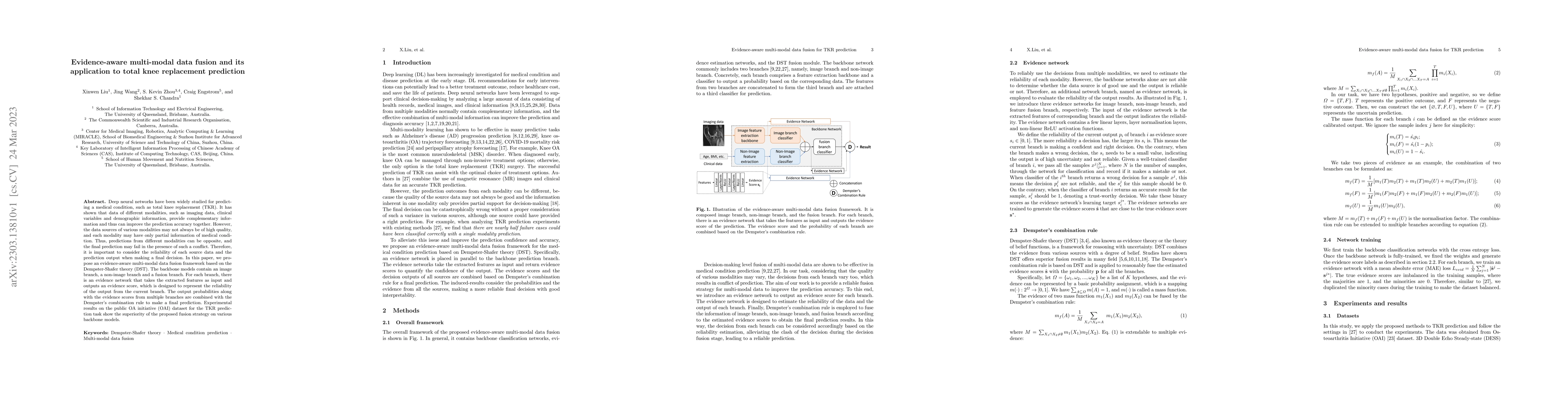
Deep neural networks have been widely studied for predicting a medical condition, such as total knee replacement (TKR). It has shown that data of different modalities, such as imaging data, clinical...
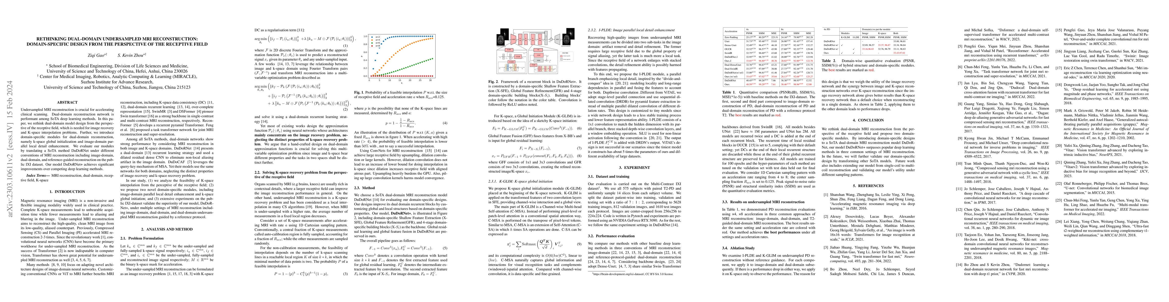
Undersampled MRI reconstruction is crucial for accelerating clinical scanning. Dual-domain reconstruction network is performant among SoTA deep learning methods. In this paper, we rethink dual-domai...
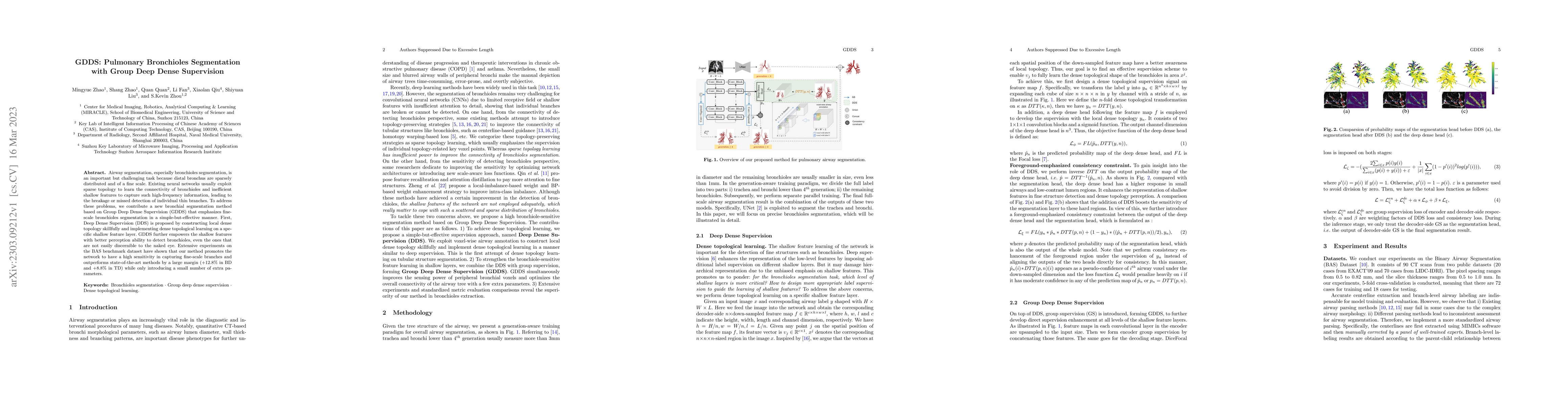
Airway segmentation, especially bronchioles segmentation, is an important but challenging task because distal bronchus are sparsely distributed and of a fine scale. Existing neural networks usually ...

Radiologists possess diverse training and clinical experiences, leading to variations in the segmentation annotations of lung nodules and resulting in segmentation uncertainty.Conventional methods t...
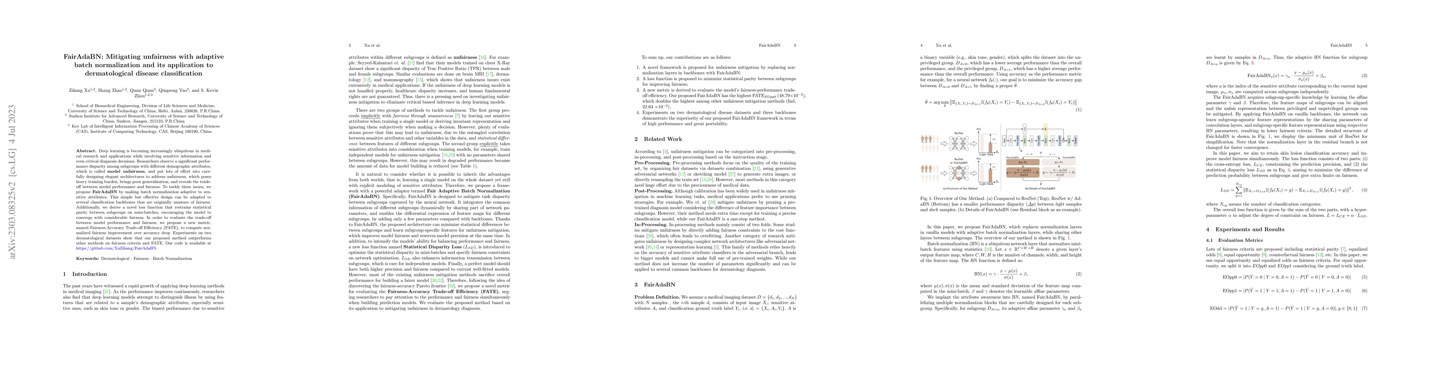
Deep learning is becoming increasingly ubiquitous in medical research and applications while involving sensitive information and even critical diagnosis decisions. Researchers observe a significant ...
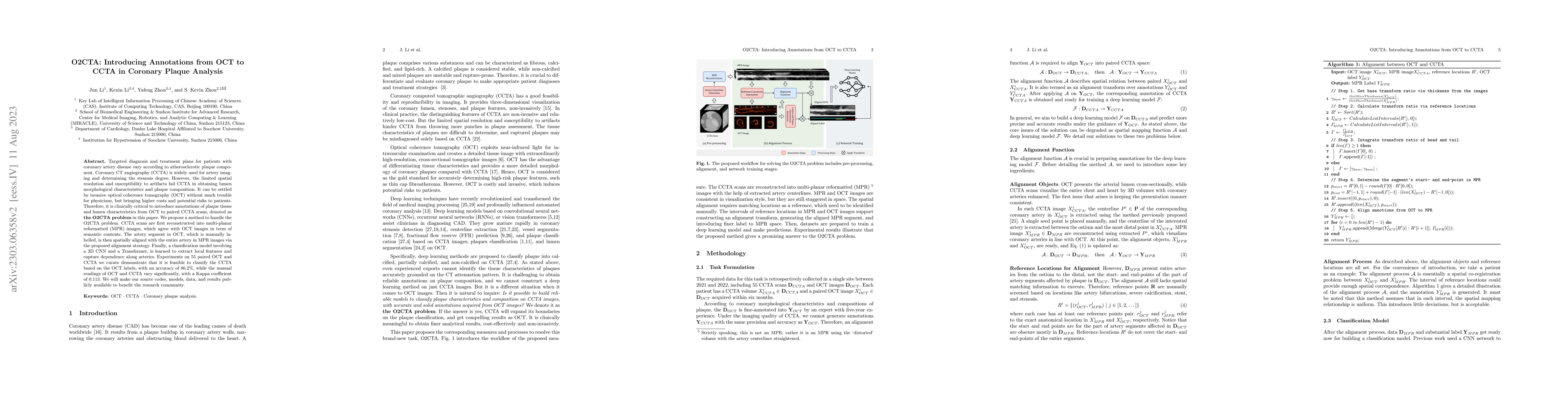
Targeted diagnosis and treatment plans for patients with coronary artery disease vary according to atherosclerotic plaque component. Coronary CT angiography (CCTA) is widely used for artery imaging ...
Self-supervised learning is well known for its remarkable performance in representation learning and various downstream computer vision tasks. Recently, Positive-pair-Only Contrastive Learning (POCL...
Self-paced curriculum learning (SCL) has demonstrated its great potential in computer vision, natural language processing, etc. During training, it implements easy-to-hard sampling based on online e...
Objective and Impact Statement: Accurate organ segmentation is critical for many clinical applications at different clinical sites, which may have their specific application requirements that concer...
Autonomous robotic surgery has advanced significantly based on analysis of visual and temporal cues in surgical workflow, but relational cues from domain knowledge remain under investigation. Comple...
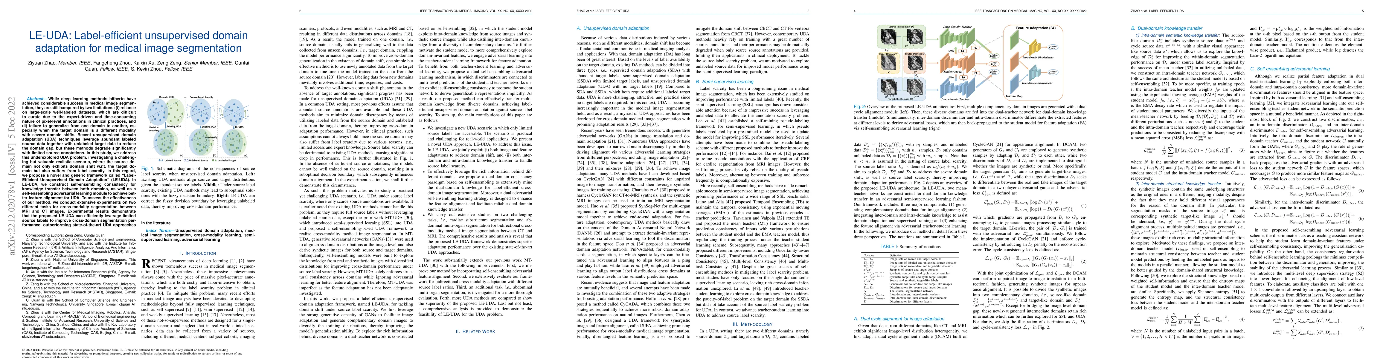
While deep learning methods hitherto have achieved considerable success in medical image segmentation, they are still hampered by two limitations: (i) reliance on large-scale well-labeled datasets, ...
Fast screening and diagnosis are critical in COVID-19 patient treatment. In addition to the gold standard RT-PCR, radiological imaging like X-ray and CT also works as an important means in patient s...
Artificial Intelligence (AI) is having a tremendous impact across most areas of science. Applications of AI in healthcare have the potential to improve our ability to detect, diagnose, prognose, and...
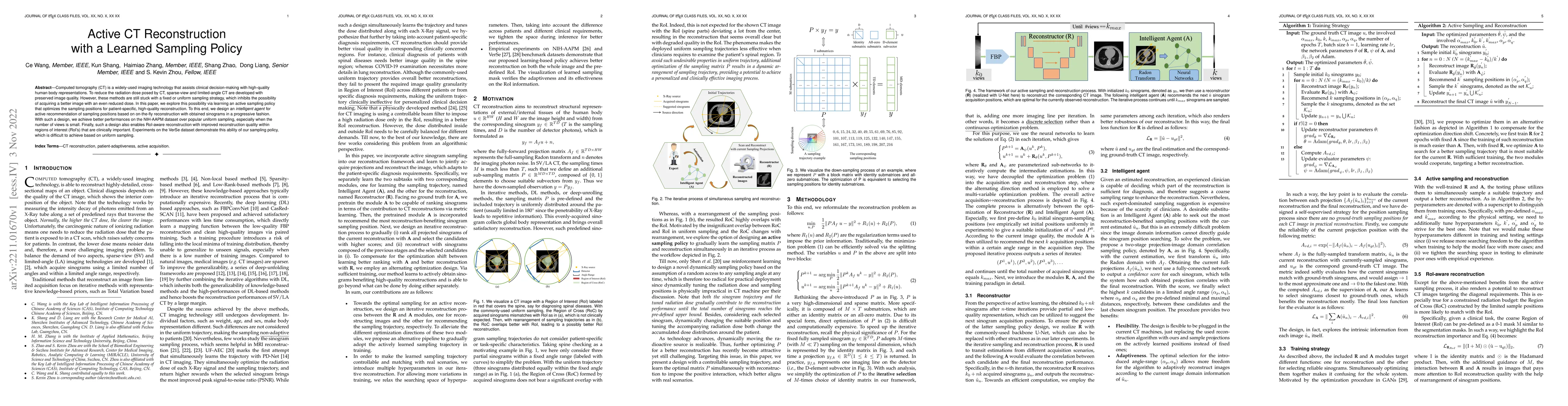
Computed tomography (CT) is a widely-used imaging technology that assists clinical decision-making with high-quality human body representations. To reduce the radiation dose posed by CT, sparse-view...
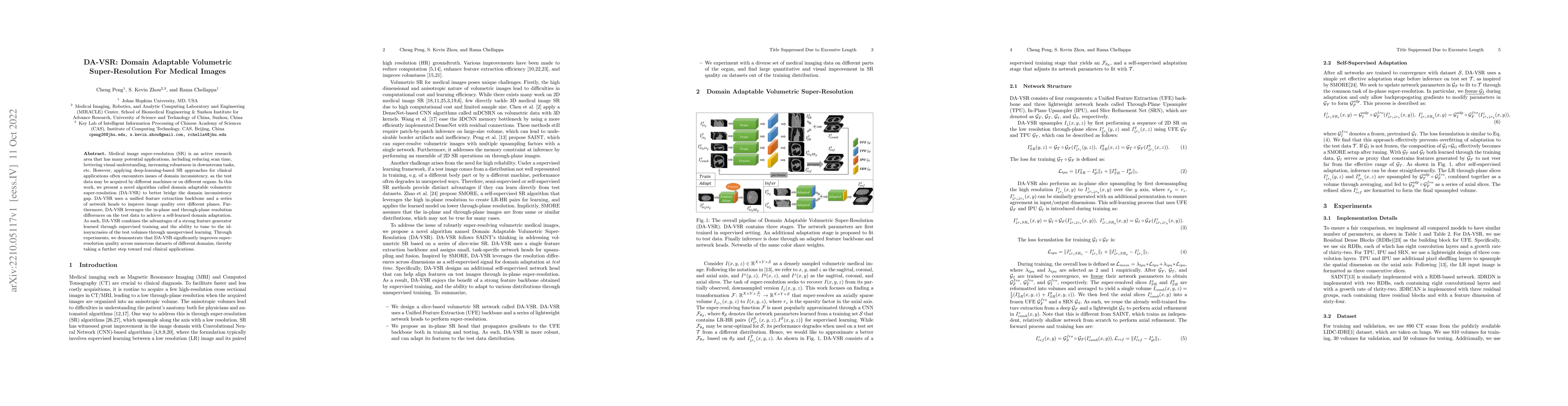
Medical image super-resolution (SR) is an active research area that has many potential applications, including reducing scan time, bettering visual understanding, increasing robustness in downstream...
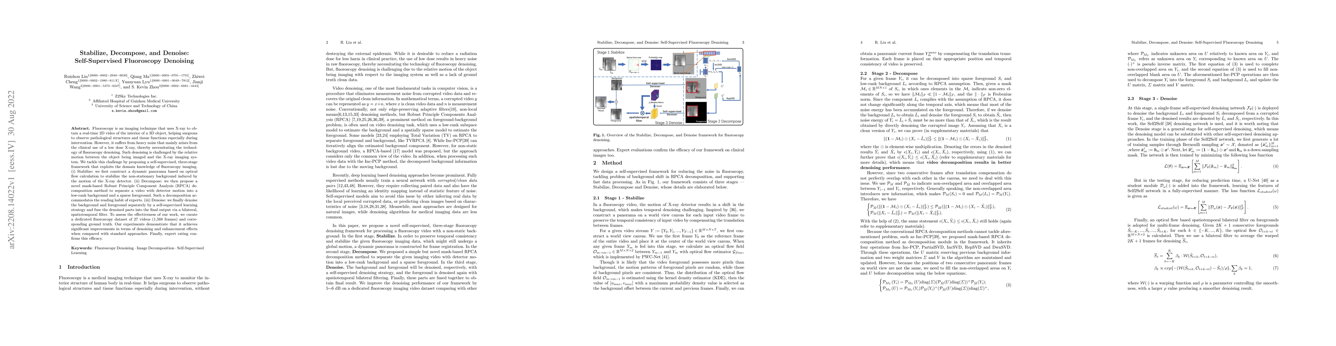
Fluoroscopy is an imaging technique that uses X-ray to obtain a real-time 2D video of the interior of a 3D object, helping surgeons to observe pathological structures and tissue functions especially...
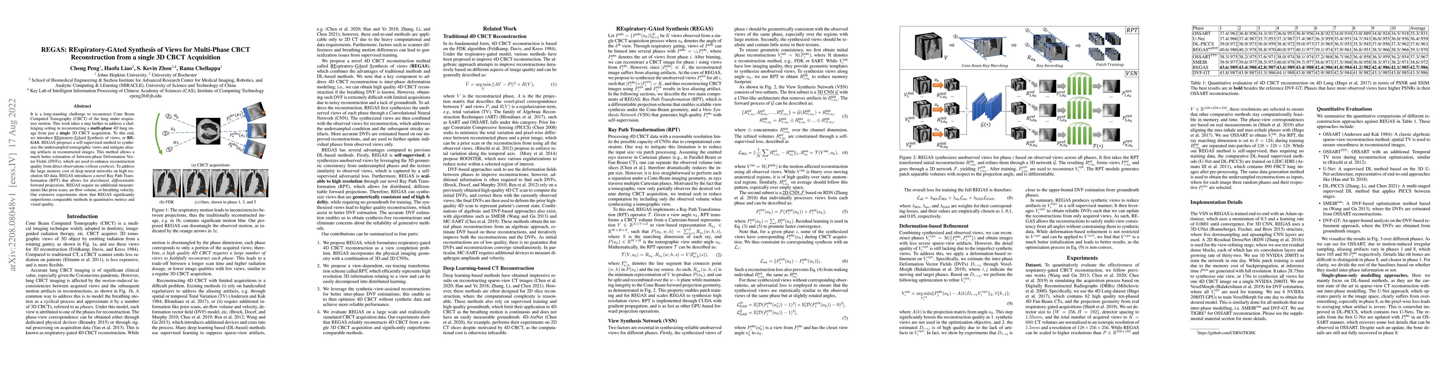
It is a long-standing challenge to reconstruct Cone Beam Computed Tomography (CBCT) of the lung under respiratory motion. This work takes a step further to address a challenging setting in reconstru...
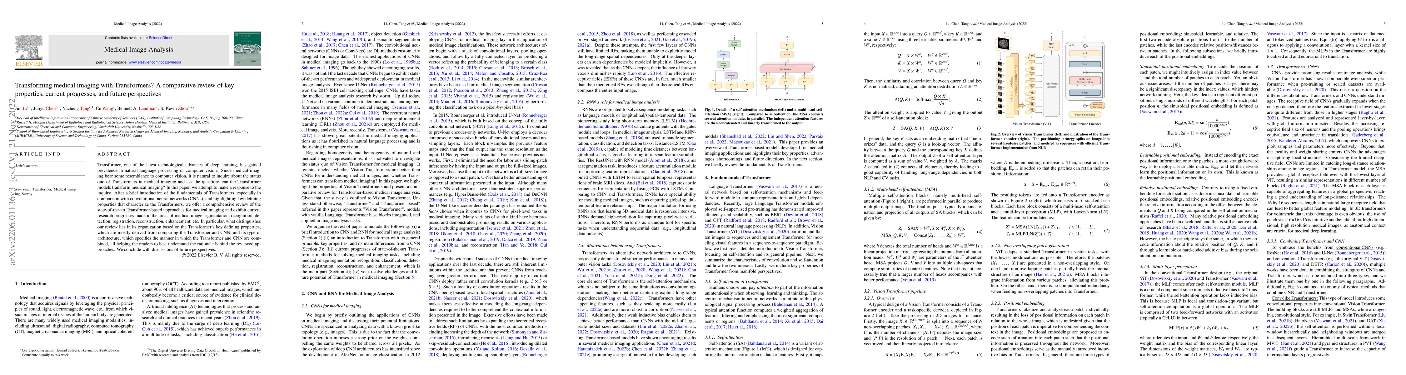
Transformer, the latest technological advance of deep learning, has gained prevalence in natural language processing or computer vision. Since medical imaging bear some resemblance to computer visio...
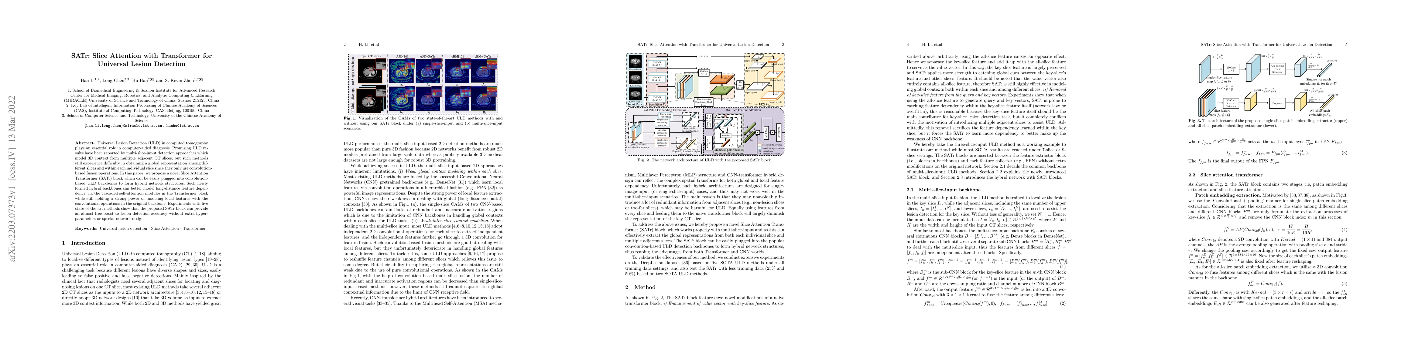
Universal Lesion Detection (ULD) in computed tomography plays an essential role in computer-aided diagnosis. Promising ULD results have been reported by multi-slice-input detection approaches which ...
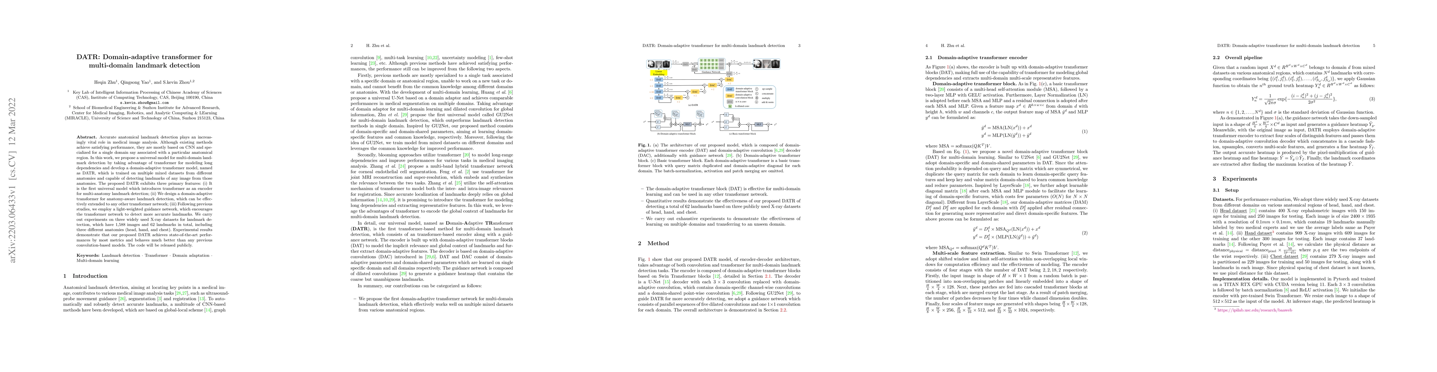
Accurate anatomical landmark detection plays an increasingly vital role in medical image analysis. Although existing methods achieve satisfying performance, they are mostly based on CNN and speciali...
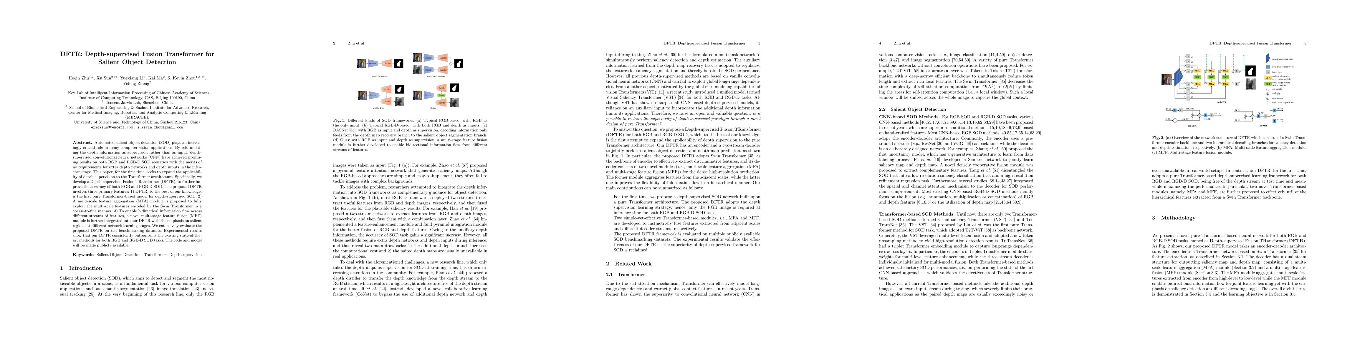
Automated salient object detection (SOD) plays an increasingly crucial role in many computer vision applications. By reformulating the depth information as supervision rather than as input, depth-su...
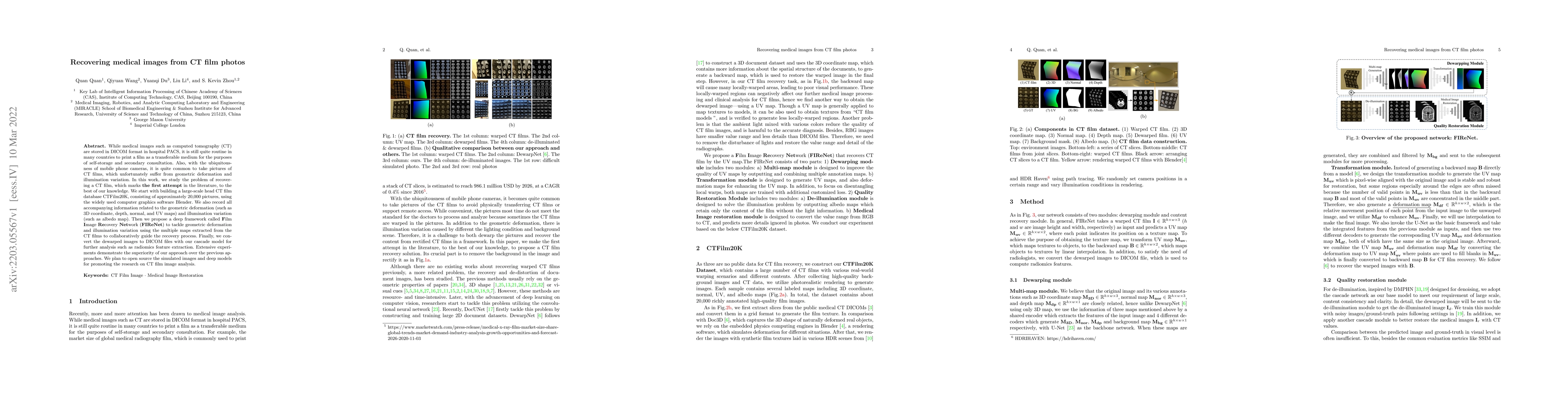
While medical images such as computed tomography (CT) are stored in DICOM format in hospital PACS, it is still quite routine in many countries to print a film as a transferable medium for the purpos...
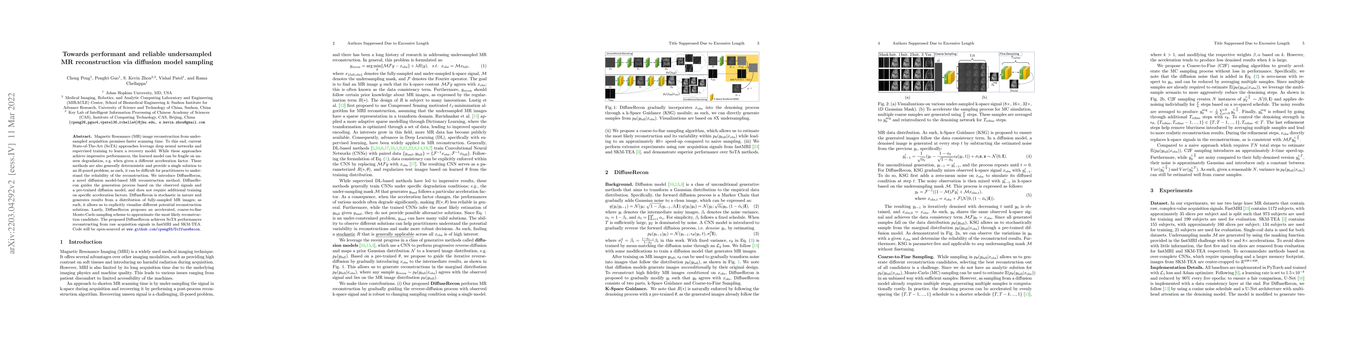
Magnetic Resonance (MR) image reconstruction from under-sampled acquisition promises faster scanning time. To this end, current State-of-The-Art (SoTA) approaches leverage deep neural networks and s...
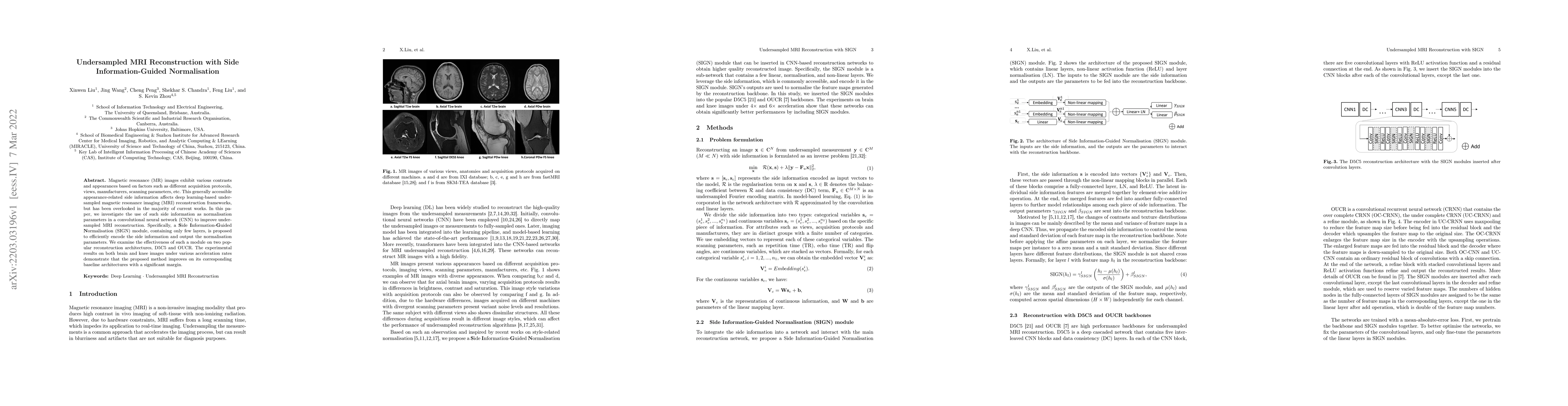
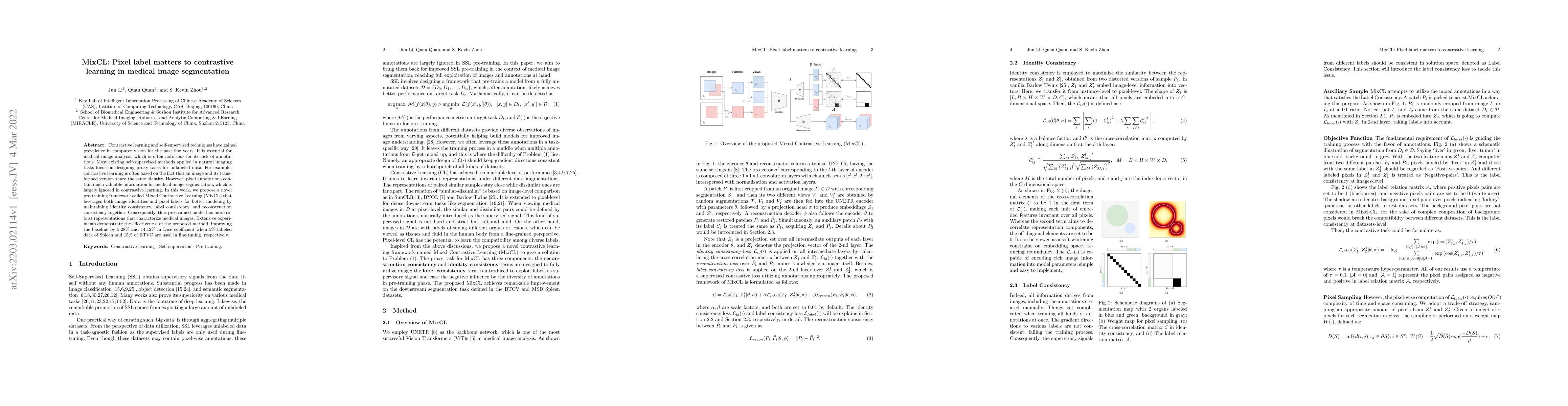
Magnetic resonance (MR) images exhibit various contrasts and appearances based on factors such as different acquisition protocols, views, manufacturers, scanning parameters, etc. This generally acce...
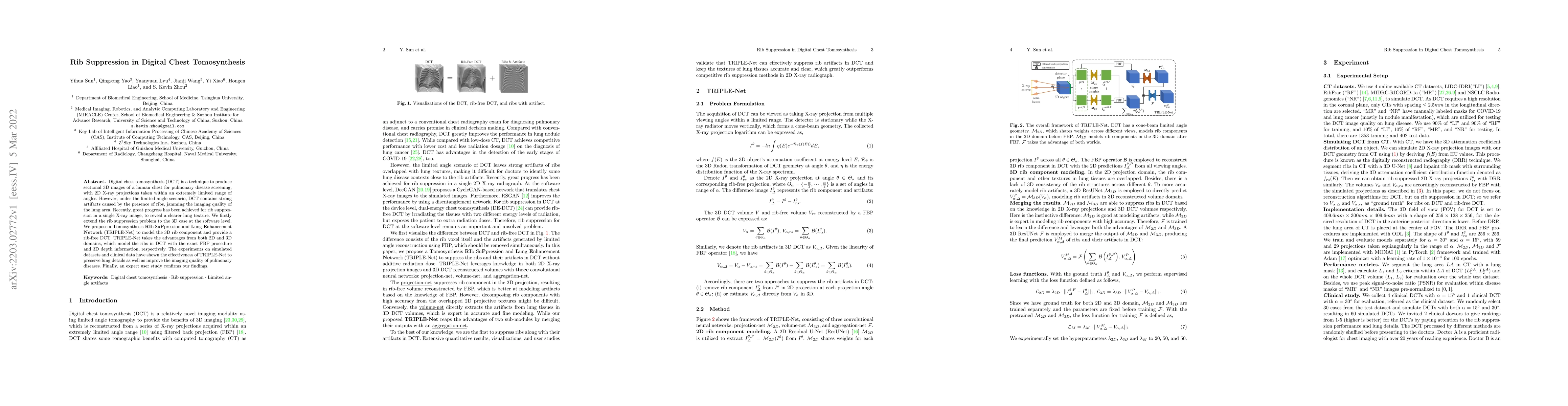
Digital chest tomosynthesis (DCT) is a technique to produce sectional 3D images of a human chest for pulmonary disease screening, with 2D X-ray projections taken within an extremely limited range of...
Contrastive learning and self-supervised techniques have gained prevalence in computer vision for the past few years. It is essential for medical image analysis, which is often notorious for its lac...
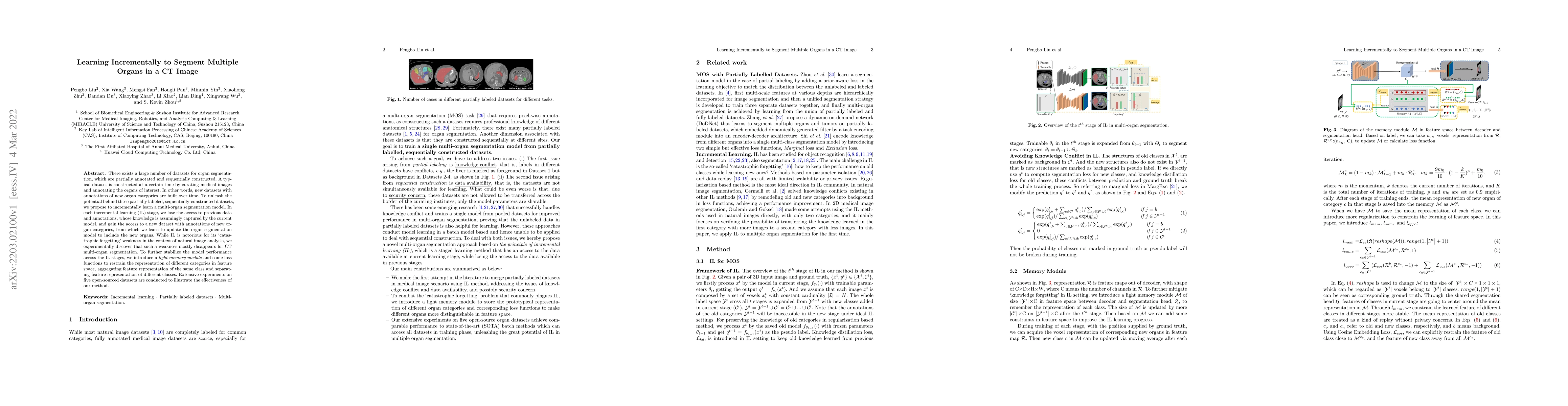
There exists a large number of datasets for organ segmentation, which are partially annotated and sequentially constructed. A typical dataset is constructed at a certain time by curating medical ima...
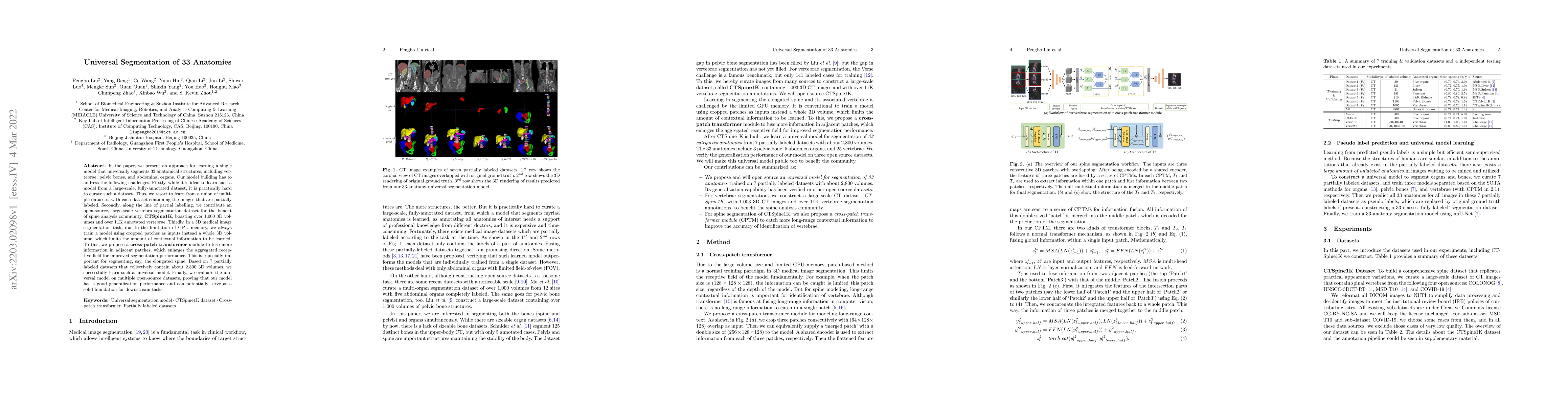
In the paper, we present an approach for learning a single model that universally segments 33 anatomical structures, including vertebrae, pelvic bones, and abdominal organs. Our model building has t...
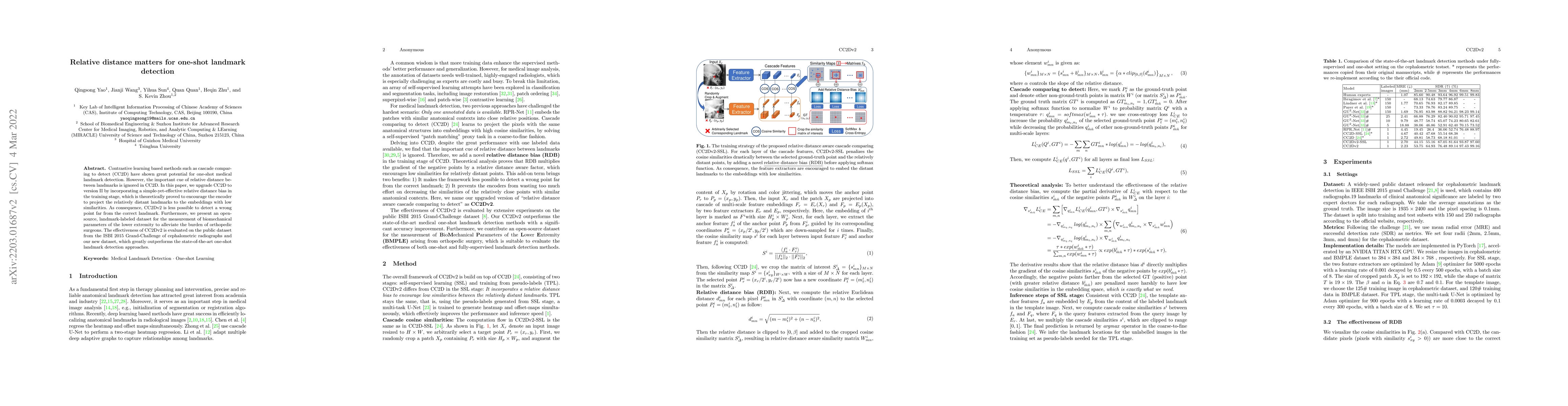
Contrastive learning based methods such as cascade comparing to detect (CC2D) have shown great potential for one-shot medical landmark detection. However, the important cue of relative distance betw...

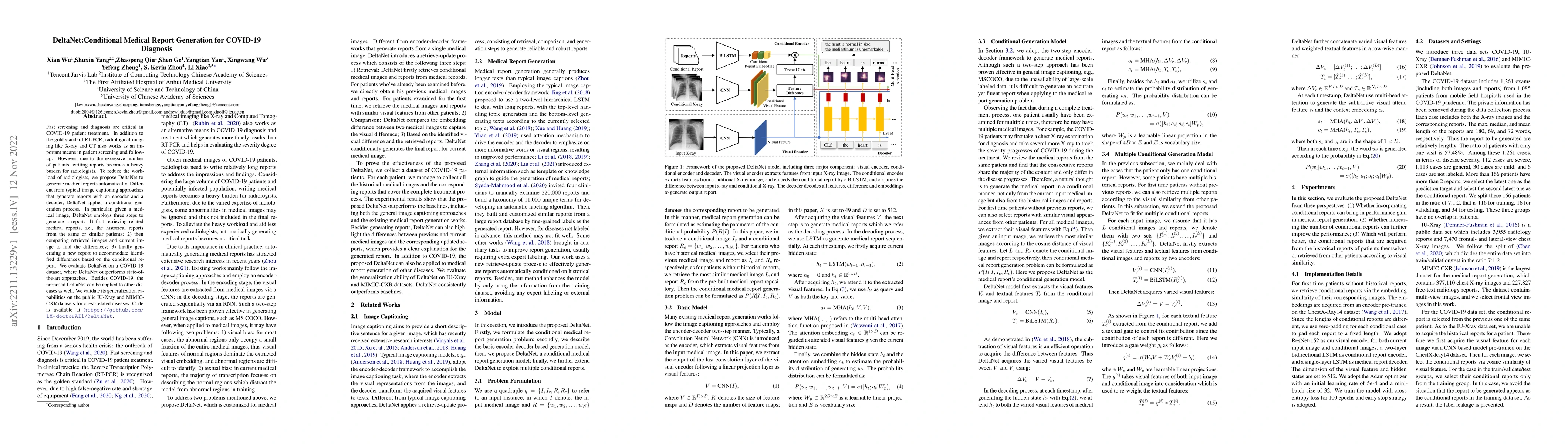
In clinics, a radiology report is crucial for guiding a patient's treatment. However, writing radiology reports is a heavy burden for radiologists. To this end, we present an automatic, multi-modal ...
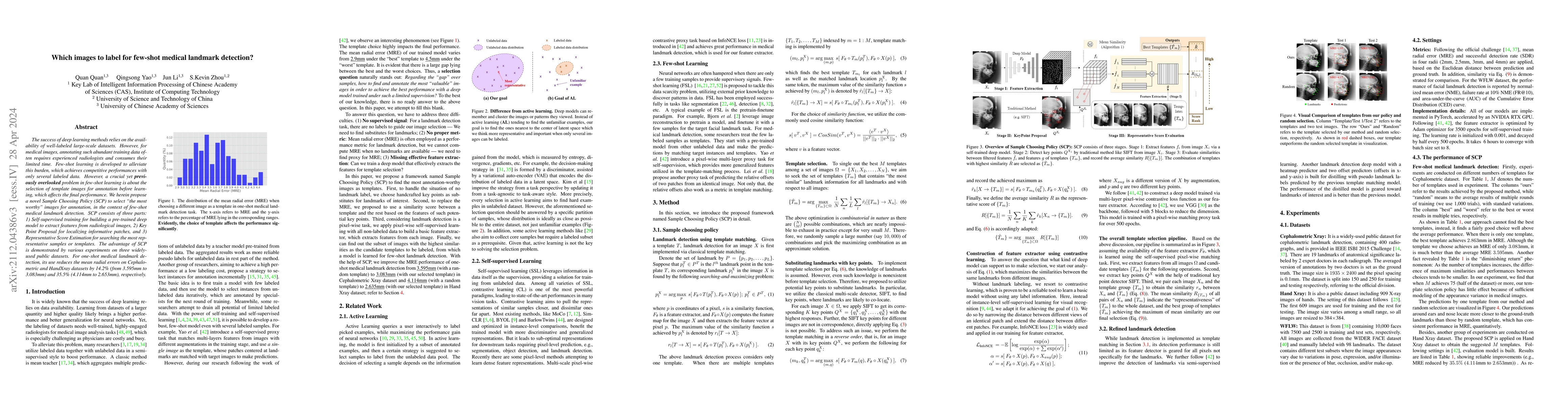
The success of deep learning methods relies on the availability of well-labeled large-scale datasets. However, for medical images, annotating such abundant training data often requires experienced r...
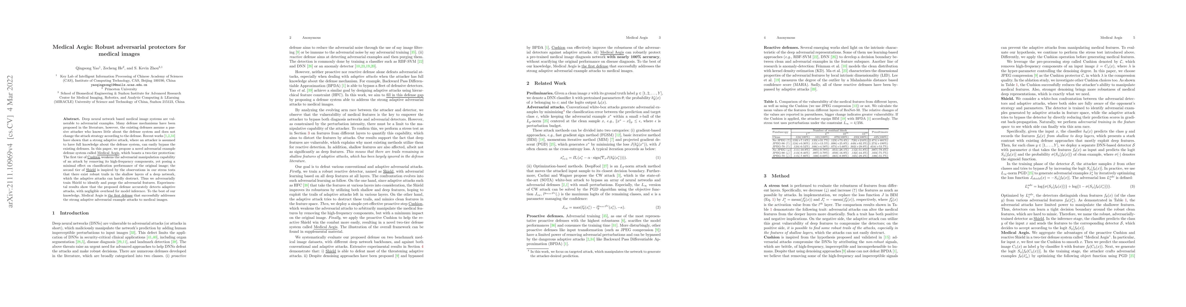
Deep neural network based medical image systems are vulnerable to adversarial examples. Many defense mechanisms have been proposed in the literature, however, the existing defenses assume a passive ...
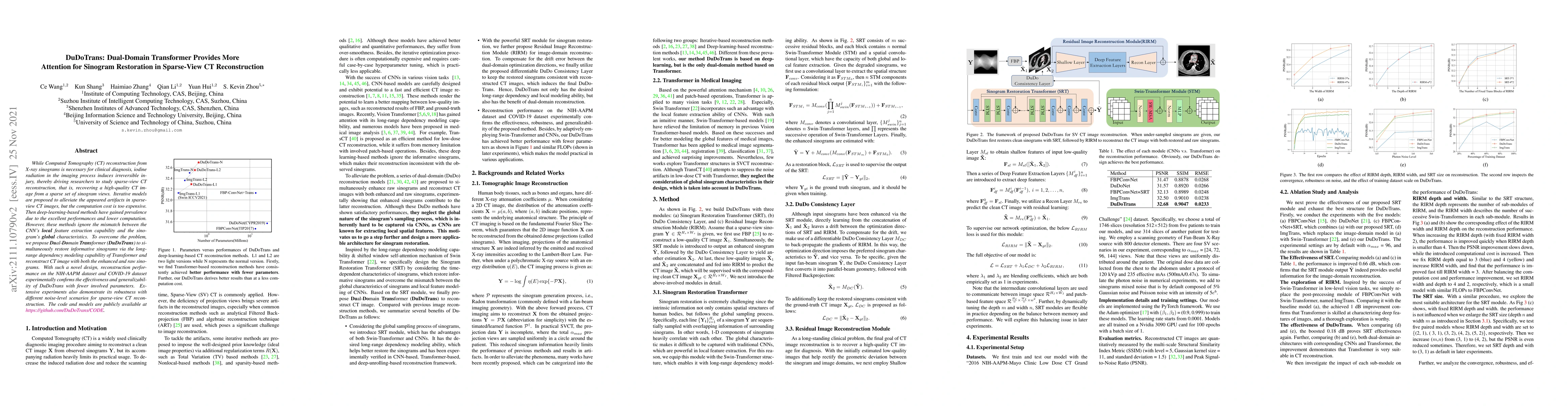
While Computed Tomography (CT) reconstruction from X-ray sinograms is necessary for clinical diagnosis, iodine radiation in the imaging process induces irreversible injury, thereby driving researche...
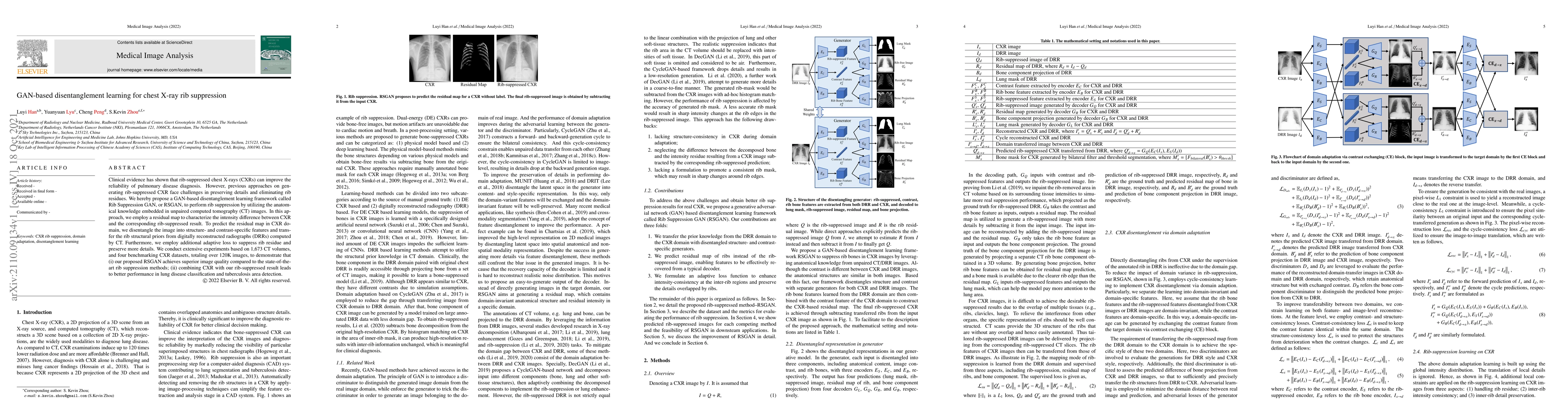
Clinical evidence has shown that rib-suppressed chest X-rays (CXRs) can improve the reliability of pulmonary disease diagnosis. However, previous approaches on generating rib-suppressed CXR face cha...
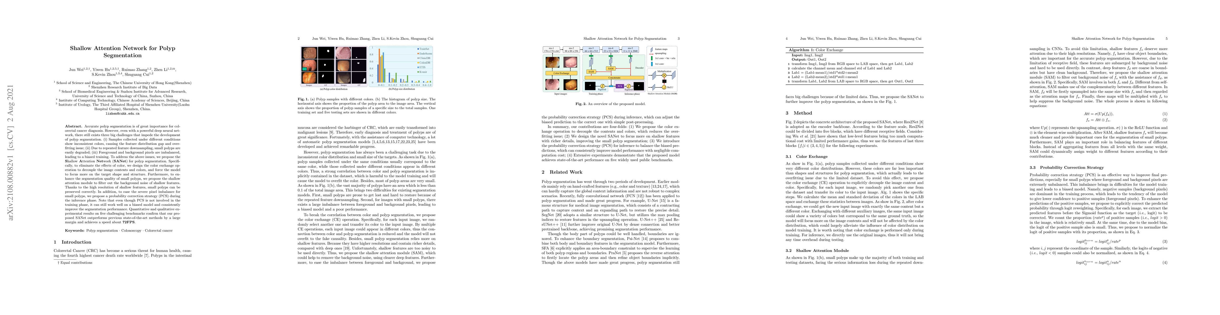
Accurate polyp segmentation is of great importance for colorectal cancer diagnosis. However, even with a powerful deep neural network, there still exists three big challenges that impede the develop...
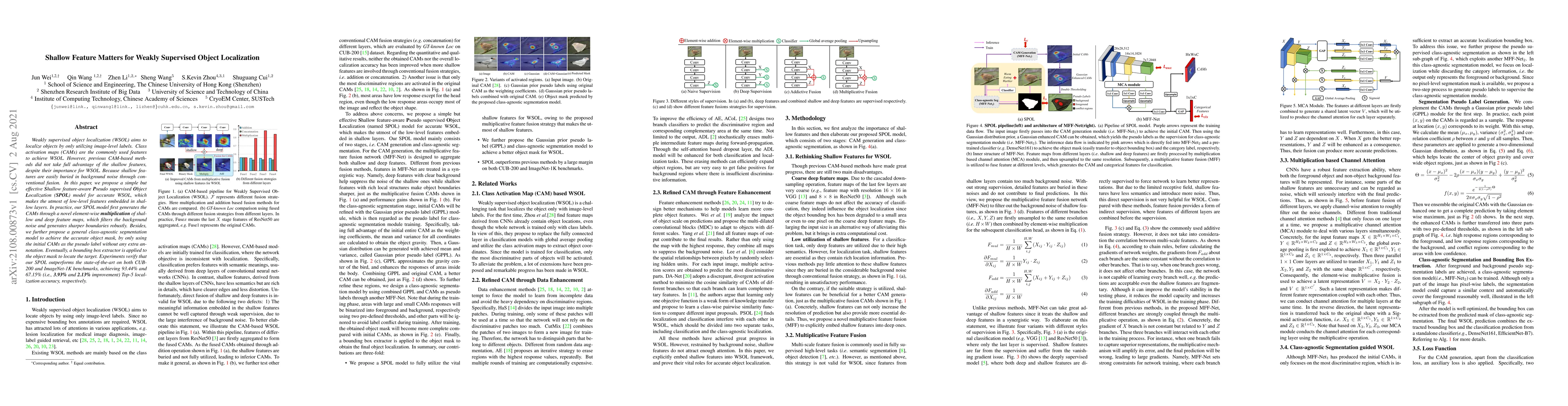
Weakly supervised object localization (WSOL) aims to localize objects by only utilizing image-level labels. Class activation maps (CAMs) are the commonly used features to achieve WSOL. However, prev...
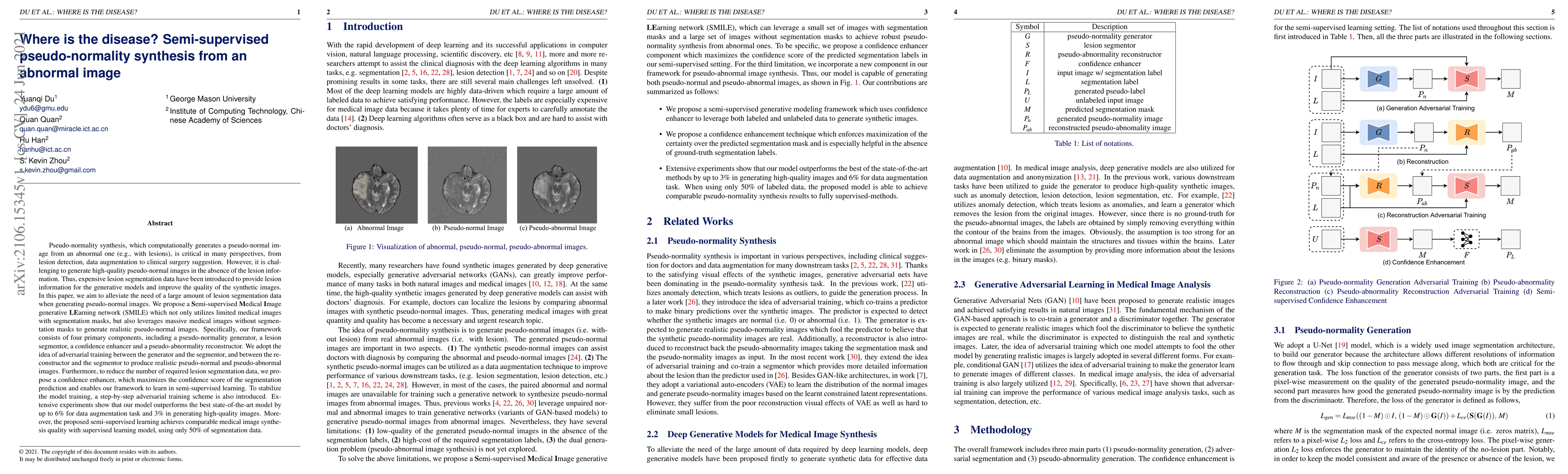
Pseudo-normality synthesis, which computationally generates a pseudo-normal image from an abnormal one (e.g., with lesions), is critical in many perspectives, from lesion detection, data augmentatio...
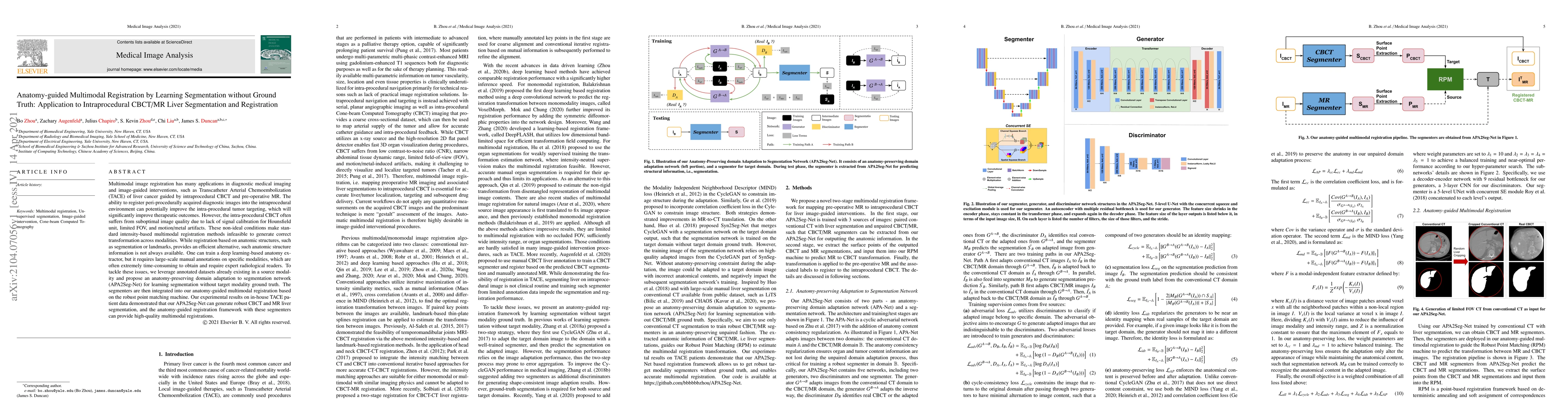
Multimodal image registration has many applications in diagnostic medical imaging and image-guided interventions, such as Transcatheter Arterial Chemoembolization (TACE) of liver cancer guided by in...
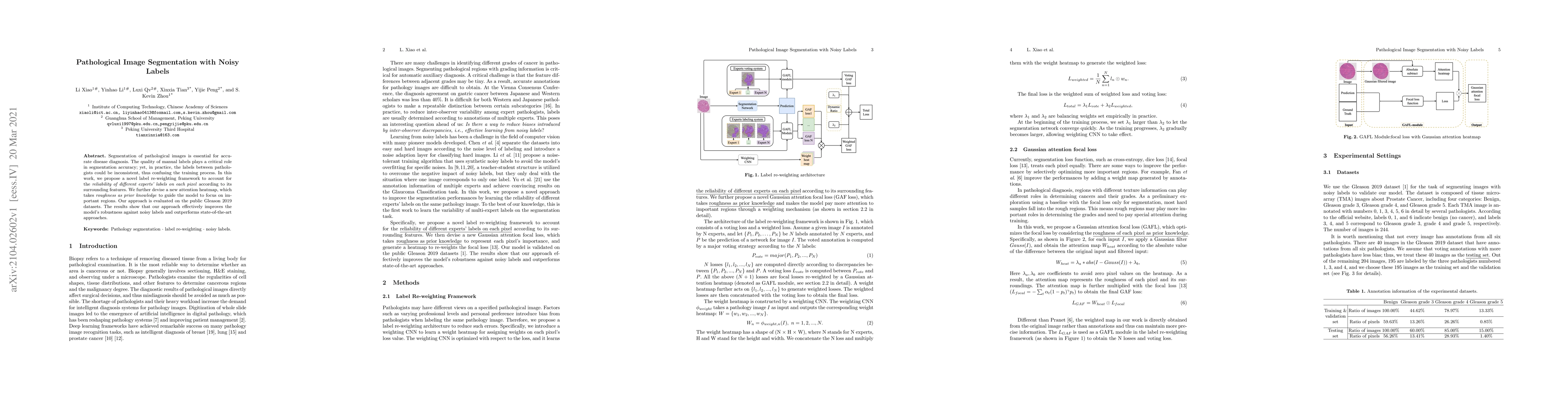
Segmentation of pathological images is essential for accurate disease diagnosis. The quality of manual labels plays a critical role in segmentation accuracy; yet, in practice, the labels between pat...
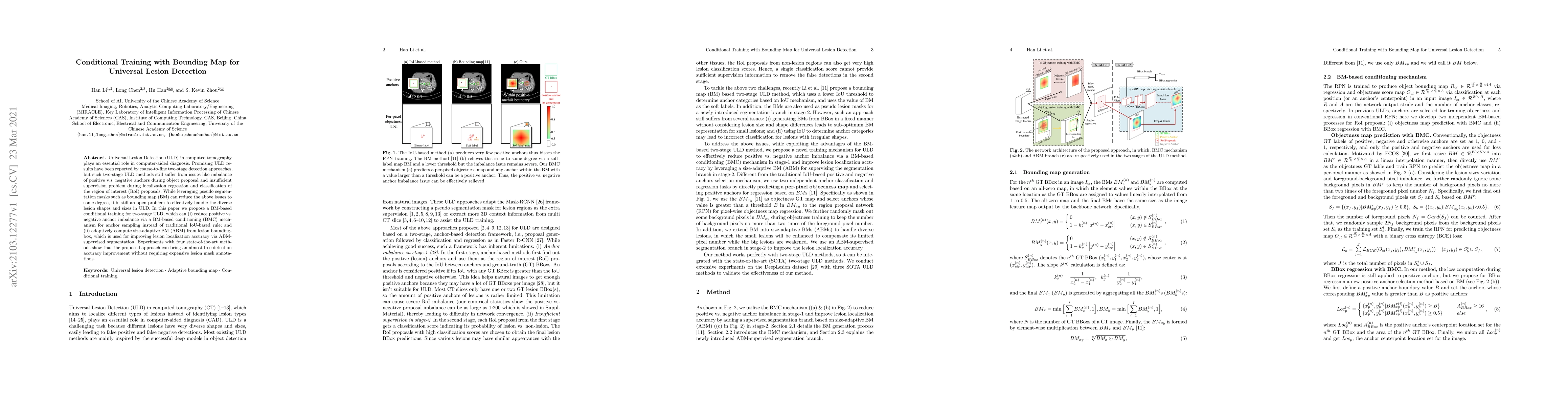
Universal Lesion Detection (ULD) in computed tomography plays an essential role in computer-aided diagnosis. Promising ULD results have been reported by coarse-to-fine two-stage detection approaches...
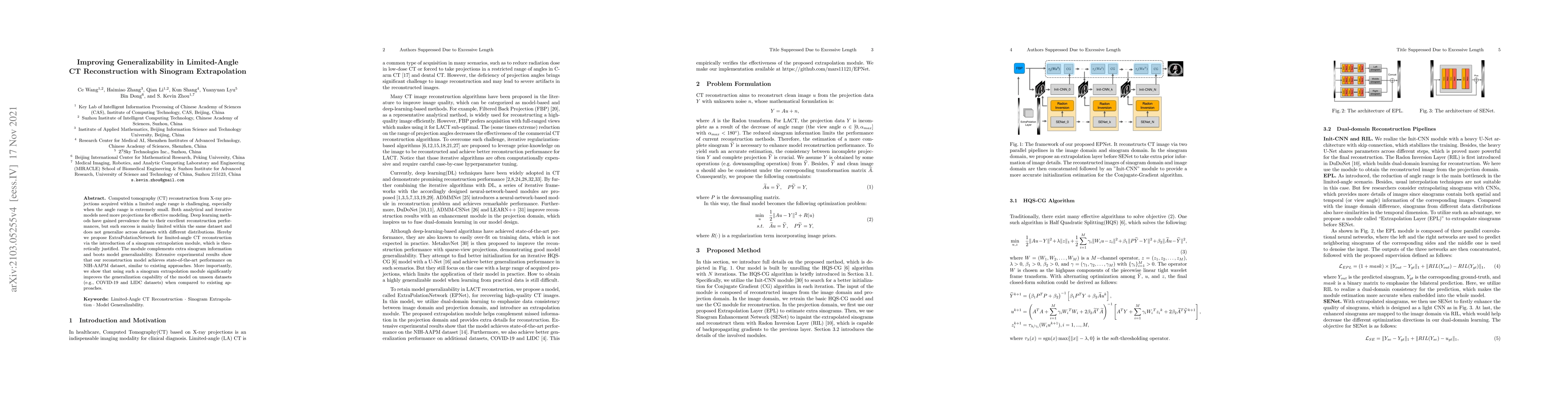
Computed tomography (CT) reconstruction from X-ray projections acquired within a limited angle range is challenging, especially when the angle range is extremely small. Both analytical and iterative...
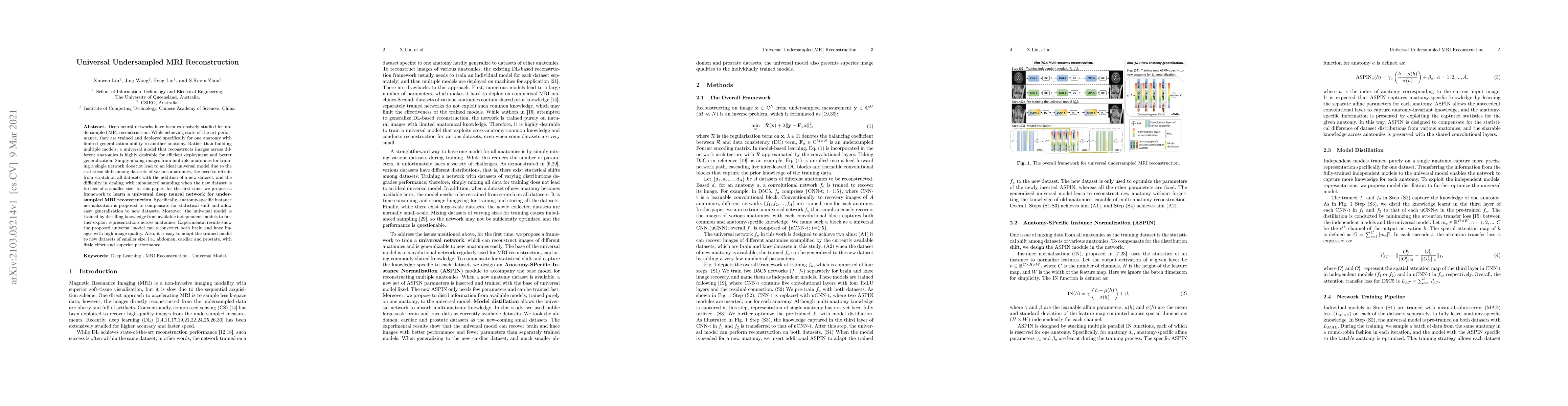
Deep neural networks have been extensively studied for undersampled MRI reconstruction. While achieving state-of-the-art performance, they are trained and deployed specifically for one anatomy with ...
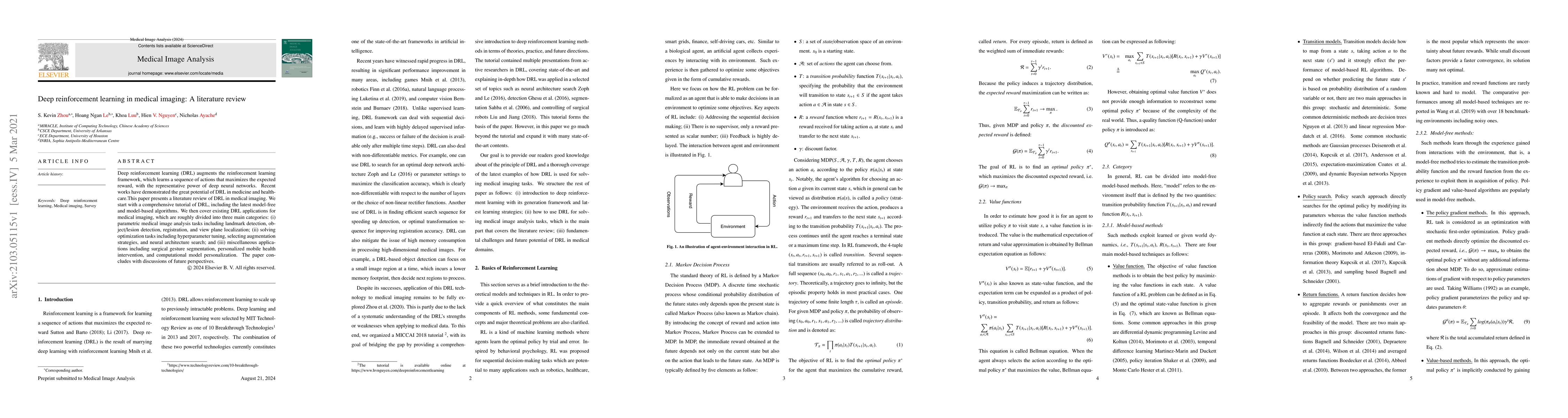
Deep reinforcement learning (DRL) augments the reinforcement learning framework, which learns a sequence of actions that maximizes the expected reward, with the representative power of deep neural n...
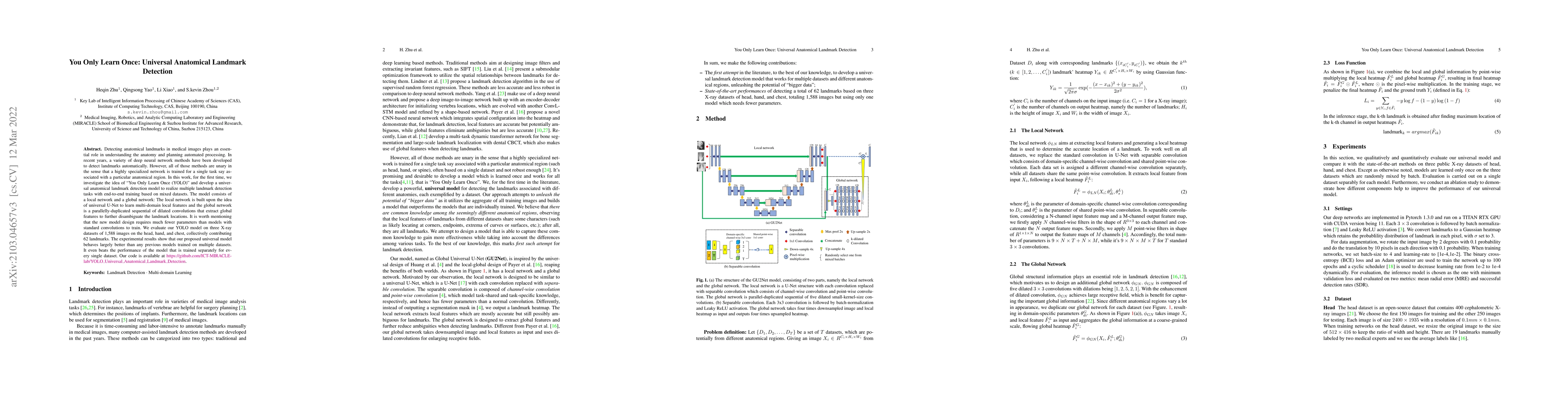
Detecting anatomical landmarks in medical images plays an essential role in understanding the anatomy and planning automated processing. In recent years, a variety of deep neural network methods hav...
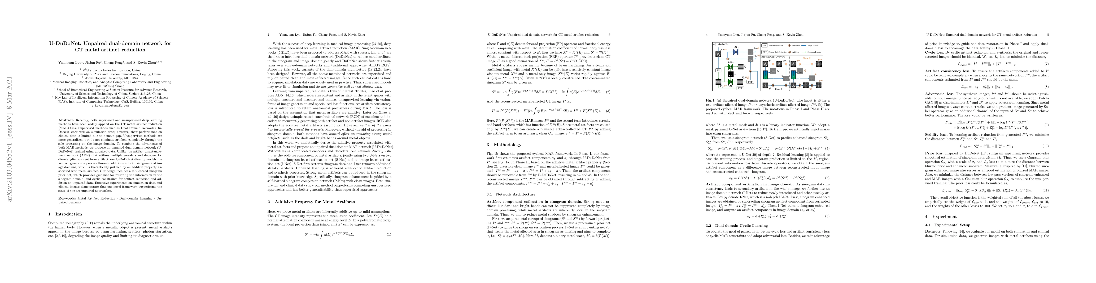
Recently, both supervised and unsupervised deep learning methods have been widely applied on the CT metal artifact reduction (MAR) task. Supervised methods such as Dual Domain Network (Du-DoNet) wor...
The success of deep learning methods relies on the availability of a large number of datasets with annotations; however, curating such datasets is burdensome, especially for medical images. To relie...
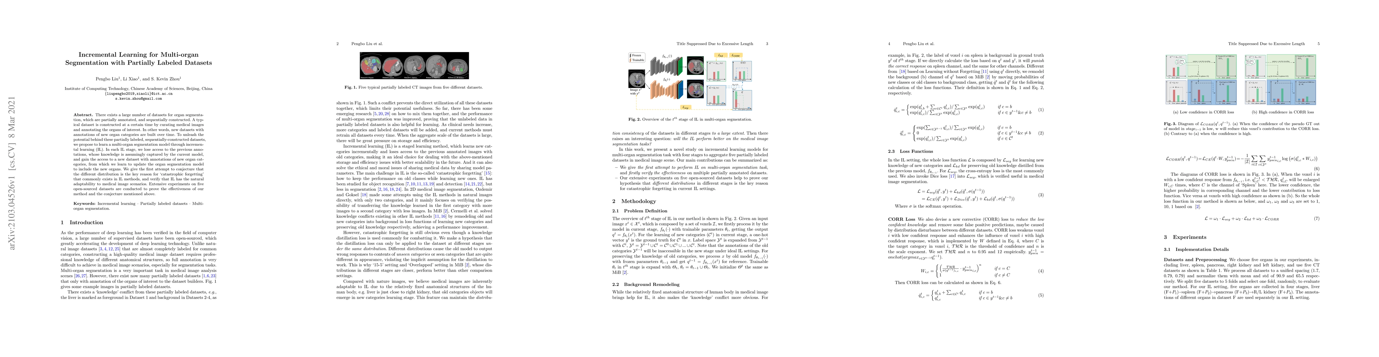
There exists a large number of datasets for organ segmentation, which are partially annotated, and sequentially constructed. A typical dataset is constructed at a certain time by curating medical im...
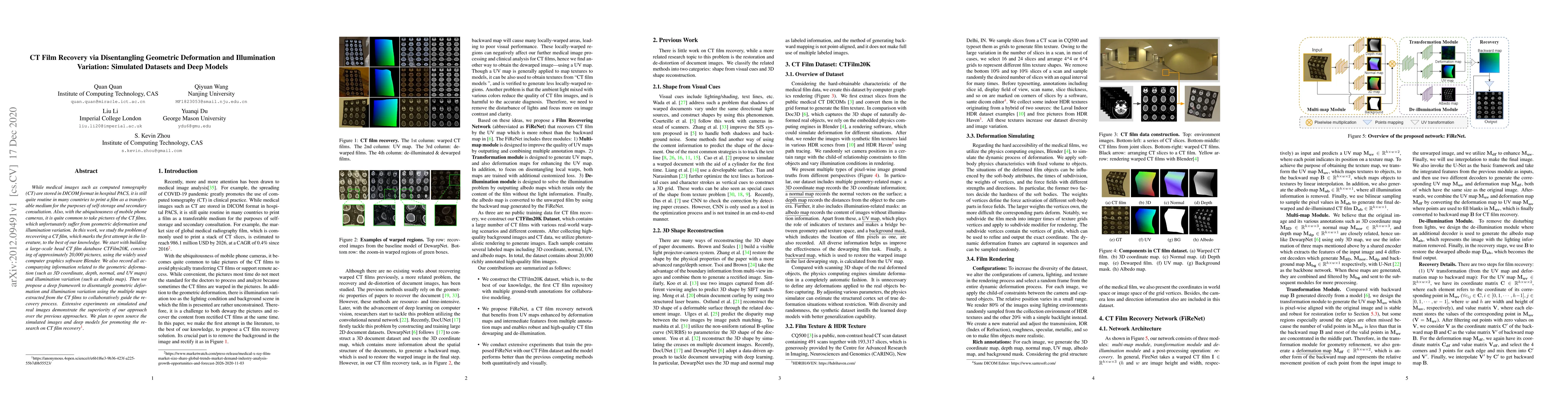
While medical images such as computed tomography (CT) are stored in DICOM format in hospital PACS, it is still quite routine in many countries to print a film as a transferable medium for the purpos...
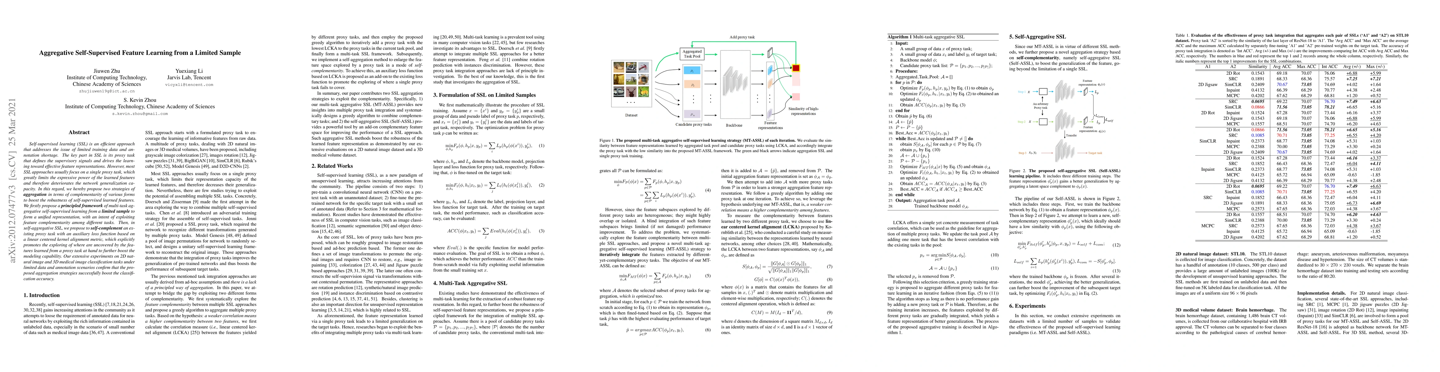
Self-supervised learning (SSL) is an efficient approach that addresses the issue of limited training data and annotation shortage. The key part in SSL is its proxy task that defines the supervisory ...
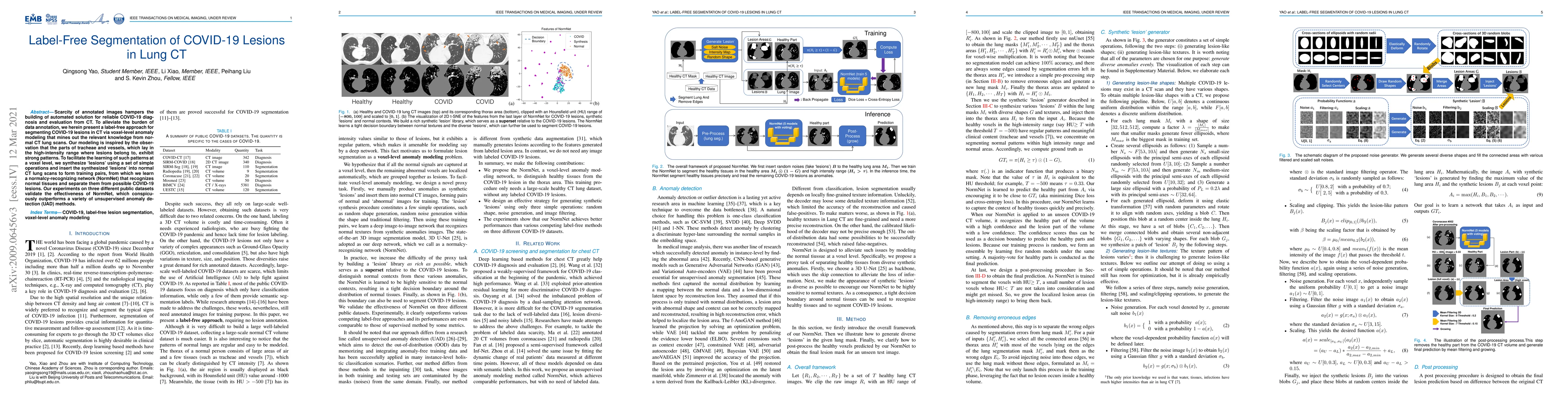
Scarcity of annotated images hampers the building of automated solution for reliable COVID-19 diagnosis and evaluation from CT. To alleviate the burden of data annotation, we herein present a label-...
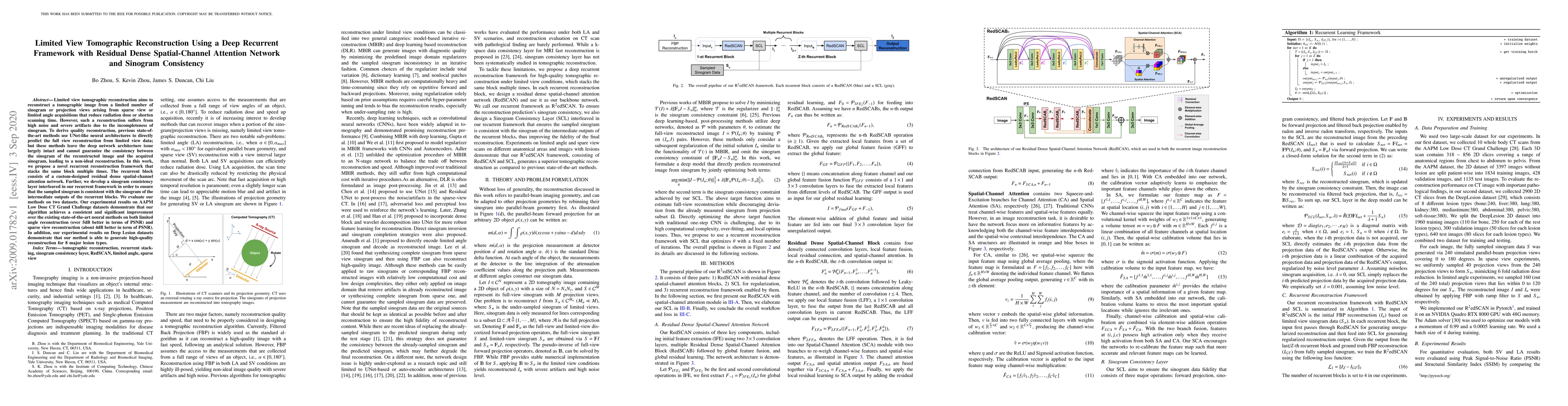
Limited view tomographic reconstruction aims to reconstruct a tomographic image from a limited number of sinogram or projection views arising from sparse view or limited angle acquisitions that redu...
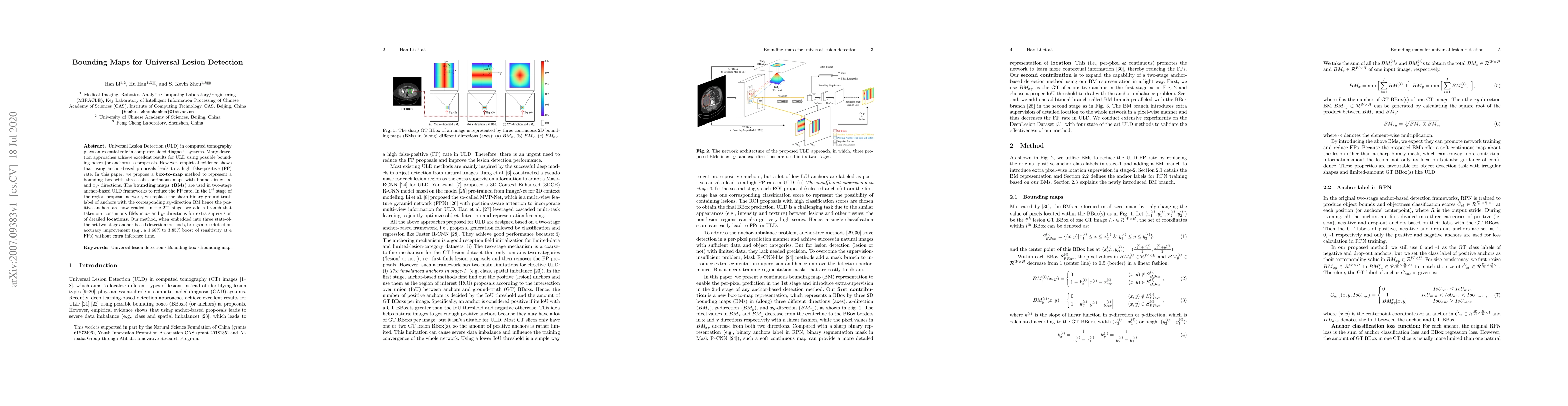
Universal Lesion Detection (ULD) in computed tomography plays an essential role in computer-aided diagnosis systems. Many detection approaches achieve excellent results for ULD using possible boundi...
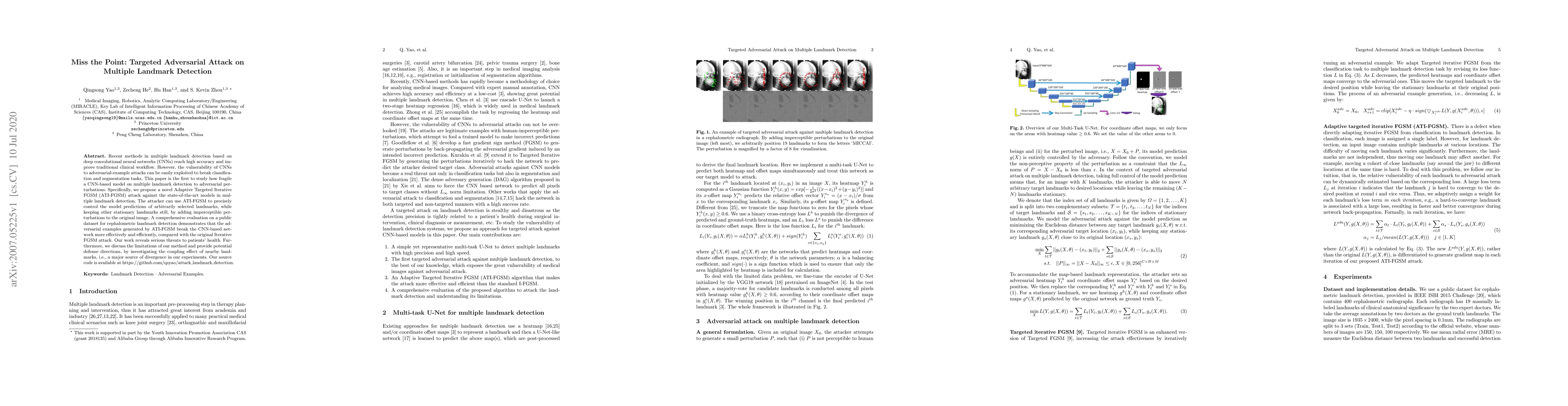
Recent methods in multiple landmark detection based on deep convolutional neural networks (CNNs) reach high accuracy and improve traditional clinical workflow. However, the vulnerability of CNNs to ...
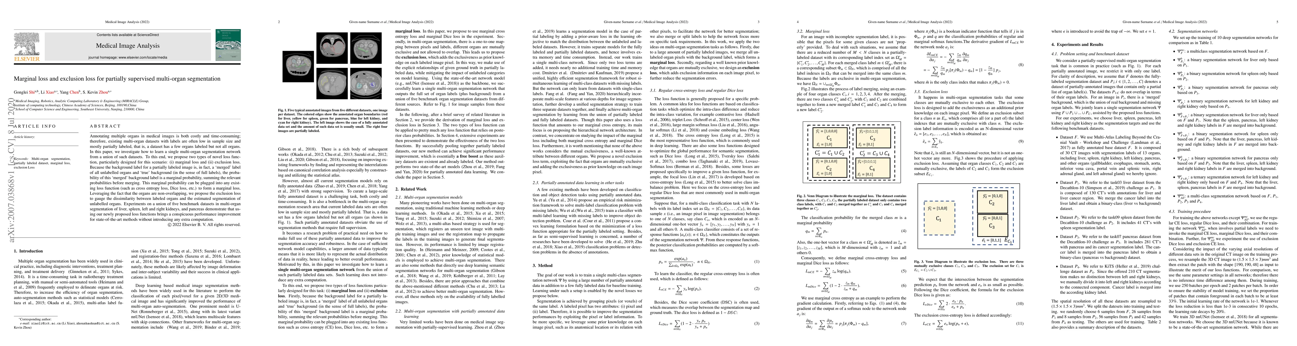
Annotating multiple organs in medical images is both costly and time-consuming; therefore, existing multi-organ datasets with labels are often low in sample size and mostly partially labeled, that i...
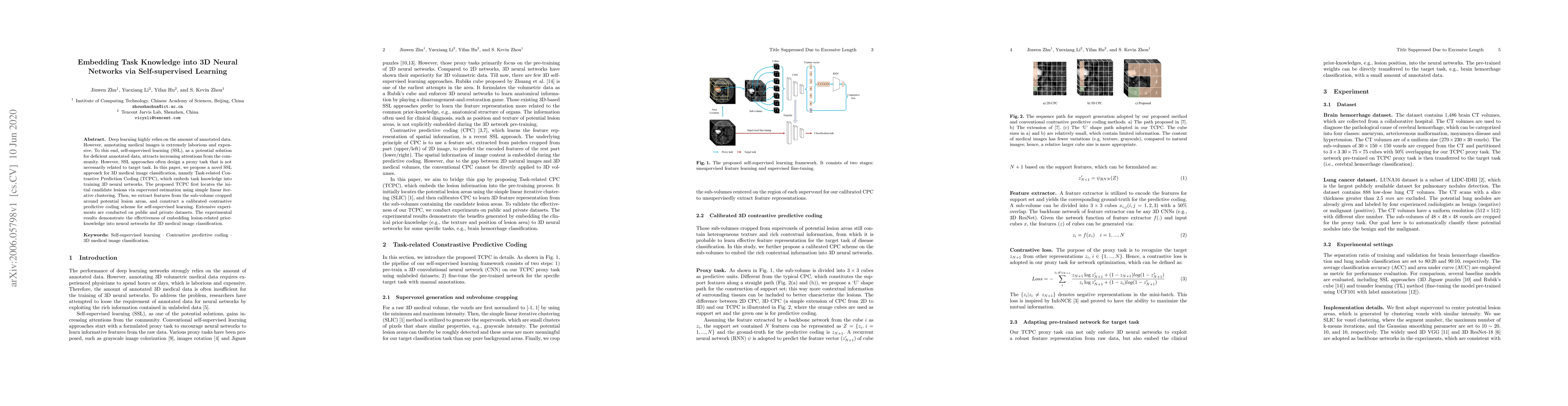
Deep learning highly relies on the amount of annotated data. However, annotating medical images is extremely laborious and expensive. To this end, self-supervised learning (SSL), as a potential solu...
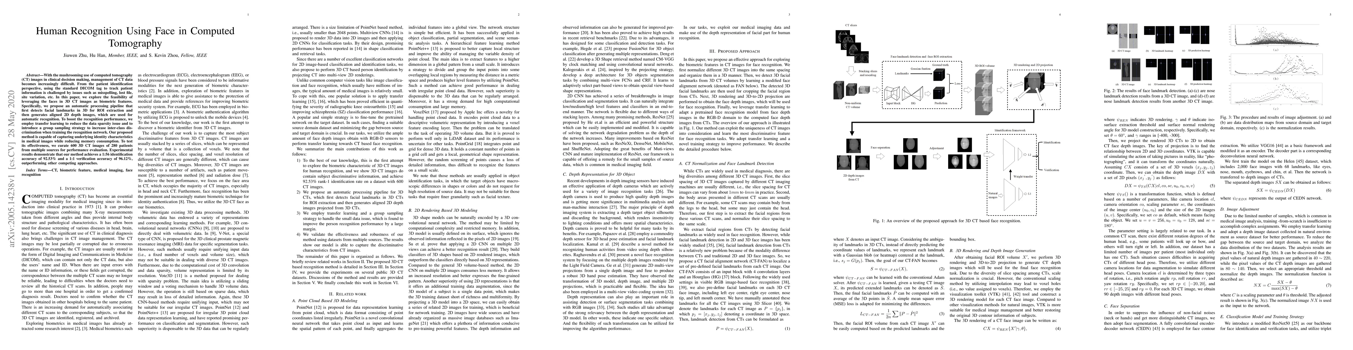
With the mushrooming use of computed tomography (CT) images in clinical decision making, management of CT data becomes increasingly difficult. From the patient identification perspective, using the ...
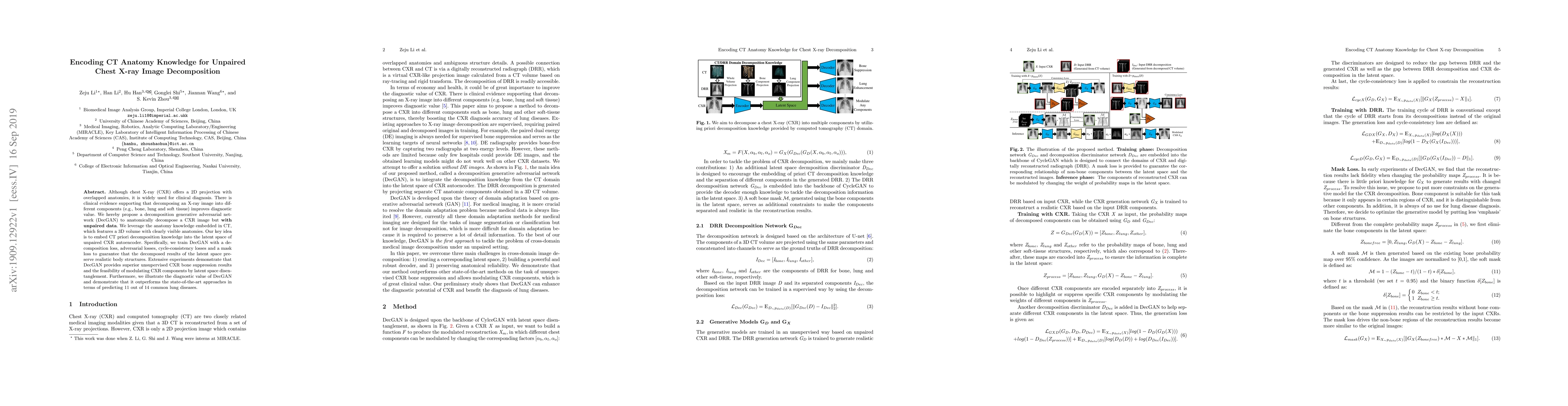
Although chest X-ray (CXR) offers a 2D projection with overlapped anatomies, it is widely used for clinical diagnosis. There is clinical evidence supporting that decomposing an X-ray image into diff...
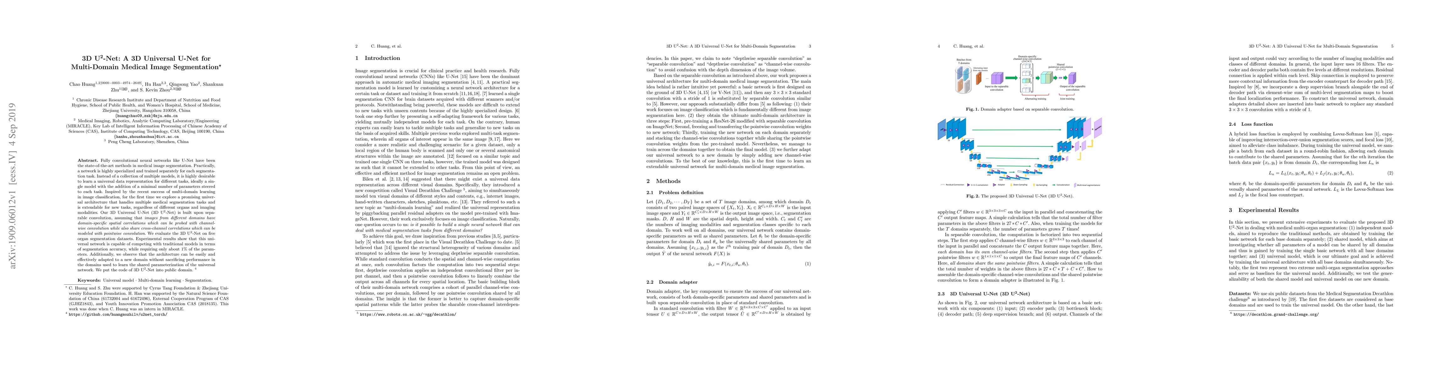
Fully convolutional neural networks like U-Net have been the state-of-the-art methods in medical image segmentation. Practically, a network is highly specialized and trained separately for each segm...
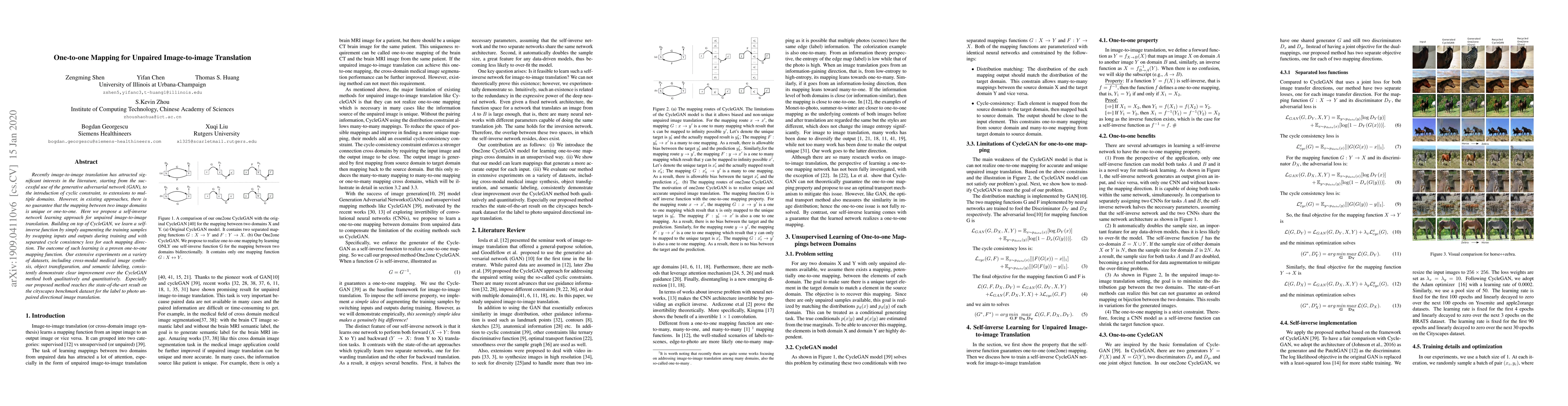
Recently image-to-image translation has attracted significant interests in the literature, starting from the successful use of the generative adversarial network (GAN), to the introduction of cyclic...
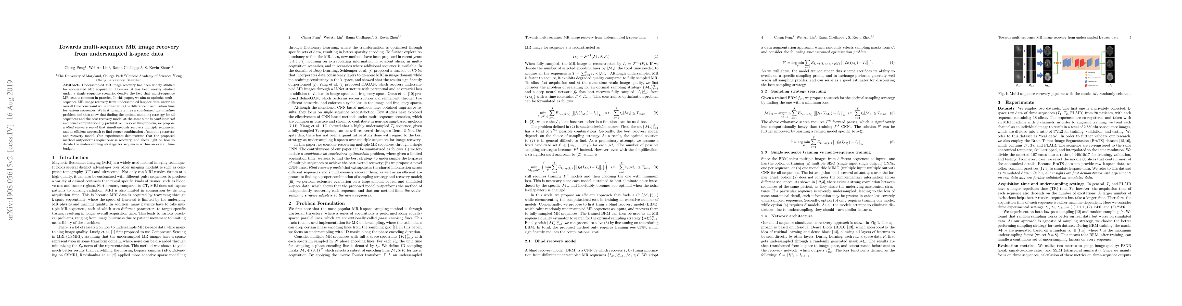
Undersampled MR image recovery has been widely studied for accelerated MR acquisition. However, it has been mostly studied under a single sequence scenario, despite the fact that multi-sequence MR s...
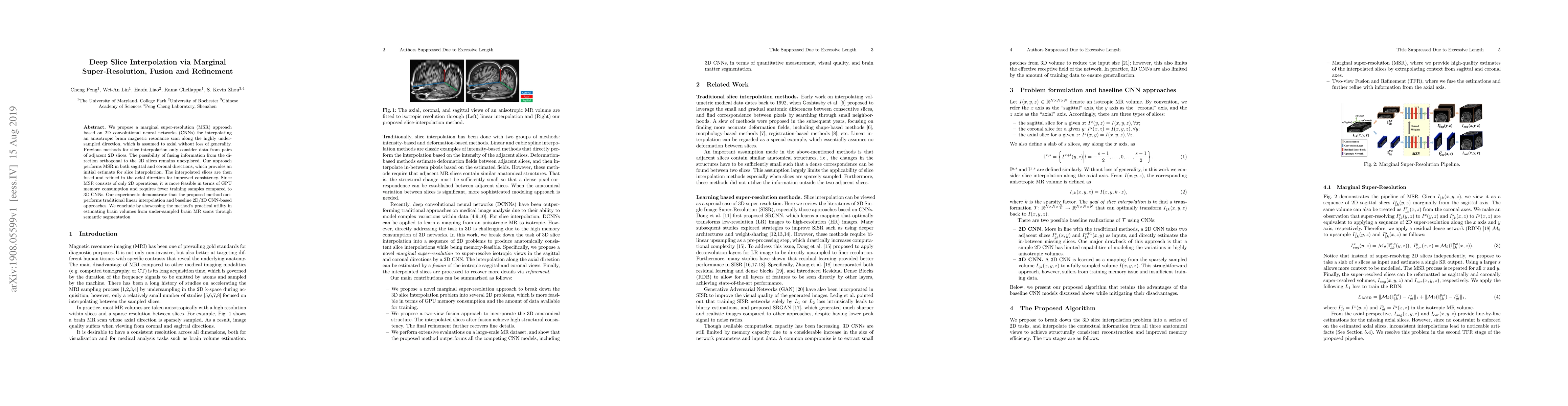
We propose a marginal super-resolution (MSR) approach based on 2D convolutional neural networks (CNNs) for interpolating an anisotropic brain magnetic resonance scan along the highly under-sampled d...
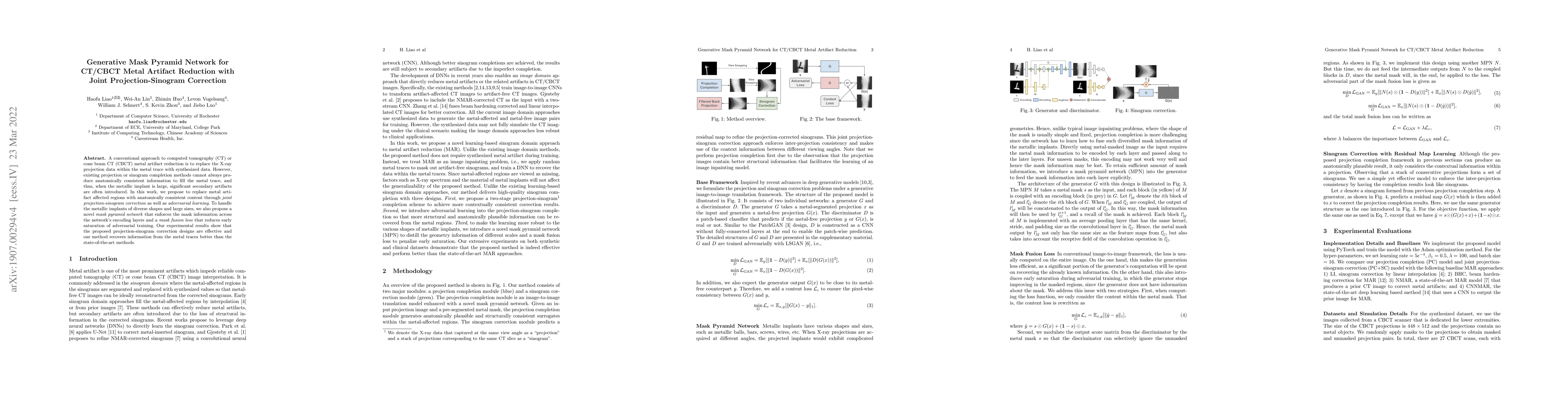
A conventional approach to computed tomography (CT) or cone beam CT (CBCT) metal artifact reduction is to replace the X-ray projection data within the metal trace with synthesized data. However, exi...
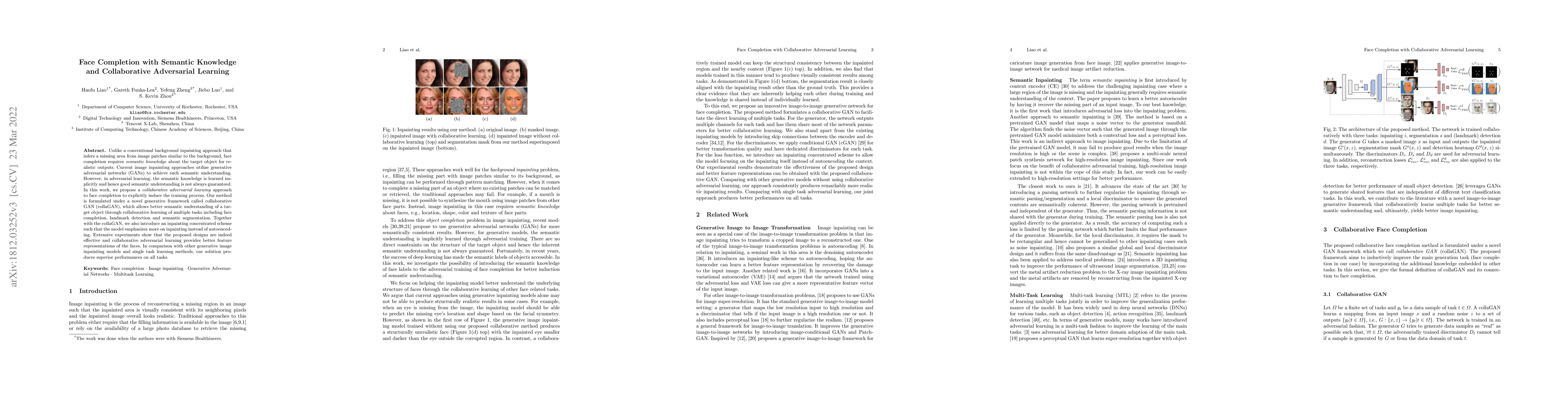
Unlike a conventional background inpainting approach that infers a missing area from image patches similar to the background, face completion requires semantic knowledge about the target object for ...
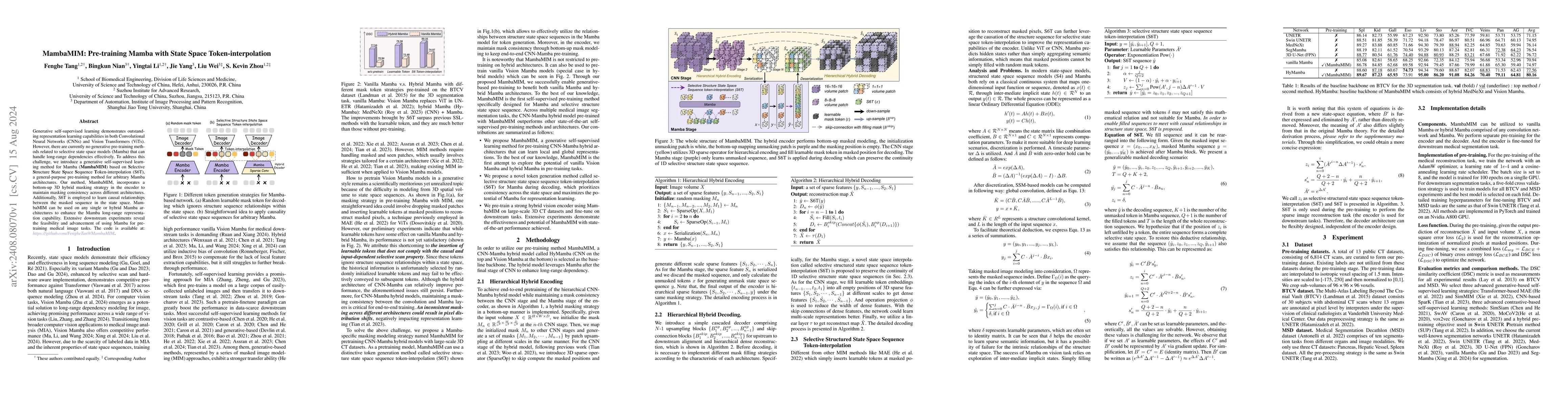
Generative self-supervised learning demonstrates outstanding representation learning capabilities in both Convolutional Neural Networks (CNNs) and Vision Transformers (ViTs). However, there are curren...
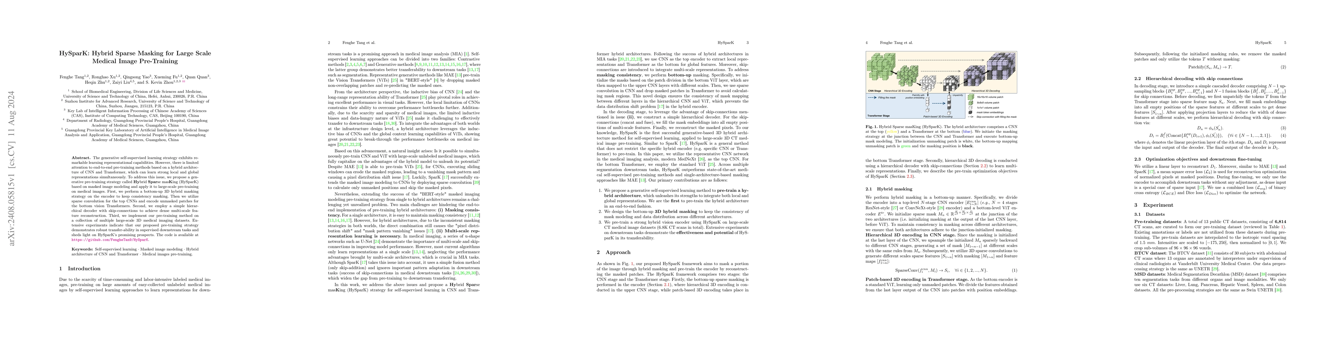
The generative self-supervised learning strategy exhibits remarkable learning representational capabilities. However, there is limited attention to end-to-end pre-training methods based on a hybrid ar...
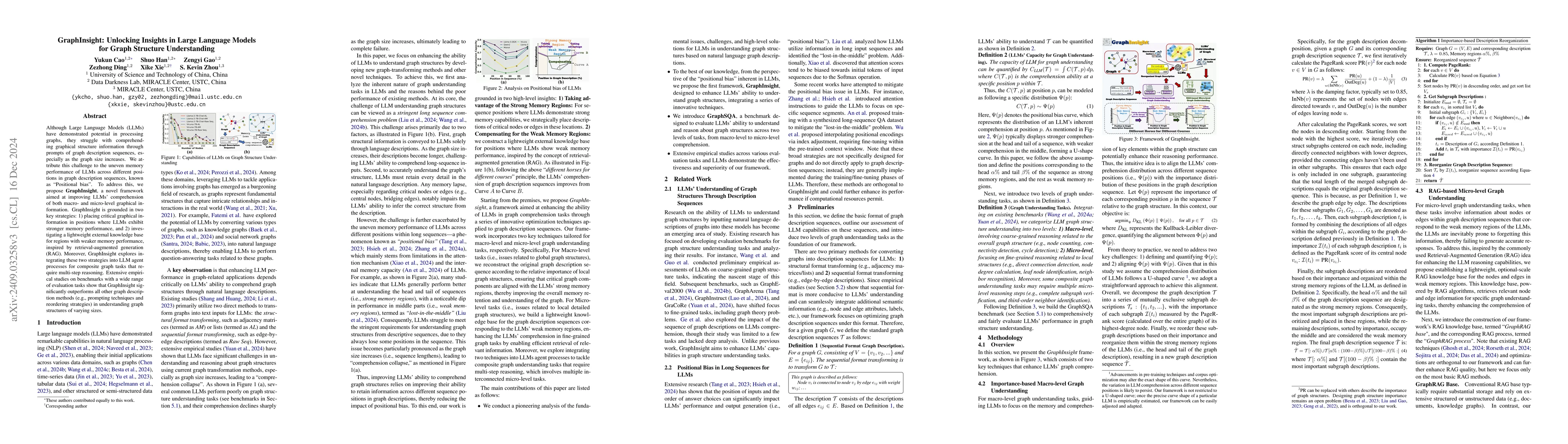
Although Large Language Models (LLMs) have demonstrated potential in processing graphs, they struggle with comprehending graphical structure information through prompts of graph description sequences,...
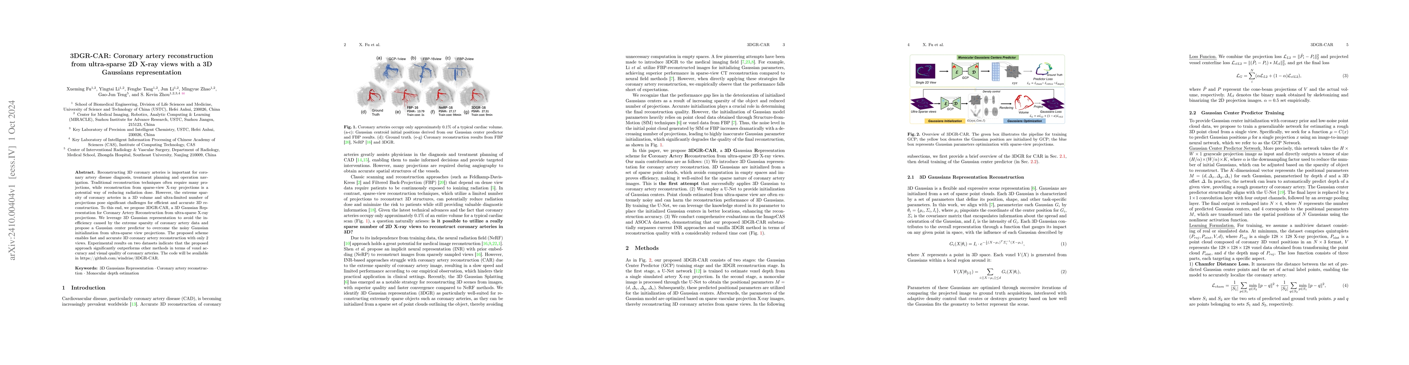
Reconstructing 3D coronary arteries is important for coronary artery disease diagnosis, treatment planning and operation navigation. Traditional reconstruction techniques often require many projection...
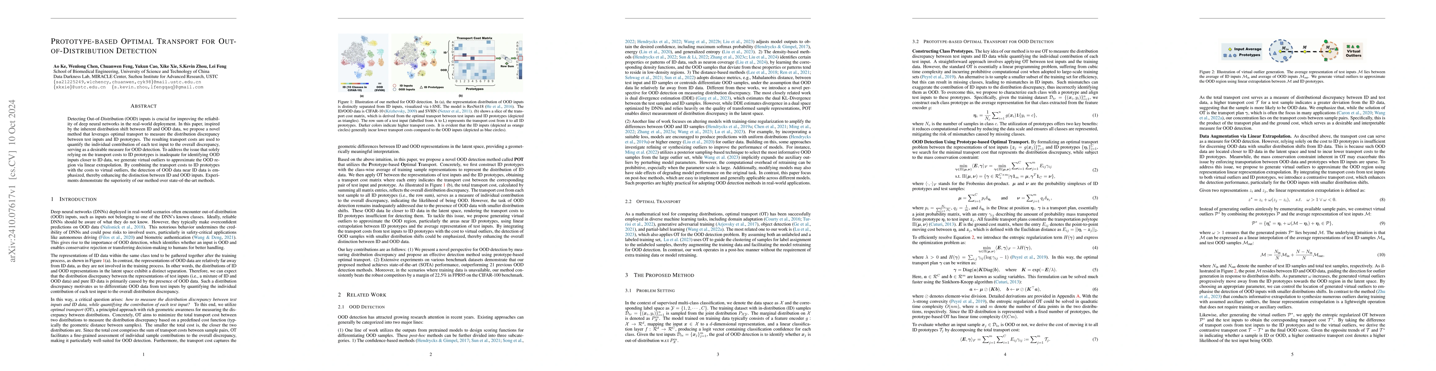
Detecting Out-of-Distribution (OOD) inputs is crucial for improving the reliability of deep neural networks in the real-world deployment. In this paper, inspired by the inherent distribution shift bet...
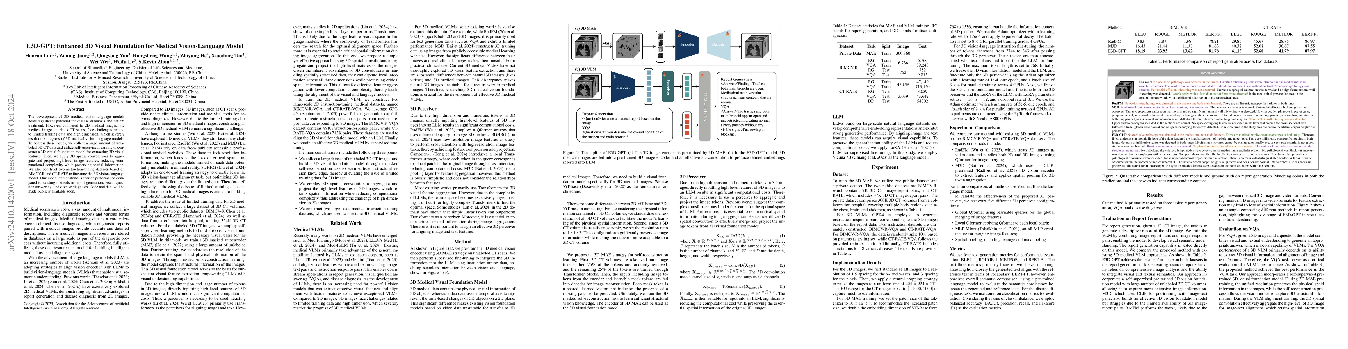
The development of 3D medical vision-language models holds significant potential for disease diagnosis and patient treatment. However, compared to 2D medical images, 3D medical images, such as CT scan...
Non-semantic context information is crucial for visual recognition, as the human visual perception system first uses global statistics to process scenes rapidly before identifying specific objects. Ho...
Accurate anatomical landmark detection in medical images is crucial for clinical applications. Existing methods often struggle to balance global context with computational efficiency, particularly wit...
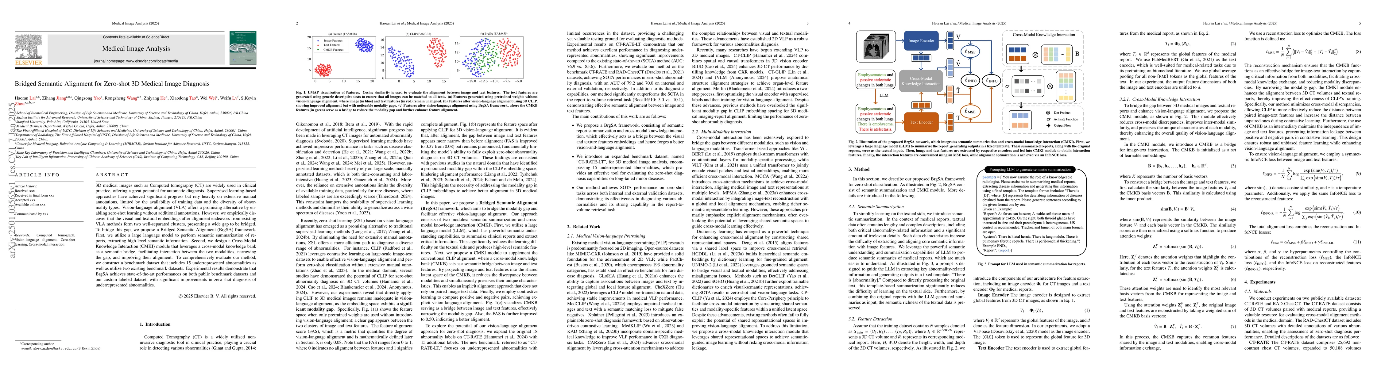
3D medical images such as Computed tomography (CT) are widely used in clinical practice, offering a great potential for automatic diagnosis. Supervised learning-based approaches have achieved signific...
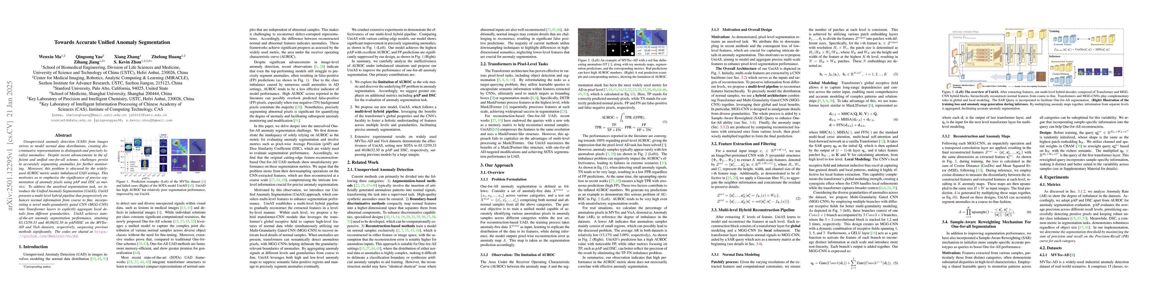
Unsupervised anomaly detection (UAD) from images strives to model normal data distributions, creating discriminative representations to distinguish and precisely localize anomalies. Despite recent adv...
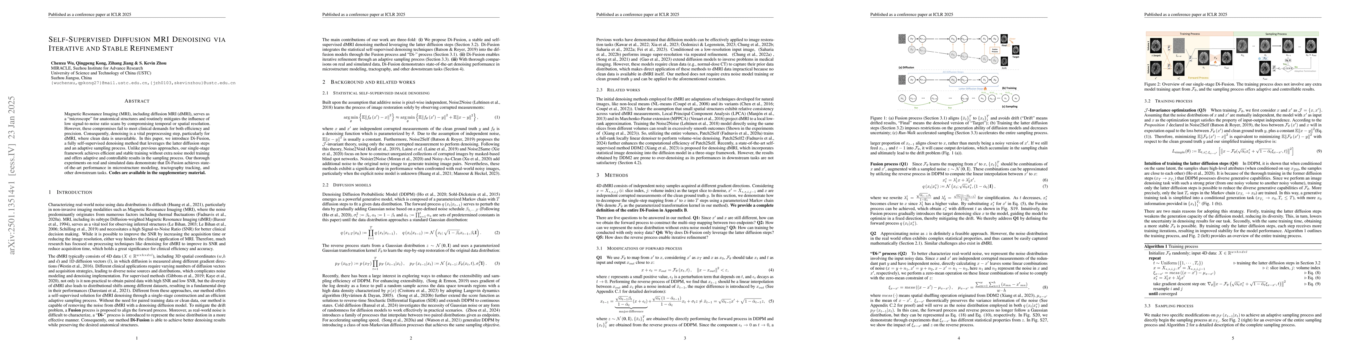
Magnetic Resonance Imaging (MRI), including diffusion MRI (dMRI), serves as a ``microscope'' for anatomical structures and routinely mitigates the influence of low signal-to-noise ratio scans by compr...
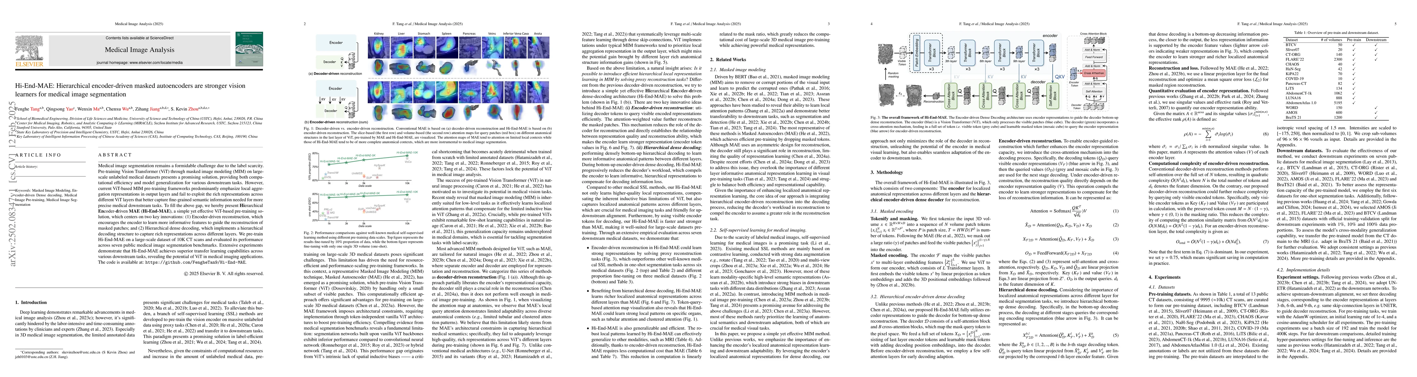
Medical image segmentation remains a formidable challenge due to the label scarcity. Pre-training Vision Transformer (ViT) through masked image modeling (MIM) on large-scale unlabeled medical datasets...
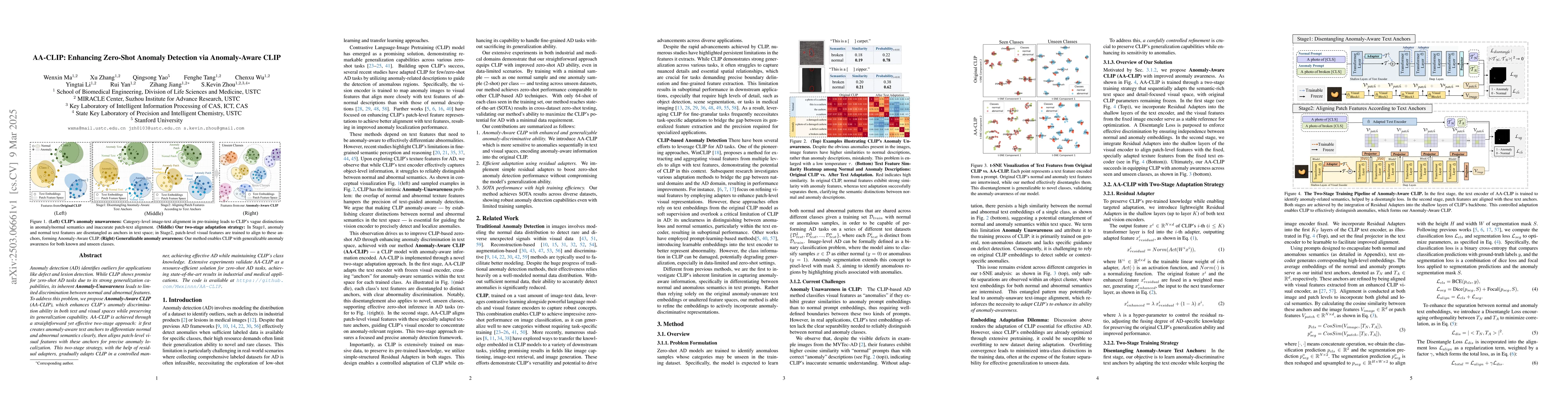
Anomaly detection (AD) identifies outliers for applications like defect and lesion detection. While CLIP shows promise for zero-shot AD tasks due to its strong generalization capabilities, its inheren...
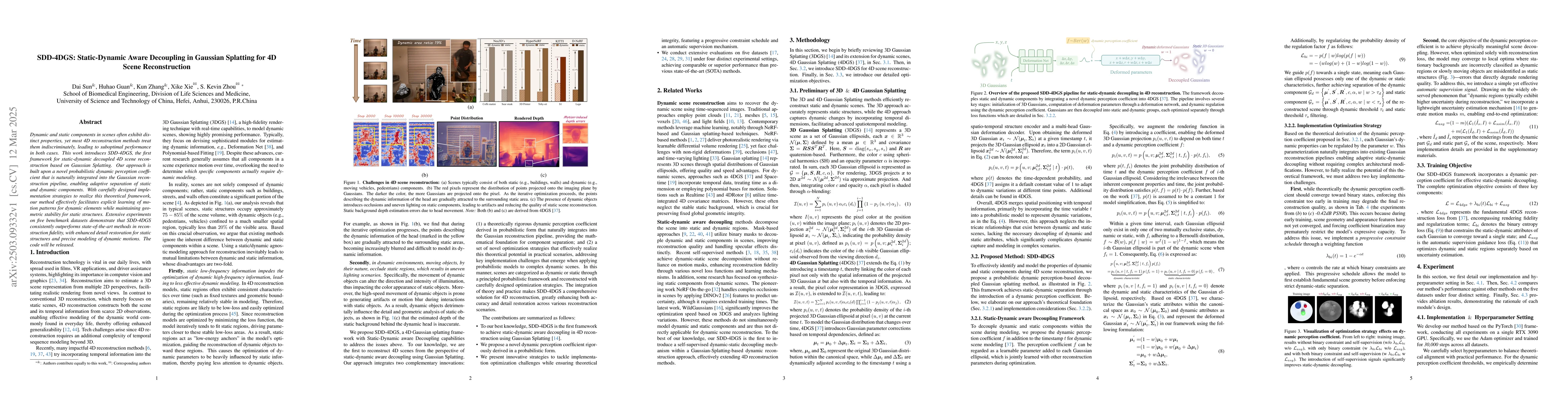
Dynamic and static components in scenes often exhibit distinct properties, yet most 4D reconstruction methods treat them indiscriminately, leading to suboptimal performance in both cases. This work in...
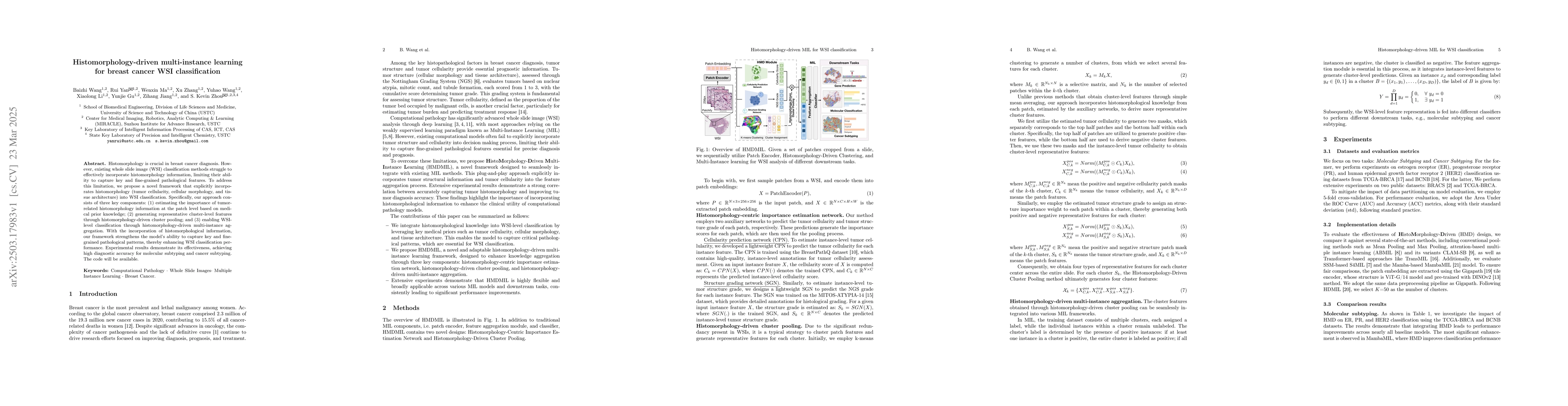
Histomorphology is crucial in breast cancer diagnosis. However, existing whole slide image (WSI) classification methods struggle to effectively incorporate histomorphology information, limiting their ...
Landmark detection plays a crucial role in medical imaging applications such as disease diagnosis, bone age estimation, and therapy planning. However, training models for detecting multiple landmarks ...
The segmentation of pelvic fracture fragments in CT and X-ray images is crucial for trauma diagnosis, surgical planning, and intraoperative guidance. However, accurately and efficiently delineating th...
Recent years have seen rapid advances in AI-driven image generation. Early diffusion models emphasized perceptual quality, while newer multimodal models like GPT-4o-image integrate high-level reasonin...
Natural images exhibit label diversity (clean vs. noisy) in noisy-labeled image classification and prevalence diversity (abundant vs. sparse) in long-tailed image classification. Similarly, medical im...
ICD Coding aims to assign a wide range of medical codes to a medical text document, which is a popular and challenging task in the healthcare domain. To alleviate the problems of long-tail distributio...
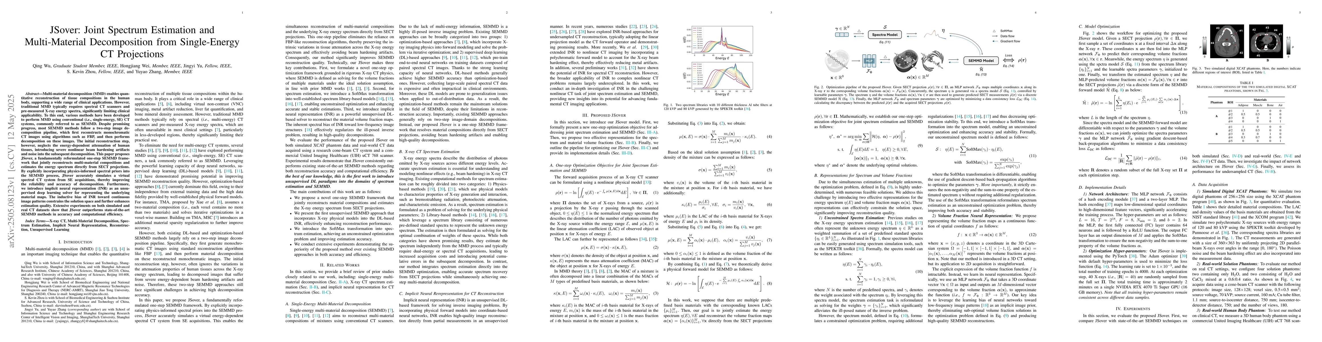
Multi-material decomposition (MMD) enables quantitative reconstruction of tissue compositions in the human body, supporting a wide range of clinical applications. However, traditional MMD typically re...
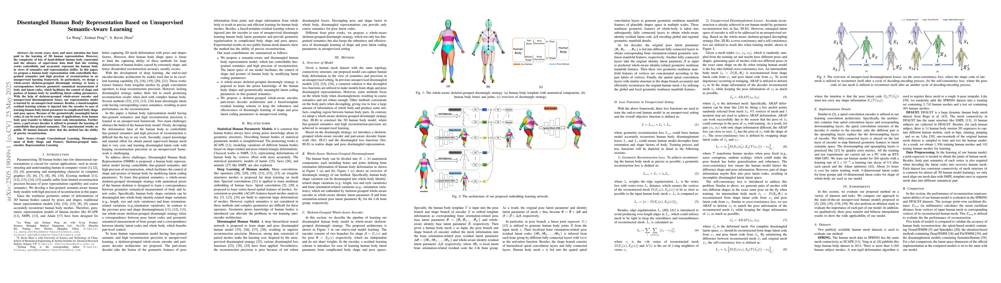
In recent years, more and more attention has been paid to the learning of 3D human representation. However, the complexity of lots of hand-defined human body constraints and the absence of supervision...
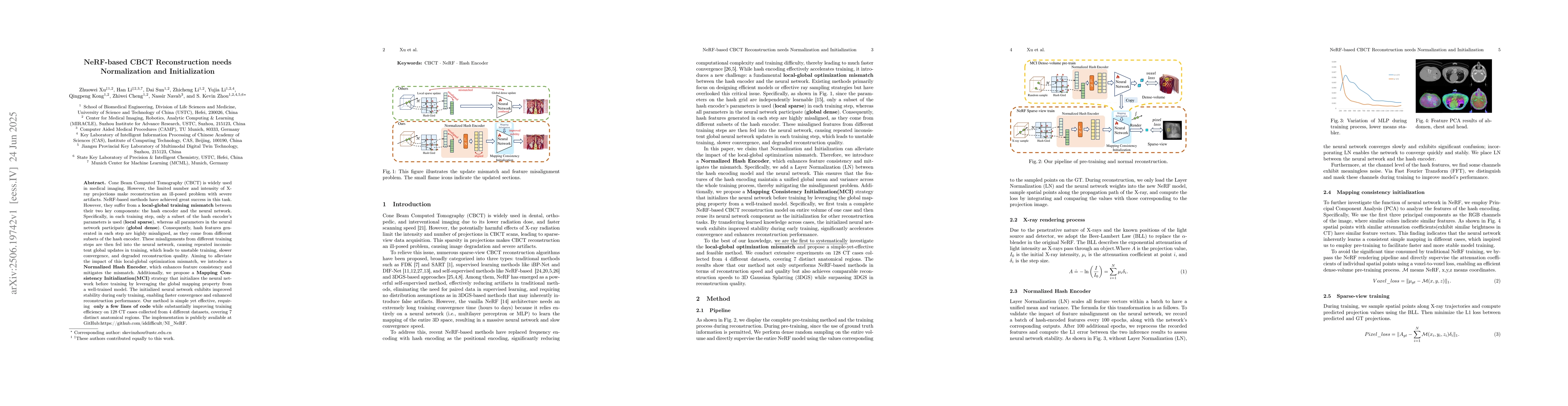
Cone Beam Computed Tomography (CBCT) is widely used in medical imaging. However, the limited number and intensity of X-ray projections make reconstruction an ill-posed problem with severe artifacts. N...
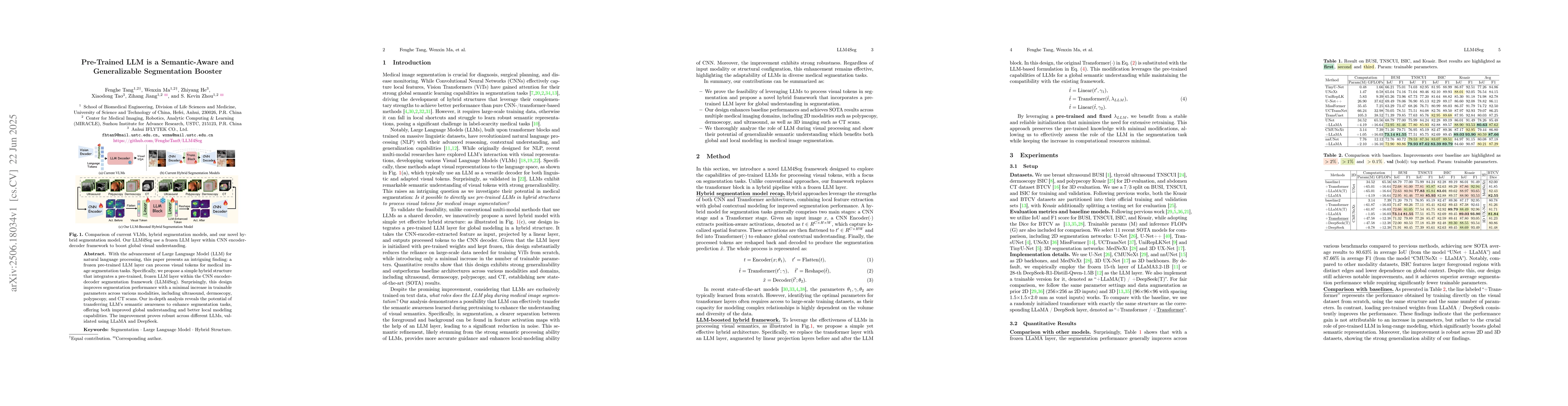
With the advancement of Large Language Model (LLM) for natural language processing, this paper presents an intriguing finding: a frozen pre-trained LLM layer can process visual tokens for medical imag...

The concept bottleneck model (CBM), as a technique improving interpretability via linking predictions to human-understandable concepts, makes high-risk and life-critical medical image classification c...
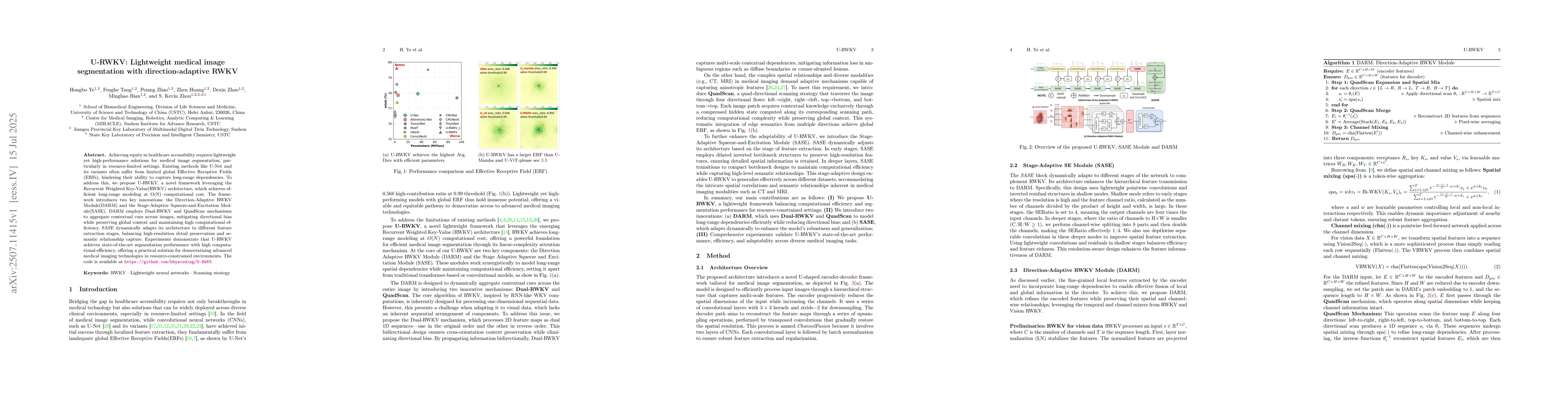
Achieving equity in healthcare accessibility requires lightweight yet high-performance solutions for medical image segmentation, particularly in resource-limited settings. Existing methods like U-Net ...
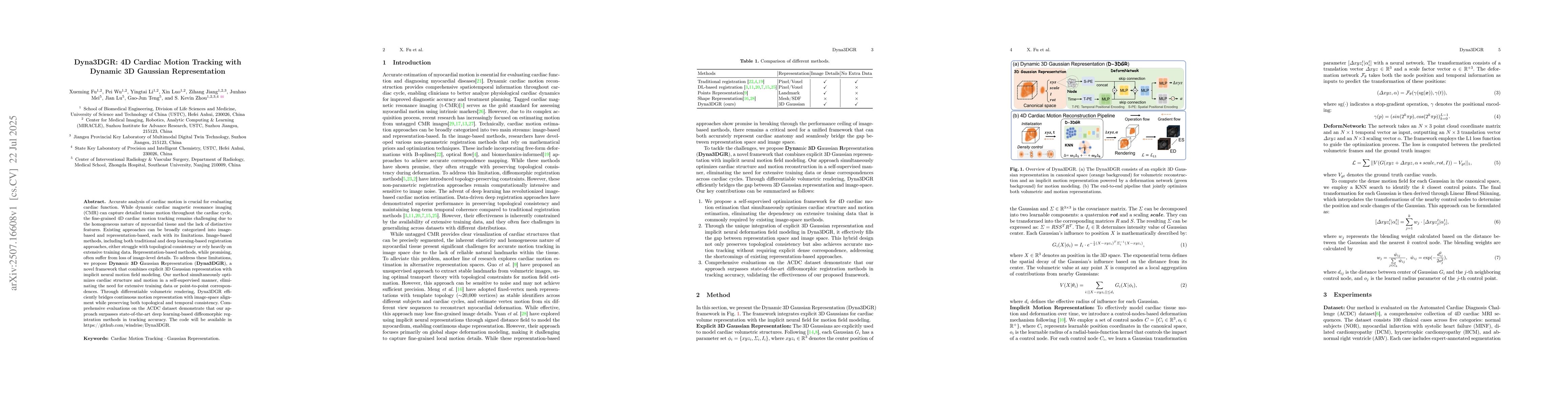
Accurate analysis of cardiac motion is crucial for evaluating cardiac function. While dynamic cardiac magnetic resonance imaging (CMR) can capture detailed tissue motion throughout the cardiac cycle, ...
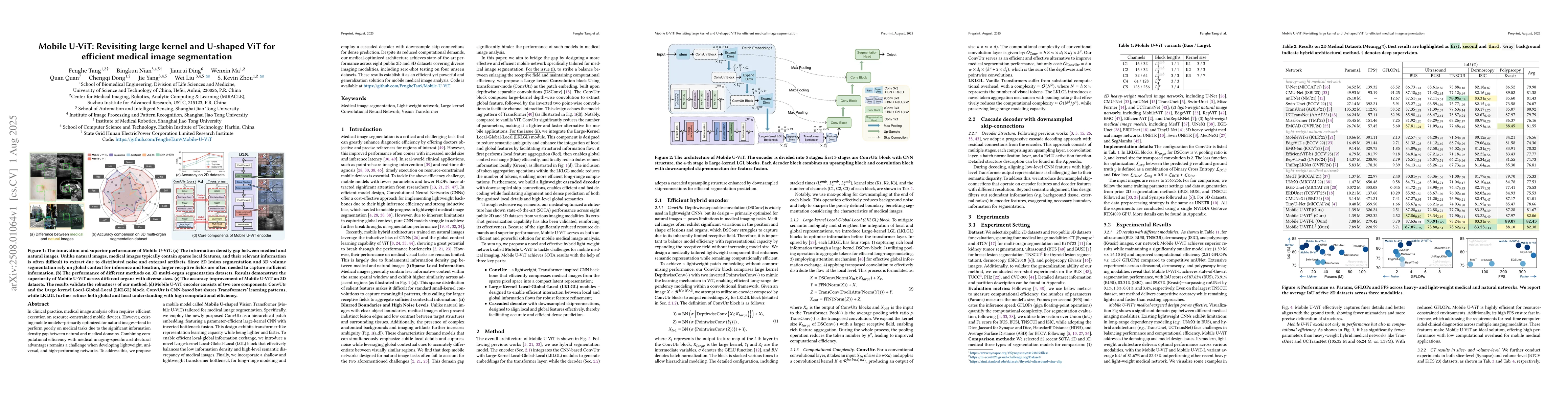
In clinical practice, medical image analysis often requires efficient execution on resource-constrained mobile devices. However, existing mobile models-primarily optimized for natural images-tend to p...
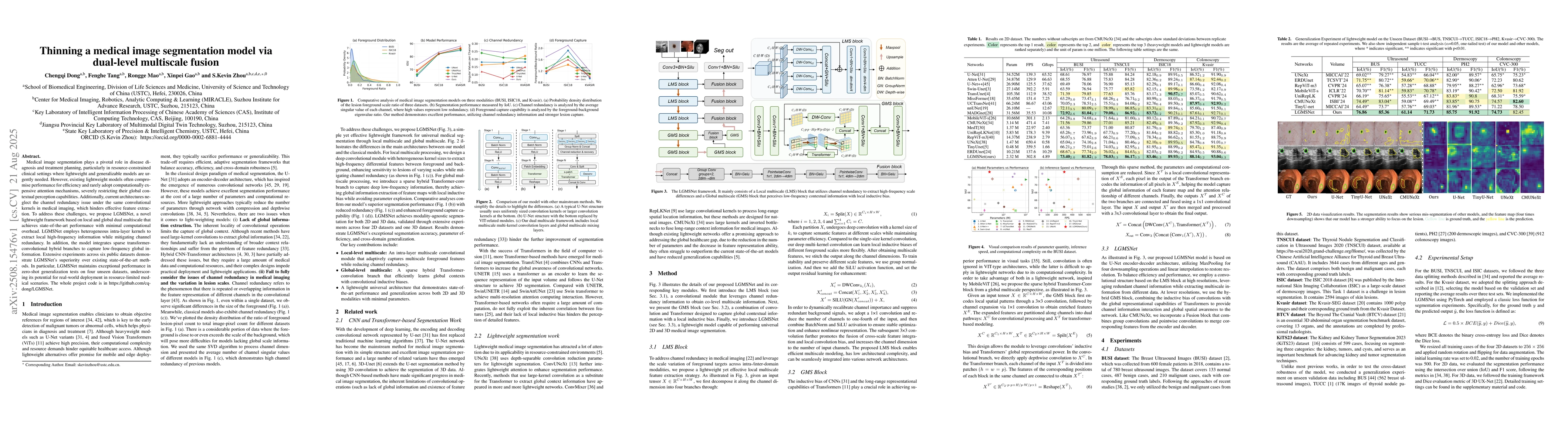
Medical image segmentation plays a pivotal role in disease diagnosis and treatment planning, particularly in resource-constrained clinical settings where lightweight and generalizable models are urgen...
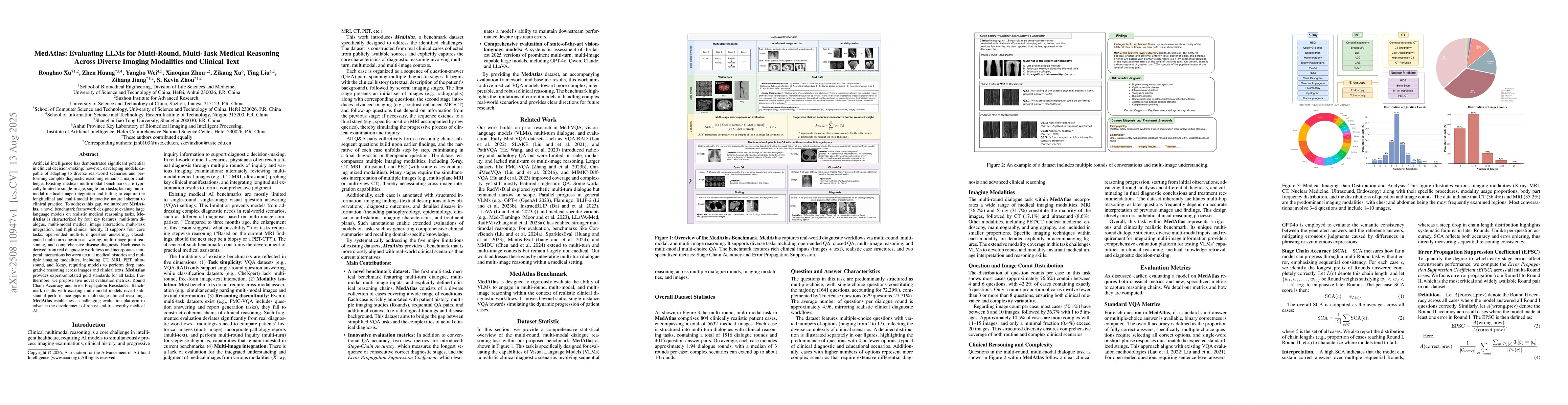
Artificial intelligence has demonstrated significant potential in clinical decision-making; however, developing models capable of adapting to diverse real-world scenarios and performing complex diagno...
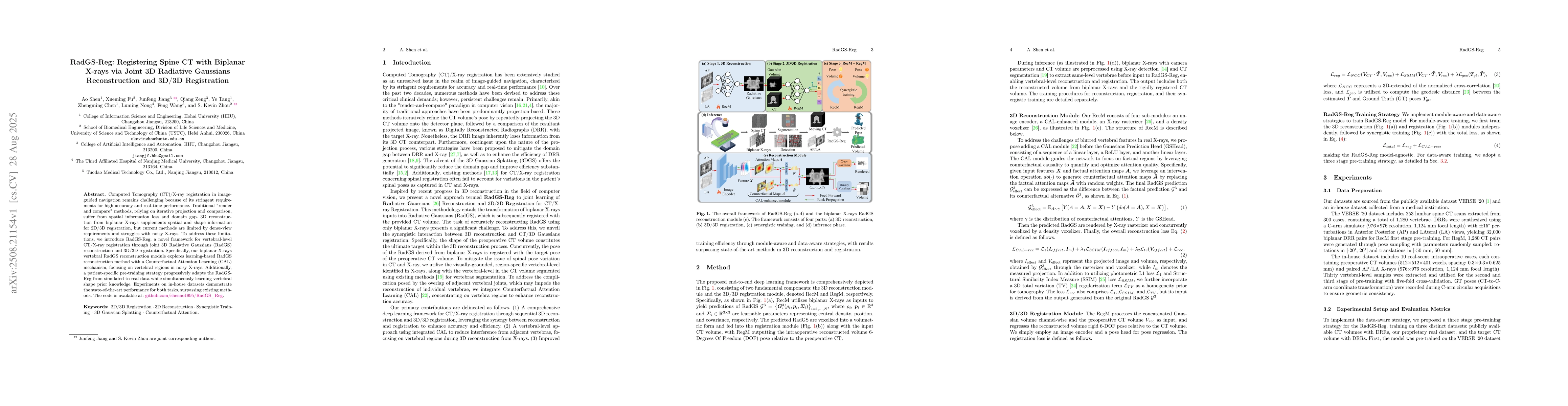
Computed Tomography (CT)/X-ray registration in image-guided navigation remains challenging because of its stringent requirements for high accuracy and real-time performance. Traditional "render and co...
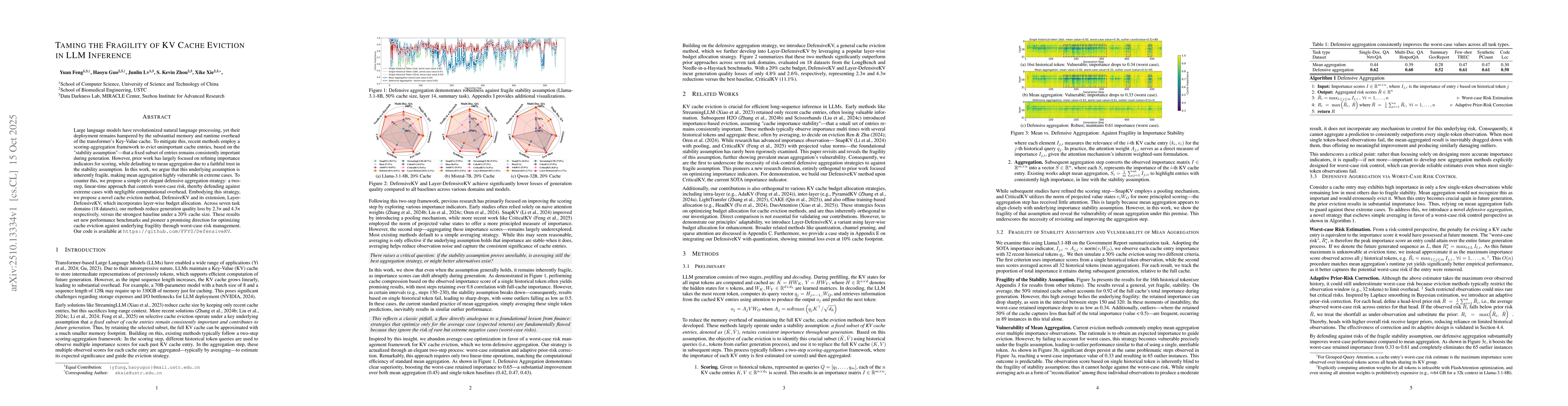
Large language models have revolutionized natural language processing, yet their deployment remains hampered by the substantial memory and runtime overhead of the transformer's Key-Value cache. To mit...
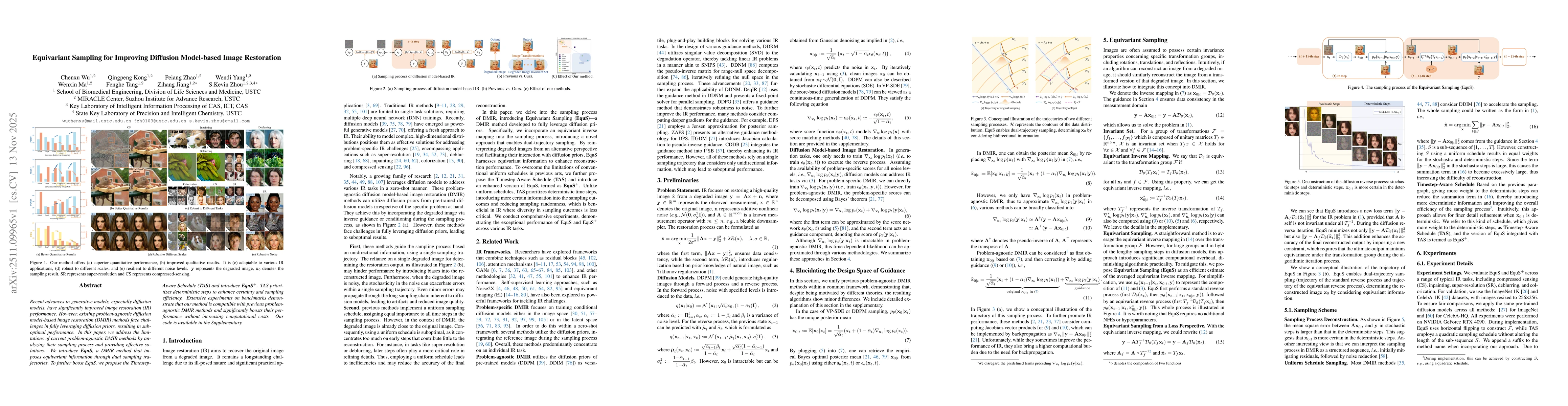
Recent advances in generative models, especially diffusion models, have significantly improved image restoration (IR) performance. However, existing problem-agnostic diffusion model-based image restor...
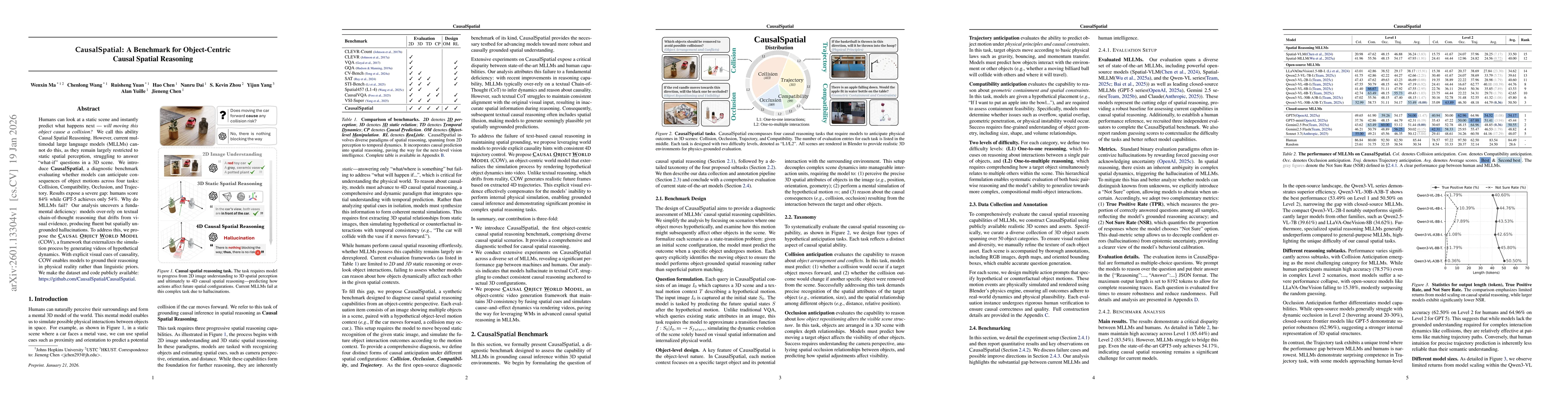
Humans can look at a static scene and instantly predict what happens next -- will moving this object cause a collision? We call this ability Causal Spatial Reasoning. However, current multimodal large...
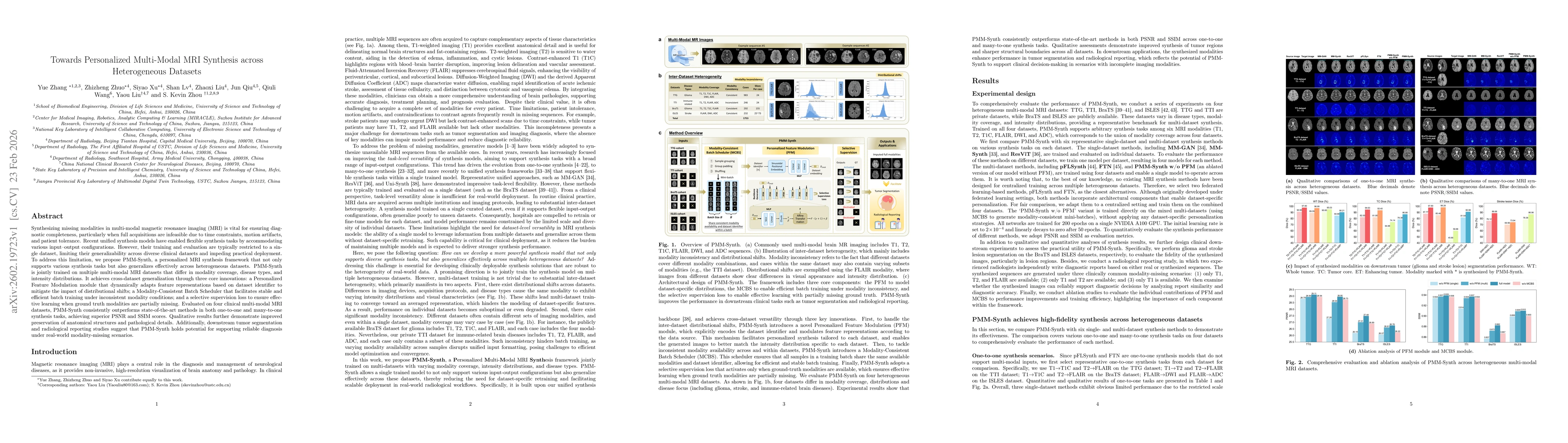
Synthesizing missing modalities in multi-modal magnetic resonance imaging (MRI) is vital for ensuring diagnostic completeness, particularly when full acquisitions are infeasible due to time constraint...
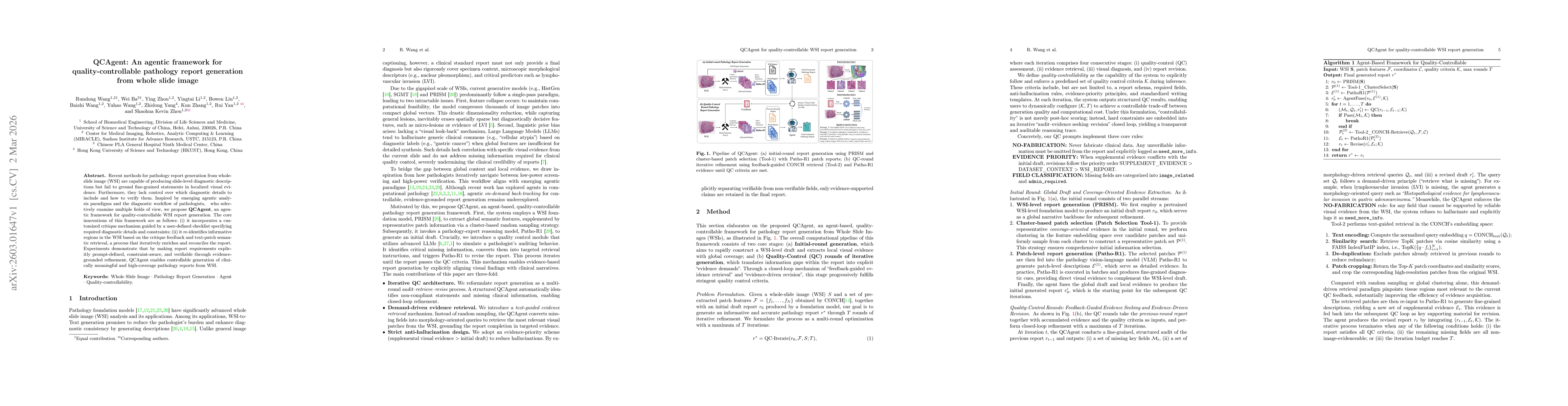
Recent methods for pathology report generation from whole-slide image (WSI) are capable of producing slide-level diagnostic descriptions but fail to ground fine-grained statements in localized visual ...
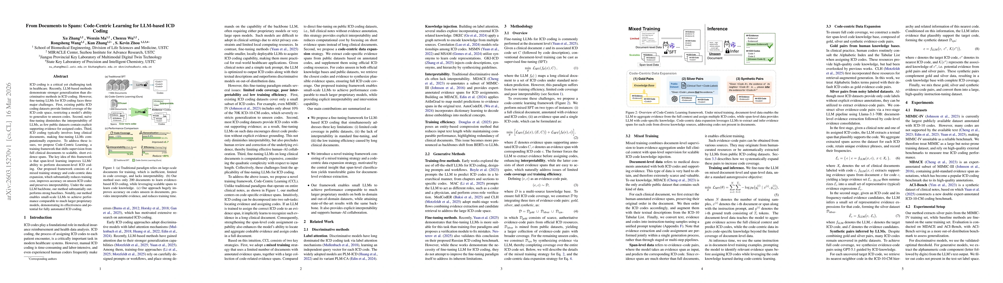
ICD coding is a critical yet challenging task in healthcare. Recently, LLM-based methods demonstrate stronger generalization than discriminative methods in ICD coding. However, fine-tuning LLMs for IC...
Uterine diseases represent an important category of gynecologic pathology and require accurate histopathological assessment for diagnosis and treatment planning. Whole-slide images (WSI) have enabled ...