Academic Profile
Statistics
Similar Authors
Papers on arXiv
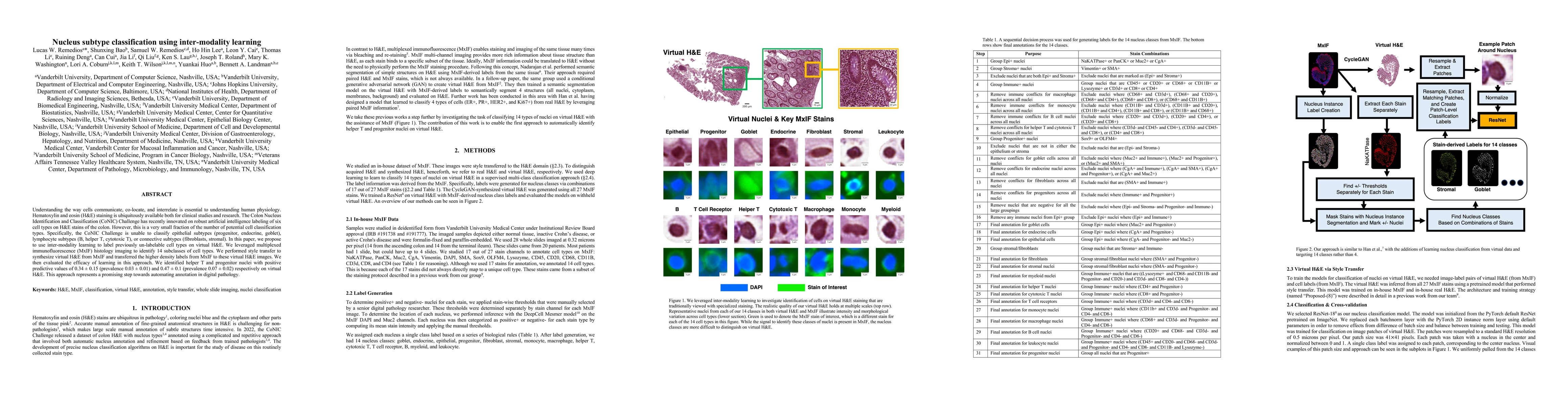
Understanding the way cells communicate, co-locate, and interrelate is essential to understanding human physiology. Hematoxylin and eosin (H&E) staining is ubiquitously available both for clinical s...
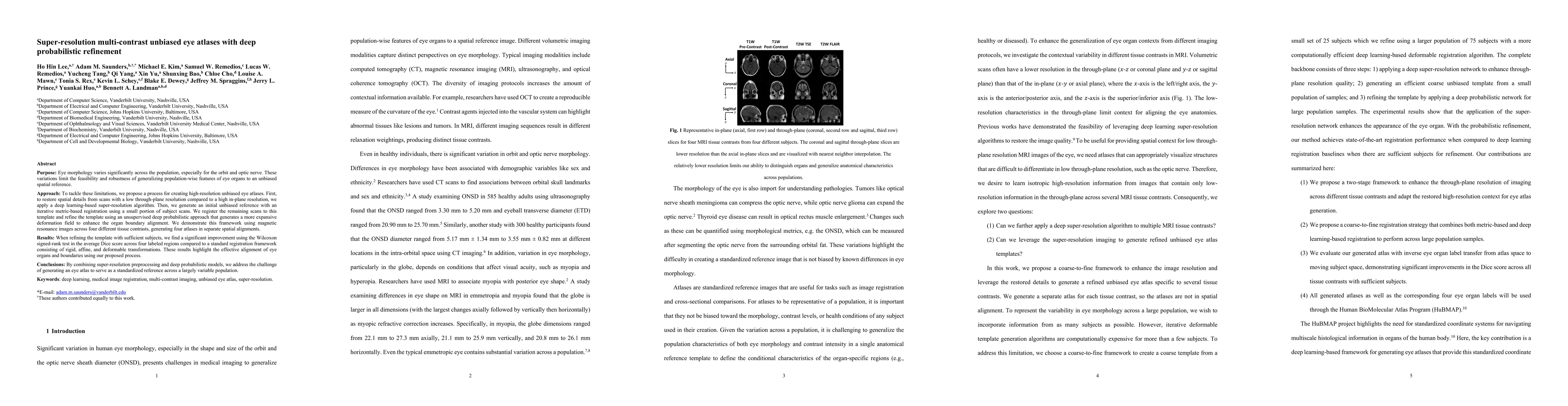
Purpose: Eye morphology varies significantly across the population, especially for the orbit and optic nerve. These variations limit the feasibility and robustness of generalizing population-wise fe...
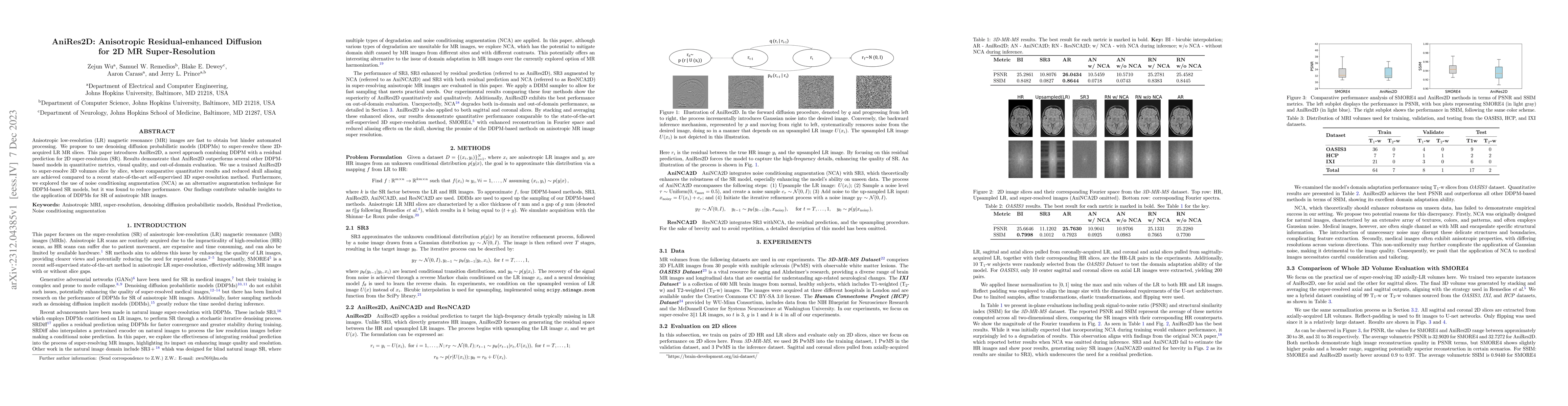
Anisotropic low-resolution (LR) magnetic resonance (MR) images are fast to obtain but hinder automated processing. We propose to use denoising diffusion probabilistic models (DDPMs) to super-resolve...
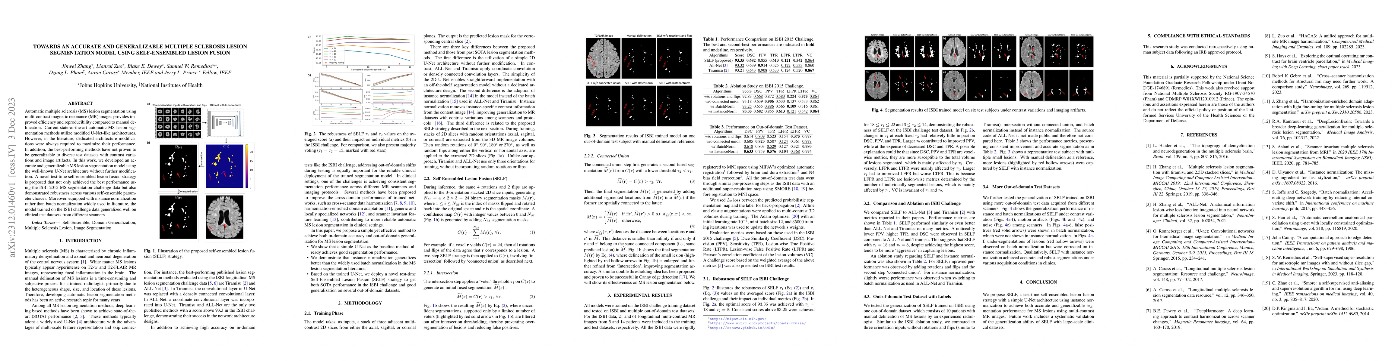
Automatic multiple sclerosis (MS) lesion segmentation using multi-contrast magnetic resonance (MR) images provides improved efficiency and reproducibility compared to manual delineation. Current sta...
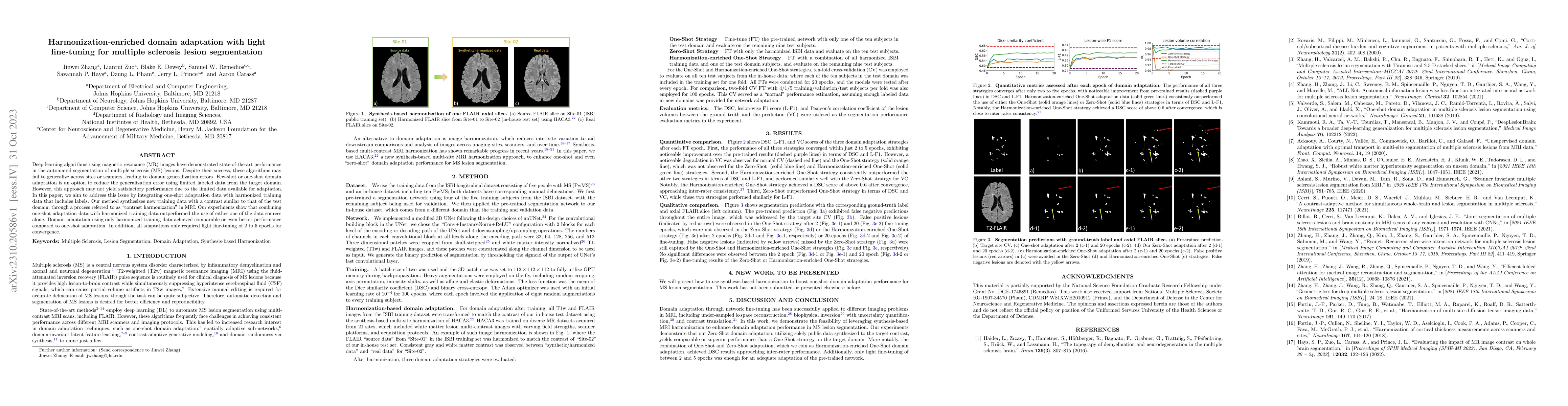
Deep learning algorithms utilizing magnetic resonance (MR) images have demonstrated cutting-edge proficiency in autonomously segmenting multiple sclerosis (MS) lesions. Despite their achievements, t...
Deep learning has made great strides in medical imaging, enabled by hardware advances in GPUs. One major constraint for the development of new models has been the saturation of GPU memory resources ...
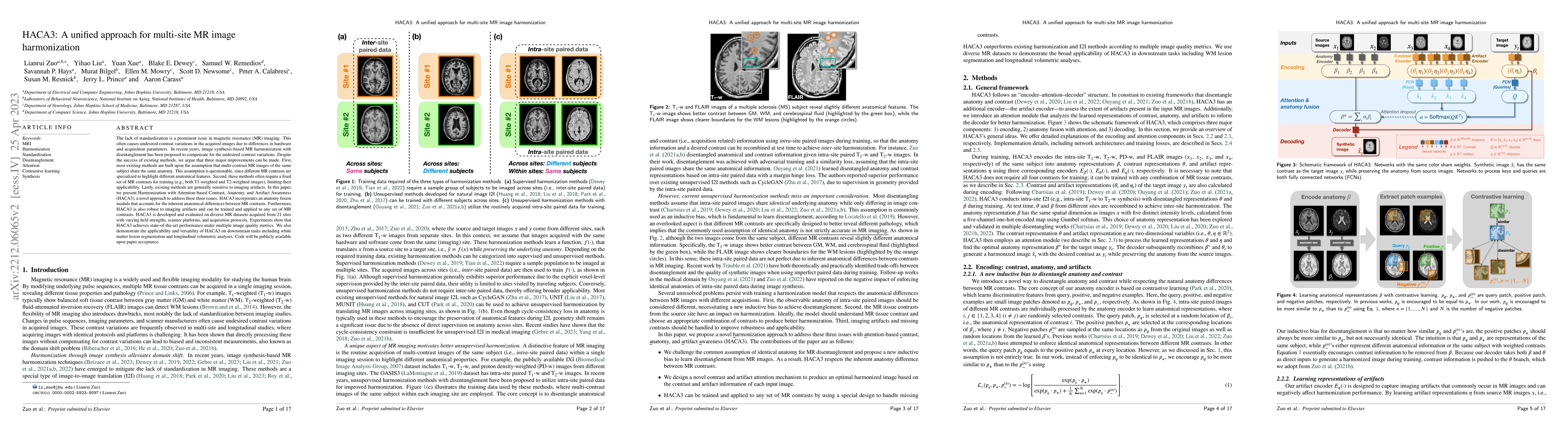
The lack of standardization is a prominent issue in magnetic resonance (MR) imaging. This often causes undesired contrast variations in the acquired images due to differences in hardware and acquisi...
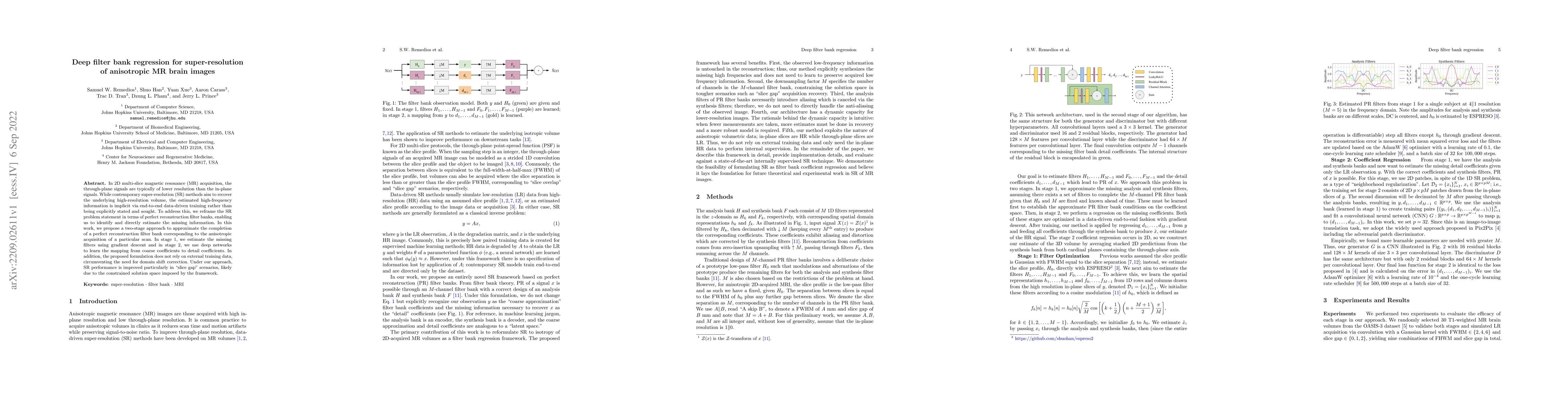
In 2D multi-slice magnetic resonance (MR) acquisition, the through-plane signals are typically of lower resolution than the in-plane signals. While contemporary super-resolution (SR) methods aim to ...
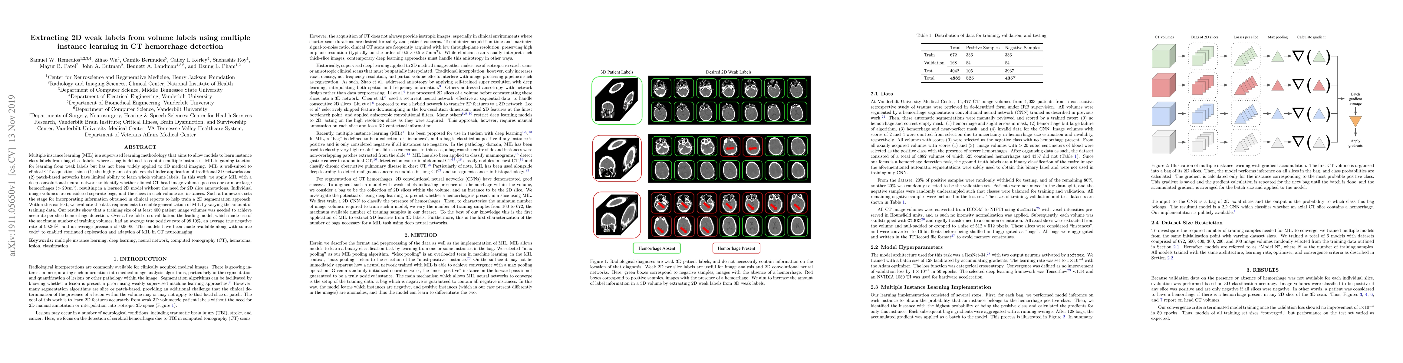
Multiple instance learning (MIL) is a supervised learning methodology that aims to allow models to learn instance class labels from bag class labels, where a bag is defined to contain multiple insta...
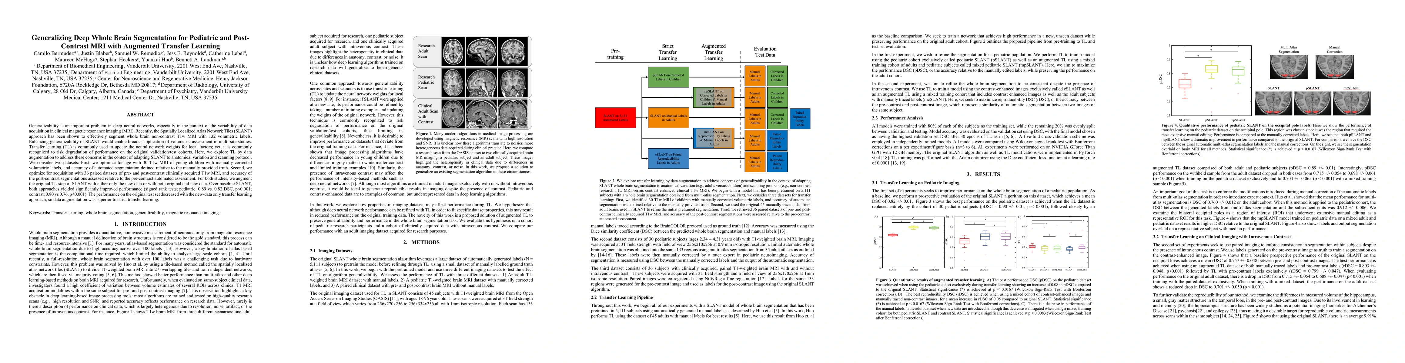
Generalizability is an important problem in deep neural networks, especially in the context of the variability of data acquisition in clinical magnetic resonance imaging (MRI). Recently, the Spatial...
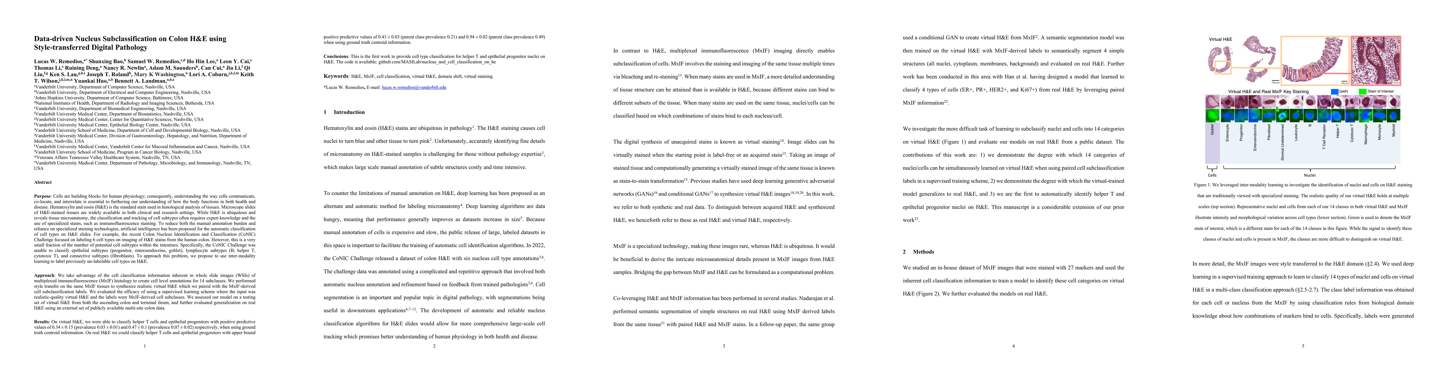
Understanding the way cells communicate, co-locate, and interrelate is essential to furthering our understanding of how the body functions. H&E is widely available, however, cell subtyping often requi...
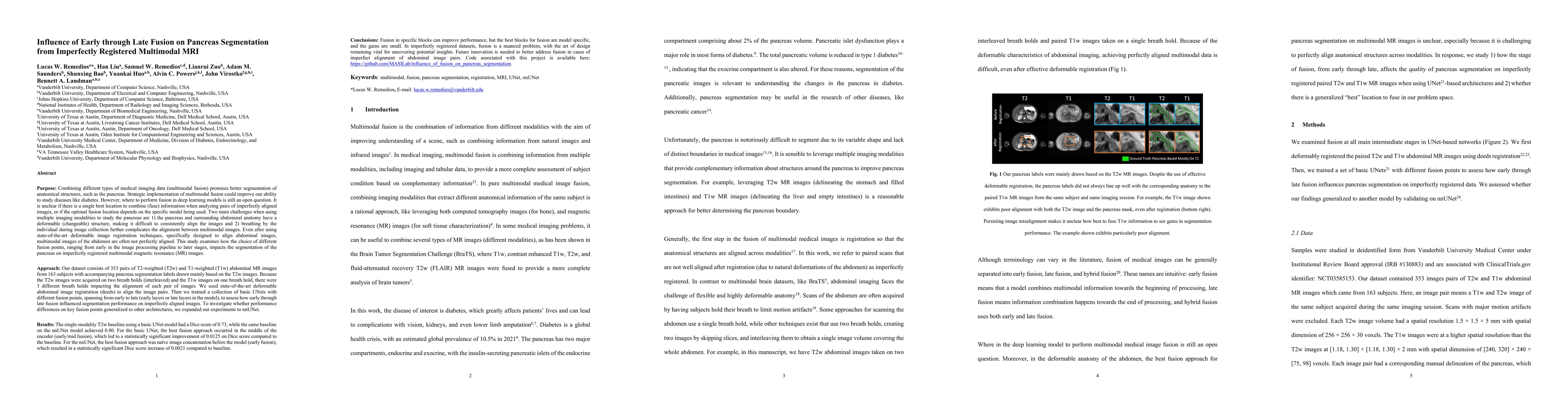
Multimodal fusion promises better pancreas segmentation. However, where to perform fusion in models is still an open question. It is unclear if there is a best location to fuse information when analyz...
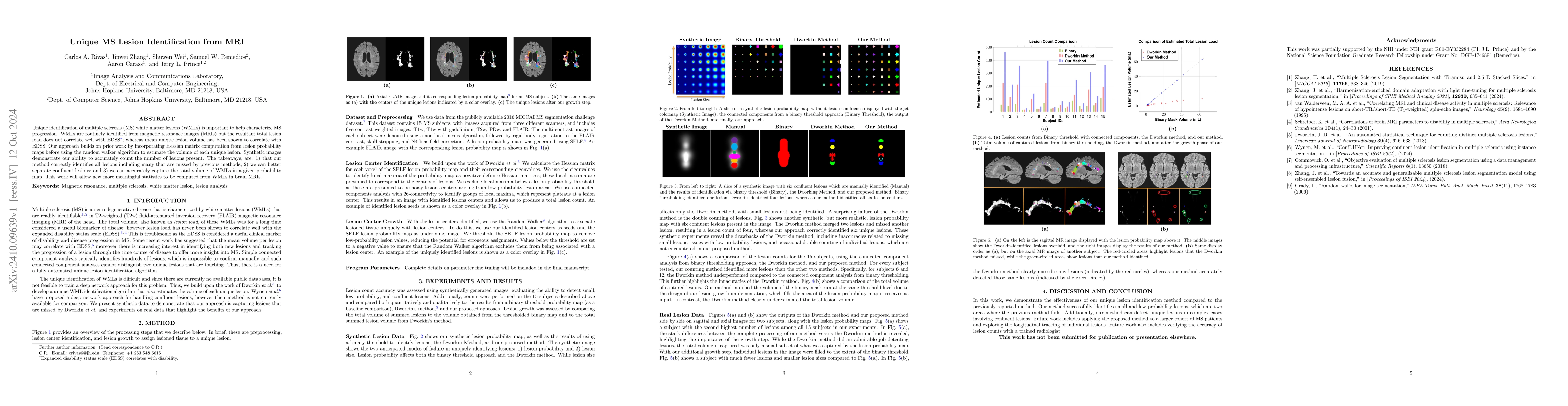
Unique identification of multiple sclerosis (MS) white matter lesions (WMLs) is important to help characterize MS progression. WMLs are routinely identified from magnetic resonance images (MRIs) but t...
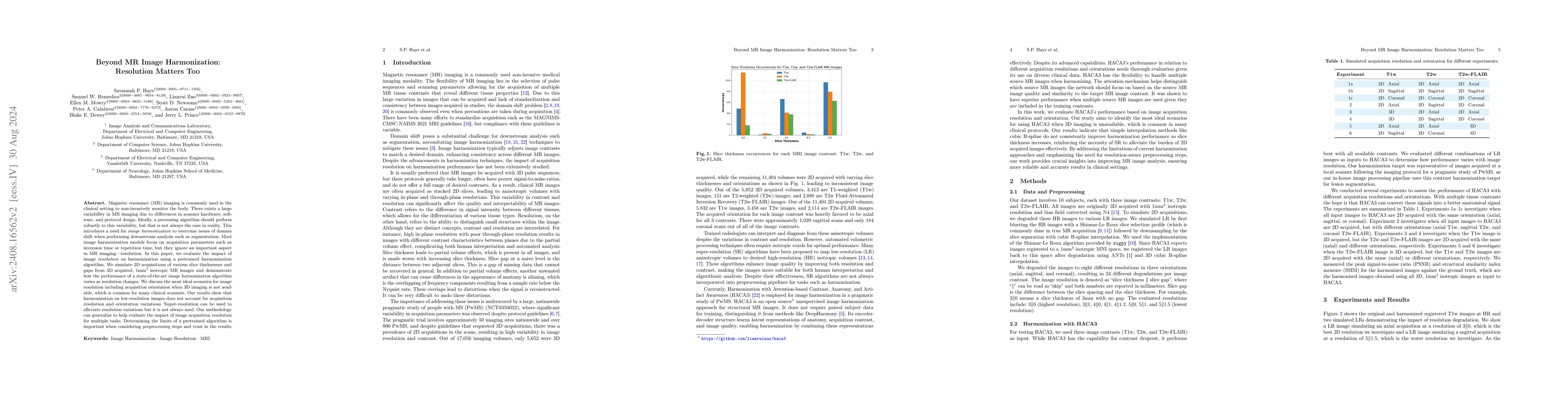
Magnetic resonance (MR) imaging is commonly used in the clinical setting to non-invasively monitor the body. There exists a large variability in MR imaging due to differences in scanner hardware, soft...
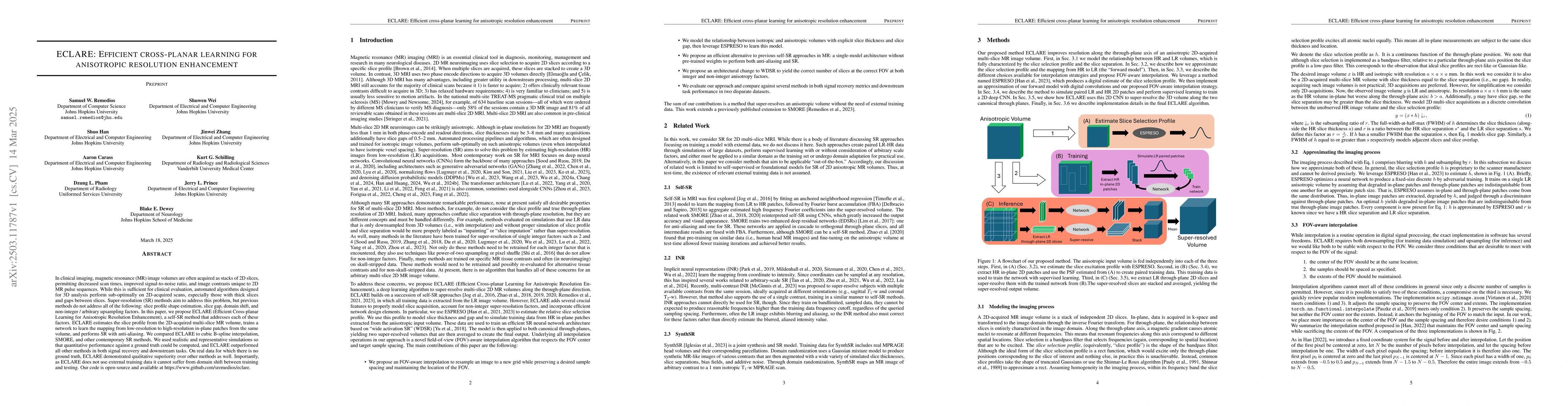
In clinical imaging, magnetic resonance (MR) image volumes are often acquired as stacks of 2D slices, permitting decreased scan times, improved signal-to-noise ratio, and image contrasts unique to 2D ...
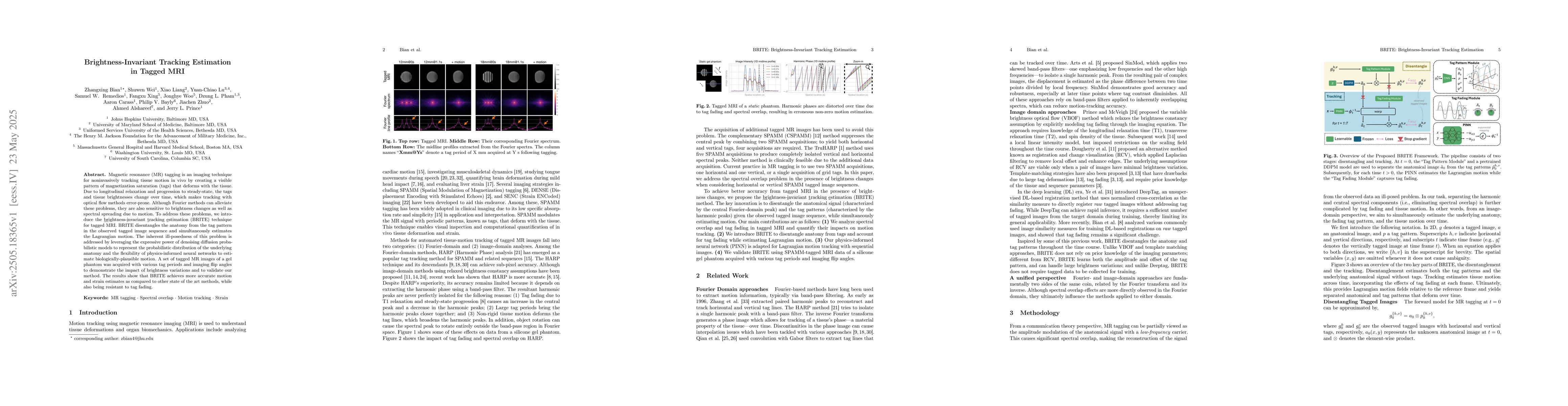
Magnetic resonance (MR) tagging is an imaging technique for noninvasively tracking tissue motion in vivo by creating a visible pattern of magnetization saturation (tags) that deforms with the tissue. ...
Automated segmentation of multiple sclerosis (MS) lesions using multicontrast magnetic resonance (MR) images improves efficiency and reproducibility compared to manual delineation, with deep learning ...
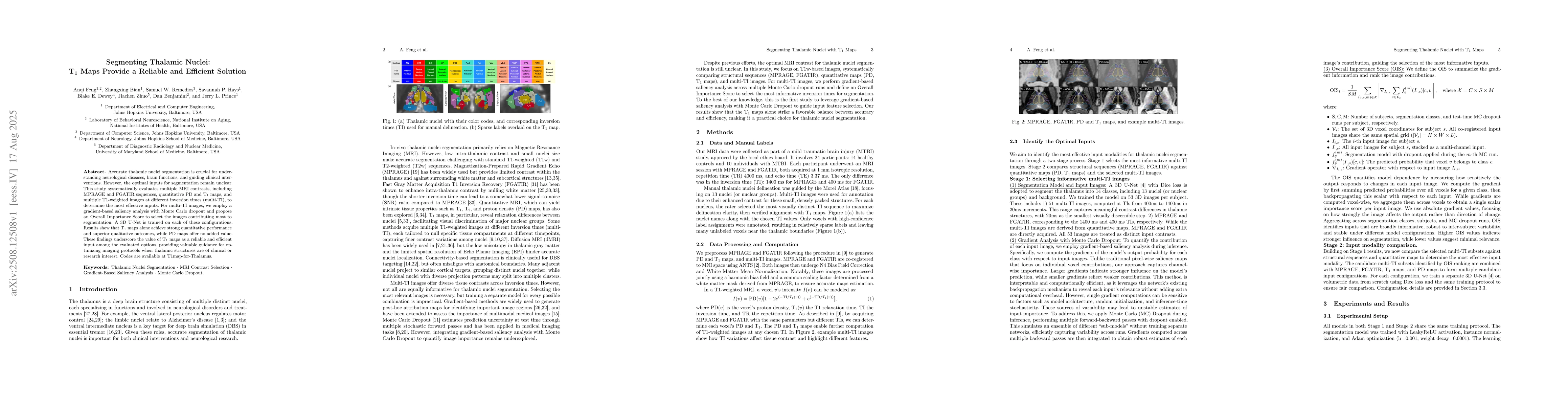
Accurate thalamic nuclei segmentation is crucial for understanding neurological diseases, brain functions, and guiding clinical interventions. However, the optimal inputs for segmentation remain uncle...
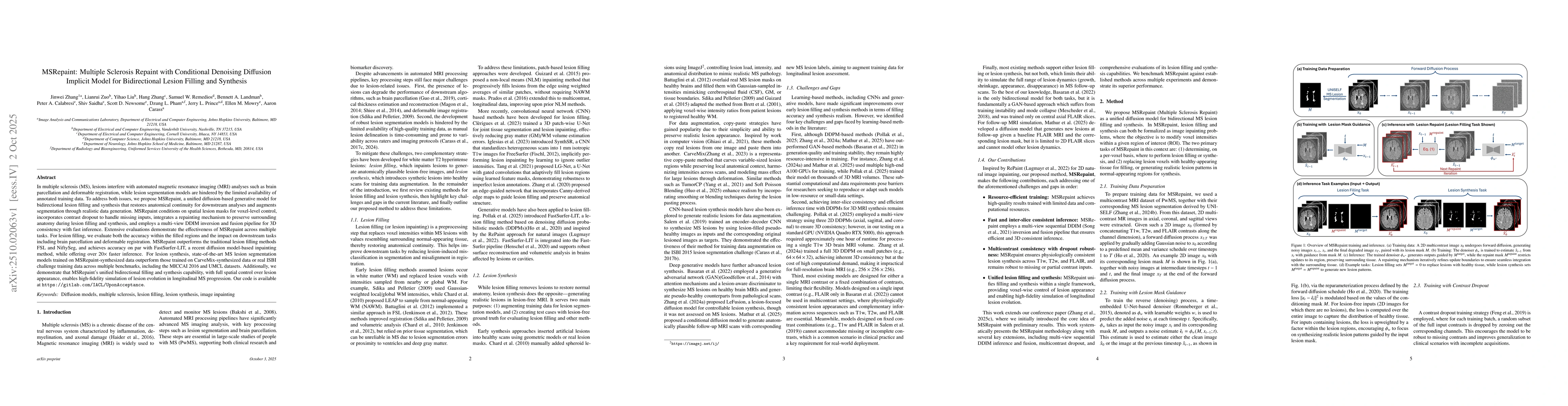
In multiple sclerosis, lesions interfere with automated magnetic resonance imaging analyses such as brain parcellation and deformable registration, while lesion segmentation models are hindered by the...
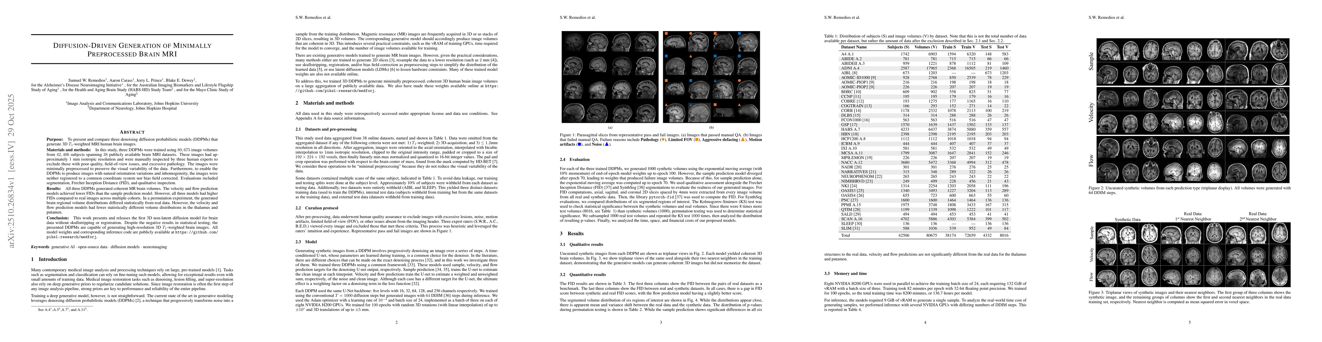
The purpose of this study is to present and compare three denoising diffusion probabilistic models (DDPMs) that generate 3D $T_1$-weighted MRI human brain images. Three DDPMs were trained using 80,675...
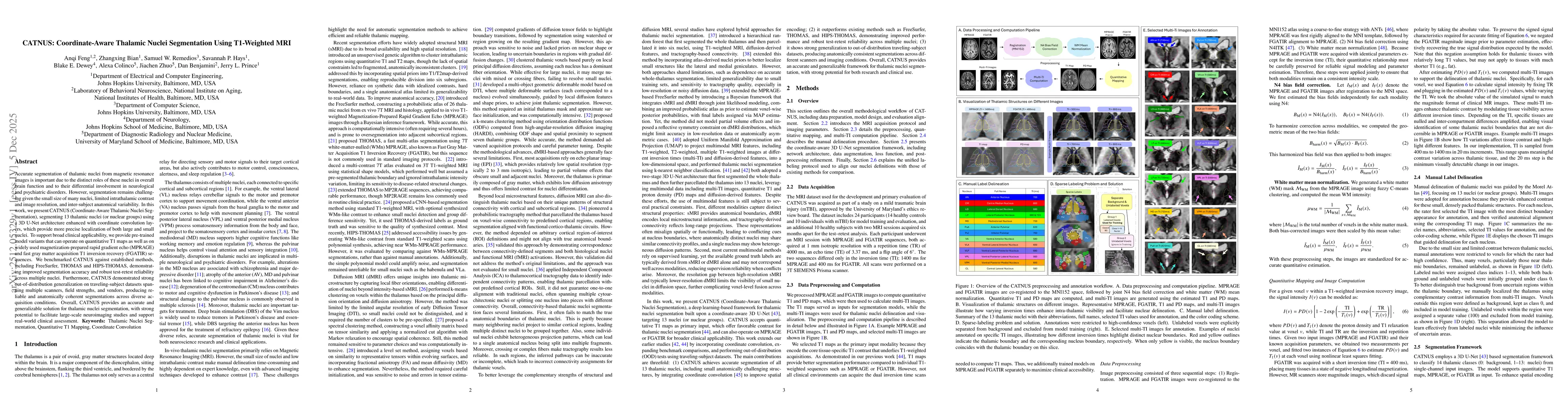
Accurate segmentation of thalamic nuclei from magnetic resonance images is important due to the distinct roles of these nuclei in overall brain function and to their differential involvement in neurol...
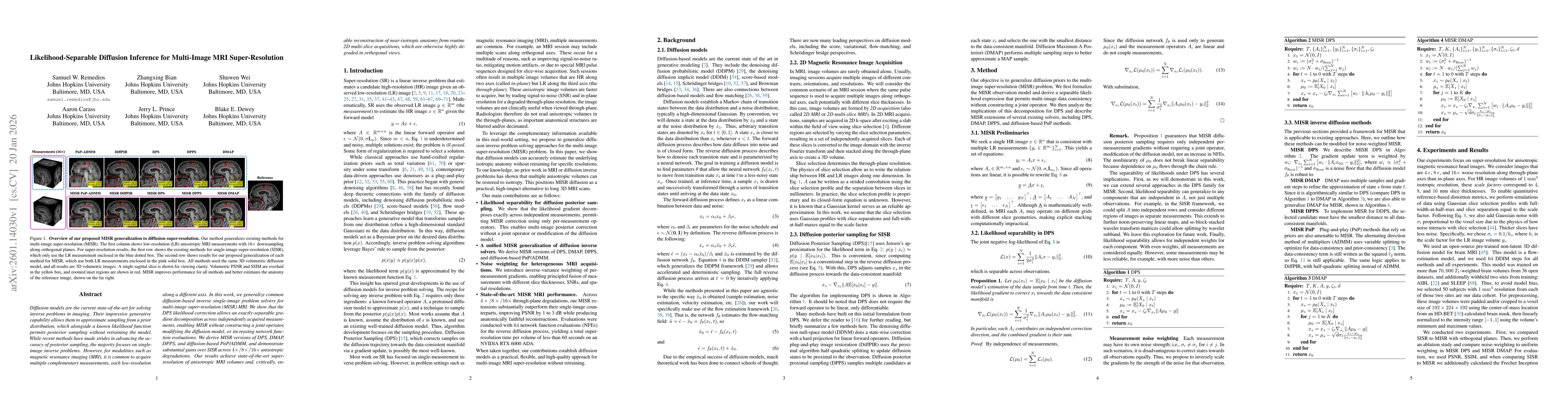
Diffusion models are the current state-of-the-art for solving inverse problems in imaging. Their impressive generative capability allows them to approximate sampling from a prior distribution, which a...
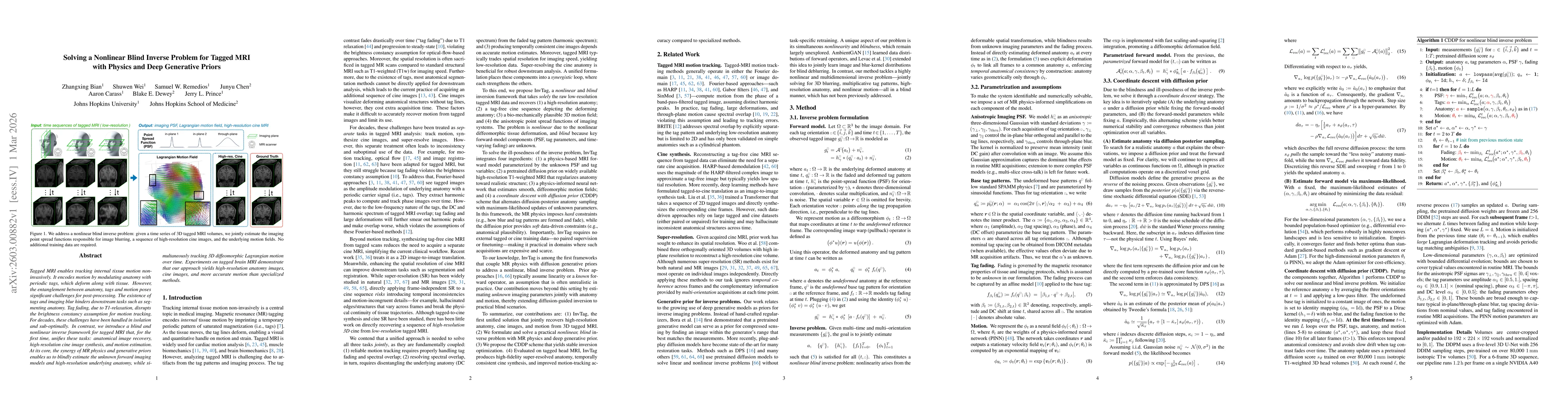
Tagged MRI enables tracking internal tissue motion non-invasively. It encodes motion by modulating anatomy with periodic tags, which deform along with tissue. However, the entanglement between anatomy...
Reliable harmonization of heterogeneous magnetic resonance~(MR) image datasets, especially those acquired in pragmatic clinical trials, is critical to advance multi-center neuroimaging studies and tra...