ArxivLens
S
Researcher

Shichuan Zhang

23 papers on arXiv

23 arXiv Papers
4 Total Publications
Profile

Academic Profile

Name Z. Shichuan
Total Papers 4
Scholar Profile View on Semantic Scholar
Metrics

Statistics

23 arXiv Papers
4 Total Publications
Publications

Papers on arXiv

Thumbnail for Combining Supervised Learning and Reinforcement Learning for Multi-Label
  Classification Tasks with Partial Labels
arXiv
Combining Supervised Learning and Reinforcement Learning for Multi-Label Classification Tasks with Partial Labels

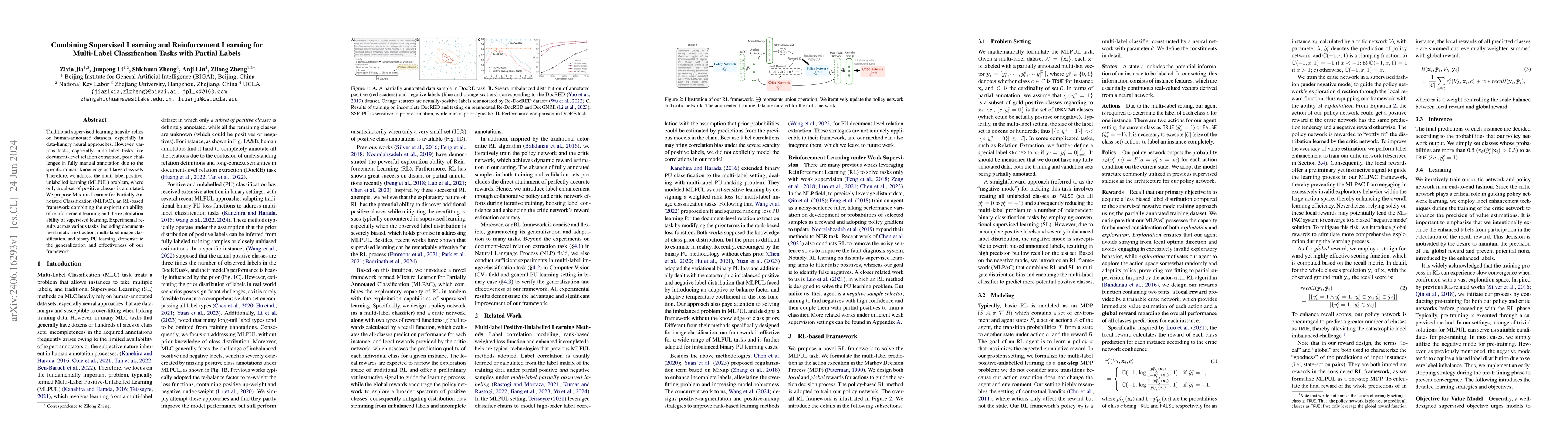
Traditional supervised learning heavily relies on human-annotated datasets, especially in data-hungry neural approaches. However, various tasks, especially multi-label tasks like document-level relati...

Thumbnail for Multi-modal Learning with Missing Modality in Predicting Axillary Lymph
  Node Metastasis
arXiv
Multi-modal Learning with Missing Modality in Predicting Axillary Lymph Node Metastasis

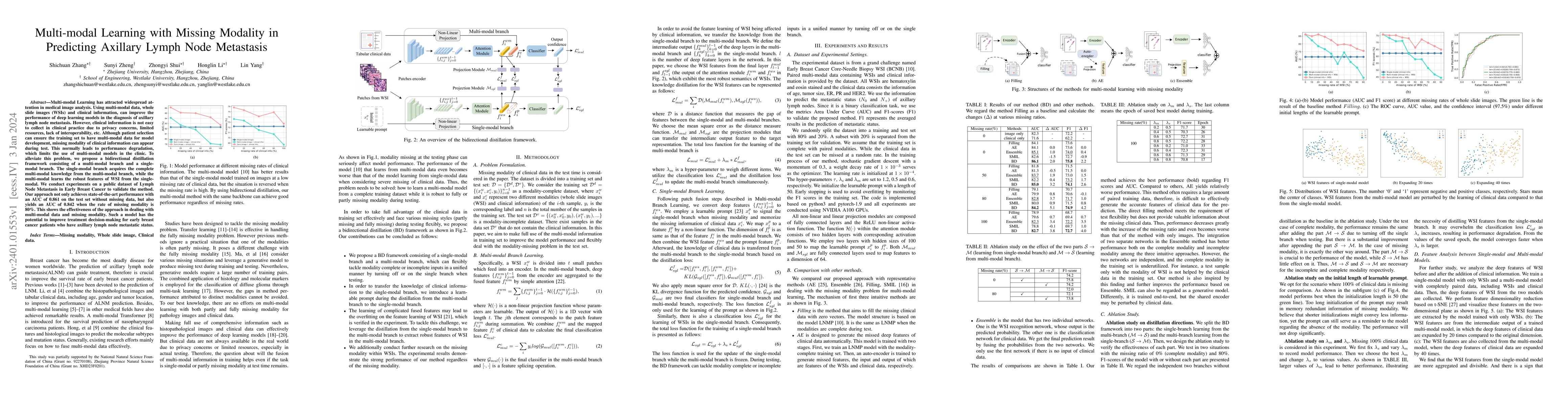
Multi-modal Learning has attracted widespread attention in medical image analysis. Using multi-modal data, whole slide images (WSIs) and clinical information, can improve the performance of deep lea...

Thumbnail for SegRap2023: A Benchmark of Organs-at-Risk and Gross Tumor Volume
  Segmentation for Radiotherapy Planning of Nasopharyngeal Carcinoma
arXiv
SegRap2023: A Benchmark of Organs-at-Risk and Gross Tumor Volume Segmentation for Radiotherapy Planning of Nasopharyngeal Carcinoma

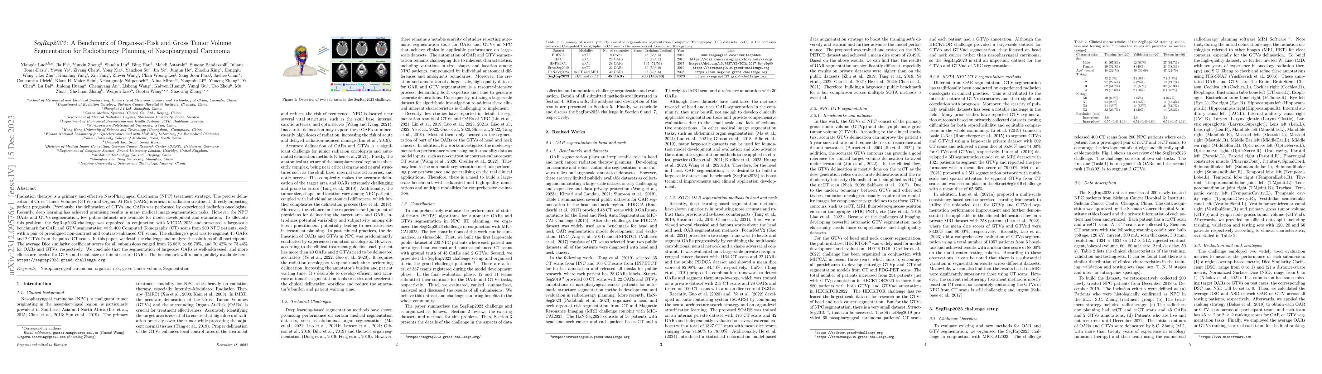
Radiation therapy is a primary and effective NasoPharyngeal Carcinoma (NPC) treatment strategy. The precise delineation of Gross Tumor Volumes (GTVs) and Organs-At-Risk (OARs) is crucial in radiatio...

Thumbnail for Scribble-based 3D Multiple Abdominal Organ Segmentation via
  Triple-branch Multi-dilated Network with Pixel- and Class-wise Consistency
arXiv
Scribble-based 3D Multiple Abdominal Organ Segmentation via Triple-branch Multi-dilated Network with Pixel- and Class-wise Consistency

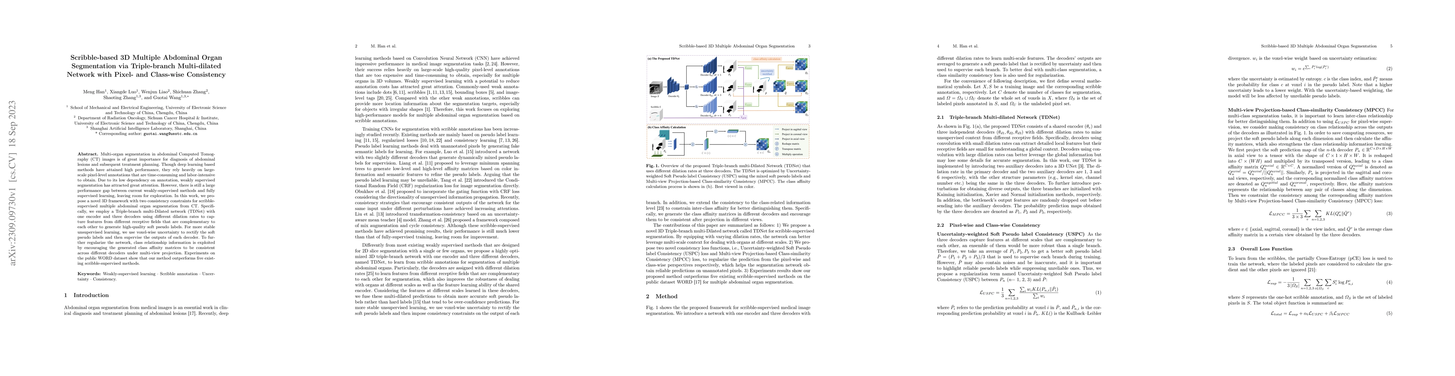
Multi-organ segmentation in abdominal Computed Tomography (CT) images is of great importance for diagnosis of abdominal lesions and subsequent treatment planning. Though deep learning based methods ...

Thumbnail for Exploring Unsupervised Cell Recognition with Prior Self-activation Maps
arXiv
Exploring Unsupervised Cell Recognition with Prior Self-activation Maps

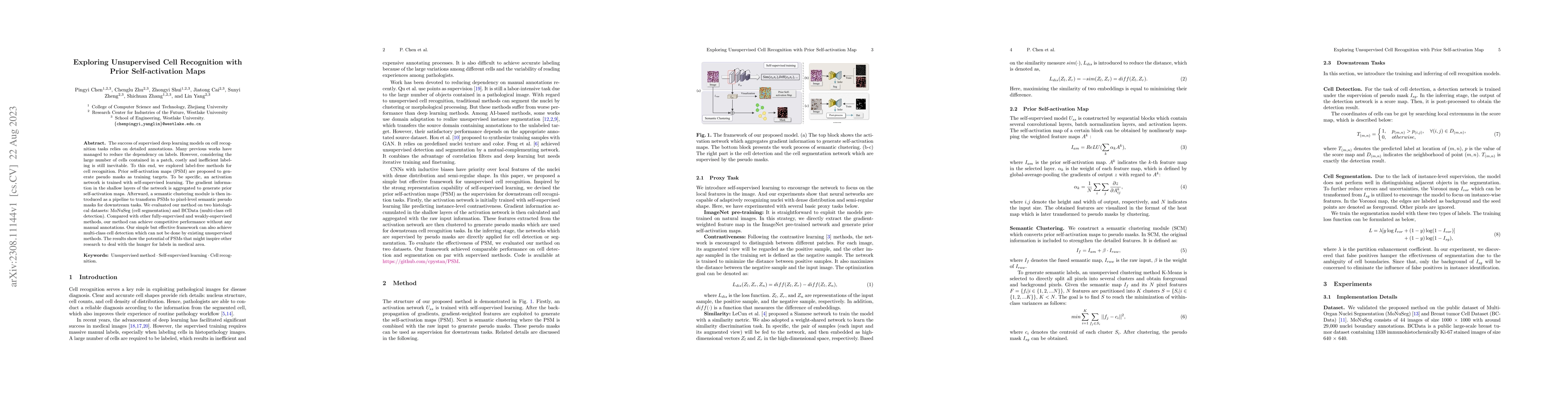
The success of supervised deep learning models on cell recognition tasks relies on detailed annotations. Many previous works have managed to reduce the dependency on labels. However, considering the...

Thumbnail for Masked conditional variational autoencoders for chromosome straightening
arXiv
Masked conditional variational autoencoders for chromosome straightening

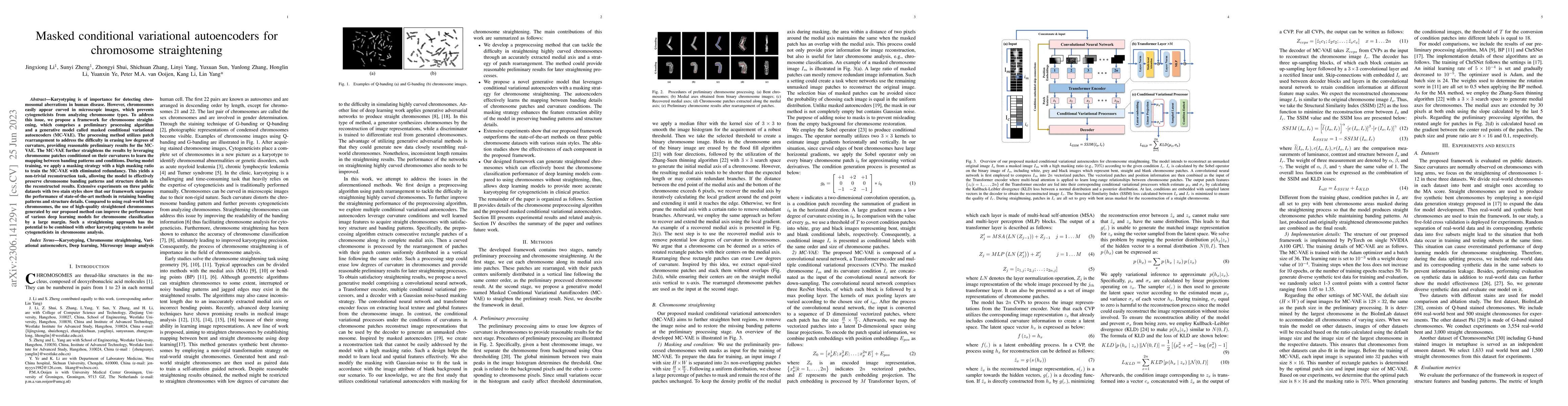
Karyotyping is of importance for detecting chromosomal aberrations in human disease. However, chromosomes easily appear curved in microscopic images, which prevents cytogeneticists from analyzing ch...

Thumbnail for Semi-supervised Cell Recognition under Point Supervision
arXiv
Semi-supervised Cell Recognition under Point Supervision

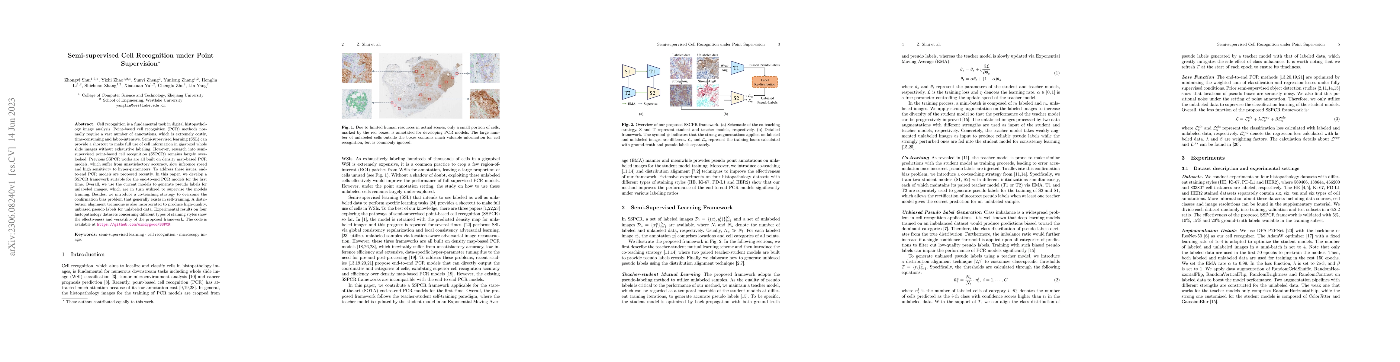
Cell recognition is a fundamental task in digital histopathology image analysis. Point-based cell recognition (PCR) methods normally require a vast number of annotations, which is extremely costly, ...

Thumbnail for DPA-P2PNet: Deformable Proposal-aware P2PNet for Accurate Point-based
  Cell Detection
arXiv
DPA-P2PNet: Deformable Proposal-aware P2PNet for Accurate Point-based Cell Detection

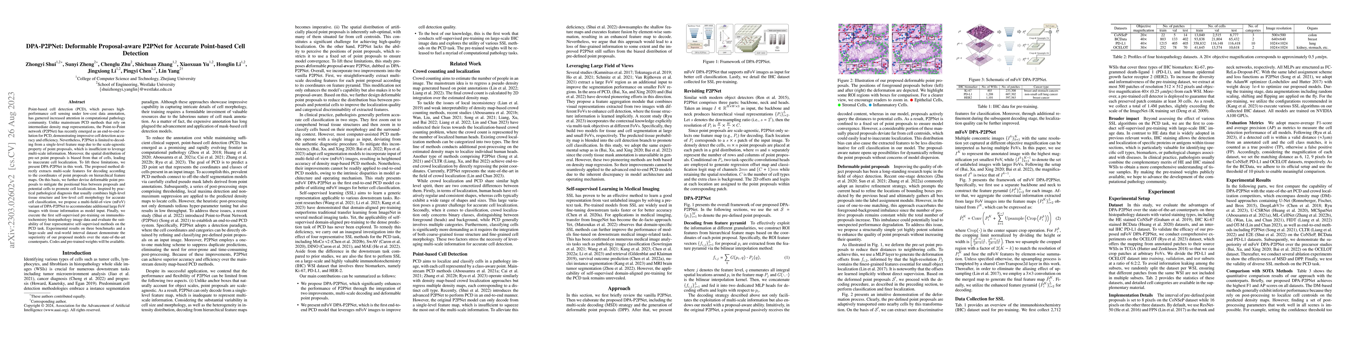
Point-based cell detection (PCD), which pursues high-performance cell sensing under low-cost data annotation, has garnered increased attention in computational pathology community. Unlike mainstream...

Thumbnail for CDDSA: Contrastive Domain Disentanglement and Style Augmentation for
  Generalizable Medical Image Segmentation
arXiv
CDDSA: Contrastive Domain Disentanglement and Style Augmentation for Generalizable Medical Image Segmentation

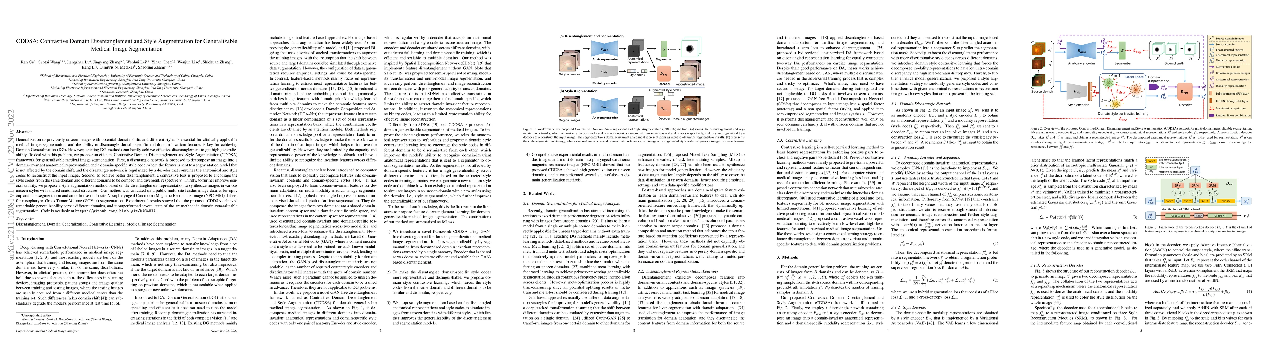
Generalization to previously unseen images with potential domain shifts and different styles is essential for clinically applicable medical image segmentation, and the ability to disentangle domain-...

Thumbnail for Unsupervised Dense Nuclei Detection and Segmentation with Prior
  Self-activation Map For Histology Images
arXiv
Unsupervised Dense Nuclei Detection and Segmentation with Prior Self-activation Map For Histology Images

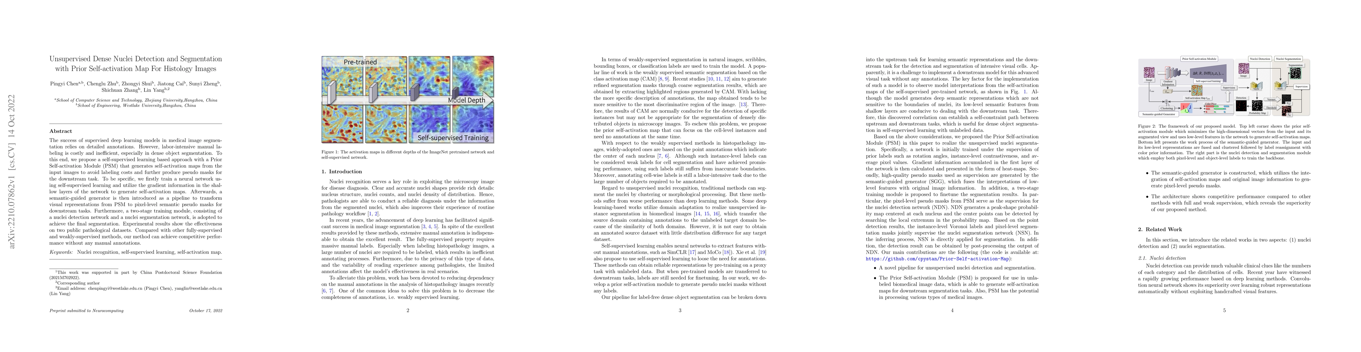
The success of supervised deep learning models in medical image segmentation relies on detailed annotations. However, labor-intensive manual labeling is costly and inefficient, especially in dense o...

Thumbnail for End-to-end cell recognition by point annotation
arXiv
End-to-end cell recognition by point annotation

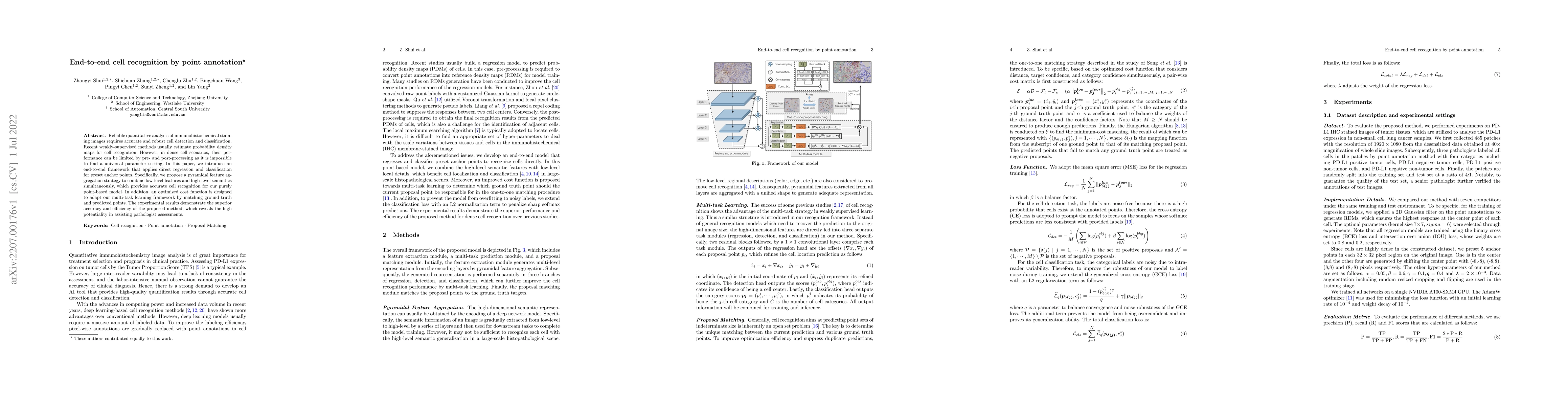
Reliable quantitative analysis of immunohistochemical staining images requires accurate and robust cell detection and classification. Recent weakly-supervised methods usually estimate probability de...

Thumbnail for HMRNet: High and Multi-Resolution Network with Bidirectional Feature
  Calibration for Brain Structure Segmentation in Radiotherapy
arXiv
HMRNet: High and Multi-Resolution Network with Bidirectional Feature Calibration for Brain Structure Segmentation in Radiotherapy

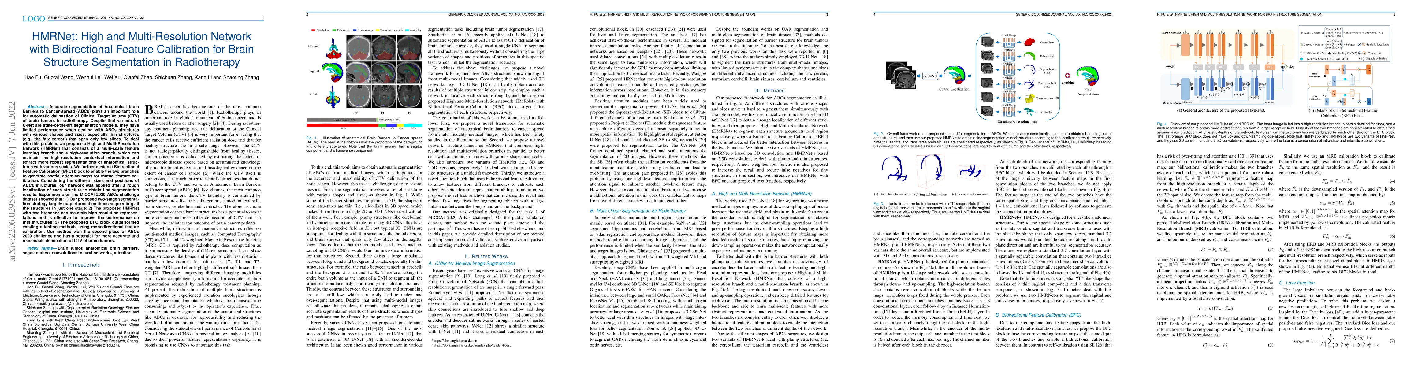
Accurate segmentation of Anatomical brain Barriers to Cancer spread (ABCs) plays an important role for automatic delineation of Clinical Target Volume (CTV) of brain tumors in radiotherapy. Despite ...

Thumbnail for Weakly Supervised Learning for cell recognition in immunohistochemical
  cytoplasm staining images
arXiv
Weakly Supervised Learning for cell recognition in immunohistochemical cytoplasm staining images

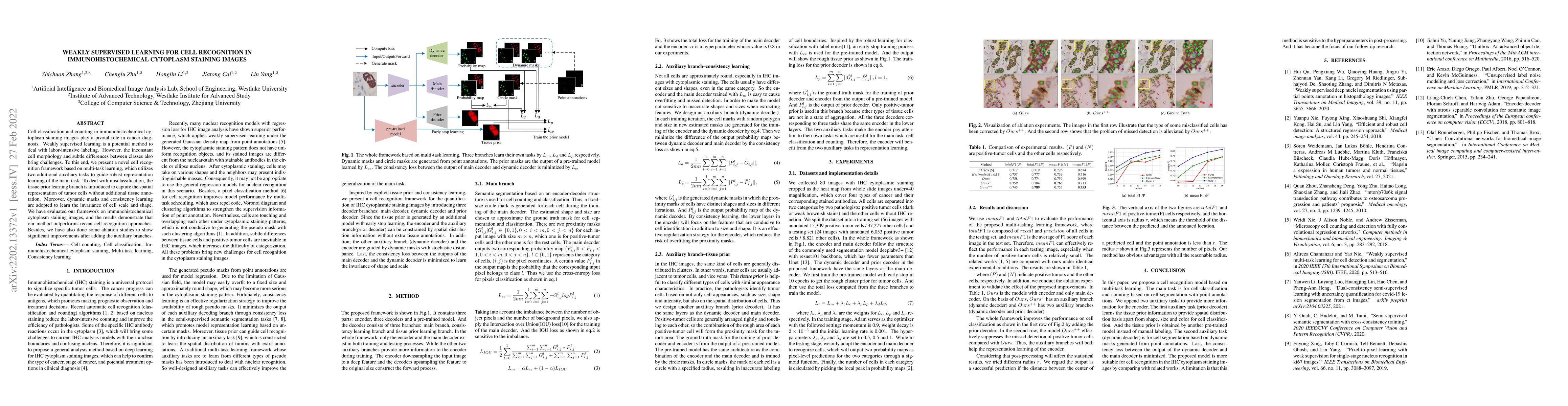
Cell classification and counting in immunohistochemical cytoplasm staining images play a pivotal role in cancer diagnosis. Weakly supervised learning is a potential method to deal with labor-intensi...

Thumbnail for Generalizing Nucleus Recognition Model in Multi-source Images via
  Pruning
arXiv
Generalizing Nucleus Recognition Model in Multi-source Images via Pruning

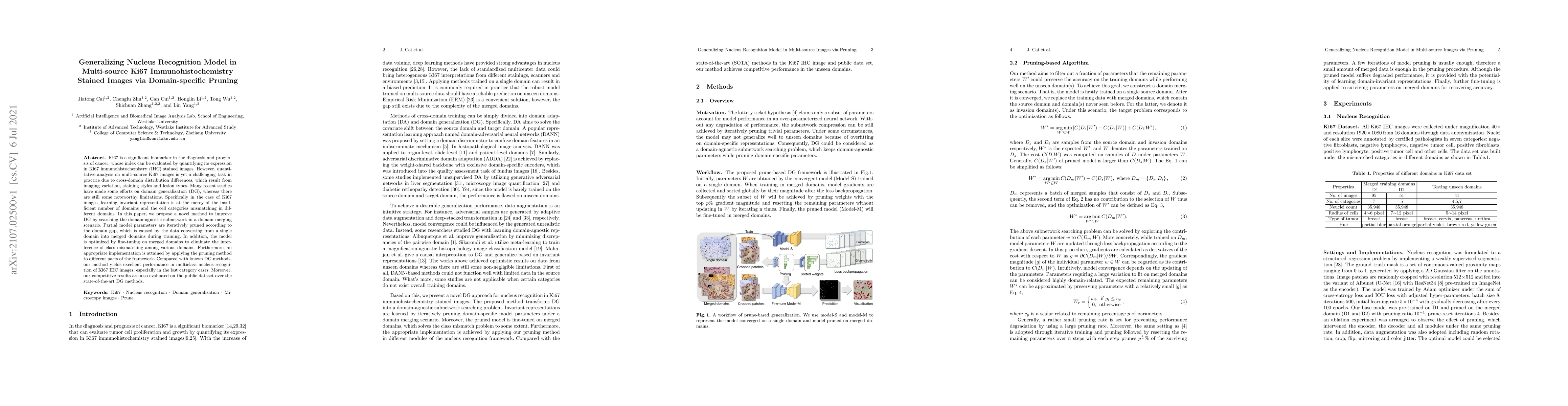
Ki67 is a significant biomarker in the diagnosis and prognosis of cancer, whose index can be evaluated by quantifying its expression in Ki67 immunohistochemistry (IHC) stained images. However, quant...

Thumbnail for Automatic Segmentation of Organs-at-Risk from Head-and-Neck CT using
  Separable Convolutional Neural Network with Hard-Region-Weighted Loss
arXiv
Automatic Segmentation of Organs-at-Risk from Head-and-Neck CT using Separable Convolutional Neural Network with Hard-Region-Weighted Loss

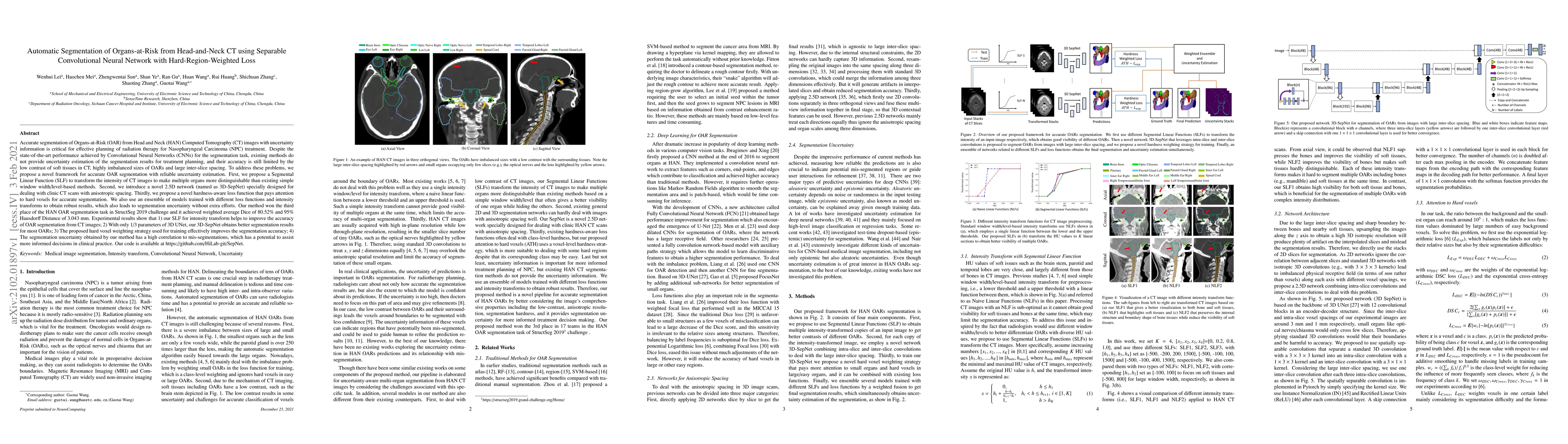
Nasopharyngeal Carcinoma (NPC) is a leading form of Head-and-Neck (HAN) cancer in the Arctic, China, Southeast Asia, and the Middle East/North Africa. Accurate segmentation of Organs-at-Risk (OAR) f...

Thumbnail for Automatic Segmentation of Gross Target Volume of Nasopharynx Cancer
  using Ensemble of Multiscale Deep Neural Networks with Spatial Attention
arXiv
Automatic Segmentation of Gross Target Volume of Nasopharynx Cancer using Ensemble of Multiscale Deep Neural Networks with Spatial Attention

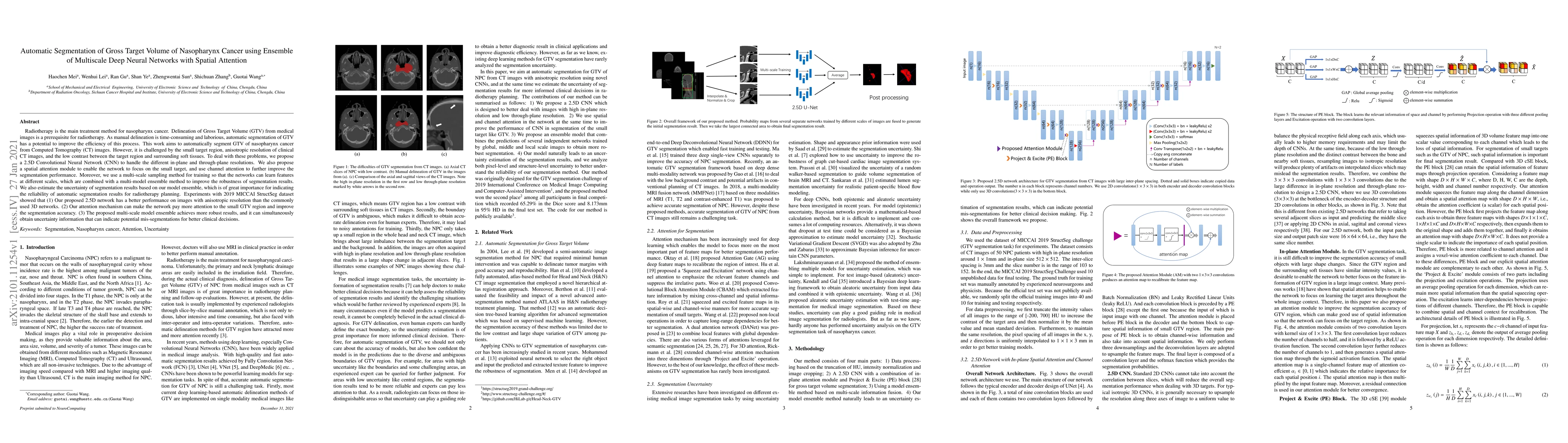
Radiotherapy is the main treatment modality for nasopharynx cancer. Delineation of Gross Target Volume (GTV) from medical images such as CT and MRI images is a prerequisite for radiotherapy. As manu...

Thumbnail for Large-scale cervical precancerous screening via AI-assisted cytology
  whole slide image analysis
arXiv
Large-scale cervical precancerous screening via AI-assisted cytology whole slide image analysis

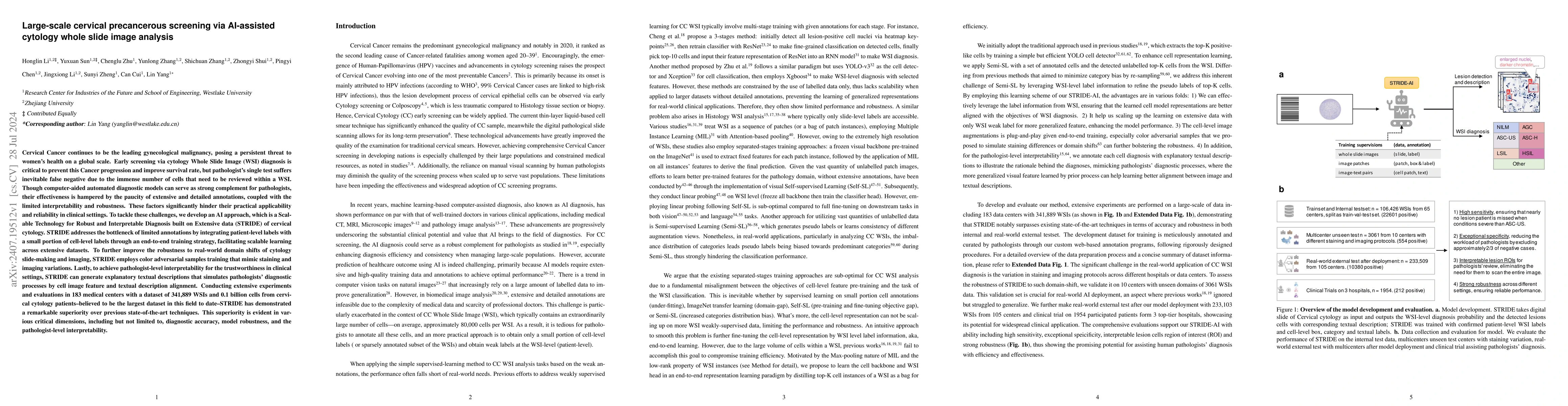
Cervical Cancer continues to be the leading gynecological malignancy, posing a persistent threat to women's health on a global scale. Early screening via cytology Whole Slide Image (WSI) diagnosis is ...

Thumbnail for Weakly Supervised Lymph Nodes Segmentation Based on Partial Instance
  Annotations with Pre-trained Dual-branch Network and Pseudo Label Learning
arXiv
Weakly Supervised Lymph Nodes Segmentation Based on Partial Instance Annotations with Pre-trained Dual-branch Network and Pseudo Label Learning

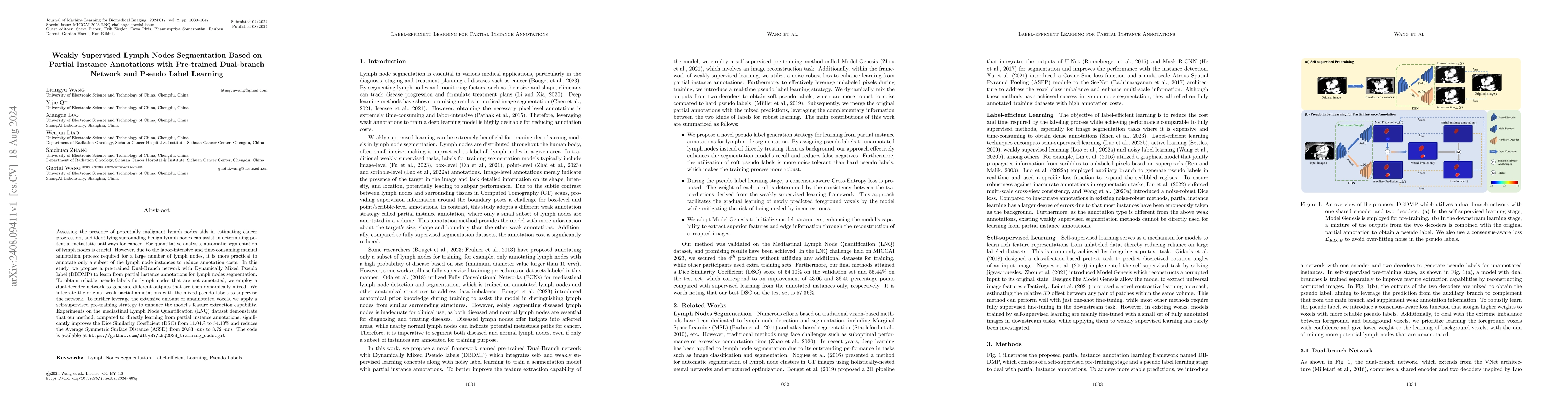
Assessing the presence of potentially malignant lymph nodes aids in estimating cancer progression, and identifying surrounding benign lymph nodes can assist in determining potential metastatic pathway...

Thumbnail for Head and Neck Tumor Segmentation of MRI from Pre- and Mid-radiotherapy
  with Pre-training, Data Augmentation and Dual Flow UNet
arXiv
Head and Neck Tumor Segmentation of MRI from Pre- and Mid-radiotherapy with Pre-training, Data Augmentation and Dual Flow UNet

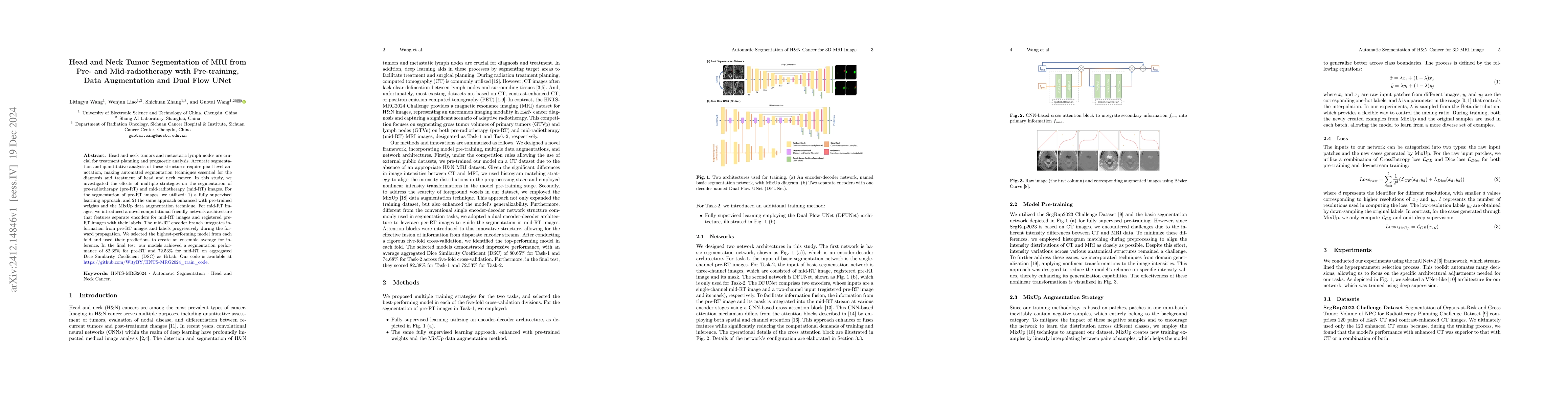
Head and neck tumors and metastatic lymph nodes are crucial for treatment planning and prognostic analysis. Accurate segmentation and quantitative analysis of these structures require pixel-level anno...

Thumbnail for CLISC: Bridging clip and sam by enhanced cam for unsupervised brain
  tumor segmentation
arXiv
CLISC: Bridging clip and sam by enhanced cam for unsupervised brain tumor segmentation

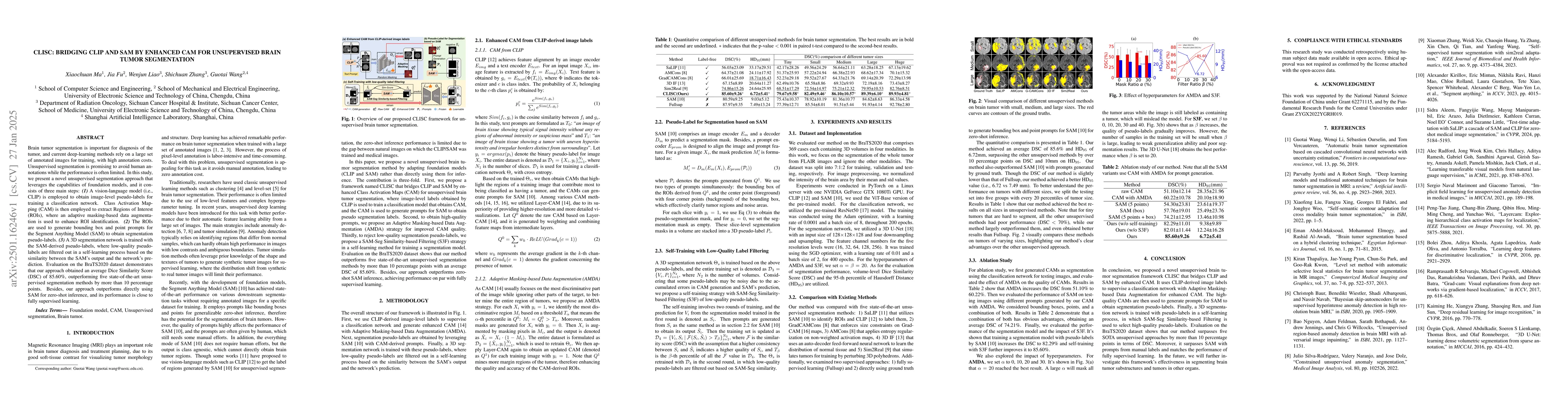
Brain tumor segmentation is important for diagnosis of the tumor, and current deep-learning methods rely on a large set of annotated images for training, with high annotation costs. Unsupervised segme...

Thumbnail for Dynamic Gradient Sparsification Training for Few-Shot Fine-tuning of CT
  Lymph Node Segmentation Foundation Model
arXiv
Dynamic Gradient Sparsification Training for Few-Shot Fine-tuning of CT Lymph Node Segmentation Foundation Model

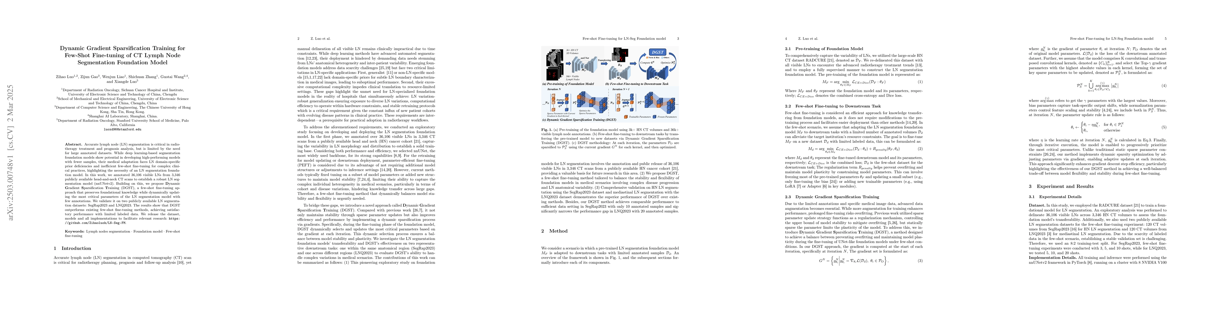
Accurate lymph node (LN) segmentation is critical in radiotherapy treatment and prognosis analysis, but is limited by the need for large annotated datasets. While deep learning-based segmentation foun...

Thumbnail for OpenPath: Open-Set Active Learning for Pathology Image Classification
  via Pre-trained Vision-Language Models
arXiv
OpenPath: Open-Set Active Learning for Pathology Image Classification via Pre-trained Vision-Language Models

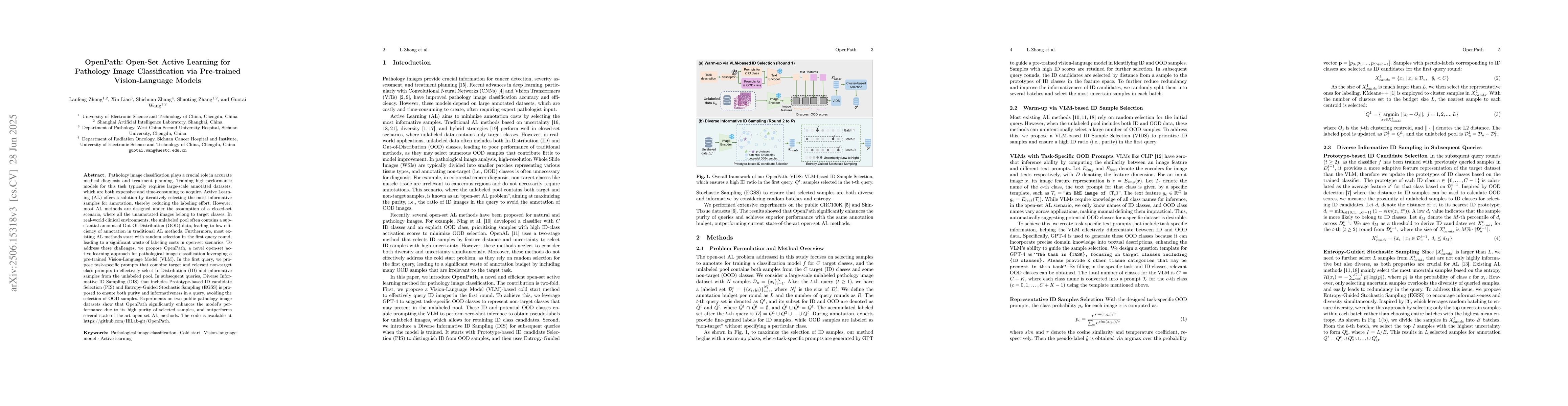
Pathology image classification plays a crucial role in accurate medical diagnosis and treatment planning. Training high-performance models for this task typically requires large-scale annotated datase...

Thumbnail for SegRap2025: A Benchmark of Gross Tumor Volume and Lymph Node Clinical Target Volume Segmentation for Radiotherapy Planning of Nasopharyngeal Carcinoma
arXiv
SegRap2025: A Benchmark of Gross Tumor Volume and Lymph Node Clinical Target Volume Segmentation for Radiotherapy Planning of Nasopharyngeal Carcinoma

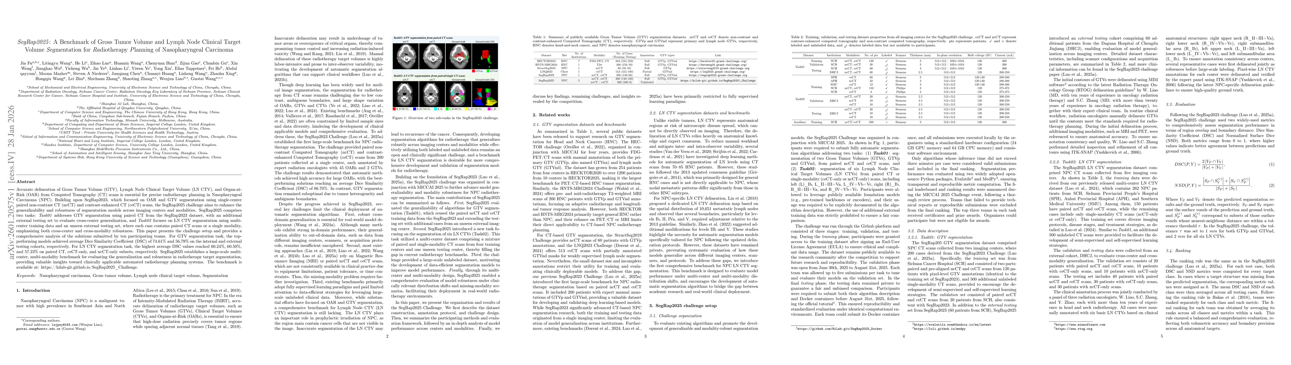
Accurate delineation of Gross Tumor Volume (GTV), Lymph Node Clinical Target Volume (LN CTV), and Organ-at-Risk (OAR) from Computed Tomography (CT) scans is essential for precise radiotherapy planning...

This website uses cookies to ensure you get the best experience. Learn more