Academic Profile
Statistics
Similar Authors
Papers on arXiv
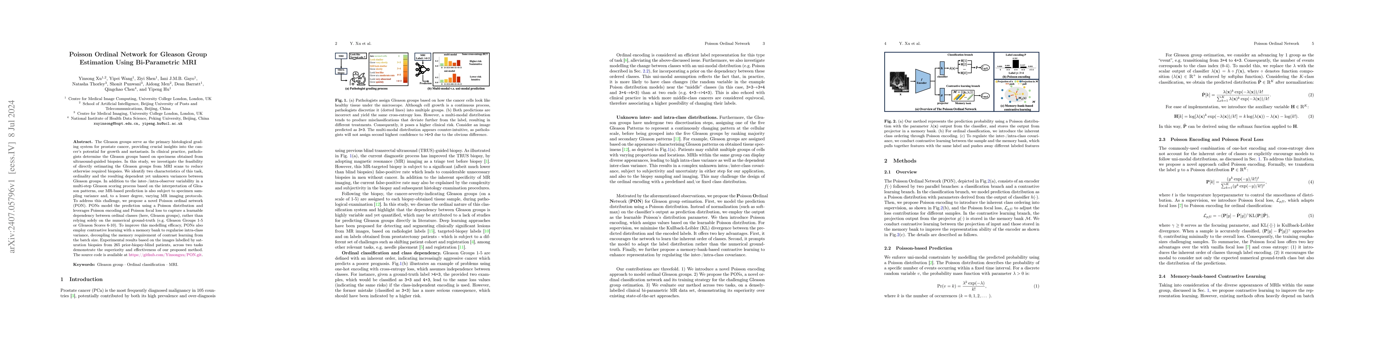
The Gleason groups serve as the primary histological grading system for prostate cancer, providing crucial insights into the cancer's potential for growth and metastasis. In clinical practice, patholo...
Purpose: Demonstrating and assessing self-supervised machine learning fitting of the VERDICT (Vascular, Extracellular and Restricted DIffusion for Cytometry in Tumours) model for prostate. Methods: ...
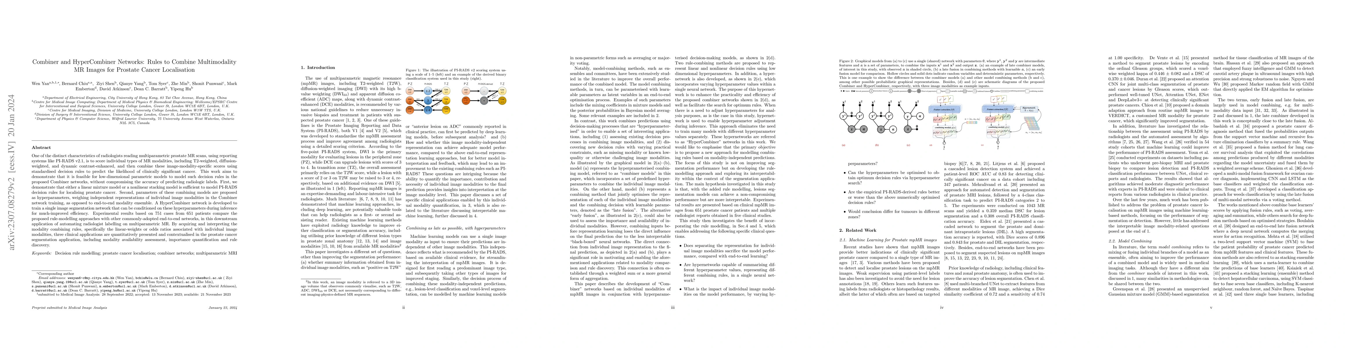
One of the distinct characteristics in radiologists' reading of multiparametric prostate MR scans, using reporting systems such as PI-RADS v2.1, is to score individual types of MR modalities, T2-wei...
We propose an image synthesis mechanism for multi-sequence prostate MR images conditioned on text, to control lesion presence and sequence, as well as to generate paired bi-parametric images conditi...
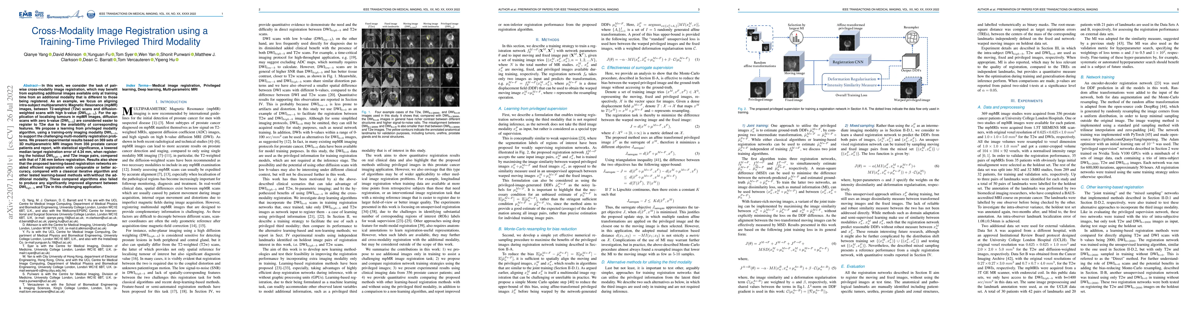
In this work, we consider the task of pairwise cross-modality image registration, which may benefit from exploiting additional images available only at training time from an additional modality that...
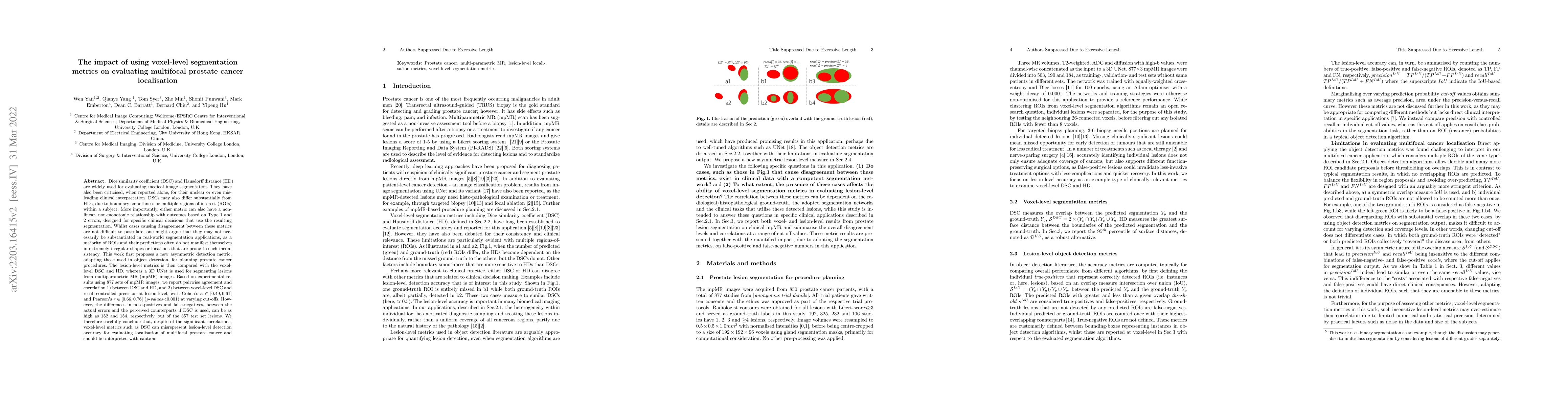
Dice similarity coefficient (DSC) and Hausdorff distance (HD) are widely used for evaluating medical image segmentation. They have also been criticised, when reported alone, for their unclear or eve...
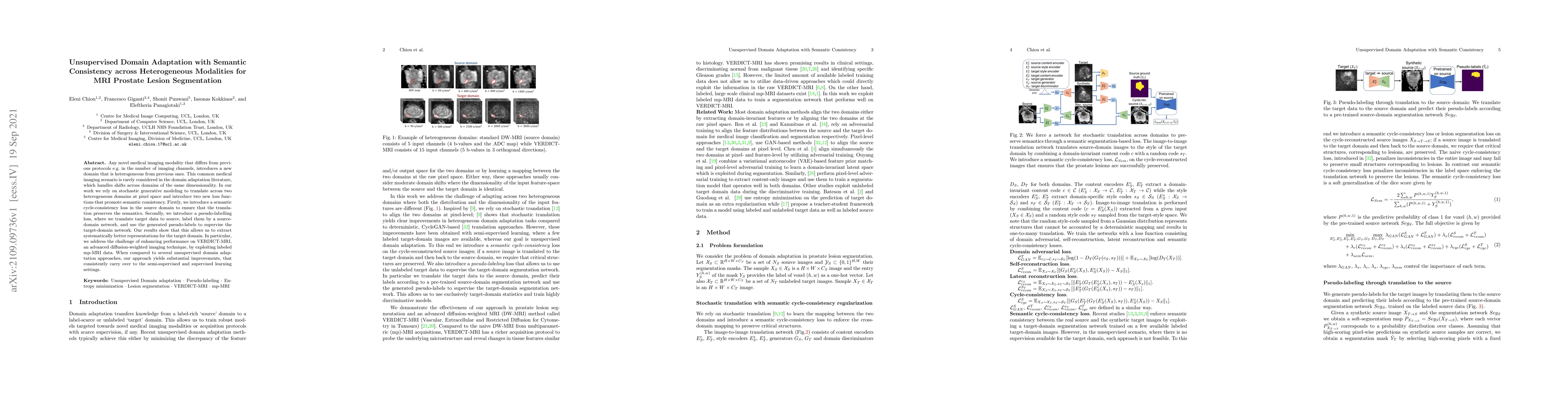
Any novel medical imaging modality that differs from previous protocols e.g. in the number of imaging channels, introduces a new domain that is heterogeneous from previous ones. This common medical ...
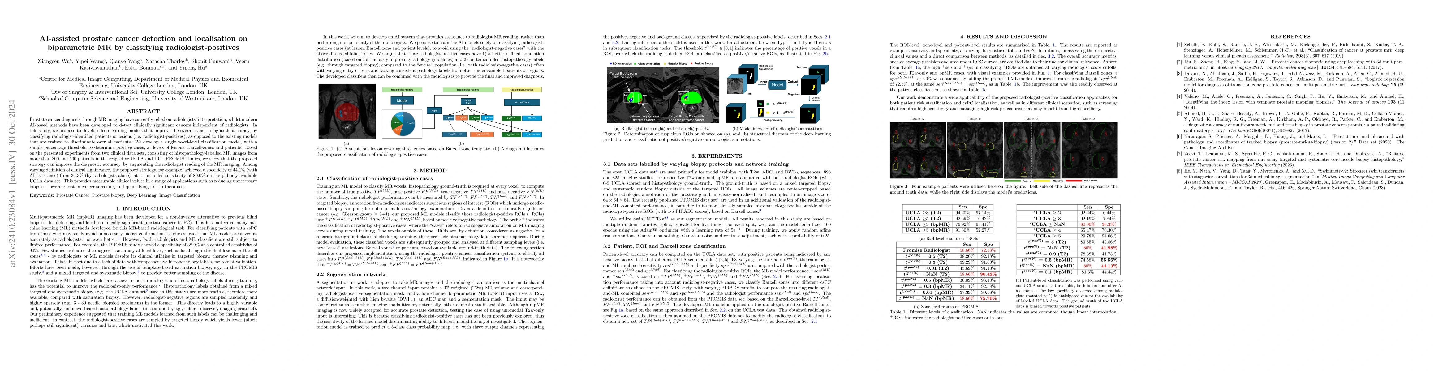
Prostate cancer diagnosis through MR imaging have currently relied on radiologists' interpretation, whilst modern AI-based methods have been developed to detect clinically significant cancers independ...
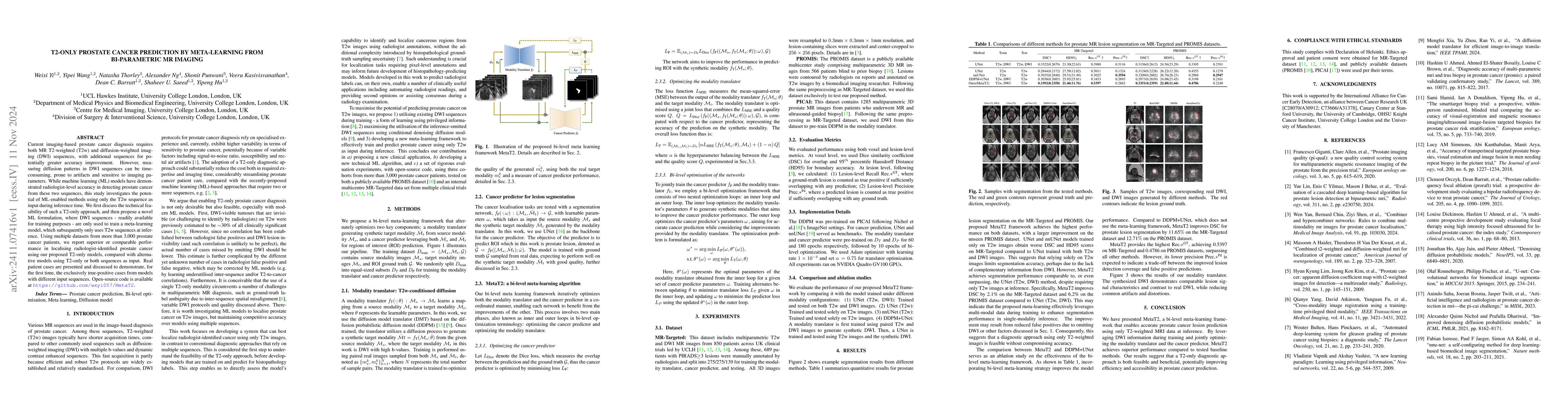
Current imaging-based prostate cancer diagnosis requires both MR T2-weighted (T2w) and diffusion-weighted imaging (DWI) sequences, with additional sequences for potentially greater accuracy improvemen...
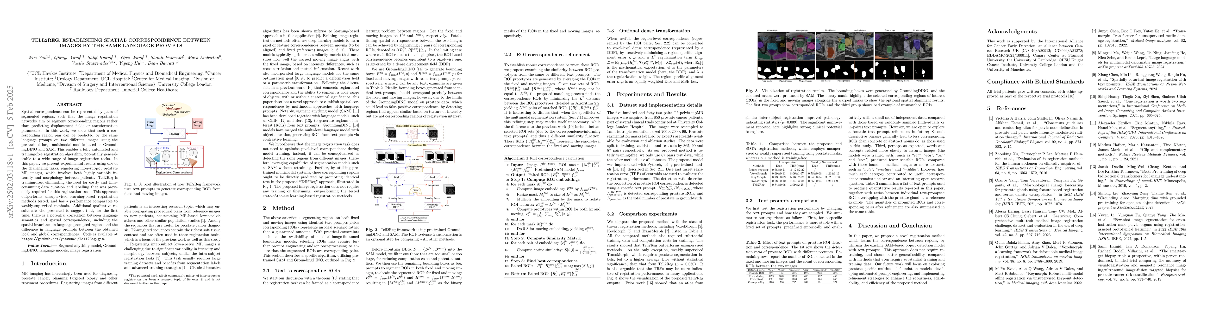
Spatial correspondence can be represented by pairs of segmented regions, such that the image registration networks aim to segment corresponding regions rather than predicting displacement fields or tr...
![Thumbnail for Consensus Recommendations for Hyperpolarized [1-13C]pyruvate MRI
Multi-center Human Studies](https://nbg1.your-objectstorage.com/arxivlens/thumbnails/2504.20440.webp)
Magnetic resonance imaging of hyperpolarized (HP) [1-13C]pyruvate allows in-vivo assessment of metabolism and has translated into human studies across diseases at 15 centers worldwide. Consensus on be...
This work aims to characterise renal tumour microstructure using diffusion MRI (dMRI); via the Vascular, Extracellular and Restricted Diffusion for Cytometry in Tumours (VERDICT)-MRI framework with se...
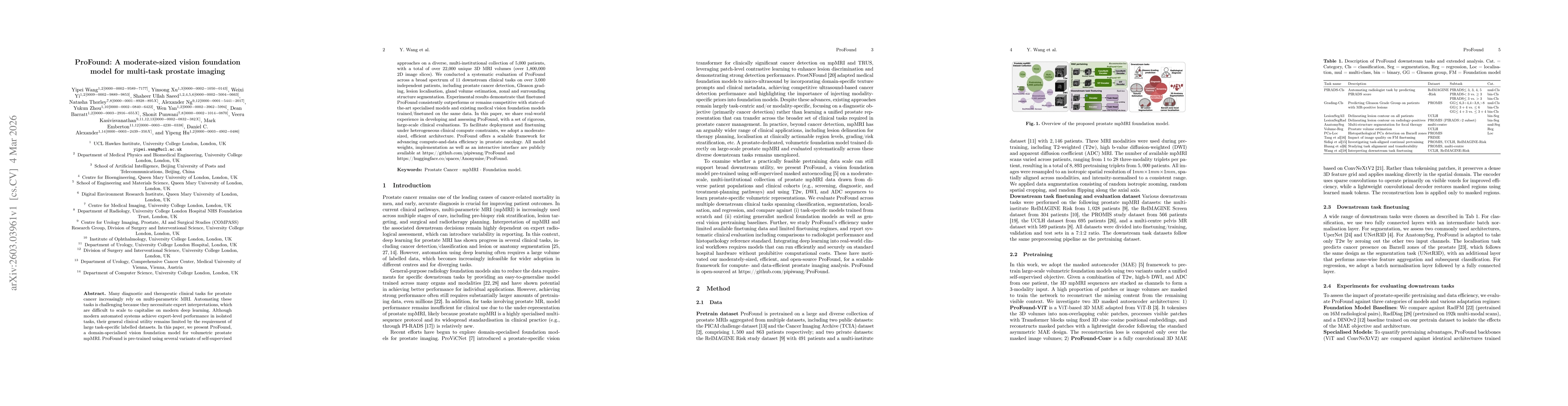
Foundation models in medical imaging have shown promising label efficiency, achieving high performance on downstream tasks using only a fraction of the annotated data otherwise required. In this study...
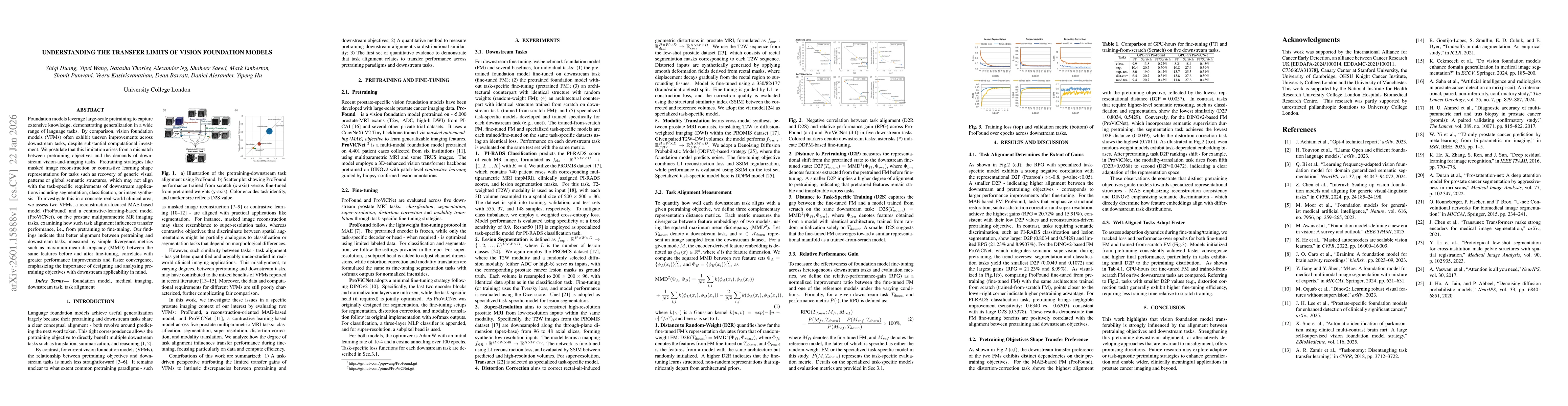
Foundation models leverage large-scale pretraining to capture extensive knowledge, demonstrating generalization in a wide range of language tasks. By comparison, vision foundation models (VFMs) often ...
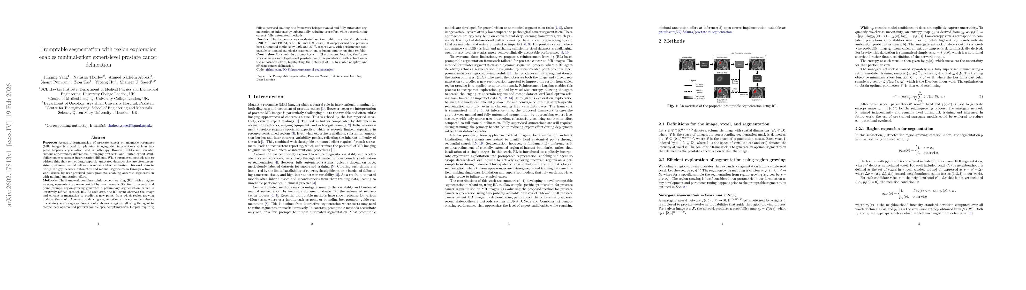
Purpose: Accurate segmentation of prostate cancer on magnetic resonance (MR) images is crucial for planning image-guided interventions such as targeted biopsies, cryoablation, and radiotherapy. Howeve...
Many diagnostic and therapeutic clinical tasks for prostate cancer increasingly rely on multi-parametric MRI. Automating these tasks is challenging because they necessitate expert interpretations, whi...
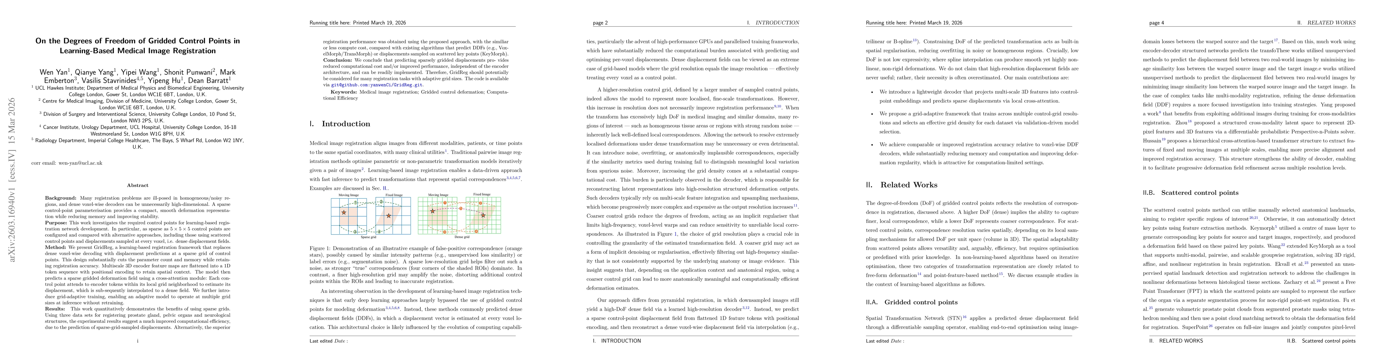
Many registration problems are ill-posed in homogeneous or noisy regions, and dense voxel-wise decoders can be unnecessarily high-dimensional. A sparse control-point parameterisation provides a compac...
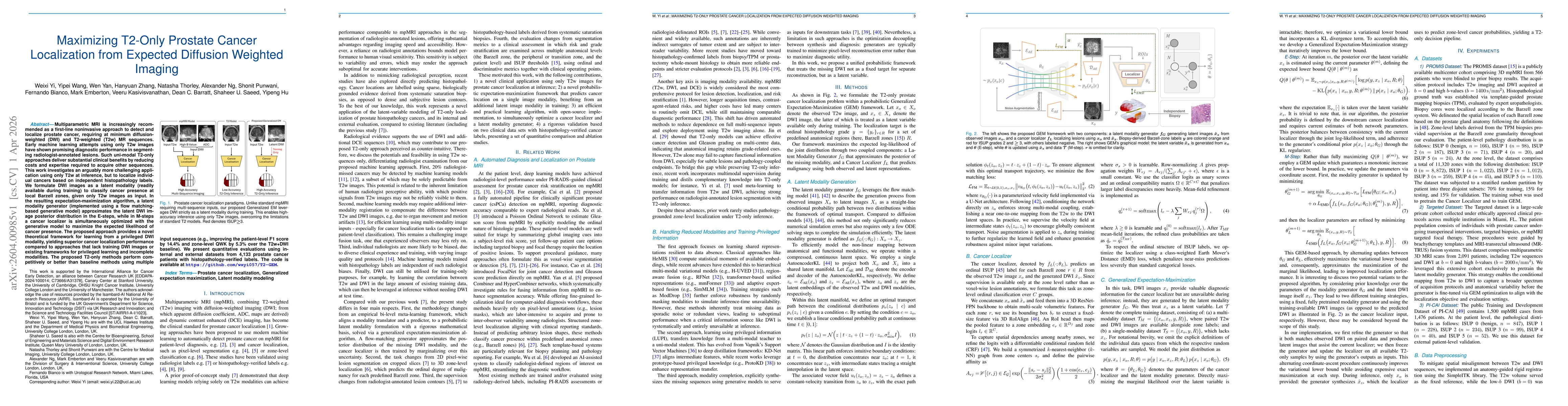
Multiparametric MRI is increasingly recommended as a first-line noninvasive approach to detect and localize prostate cancer, requiring at minimum diffusion-weighted (DWI) and T2-weighted (T2w) MR sequ...
Clinical prostate multi-parametric MRI relies heavily on high-quality diffusion-weighted imaging (DWI), yet reading DWI is frequently compromised by geometric distortion, often caused by rectal air. A...
Single-shot echo-planar prostate diffusion-weighted imaging (DWI) is frequently complicated by geometric distortions, which impact the ability to derive reliable diagnoses from such images. Developing...